

Sir,
Granular cell tumours (GCTs) are uncommonly encountered and can occur in various organs including the skin. Frequent locations are the tongue (40%), breast (15%), respiratory tract (10%), and oesophagus (2%). Solitary tumour is most common but multiple tumours can occur in about 10% of patients.[1] Though natural history of this tumor usually follows a benign course, malignant transformation has been reported in a few cases.[2] Herein we report a case of multifocal cutaneous GCT in a young female for the rarity of the entity.
A 20-year-old woman consulted us for multiple firm to hard, skin coloured to brownish, asymptomatic swellings present over different areas of her body for the preceding 15 years. A few lesions were excised 8 years back. The excised lesions didn’t recur but newer lesions continued to develop. There was no other systemic complaint. Her past medical history and family history was unremarkable. Examination revealed seven skin-colored to brownish, 1–3 cm, firm, smooth-surfaced nodules over extremity, neck, abdomen, back and perineum [Figures 1–3]. There was no other mucocutaneous or systemic finding. We considered the differentials of neurofibromatosis, eruptive dermatofibroma and xanthoma. Routine blood and biochemistry panel were noncontributory. Histopathology from a lesion from the left forearm stained with hematoxylin and eosin showed sheets of polygonal cells having abundant granular eosinophilic cytoplasm and central hyperchromatic nuclei [Figures 4 and 5]. These findings were suggestive of GCT. On immunohistochemistry, the tumor cells were found to express S-100 protein, inhibin, and protein gene product (PGP) 9.5 thus confirming the diagnosis of GCT. Apart from aesthetic concerns, the lesions were not troublesome, thus a conservative approach was preferred by the patient and she was advised to consider surgical option again if any of the present lesions becomes larger.
Figure 1.
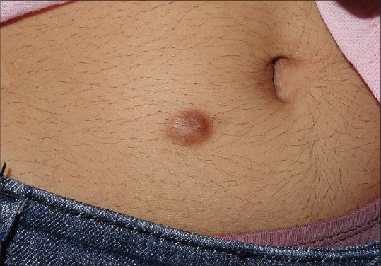
A brownish dome-shaped nodule on the periumbilical skin
Figure 3.

Skin-coloured dome-shaped nodule on front of neck
Figure 4.

Sheets of polygonal cells with pink cytoplasm and central hyperchromatic nuclei. (H and E, ×100)
Figure 5.

High-power view showing polygonal cells with granular eosinophilic cytoplasm and hyperchromatic nuclei. (H and E, ×400)
Figure 2.

Subcutaneous nodule on the back
Granular cell tumour is a neoplasm probably involving structures of neural derivation that can arise at virtually any body site. GCTs are usually asymptomatic and present as smooth, slow-growing, solitary nodules in subcutaneous, intradermal, or submucosal regions. Their clinical presentation ranges from large verrucous nodules to small, nonspecific, subcutaneous lesions. Multiple GCT can occur as isolated disorder without any other syndrome or diseases as in our case or can be associated with conditions such as Noonan syndrome and LEOPARD syndrome.[3,4] Histopathology of GCT shows a nonencapsulated tumor composed of large polyhedral cells containing faintly eosinophilic, uniform fine to coarse granules. Nuclei are small, hyperchromatic and centrally located. Mitoses are uncommon. Sometimes neurotropic spread along the peripheral nerves or infiltration of skeletal muscles could be appreciated.[5] Although the routine histologic features in our case were sufficiently diagnostic, the immunohistochemistry panel further consolidated our diagnosis.
The treatment of choice for GCT is surgical resection. Our patient's lesions were nearly asymptomatic and she preferred the conservative approach with periodic follow up. To conclude, although GCTs are uncommonly encountered its possibility should be considered in the differential diagnosis of solitary as well as multiple skin tumours and nodules.
References
- 1.Lack EE, Worsham GF, Callihan MD, Crawford BE, Klappenbach S, Rowden G, et al. Granular cell tumor: A clinicopathologic study of 110 patients. J Surg Oncol. 1980;13:301–16. doi: 10.1002/jso.2930130405. [DOI] [PubMed] [Google Scholar]
- 2.Gomard-Mennesson E, Isaac S, Freymond N, Guibert B, Pacheco Y, Devouassoux G. Pulmonary metastases from Abrikossoff's tumour. Transformation capability of a benign granular cell tumour. Rev Mal Respir. 2007;24:900–4. doi: 10.1016/s0761-8425(07)91395-9. [DOI] [PubMed] [Google Scholar]
- 3.Ramaswamy PV, Storm CA, Filiano JJ, Dinulos JG. Multiple granular cell tumors in a child with Noonan syndrome. Pediatr Dermatol. 2010;27:209–11. doi: 10.1111/j.1525-1470.2010.01111.x. [DOI] [PubMed] [Google Scholar]
- 4.Gunson TH, Hashim N, Sharpe GR. Generalized lentiginosis, short stature, and multiple cutaneous nodules – Quiz case. LEOPARD syndrome (LS) associated with multiple granular cell tumors (GCTs) Arch Dermatol. 2010;146:337–42. doi: 10.1001/archdermatol.2010.7-a. [DOI] [PubMed] [Google Scholar]
- 5.Reed RJ, Pulitzer DR. Tumors of neural tissue. In: Elder DE, editor. Lever's Histopathology of the Skin. 10th ed. Philadelphia: Lippincott Williams and Wilkins; 2009. pp. 1107–49. [Google Scholar]