Abstract
Sarcomatoid carcinomas are biphasic tumours, which occur at any site in the human body. It rarely affects the penis, with only 38 cases being reported in literature. It may be considered as a variant of squamous cell carcinoma or a dedifferentiated tumour. We report a 60-year old gentleman who presented with a swelling in the glans penis. He underwent a partial penectomy. Histopathology revealed sarcomatoid carcinoma of the penis, which was confirmed by immunohistochemistry. The rarity of this clinical entity makes its diagnosis difficult.
Keywords: Biphasic tumours, Dedifferentiated tumours, Immunohistochemistry, Partial penectomy
Introduction
The incidence of penile cancer in the Indian subcontinent is 0.7-3/100,000 men [1, 2]. Majority of these tumours are squamous cell carcinomas. Sarcomatoid carcinoma is a very rare variant of penile cancer. It has also been called as spindle cell carcinoma, metaplastic carcinoma, or biphasic squamous cell carcinoma. It is considered as a variant of squamous cell carcinoma or as a metaplastic differentiation of the mesenchyme. Only 38 cases of sarcomatoid carcinoma of the penis have been reported in literature.
We report a patient with penile sarcomatoid carcinoma, and have reviewed the literature.
Case Report
A 60 year old male gentleman presented with a history of a fleshy growth on the glans penis since 2 months. The growth was bleeding for the last 1 week. Clinical examination revealed a 3 × 4 cm ulcero-proliferative growth involving the glans penis, the shaft was not involved by the tumour (Fig. 1). The urethral meatus was free. There were no lymph nodes palpable in the inguinal region.
Fig. 1.
The fleshy growth on the glans penis
An initial biopsy was done that revealed the presence of spindle cells. A provisional diagnosis of a spindle cell tumour was made. An ultrasound abdomen and a chest radiograph was within normal limits. A decision to proceed with surgery was taken. The patient underwent a partial amputation of the penis.
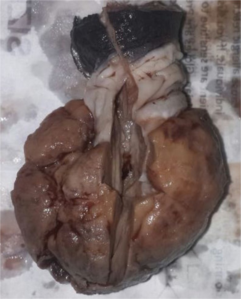
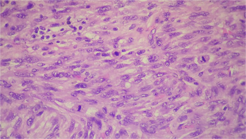
On gross examination a fleshy proliferative growth was arising from the glans penis (Fig. 2). Microscopic examination under hematoxylin and eosin (H and E) sections revealed features of a high grade pleomorphic spindle cell neoplasm (Mitosis 15–20 / 10 hpf) (Fig. 3). Immunohistochemistry was sought for further categorisation. The cells were negative for epithelial membrane antigen (EMA), cytokeratin (CK) and p63. The cells were also negative for HMB45, smooth muscle antigen (SMA), CD 34 and desmin.
Fig. 2.
Gross histopathological specimen of partial penectomy. The urethra is not involved by the tumour
Fig. 3.
H and E X 10. Spindle cells with brisk mitosis
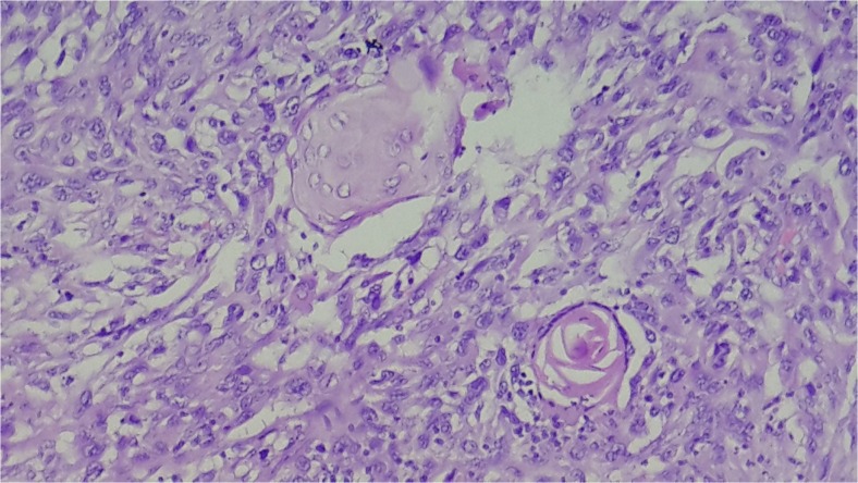
The entire tumour was subsequently processed for further H and E sections. A focus of squamous cell differentiation was present, (Fig. 4) from which we came to the diagnosis of a sarcomatoid carcinoma.
Fig. 4.
H and E X 10. Small foci of squamous differentiation with a well formed keratin pearl amongst the spindle cells
On his first follow up in the postoperative period he underwent a cross sectional imaging of the chest, abdomen and pelvis which was normal.
He had an uneventful postoperative recovery. At 1 year of followup patient is doing well.
Discussion
Penile carcinoma is an uncommon malignancy in the west with an incidence of less than one per 100,000 males. However there is significant geographical variation with a higher incidence in areas where human papilloma virus infection is common and there is prevalence of unhygienic practice, especially in the developing countries. Squamous cell carcinoma constitute the majority (95 %) of the penile cancers. Sarcomatoid carcinoma of the penis representing only 1–2 % of the penile cancers, is considered a variant of squamous cell carcinoma with a poor prognosis [3].
Sarcomatoid carcinomas are biphasic tumours with an intimate admixture of both carcinomatous and sarcomatous elements [4]. As they have both the components ( epithelial and mesenchymal ) they are capable of both a lymphatic and hematogenous dissemination leading to both regional and distant metastasis [5]. Sarcomatoid carcinomas of the penis were initially reported only sporadically and as case reports in literature. Lont et al. in 2004 retrospectively reviewed the clinical, morphological and immunohistochemical features of five cases over a span of 46 years [6]. Velazquez et al. had described 15 cases of sarcomatoid carcinoma on a retrospective analysis of 400 patients with penile carcinoma with an incidence of 4 % [7]. Majority of the patients had either regional (60–91 %) or distant metastasis at presentation or developed them on follow up and was associated with a high mortality (50–80 % ) [6, 7].
Sarcomatoid carcinoma of the penis probably originates from the epithelial cell, hence retaining their immuno-histochemistry. Unless the tumour has dedifferentiated into a sarcomatous variant, like our case where there was only a small foci of epitheloid tumour where the immuno-histochemistry would not be contributory. Based on this both the components are derived from the same stem cell. Histologically these cells have the potential to differentiate into muscle, bone or cartilage, benign or malignant.
Among the cases reported in literature, majority had lymph node or distant metastasis, suggesting marked vascular infiltration as a cause of poor prognosis [8]. Sarcomatoid carcinoma of the penis described in the literature has a very dismal prognosis with majority of the patients having inguinal lymph node metastasis at presentation or on follow up within 6 months of their primary surgery. These tumours have a very high metastatic potential, developing systemic metastasis on follow up. Palliative surgery may be considered in advanced, ulcerated tumours providing temporary tumour regression and decreasing pain and bleeding.
To conclude, sarcomatoid carcinoma of the penis, a variant of squamous cell carcinoma is an unusual rapidly growing tumour of the penis associated with the development of early metastatic spread and a poor prognosis. Differential diagnosis would include other mesenchymal tumours like synovial sarcoma. It is a rare malignancy that is often difficult to diagnose. Complete examination of the tumour in its entirety along with immuno-histochemistry would help in diagnosing this entity.
Contributor Information
Kiran Shankar, Phone: +919900933982, Email: kiranshankar1984@gmail.com.
M. Vijaya Kumar, Phone: +919448467765, Email: mvijai2013@gmail.com.
References
- 1.Parkin DM, Whelan SL, Ferlay J, et al. Cancer incidence in five continents. IARC: Lyon; 2002. [Google Scholar]
- 2.Parkin DM, Bray F (2006) Chapter 2: The burden of HPV-related cancers. Vaccine 24(Suppl 3):S3/11–25 [DOI] [PubMed]
- 3.Ranganath R, Singh SS, Sateeshan B. Sarcomatous carcinoma of the penis: clinicopathological features. Indian J Urol. 2008;24(2):267–268. doi: 10.4103/0970-1591.40630. [DOI] [PMC free article] [PubMed] [Google Scholar]
- 4.Reuter VE. Sarcomatous lesions of the urogenital tract. Semin Diagn Pathol. 1993;10:188–201. [PubMed] [Google Scholar]
- 5.Challa VR, Swamyvelu K, Shivappa P, Amirtram U. Sarcomatous carcinoma of the penis with bilateral inguinal metastasis- a case report and review of literature. Indian J Surg. 2014;76(4):316–318. doi: 10.1007/s12262-013-0965-6. [DOI] [PMC free article] [PubMed] [Google Scholar]
- 6.Lont AP, Galle MPW, Snijders P, Horenblas S. Sarcomatoid squamous cell carcinoma of the penis: a clinical and pathological study of 5 cases. J Urol. 2004;172:932–935. doi: 10.1097/01.ju.0000136363.90911.e5. [DOI] [PubMed] [Google Scholar]
- 7.Velazquez EF, Melamed J, Barreto JE, Aguero F, Cubilla AL. Sarcomatoid carcinoma of the penis: a clinicopathological study of 15 cases. Am J Surg Pathol. 2005;29:1152–1158. doi: 10.1097/01.pas.0000160440.46394.a8. [DOI] [PubMed] [Google Scholar]
- 8.Kuroda I, Ishida T, Aoyagi T. Sarcomatoid carcinoma of the penis. Case Rep Clin Med. 2014;3:10–12. doi: 10.4236/crcm.2014.31003. [DOI] [Google Scholar]


