

Abstract
Fiessinger-Leroy's disease (Reiter's disease) is rare and not commonly reported in India. This paper reports a case of a 35-year-old male with Fiessinger-Leroy's disease, occurring after balanitis. The patient presented with symmetrical polyarthritis, pruritic scaly plaques, keratoderma blenorrhagicum, and hematuria with histopathology of skin suggestive of Fiessinger-Leroy's disease. The case is presented with a view to document the occurrence of the same in India.
Key words: Arthritis, Fiessinger-Leroy's disease, keratoderma blenorrhagicum balanitis, Reiter's disease
INTRODUCTION
Fiessinger-Leroy's disease is a rare multisystem autoimmune spondyloarthropathic disease, first described by Hans Reiter in 1916[1] in a Prussian soldier, as Reiter's disease, and by Ahvonen, Sievers, and Aho[2] in 1969 as “Reactive Arthritis” with an incidence of 3.5–5/100,000 across the world. It is characterized by a triad of nongonococcal urethritis, conjunctivitis, and nonsuppurative, predominantly lower limb asymmetrical oligoarthritis (an average of four joints are affected).[3,4,5]
The fact that only one-third of patients show the complete triad prompted the American Rheumatism Association to define Reiter's syndrome as “an episode of peripheral arthritis of more than one month's duration occurring in association with urethritis and/or cervicitis.”[6,7]
Symptoms generally appear within 1–3 weeks but can range from 4 to 35 days from the onset of inciting episode of urethritis/cervicitis/diarrhea. Signs and symptoms usually remit within 6 months.[8] It is probable that a preceding infection, usually a urethritis or dysentery, serves as a trigger in a genetically predisposed individual and that the disease may then persist or recur despite eradication of the infection.
The postulated triggering agents are Chlamydia trachomatis, Shigella flexneri, Salmonella spp., Yersinia enterocolitica, Campylobacter spp., Neisseria gonorrhoea, and Borrelia burgdorferi.[4]
There is a high incidence of HLA B27 positivity (60%–90%) in patients with Reiter's disease.[5,6]
We report a case of Fiessinger-Leroy's disease with an unusual rheumatoid-like articular presentation.
CASE REPORT
A 35-year-old male presented with pain and stiffness in multiple joints, crusted lesions over the trunk, both extremities, palms and soles, and intermittent hematuria for 2 years. The patient had a history of redness in the right eye before 1 year. Despite treatment from an outside hospital for 2 years, his joints became increasingly painful and the whole course was punctuated with recurrences of skin lesions associated with fever, chills, malaise, and headache.
The patient had a history of genital ulceration following an unprotected sexual exposure with a commercial sex worker (CSW) which was diagnosed as balanitis from a local doctor. The patient had repeated extramarital unprotected sexual exposures with multiple partners, also with CSWs, with duration of 2 years from the last exposure.
The patient had a treatment history of tablet methotrexate 20 mg/week for 4 months and varying tapering doses of systemic steroids as tablet prednisolone and tablet betamethasone over a period of 1 year. There was a history of improvement in the lesions and joint pain with ongoing treatment, but the lesions again flared up with self-stoppage of treatment for 6 months.
On examination, the patient was a thin-built middle-aged man. Cutaneous examination revealed multiple erythematous dry crusted plaques of variable size and shapes ranging from smallest < 0.5 cm to largest 10 cm × 10 cm over the trunk, distributed mainly over the trunk [Figures 1 and 2], both extremities, both palms and soles (keratoderma blenorrhagicum) [Figure 3], with sparing the scalp, face, and flexures. There was single erosion with crusting of 1 cm × 1 cm size over the glans penis [Figure 4] with surrounding erythematous skin and sparing other mucosae. The striking feature was the symmetrical polyarthritis with painful joints, resulting in morning stiffness and limited joint mobility throughout the day. The hip joint and lower extremities were predominantly affected. The upper limbs were also affected though to a lesser extent.
Figure 1.
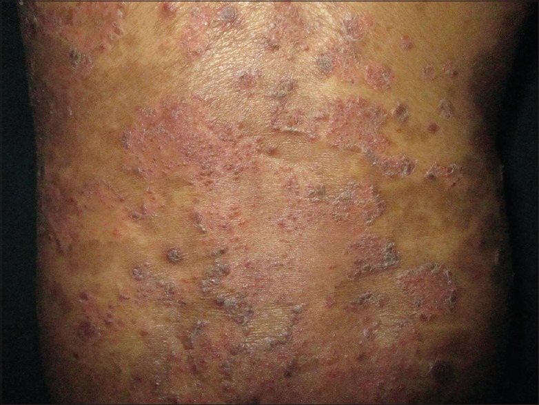
Multiple crusted plaques over trunk
Figure 2.
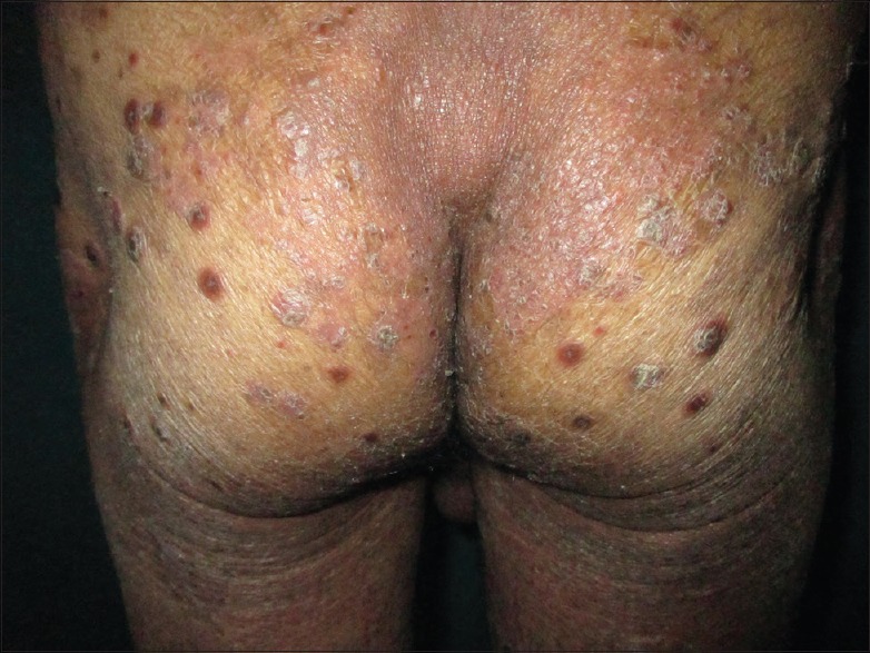
Multiple crusted plaques over buttocks
Figure 3.

Keratoderma blenorrhagicum - soles
Figure 4.

Circinate balanitis
Nail examination revealed coarse pitting, yellowish discoloration, transverse ridges, subungual hyperkeratosis, friability, and dystrophy [Figure 5].
Figure 5.

Histopathology showing marked hyperkeratosis, focal parakeratosis, with spongiosis, irregular rete ridges and few micro abscesses in the epidermis and predominantly lymphocytic infiltrate in perivascular area in the dermis
Other systemic examinations revealed no specific finding.
Routine hemogram revealed hypochromic, microcytic anemia with hemoglobin – 8 g/dl, leukocytosis – 18,600 cells/cumm, differential count – neutrophilia (80%), and elevated erythrocyte sedimentation rate – 115 mm/1st h. Urine routine examination, blood sugar level, renal function test, and liver function test were within normal limits. Antistreptolysin-O titer and C-reactive protein were positive and rheumatoid factor was negative. Rapid plasma reagent and HIV serology were nonreactive.[9] Urine and stool cultures were negative. Electrocardiogram showed no abnormality. Human leukocyte antigen studies could not be carried out because of unavailability of the test.
On histopathology of skin biopsy, there were marked hyperkeratosis, focal parakeratosis, spongiosis, irregular elongation of rete ridges, and few microabscesses in the epidermis and predominantly perivascular lymphocytic infiltrate in the dermis [Figure 6]. Radiographic study of hip joint revealed reduced joint space and that of spine revealed calcification of the anterior ligament [Figures 7 and 8].
Figure 6.

Finger nails showing coarse pitting, yellowish discoloration, transverse ridges and subungual hyperkeratosis
Figure 7.

X-ray hip joint (AP view) showing reduced joint space
Figure 8.

X-ray spine (AP view) showing calcification in spine
Based on the history, clinical examination, and investigations, it was diagnosed as a case of Fiessinger-Leroy's disease. The treatment given included tablet methotrexate 15 mg weekly and capsule doxycycline 100 mg BD for 1 month. The patient returned to his native state and continued further treatment from there.
DISCUSSION
The name Reiter's syndrome was popularized in English texts by Bauer and Engelman in 1942, who thought that Reiter was the first to describe it. At the same time of the disease's description by Reiter in 1916, two French doctors, Fiessinger and Leroy, attributed a case of reactive arthritis to a Shigella infection. In 1818, Brodie published a case of Reiter's syndrome after venereal infection, followed by Stoll, who described a case of the disease in 1869.[4,10]
Fiessinger-Leroy's disease is a genetically determined seronegative spondyloarthropathy characterized by a triad of urethritis, arthritis, and conjunctivitis, after an episode of urogenital or gastrointestinal infection.[3,4,5,10]
The mechanism by which the organisms causing infection in the urogenital or gastrointestinal tract lead to Fiessinger-Leroy's disease in some individuals is not clear. It has been proposed that damaged exogenous pathogen-associated molecular patterns, derived from microbes, can disseminate through the pelvic and spinal lymphatic pathways and activate toll-like receptors (TLRs). Their activation triggers signaling pathways that result in the expression of immune response genes and cytokine production. First, it was thought that TLR-4 expression by neutrophil was responsible for host clearance. However, recent human data suggest that TLR-2, not TLR-4, is important in determining reactive arthritis susceptibility after Salmonella infection.[4,10,11] Severe cases of Fiessinger-Leroy's disease can occur as a late manifestation of HIV. Other rare factors, which can induce the disease, are immunotherapy with Bacillus Calmette–Guerin and interferon α, following hepatitis B vaccination.[4] Caucasians were more commonly affected, probably because of higher prevalence of HLA-B27 in this population group.[4]
Diagnosing Fiessinger-Leroy's disease can be difficult because of the great variation of the clinical features and because the classical triad is present in only one-third of the patients. Reactive arthritis usually has a self-limited course of 3–12 months, but up to 50% of patients experience recurrent bouts of arthritis and 15%–30% of them develop chronic symptoms of the disease. Most frequent presentation is nondestructive acute oligoarthritis of large lower limb joints (an average of four joints are affected).[6] The average duration of the arthritis is 4–5 months, but two-thirds of patients have mild musculoskeletal symptoms that persist for more than 1 year. Recurrent attacks are more common in patients with Chlamydia-induced reactive arthritis. Secondary to enthesitis, patients may have heel pain. Diffuse swelling of an entire finger/toe (sausage digit) can be seen. Erythematous macules and plaques, diffuse erythema, erosions, and bleeding can appear on the oral mucosae. Circinate balanitis (20%–40%) is a well-defined, painless, erythematous lesion with small, shallow ulcers of the glans penis and urethral meatus.[3,4]
Keratoderma blenorrhagicum is the characteristic cutaneous manifestation of the disease although it occurs in only about 10% of patients. It usually appears 1–2 months after the onset of arthritis as in our case. However, it may accompany or rarely precede the initial manifestations. The soles of the feet are almost always involved, but the extensor surfaces such as legs, dorsal aspects of the toes, feet, fingers, nails, and scalp are the common sites. Occasionally, the eruption is very widespread and it may evolve into generalized exfoliative dermatitis (erythroderma). The initial lesion is a dull, red macule which rapidly becomes papular and pseudovesicular. Its color changes from yellow to orange-red as the roof thickens to form a hyperkeratotic plaque.[4,10]
In our case, the differential diagnoses considered were pustular psoriasis, secondary syphilis, and Behcet's disease.[12]
In our case, the presentation of the patient with pain and stiffness in multiple joints predominantly affecting hip joint and lower limbs with changes of reduced joint space and calcification on radiographic examination, history of genital ulceration and redness in eyes prior to joint involvement, psoriasiform plaques over the body, keratoderma blenorrhagicum over the soles, and circinate balanitis on the glans penis favored the diagnosis of Fiessinger-Leroy's disease.
To conclude, Fiessinger-Leroy's disease is a rare disease worldwide and hence we are reporting this case for documentation.
Financial support and sponsorship
Nil.
Conflicts of interest
There are no conflicts of interest.
REFERENCES
- 1.Sawhney MP, Parihar JK. Macular degeneration in a case of Reiter's disease. Indian J Dermatol Venereol Leprol. 2006;72:227–30. doi: 10.4103/0378-6323.25787. [DOI] [PubMed] [Google Scholar]
- 2.Rao A, Gupta V, Bhatia R, Malhotra A, Gupta S. Urethritis pelvic inflammatory disease and Reiter's syndrome. In: Sacchidanand S, editor. IADVL Textbook of Dermatology. 4th ed. Mumbai: Bhalani Publishing House; 2013. p. 2919. Ch. 90. [Google Scholar]
- 3.Rathod T, Chandanwale A, Chavan S, Shah M. Polyarthritic, symmetric arthropathy in reactive arthritis. J Nat Sci Biol Med. 2011;2:216–8. doi: 10.4103/0976-9668.92312. [DOI] [PMC free article] [PubMed] [Google Scholar]
- 4.Kumar S, Mahajan BB, Ahluwalia RS, Boparai AS. Reiter's disease: Circinate balanitis as alone preceding presentation – Successfully treated with pimecrolimus 1% cream. Indian J Sex Transm Dis. 2015;36:70–3. doi: 10.4103/2589-0557.156733. [DOI] [PMC free article] [PubMed] [Google Scholar]
- 5.Monteiro RC, Bhat RM, Sukumar D, Srinath MK. A case of Reiter's disease exacerbated by lithium. Indian J Sex Transm Dis. 2011;32:121–3. doi: 10.4103/2589-0557.85422. [DOI] [PMC free article] [PubMed] [Google Scholar]
- 6.Madavamurthy P, Hanish VB. Reiter's syndrome. Indian J Dermatol Venereol Leprol. 1993;59:197–200. [Google Scholar]
- 7.Arora S, Arora G. Reiter's disease in a six-year-old girl. Indian J Dermatol Venereol Leprol. 2005;71:285–6. doi: 10.4103/0378-6323.16626. [DOI] [PubMed] [Google Scholar]
- 8.Alebiosu CO, Raimi TH, Badru AI, Amore OO, Ogunkoya JO, Odusan O. Reiters syndrome – A case report and review of literature. Afr Health Sci. 2004;4:136–7. [PMC free article] [PubMed] [Google Scholar]
- 9.Gaurkar SP, Parmar KS, Shah BJ. Sequential development of psoriasis, alopecia universalis, and vitiligo vulgaris in a human immunodeficiency virus seropositive patient: A unique case report. Indian J Sex Transm Dis. 2014;35:167–9. doi: 10.4103/2589-0557.142424. [DOI] [PMC free article] [PubMed] [Google Scholar]
- 10.Dimitrova V, Valtchev V, Yordanova I, Haidudova H, Gospodinov D, Tisheva S. Keratoderma blenorrhagicum in a patient with Reiter syndrome. J IMAB. 2008;14:68–71. [Google Scholar]
- 11.Tsui FW, Xi N, Rohekar S, Riarh R, Bilotta R, Tsui HW, et al. Toll-like receptor 2 variants are associated with acute reactive arthritis. Arthritis Rheum. 2008;58:3436–8. doi: 10.1002/art.23967. [DOI] [PubMed] [Google Scholar]
- 12.Kharkar V, Yeole S, Mahajan S, Gutte R, Khopkar U, Sujatha MR. Behcet's disease simulating secondary syphilis in an HIV-infected patient. Indian J Sex Transm Dis. 2013;34:62–4. doi: 10.4103/2589-0557.112950. [DOI] [PMC free article] [PubMed] [Google Scholar]