

Abstract
Knowledge of variations of azygos and hemiazygos veins is of importance to cardiothoracic surgeons and radiologists during various surgical, radiological, and echography techniques. We report some unique variations of azygos system of veins observed during dissection classes for undergraduate medical students. The azygos vein was formed as usual by the union of right subcostal and ascending lumbar veins. The vein ascended upward and to the left to reach the midline at the level of the 9th thoracic vertebra. After ascending till 5th thoracic vertebra, it gradually inclined to the right of midline and terminated by opening into the superior vena cava at the level of the 3rd thoracic vertebra. There was no major variation in the tributaries of the azygos vein on the right side, except that the right superior intercostal vein crossed behind the azygos vein from right to left and opened into the left side of the azygos vein. Further, two anastomotic veins connected the 10th, 11th and 12th posterior intercostal veins with each other to form two anastomotic circles on the right side of 10th to 12th thoracic vertebrae. The hemiazygos vein bifurcated on the left side of the 10th thoracic vertebra and the two ends opened into the azygos vein at the level of 9th and 10th thoracic vertebrae forming a venous circle in front of the 10th thoracic vertebra. The course of accessory hemiazygos vein was noteworthy. Instead of its classic descending course, the vein ascended upward from the left side of the 8th thoracic vertebra till the 6th thoracic vertebra before opening into the azygos vein.
Key Words: Accessory hemiazygos vein, azygos vein, hemiazygos vein, intercostal vein, posterior mediastinum
INTRODUCTION
Azygos vein is the vein that drains the venous blood from the upper part of the posterior abdominal wall and the thoracic wall. It is very important for the reason that it connects the superior vena cava with the inferior vena cava, and it has very few to no valves. It helps to shunt the blood in both ways due to this reason.[1] Although its formation is variable, generally it is formed at the level of the 1st lumbar vertebra by the union of right ascending lumbar vein and the right subcostal vein. It ascends on the right side of the vertebral column and terminates by opening into the superior vena cava. It receives right superior intercostal vein, 5th to 11th right posterior intercostal veins, hemiazygos vein, accessory hemiazygos vein, pericardial, bronchial, and esophageal veins.[1] The hemiazygos vein looks like the mirror image of the lower part of the azygos vein. It is formed by the union of left ascending lumbar vein and subcostal vein. It ascends to the level of the 8th thoracic vertebra and opens into the azygos vein. On its way, it receives 9th, 10th, and 11th left posterior intercostal veins. The accessory hemiazygos vein collects blood from 4th or 5th intercostal spaces to 8th intercostal spaces through posterior intercostal veins and drains into azygos vein at the level of 7th thoracic vertebra. Sometimes, the hemiazygos and accessory hemiazygos veins drain into azygos vein through a common trunk. We report a unique pattern of azygos system of veins, the knowledge of which could be very useful to cardiothoracic surgeons, vascular surgeons, radiologists, and other clinical disciplines.
CASE REPORT
During dissection classes for medical students, we found some venous variations in the posterior mediastinum of and adult male cadaver aged approximately 70 years. Variations were noted in the course and tributaries of the azygos vein. The azygos vein was formed as usual by the union of right subcostal and ascending lumbar veins on the right side of the 12th thoracic vertebra. The vein ascended upward and to the left to reach the midline at the level of 9th thoracic vertebra. Keeping its median position, it ascended till 5th thoracic vertebra and then gradually inclined to the right of midline [Figure 1]. It ascended till the right side of 3rd thoracic vertebra, where it terminated by opening into the superior vena cava. There was no major variation in the tributaries of the azygos vein on the right side, except that the right superior intercostal vein crossed behind the azygos vein from right to left and opened into the left side of the azygos vein [Figure 1]. Further, two anastomotic veins connected the 10th, 11th and 12th posterior intercostal veins with each other to form two anastomotic circles on the right side of 10th to 12th thoracic vertebrae [Figure 2]. The hemiazygos vein received 9th to 12th left posterior intercostal veins. It bifurcated on the left side of the 10th thoracic vertebra, and the two ends opened into the azygos vein at the level of 9th and 10th thoracic vertebrae forming a venous circle in front of the 10th thoracic vertebra [Figure 3]. The accessory hemiazygos vein received 5th to 8th left posterior intercostal veins and opened into the azygos vein at the level of the 6th thoracic vertebra. The course of this vein was noteworthy. Instead of its classic descending course, the vein ascended upwards from the left side of 8th thoracic vertebra till the 6th thoracic vertebra before opening into the azygos vein [Figure 3]. A schematic representation of all these variations is collectively shown in Figure 4.
Figure 1.

Photograph of dissection of posterior thoracic wall showing the azygos system of veins. AV = Azygos vein, TD = Thoracic duct, SICV = Left superior intercostal vein, HAV = Hemiazygos vein, S = Superior, I = Inferior, L = Left, R = Right
Figure 2.
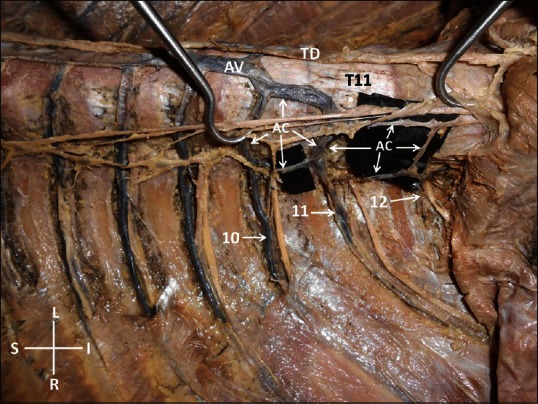
Photograph of dissection of lower part of the posterior thoracic wall as viewed from the right side. AV = Azygos vein, TD = Thoracic duct, T11 = Eleventh thoracic vertebra, AC = Anastomotic venous circle, 10 = Right tenth posterior intercostal vein, 11 = Right eleventh posterior intercostal vein, 12 = Right subcostal vein, S = Superior, I = Inferior, L = Left, R = Right
Figure 3.

Photograph of dissection of the posterior thoracic wall as viewed from the left side. AV = Azygos vein, T11 = Eleventh thoracic vertebra, T10 = Tenth thoracic vertebra, T6 = Sixth thoracic vertebra, HAV = Hemiazygos vein, AHAV = Accessory hemiazygos vein, S = Superior, I = Inferior, L = Left, R = Right
Figure 4.

A schematic drawing of the azygos vein variation. AV = Azygos vein, HAV = Hemiazygos vein, AHAV = Accessory hemiazygos vein, SICV = Superior intercostal vein, LA = Ascending lumbar vein, AC = Anastomotic venous circle, 1-11 = Posterior intercostal veins, 12 = Subcostal veins
DISCUSSION
Azygos system of veins is subjected to wide range of variations. Knowledge of its variations is especially important for cardiothoracic surgeons and radiologists. Arslan et al. have reported a case of total absence of azygos vein.[2] Anomalies of azygos vein are often associated with anomalies of superior and inferior venae cavae. A case of absence of azygos vein with the presence of a left superior vena cava has also been diagnosed through computed tomography.[3] Absent left and right superior vena cava and azygos continuation of inferior vena cava have also been reported.[4] Abnormal location of the azygos vein might cause difficulties to surgeons. An anomalous azygos vein covering the sympathetic chain has been reported. Such a vein might pose a significant risk in endoscopic thoracic sympathectomy.[5] Azygos vein usually lies on the right side of the vertebral column, but it may be shifted to the midline or the left of midline as age advances.[6] Hence, the midline position of the azygos vein in the current case was possibly due to old age.
Hemiazygos and accessory hemiazygos veins can also be absent of show variation in their formation, course, termination or tributaries. A ladder pattern of hemiazygos veins has been reported.[7] Transverse segment of hemiazygos vein crossing in front of the vertebral column is called “interazygos vein.” An anomalous course of interazygos vein, between the esophagus and descending thoracic aorta has been reported.[8] The absence of hemiazygos vein and presence of preaortic interazygos vein has also been reported.[9] Our literature survey also revealed a case where the hemiazygos vein was absent with the presence of right and left azygos veins.[10] Another very rare variation associated with the azygos system of veins is opening of all pulmonary veins into the azygos vein. One such case has been diagnosed by echocardiographic technique.[11]
Current case is unique because of the following reasons; (1) to the best of our knowledge, there is no report on the venous circle formed in front of the vertebral column by the bifurcated hemiazygos vein. (2) There is no report on anastomotic venous circles on the right side of lower thoracic vertebrae. (3) Usually, the hemiazygos vein descends down on the left side of the vertebral column from 4th to 8th thoracic vertebra. However in the current case, it ascended to the 6th thoracic vertebra from the level of the 8th thoracic vertebra. Apart from knowing the normal anatomy of azygos system of veins, its possible variations such as the one being reported here, have to be kept in mind during clinical procedures such as cardiothoracic surgery, echocardiography, and other radiological techniques. The variations of azygos system may lead to iatrogenic bleeding or faulty radiological diagnosis. Idiopathic azygos aneurysms might mimic mediastinal masses and have to be confirmed radiographically.[12] Misplacement of venous catheters can happen when there are variations of azygos veins, especially when the terminal parts of the veins are dilated. The misplacement might lead to severe bleeding.[13] The knowledge of variation of the azygos system is also important during the surgery to repair the esophageal atresia.[14] It is possible to identify the gross anomalies like azygos continuation of inferior vena cava[15] and absence of superior vena cava.[16] However the less frequent, uncommon variations might lead to confusions during echocardiography or any other procedures.
CONCLUSIONS
The knowledge of unique variations of azygos system, being reported here is of importance to radiologists performing venous cannulations, cardiac echography, and other vascular radiology procedures. It is also important to cardiothoracic surgeons to minimize the chances of iatrogenic hemorrhage.
Financial support and sponsorship
Nil.
Conflicts of interest
There are no conflicts of interest.
REFERENCES
- 1.Alves EC, Porciúncula Junior RW, Monte Bispo RF, de Sousa-Rodrigues CF, da Rocha AC. Formation of the azygos vein. Int J Morphol. 2011;29:140–3. [Google Scholar]
- 2.Arslan G, Cubuk M, Ozkaynak C, Sindel T, Lüleci E. Absence of the azygos vein. Clin Imaging. 2000;24:157–8. doi: 10.1016/s0899-7071(00)00191-1. [DOI] [PubMed] [Google Scholar]
- 3.Kullnig P, Melzer G, Hausegger K, Einspieler R. Computed tomographic diagnosis of left superior vena cava and absence of the azygos vein: Case report. Cardiovasc Intervent Radiol. 1990;13:47–9. doi: 10.1007/BF02576939. [DOI] [PubMed] [Google Scholar]
- 4.Quraishi MB, Mufti O, Wase A. Absent left and right superior vena cava and azygos continuation of inferior vena cava: A rare anomaly of systemic venous return. J Invasive Cardiol. 2010;22:E159–61. [PubMed] [Google Scholar]
- 5.Sieunarine K, May J, White GH, Harris JP. Anomalous azygos vein: A potential danger during endoscopic thoracic sympathectomy. Aust N Z J Surg. 1997;67:578–9. doi: 10.1111/j.1445-2197.1997.tb02046.x. [DOI] [PubMed] [Google Scholar]
- 6.Saito A, Murakami M, Tomioka K, Ezure H, Moriyama H, Mori R, et al. The impact of aging on the course of the azygos vein. Okajimas Folia Anat Jpn. 2015;92:7–10. doi: 10.2535/ofaj.92.7. [DOI] [PubMed] [Google Scholar]
- 7.Kumar N, Nayak SB, Shetty S, Somayaji SN. Azygos ladder and looped thoracic duct - A case report. Int J Anat Variat. 2011;4:80–2. [Google Scholar]
- 8.Celik HH, Sargon MF, Aldur MM, Cumhur M. An anomalous course of the interazygos vein. Surg Radiol Anat. 1996;18:61–2. doi: 10.1007/BF03207766. [DOI] [PubMed] [Google Scholar]
- 9.Ozdemir B, Aldur MM, Celik HH. Multiple variations in the azygos venous system: A preaortic interazygos vein and the absence of hemiazygos vein. Surg Radiol Anat. 2002;24:68–70. doi: 10.1007/s00276-002-0008-7. [DOI] [PubMed] [Google Scholar]
- 10.Keskin S, Keskin Z, Sekmenli N. The independent right and left azygos veins with hemiazygos absence: A rare case presentation. Case Rep Vasc Med 2013. 2013 doi: 10.1155/2013/282416. 282416. [DOI] [PMC free article] [PubMed] [Google Scholar]
- 11.Tomar M, Radhakrishnan S, Shrivastava S, Iyer KS. Total anomalous pulmonary venous connection to azygos vein: Echocardiographic recognition of a rare variant. Indian Heart J. 2006;58:54–6. [PubMed] [Google Scholar]
- 12.Ichiki Y, Hamatsu T, Suehiro T, Koike M, Tanaka F, Sugimachi K. An idiopathic azygos vein aneurysm mimicking a mediastinal mass. Ann Thorac Surg. 2014;98:338–40. doi: 10.1016/j.athoracsur.2013.09.024. [DOI] [PubMed] [Google Scholar]
- 13.Currarino G. Migration of jugular or subclavian venous catheters into inferior tributaries of the brachiocephalic veins or into the azygos vein, with possible complications. Pediatr Radiol. 1996;26:439–49. doi: 10.1007/BF01377198. [DOI] [PubMed] [Google Scholar]
- 14.Fathi M, Joudi M, Morteza A. Evaluating necessity of azygos vein ligation in primary repair of esophageal atresia. Indian J Surg. 2015;77(Suppl 2):543–5. doi: 10.1007/s12262-013-0917-1. [DOI] [PMC free article] [PubMed] [Google Scholar]
- 15.Pantin EJ, Naftalovich R, Denny J. Echocardiographic identification of an interrupted inferior vena cava with dilated azygos vein during coronary artery bypass graft surgery. Anesth Analg. 2016;122:358–60. doi: 10.1213/ANE.0000000000001075. [DOI] [PubMed] [Google Scholar]
- 16.Gibelli G, Biasi S. Persistent left superior vena cava and absent right superior vena cava: Not only an anatomic variant. J Cardiovasc Echogr. 2013;23:42–4. doi: 10.4103/2211-4122.117985. [DOI] [PMC free article] [PubMed] [Google Scholar]