Abstract
Introduction
We describe the youngest case of enhanced S-cone syndrome (ESCS) associated with choroidal neovascularization (CNV) successfully treated with intravitreal ranibizumab injections.
Case Report
A 5-year-old boy presented with round-shaped fibrotic subretinal lesions in both eyes with surrounding subretinal fluid and progressive visual deterioration in the right eye. Fine foci of increased autofluorescence were observed along the arcades in both eyes. Fluorescein angiography revealed the presence of CNV in his right eye, and treatment with ranibizumab was initiated, with significant improvement in vision. Subsequent electroretinogram examination and genetic studies of the patient and his two younger siblings confirmed the diagnosis of ESCS.
Conclusion
CNV has been reported to occur in different inherited retinal degenerations, including ESCS. Our experience confirms that treatment with ranibizumab in patients with CNV-complicated ESCS can be potentially vision-saving.
Keywords: Enhanced S-cone syndrome, Choroidal neovascularization, Ranibizumab, Anti-VEGF, NR2E3, Electroretinogram
Introduction
First described in 1990, enhanced S-cone syndrome (ESCS) is a rare, autosomal recessive retinal dystrophy associated with pathognomonic electrophysiological findings and NR2E3 gene mutations. This gene encodes a transcription factor that regulates the proper differentiation of photoreceptor cells. In fact, mutations in NR2E3 leading to loss of function of the transcription factor are thought to be responsible for altering photoreceptor precursor cells fate from rod to S cone. This leads to a disorganized retina with lack of structurally recognizable rods and approximately twice the number of cones, most of which are S cones (short wavelength), responsible for the detection of blue light. To date, at least 49 mutations in NR2E3 have been described with resultant phenotypes of ESCS, Goldmann-Favre syndrome, and autosomal dominant and autosomal recessive retinitis pigmentosa [1, 2, 3]. Characteristic features of electroretinograms in ESCS include undetectable rod-specific response, relatively increased responses to the shorter wavelengths of light, similar waveform response to a standard single flash under photopic and scotopic conditions, and profoundly delayed 30-Hz flicker ERG with lower amplitude than the single-flash light-adapted 3.0 ERG a-wave [4].
Choroidal neovascularization (CNV) has been reported to occur in a number of inherited retinal degenerations, including ESCS [5, 6, 7]. The prevalence of this latter association has not been estimated yet. Although uncommon, neovascularization can occur early in the course of the disease accelerating the loss of visual function. Published clinical reports of CNV-complicated ESCS showed a relatively favorable visual prognosis with common tendency to a spontaneous involution of the low-active CNV to a small, circumscribed fibrotic subretinal scar. Being a rare clinical condition, there is still little information on the natural history and management of these lesions; however, few cases reported in the literature have shown that timely anti-VEGF treatment can prevent visual deterioration and leads to a better visual outcome in these patients [5, 8, 9, 10]. Here, we describe the efficacy of ranibizumab in the treatment of a case of CNV associated with ESCS.
Clinical Case
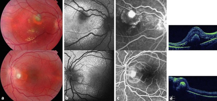
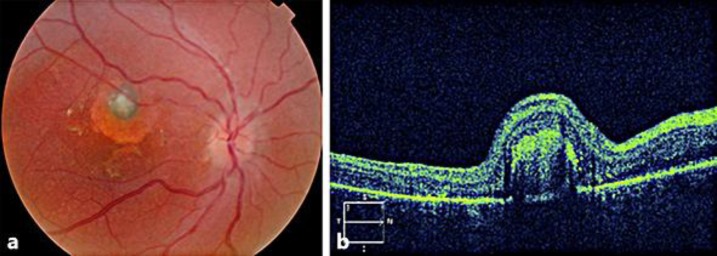
We present a case of a 5-year-old boy who came to our service in May 2011 due to esotropia and vision loss in the right eye. He presented with a best corrected visual acuity (BCVA) of 20/80 in the right eye and 20/32 in the left eye. He had a cycloplegic hyperopic refractive error of 5.75 diopters in the right eye and 7.00 diopters in the left eye. Dilated fundus examination revealed a round-shaped pigmented subretinal lesion along the superior arcade with serous retinal epithelium detachment and retinal exudates in his right eye and a smaller round-shaped pigmented subretinal peripapillary lesion in his left eye. Pale white dots along the arcades were also observed in both eyes. Fluorescein angiography revealed the presence of CNV in the right eye and retinochoroidal anastomosis in the left eye. Optical coherence tomography showed an hyperreflective lesion with subretinal fluid and intraretinal cystic changes in the right eye and a peripapillary hyperreflective lesion in the left eye without exudation. Fundus autofluorescence showed several fine foci of increased autofluorescence along the arcades in both eyes (Fig. 1). In June 2011, the patient received in his right eye two intravitreal injections of ranibizumab over a 2-month period, and BCVA improved to 20/32 with complete disappearance of subretinal fluid, which remained stable over the years (Fig. 2).
Fig. 1.
Color fundus photography (a), fundus autofluorescence imaging (b), fluorescein angiography (c), and OCT images (d) of the patient in May 2011 (right eye in the upper line, left eye in the lower line).
Fig. 2.
Color fundus photography (a) and OCT images (b) of the patient in the right eye after two ranibizumab injections.
In the meantime, we decided to investigate his little sister and brother who were 3 and 1 years old, respectively. Both of them presented with hyperopia and age-appropriate visual acuity. Barely visible white dots were present along the arcades at fundus examination in both eyes of the two siblings, and autofluorescence imaging revealed the presence of multiple small hyperfluorescent foci along the arcades.
Electroretinography of our patient displayed normal rod-specific ERG and normal oscillatory potentials. Dark-adapted maximal response and light-adapted cone response were normal and had a similar waveform. The 30-Hz flicker was delayed with very low amplitude. Full-field ERG also showed a greatly magnified blue-cone signal.
The standard full-field ERGs of his two siblings were very similar to each other and showed characteristic features. The rod-specific ERG was undetectable. The dark-adapted maximal response was reduced and had a similar waveform to the light-adapted cone response, which had a normal amplitude. The 30-Hz flicker was severely reduced and delayed. We also observed an increased sensitivity to short wavelength light stimulus.
The parents of the three children were also investigated. They both had a 20/20 visual acuity with normal fundus appearance and normal autofluorescence imaging.
Genetic studies identified a homozygous splice site mutation, c.119–2A>C, in the NR2E3 gene in all 3 affected siblings, confirming the diagnosis of ESCS. The parents and the paternal grandmother carried the variant heterozygously.
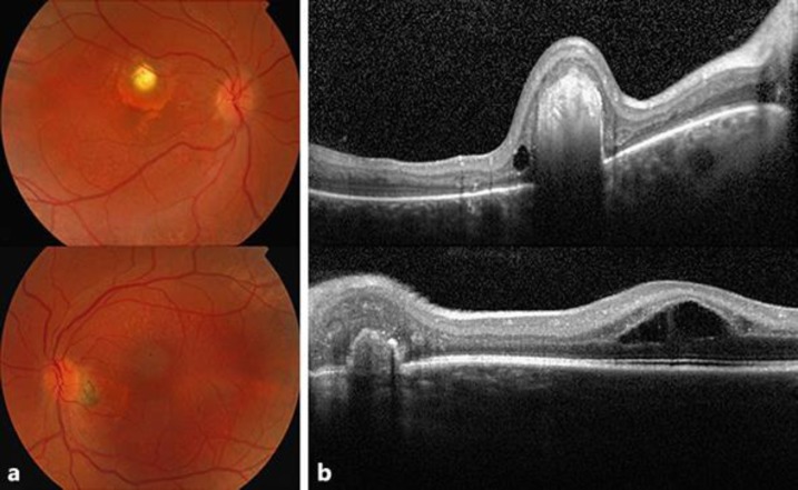
After a 7-year follow-up, the patient's BCVA is 20/20 in both eyes. In the right eye, the fibrotic lesion is stationary with adjacent intraretinal cysts and no signs of exudation, while in the left eye, a schisis-like maculopathy with intraretinal cystic changes has appeared (Fig. 3). His siblings also have a 20/20 BCVA with stationary fundus appearance in both eyes.
Fig. 3.
Color fundus photography (a) and OCT images (b) of the patient in 2018 (right eye in the upper line, left eye in the lower line).
Discussion
CNV occurs in several retinal dystrophies. The association between CNV and ESCS is already well known [5, 6, 7, 8, 9, 10, 11], and very few of the previously reported cases have been treated with anti-VEGF [5, 8, 9]. To our knowledge, this is the youngest case of ESCS-associated CNV described in literature.
The pathological pathway that promotes the evolution of CNV in ESCS patients has not been completely understood yet. There is evidence that retinal pigment epithelium (RPE) changes, photoreceptor dysfunction, and choriocapillaris damage, which are constantly present in retinal dystrophies, may create weakness zones through which choroidal vessels can grow, leading to neovascularization. Furthermore, dysfunctional RPE may become hypoxic and produce VEGF that promotes angiogenesis [5, 10]. Other authors hypothesized that the increased number of cones in ESCS patients, which are known to have a higher oxygen demand than rods, leads to an ischemic condition with upregulation of VEGF production and subsequent vascular abnormalities [8, 12].
CNV can trigger acute changes in central vision that are atypical in patients with hereditary retinal dystrophies, and CNV-related visual loss can be the first clinical sign of ESCS. Therefore, any rapid decrease in visual acuity in these patients warrants urgent investigation for potential CNV [5, 10]. Young children rarely complain of vision loss, but indirect warning signs can be identified. Our patient did not suffer from night blindness or any other visual symptom except for acute onset esotropia in the affected eye secondary to visual loss due to subretinal fluid.
Although CNV associated with inherited retinal dystrophies seems to have a relatively favorable visual prognosis, if left untreated, it can lead to persistent visual loss due to the development of fibrotic subretinal scars [10, 13]. Furthermore, in young children, it can affect the development of the visual system and lead to deprivation amblyopia. Despite the high possibility of spontaneous regression of CNV in children, the mean visual acuity in eyes treated with anti-VEGF agents seems to be higher than in eyes with spontaneously regressed CNV. Therefore, early diagnosis and treatment of this complication is of utmost importance [14]. The fibrotic subretinal peripapillary lesion associated with retinochoroidal anastomosis in the fellow eye of our patient might suggest that an early neovascular membrane had occurred and involuted. These findings could confirm that CNV associated with ESCS presents early in the course of the disease and regresses spontaneously, leading to a circumscribed subretinal scar [7, 9, 10].
Back in 2011, the efficacy of both bevacizumab and ranibizumab in treating pediatric CNV was reported. Moreover, no adverse effects were observed in clinical practice with pediatric use of both anti-VEGF agents [15]. Rishi et al. [14] reported that since ranibizumab has a shorter serum half-life than bevacizumab, it may lower systemic exposure in children. Nowadays, considering the increasing use of anti-VEGF agents in pediatric patients, long-term safety results need to be further evaluated. In our patient, treatment with ranibizumab led to a complete resolution of subretinal fluid and a great improvement in visual acuity, which remained stationary after a 7-year follow-up. We did not observe any short-term ocular or systemic adverse effects of treatment with intravitreal ranibizumab.
It is important to note that intraretinal cysts in ESCS are mainly attributable to chronic degeneration and disorganization of the retina associated with loss of photoreceptos and RPE pump dysfunction rather than neovascularization. Cystic changes are considered a pathognomonic finding of the disease and should not be confused with a sign of neovascularization; fluorescein angiography can be a useful tool to distinguish between these two conditions [5, 11].
Fundus hyperautofluorescent spots, typically observed in ESCS, seem to be generated by chromophores within the cytoplasm of macrophages, recruited into the retina to support the role of RPE in removing damaged photoreceptors and outer segment debris [7].
In conclusion, considering the previously reported clinical cases of anti-VEGF treatment for CNV in ESCS [5, 6, 8, 9], we described several novel features. First, in young children, the disease can present with atypical or indirect symptoms: our patient did not complain of night blindness, but he suffered from acute-onset esotropia as a consequence of vision impairment. Second, the number of ranibizumab injections needed to obtain a complete regression of the CNV was the lowest described in the literature, and our patient was the youngest child to be treated for this condition. Furthermore, to our knowledge, this is the longest follow-up of a patient successfully treated with ranibizumab for this disease. Our experience confirms that anti-VEGF therapy in ESCS patients can be potentially vision-saving and can prevent chronic visual deterioration secondary to CNV.
Statement of Ethics
The authors have no ethical conflicts to disclose. Informed consent was obtained from our patients, and the study has been approved by the institute's committee on human research.
Disclosure Statement
Federica Bertoli, MD, Silvia Pignatto, MD, and Francesca Rizzetto, MD, have no financial disclosure to report. Paolo Lanzetta, MD, is consultant for Bayer, Centervue, Novartis, and Topcon.
References
- 1.Hull S, Arno G, Sergouniotis PI, Tiffin P, Borman AD, Chandra A, et al. Clinical and molecular characterization of enhanced S-cone syndrome in children. JAMA Ophthalmol. 2014 Nov;132((11)):1341–9. doi: 10.1001/jamaophthalmol.2014.2343. [DOI] [PubMed] [Google Scholar]
- 2.Milam AH, Rose L, Cideciyan AV, Barakat MR, Tang WX, Gupta N, et al. The nuclear receptor NR2E3 plays a role in human retinal photoreceptor differentiation and degeneration. Proc Natl Acad Sci USA. 2002 Jan;99((1)):473–8. doi: 10.1073/pnas.022533099. [DOI] [PMC free article] [PubMed] [Google Scholar]
- 3.Schorderet DF, Escher P. NR2E3 mutations in enhanced S-cone sensitivity syndrome (ESCS), Goldmann-Favre syndrome (GFS), clumped pigmentary retinal degeneration (CPRD), and retinitis pigmentosa (RP) Hum Mutat. 2009 Nov;30((11)):1475–85. doi: 10.1002/humu.21096. [DOI] [PubMed] [Google Scholar]
- 4.Sustar M, Perovšek D, Cima I, Stirn-Kranjc B, Hawlina M, Brecelj J. Electroretinography and optical coherence tomography reveal abnormal post-photoreceptoral activity and altered retinal lamination in patients with enhanced S-cone syndrome. Doc Ophthalmol. 2015 Jun;130((3)):165–77. doi: 10.1007/s10633-015-9487-9. [DOI] [PubMed] [Google Scholar]
- 5.Broadhead GK, Grigg JR, McCluskey P, Korsakova M, Chang AA. Bevacizumab for choroidal neovascularisation in enhanced S-cone syndrome. Doc Ophthalmol. 2016 Oct;133((2)):139–43. doi: 10.1007/s10633-016-9555-9. [DOI] [PubMed] [Google Scholar]
- 6.Nakamura M, Hotta Y, Piao CH, Kondo M, Terasaki H, Miyake Y. Enhanced S-cone syndrome with subfoveal neovascularization. Am J Ophthalmol. 2002 Apr;133((4)):575–7. doi: 10.1016/s0002-9394(01)01428-3. [DOI] [PubMed] [Google Scholar]
- 7.Sambricio J, Tejada-Palacios P, Barceló-Mendiguchía A. Choroidal neovascularization, outer retinal tubulation and fundus autofluorescence findings in a patient with enhanced S-cone syndrome. Clin Exp Ophthalmol. 2016 Jan-Feb;44((1)):69–71. doi: 10.1111/ceo.12578. [DOI] [PubMed] [Google Scholar]
- 8.Badami K, Sultana A, Sinha B, Ijeri R, Jayadev C, Tsang SH. Diagnostic and Therapeutic Challenges. Retina. 2015 Sep;35((9)):1911–8. doi: 10.1097/IAE.0000000000000407. [DOI] [PubMed] [Google Scholar]
- 9.Zerbib J, Blanco Garavito R, Gerber S, Oubraham H, Sikorav A, Audo I, et al. Retinochoroidal anastomosis associated with enhanced S-cone syndrome. Retin Cases Brief Rep. 2017 May;:1. doi: 10.1097/ICB.0000000000000594. [DOI] [PubMed] [Google Scholar]
- 10.Marano F, Deutman AF, Leys A, Aandekerk AL. Hereditary retinal dystrophies and choroidal neovascularization. Graefes Arch Clin Exp Ophthalmol. 2000 Sep;238((9)):760–4. doi: 10.1007/s004170000186. [DOI] [PubMed] [Google Scholar]
- 11.Lam BL, Goldberg JL, Hartley KL, Stone EM, Liu M. Atypical mild enhanced S-cone syndrome with novel compound heterozygosity of the NR2E3 gene. Am J Ophthalmol. 2007 Jul;144((1)):157–9. doi: 10.1016/j.ajo.2007.03.012. [DOI] [PubMed] [Google Scholar]
- 12.Wong-Riley MT. Energy metabolism of the visual system. Eye Brain. 2010;2:99–116. doi: 10.2147/EB.S9078. [DOI] [PMC free article] [PubMed] [Google Scholar]
- 13.Patel RC, Gao SS, Zhang M, Alabduljalil T, Al-Qahtani A, Weleber RG, et al. at al. Optical coherence tomography angiography of choroidal neovasularization in four inherited retinal distrophies. Retina. 2016 Dec;36((12)):2339–47. doi: 10.1097/IAE.0000000000001159. [DOI] [PMC free article] [PubMed] [Google Scholar]
- 14.Rishi P, Gupta A, Rishi E, Shah BJ. Choroidal neovascularization in 36 eyes of children and adolescents. Eye (Lond) 2013 Oct;27((10)):1158–68. doi: 10.1038/eye.2013.155. [DOI] [PMC free article] [PubMed] [Google Scholar]
- 15.Kohly RP, Muni RH, Kertes PJ, Lam WC. Management of pediatric choroidal neovascular membranes with intravitreal anti-VEGF agents: a retrospective consecutive case series. Can J Ophthalmol. 2011 Feb;46((1)):46–50. doi: 10.3129/i10-123. [DOI] [PubMed] [Google Scholar]


