

Abstract
Ulnar nerve injury as a result of closed forearm fractures in children is a rare but disastrous complication, affecting significantly the function of the upper extremity. The management of these injuries is still controversial in the literature. This is a review of small case series and case reports, in which an algorithm of treatment is proposed. A brief description of a relative case, which was treated successfully in our department, is presented as well. This case motivated the authors to perform this study.
Keywords: Forearm fractures, ulnar nerve injuries, paediatric orthopaedics
Introduction
Paediatric forearm fractures are rather common injuries. Concurrent ulnar nerve injury, however, is a rare complication. A small number of cases has been described and published in the literature. There is not yet a clear consensus regarding the correct timing of surgical exploration.
According to Chung and Spilson1 in 1998, hand/forearm fractures accounted for 1.5% of all emergency department cases. The largest proportion of the fractures consisted of radius and/or ulna fractures (44%). The majority of the fractures (26%) occurred in the 5 to 14 age group and were caused mostly (47%) by accidental falls. Rasulić et al2 reported that 15.38% of the patients with ulnar nerve injury in this series had also suffered a forearm fracture.
Ulnar nerve injuries may be rare, but they can be quite severe. As hand plays a very important role in everyday life, loss of its function is psychologically detrimental for the patients, especially for the paediatric population. This review tends to propose a guide of managing these injuries. A brief description of a relative case, which was treated successfully in our department, is presented as well.
Mechanism of Injury
According to the literature, the following mechanisms of ulnar nerve injury post forearm fractures are described: (1) nerve contusion, (2) nerve laceration, (3) nerve entrapment between the edges of the fracture, and (4) nerve irritation by scar tissue or callus.3 Most common cause of injury is contusion and the nerve’s function resolves spontaneously within a few weeks post fracture.4
Materials and Methods
A search was performed in PubMed for studies including ulnar nerve injuries in the presence of forearm fracture. Two small case series and 5 case reports met the criteria of our search and they were included in the study. Table 1 demonstrates all the cases published in the literature so far, regarding these injuries, the way of management, timing of surgery, and period of time until recovery.
Table 1.
Literature Review.
| Author | Patient age | Initial treatment | Time until neurolysis (weeks) | Pathogenesis | Time until nerve recovery (weeks) |
|---|---|---|---|---|---|
| Prosser and Hooper5 | 13 | Internal fixation of the radius | 10 | Bony entrapment | 6 |
| Stahl et al3 | 10 | Manual reduction | 10 | Scar entrapment | 4 |
| 15 | Manual reduction | 16 | Scar entrapment | 4 | |
| 9 | Open reduction and nerve repair | N/A | Partial laceration | 24 | |
| Hirasawa et al6 | 13 | Manual reduction | 13 | Bony entrapment | 16 |
| Suganuma et al7 | 12 | Manual reduction and intramedullary pinning | 9 | Scar entrapment | 12 |
| Amit et al8 | Manual reduction | No nerve exploration was performed | Neuropraxia | 24 | |
| Schwartsmann et al9 | 17 | Manual reduction Neurolysis plus repair |
N/A | Partial laceration | N/A |
| Neiman et al10 | 12 | Manual reduction | No nerve exploration was performed | Unknown | 20 |
| 11 | Manual reduction | No nerve exploration was performed | Unknown | 20 |
Abbreviation: N/A, not applicable.
Results
Only small case series or case reports have been published in the literature concerning the management of ulnar nerve injuries post forearm fractures. There is significant controversy so far regarding the correct timing of exploration, as well as whether a surgical intervention is needed.
Prosser and Hooper5 reported a case of ulnar nerve entrapment after a forearm fracture. The patient was a 13-year-old schoolboy, who suffered a fracture of the distal radius and ulna. Neurological examination initially was normal. The patient underwent open reduction and internal fixation of the radial fracture. The fracture of the ulna was in an acceptable position and a plaster was applied. Exploration of the ulnar nerve was not performed. Three days post operation, complete ulnar nerve palsy was noticed. After 10 weeks of no recovery, the ulnar nerve was explored, and it was found entrapped at the site of the fracture. The nerve was released and 3 weeks later, there was evidence of ulnar nerve recovery. Six months later, slight weakness of the first dorsal interosseous muscle was the only clinical finding of deficit.
Stahl et al3 reported 3 cases of forearm fractures with a concomitant ulnar nerve injury. In the first case, a 10-year-old boy sustained a closed forearm fracture and on examination ulnar nerve palsy was detected, which was considered to be a neuropraxia. X-ray revealed a midshaft fracture of the radius and ulna with anterior displacement and angulation. The fracture was reduced under general anaesthesia, and a cast was applied. After 10 weeks, no nerve recovery had occurred. Examination revealed motor and sensory deficits and Tinel sign was positive at the level of the fracture. The patient underwent surgery, where the ulnar nerve was explored and found to be trapped in a hypertrophic scar. Neurolysis was performed under the microscope. Four weeks after surgery, signs of ulnar nerve function improvement appeared. One year later, there was full recovery of the nerve, apart from a slight weakness of the ulnar innervated intrinsic muscles.
In the second case, a 15-year-old boy sustained a closed midshaft fracture of the right ulna and radius, with significant angulation and slight displacement of the radius and ulna. Closed reduction was performed and a cast was applied. Twenty-four hours later, a diagnosis of ulnar nerve palsy was made, which did not improve in the next 4 months. The patient complained of numbness and weakness, which had been present since the accident. There was loss of function of the ulnar intrinsic muscles and also complete absence of sensation in the ulnar nerve sensory area. Surgery was performed, and the nerve was found to be trapped in scar tissue at the healed fracture site. It was released and minimal internal neurolysis was performed under the microscope. Improvement was noted 2 weeks later with significant improvement of the sensory loss. A month after surgery, full nerve function was recovered.
In the third case, a 9-year-old boy suffered a fracture of the left radius and ulna. X-ray showed midshaft anterior displacement and angulation of the ulna along with a distal ulnar spike. The patient complained of numbness of the ulnar digits with muscle weakness. After the failure of achieving anatomical alignment with closed reduction, open reduction was performed. During the operation, the ulnar nerve was found to be entrapped in the spike and partially lacerated. It was released, and the fractures were reduced and fixed by intramedullary nails. The nerve was repaired. Six months later, full clinical return of ulnar nerve function had occurred.
Hirasawa et al6 in 2004 reported a case of ulnar nerve entrapment. The patient was a 13-year-old girl with numbness in the ring and little fingers, and limitation in forearm rotation, 2 months after a displaced diaphyseal fracture of the left radius and ulna. Hypoesthesia was noticed in the little finger and the ulnar half of the ring finger, and Tinel sign was positive at the ulnar fracture site. Three months after the fracture, atrophy of the interosseous muscles and claw hand deformity of the ring and little fingers developed. Neurolysis of the ulnar nerve was performed. The nerve was found to be trapped between the fragments of the ulna. Corrective osteotomy of the radius was also performed. Sensation and motor function recovered 4 months later.
Suganuma et al7 published a case in 2013 where a 12-year-old girl underwent a second operation 9 weeks after the initial one, because of gradually worsening ulnar nerve palsy. She sustained a midshaft forearm fracture, which was reduced and fixed with Kirschner wires as a primary operation. Nine weeks later, she underwent a second operation because of ulnar nerve palsy. The nerve was released, as it was adhered to the surrounding scar tissue. Six months later, she had no motor dysfunction.
Amit et al8 in 2013 reported a case of a missed ulnar nerve injury from a closed forearm fracture in a child. The patient presented with a history of a fall on the floor while playing. There was swelling in distal forearm and inability to move the left hand. There were some abrasions on left hand and forearm. The radiographs revealed fractures of both bones of left forearm with significant displacement of distal fragments dorsally. The fracture was reduced under general anaesthesia. After 3 weeks, there were complaints of numbness in little and ring fingers of the child. Ulnar clawing was clearly evident. The rest of neurological examination was normal. Nerve conduction velocity (NCV) examination revealed ulnar nerve injury at the site of fracture. Magnetic resonance imaging (MRI) scan ruled out entrapment or laceration of the ulnar nerve and delineated the normal course of the nerve. At 3-month follow-up, the numbness of the fingers was long gone, as well as marked subsidence of the clawing was noticed. The limb was nearly fully functional with fracture union of both bones.
Schwartsmann et al9 reported a case of a 17-year-old male who suffered a forearm fracture after a skateboard fall. The patient complained of ‘tingling’ of the fifth finger. After a satisfactory closed reduction, the ‘tingling’ worsened. Exploration of the ulnar nerve was performed, and it was trapped at the fracture site. The nerve was released and the laceration was sutured.
In most cases, the ulnar nerve palsy required operation so that satisfactory recovery could be achieved. However, Neiman et al10 reported 2 cases where the ulnar nerve palsy was resolved without operation after 20 weeks. The nerve injury was identified at the time of fracture by neurological examination. Thus, the possibility of a post reduction nerve injury was eliminated. Neiman suggested that the ulnar nerve palsy is probably a result of nerve contusion and it should resolve without operation.
Discussion
Forearm fractures are common in paediatric population. Concomitant nerve injuries, however, are rare, but they are detrimental to the functional outcome of those fractures.8 Ulnar nerve function particularly can be affected by forearm fractures, especially if they are located in the middle and distal third of the diaphysis, probably due to the close proximity between the nerve and the ulna.7 Ulnar nerve injuries are frequently missed because most of the times, it is difficult to evaluate the neurological status of the upper limb due to severe pain.8 It is crucial, however, to determine that before any reduction manoeuvre is attempted, because any manipulation of the fracture can potentially harm the ulnar nerve.
There are a few questions which need to be answered regarding ulnar nerve injuries post forearm fractures, such as: what kind of nerve injury is it in each case? Is it neurapraxia, neurotmesis, or axonotmesis? Is there any role of further imaging such as MRI scan? Should the Orthopaedic Surgeon explore the nerve and if this is the case, what is the correct timing to proceed? According to our knowledge, there is no review in the literature regarding those injuries. We gathered the most important relative articles published so far, and this is an effort to answer the previous questions and recommend a way to manage these injuries properly. All the collected data are from small case series of patients and case reports.
We were motivated by a case, which we managed, of a 13-year-old patient, who sustained a left distal shaft forearm closed fracture, severely displaced, after a fall from a small height (Figure 1). The neurovascular status of the upper limb was thoroughly examined, prior to any manipulation and an ulnar nerve injury was detected, as there was ring and small finger clawing plus pins and needles around the same area. As the fracture was severely displaced, a gentle closed manipulation was performed, without any worsening of the neurological signs and the upper extremity was splinted. The patient was operated the next day with open reduction and internal fixation of radial and ulnar fractures (Figure 2). No attempt of exploring the ulnar nerve was performed. Ulnar nerve palsy persisted after operation, and 4 weeks later, hypothenar atrophy as well as ring and small finger numbness and clawing was noticed. Six weeks post operation, the fracture was healed and signs of ulnar nerve recovery were detected, as Tinel sign was positive plus some intrinsic muscles’ function was identified. Full ulnar nerve recovery was noticed 17 weeks post operation. This case is an example of full ulnar nerve recovery, despite initial worsening during the first 4 weeks. Surgeon’s decision was to wait and see, as the fracture was anatomically reduced, the nerve palsy was also detected before any reduction manoeuvre, and some nerve function improvement was evident 6 weeks post operation.
Figure 1.
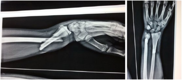
Pre-operation left forearm fracture.
Figure 2.
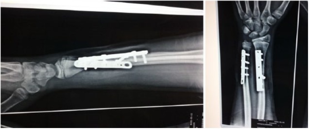
Post-operation image.
Peripheral nerve injuries are classified based on Seddon classification as neurapraxia, axonotmesis, and neurotmesis, with the last type being the worse in terms of prognosis. Sunderland added 3 more subtypes in the axonotmesis category: type I, axonotmesis with intact connective tissue; type II, axonotmesis with damage to the endoneurium; and type III, axonotmesis which includes damage to the endoneurium and perineurium. The muscle, which is the target organ of the reinnervation, should maintain its ability to accept neurological stimulation, in order for the nerve recovery to achieve a good functional outcome. It is well known that the structural architecture of the muscle and the end-plate integrity can be maintained for up to 1 year post denervation. After 2 years, irreversible muscle fibrosis has occurred along with muscle degeneration, leading to a permanent loss of functional muscle tissue.11 Thus, we question the suggestion of some authors to explore the nerve at most 10 weeks post injury, supporting that after that time, recovery is unlikely to occur.7 Ulnar nerve injuries usually concern the midshaft or the distal shaft of the forearm. Considering the fact that reinnervation mechanism proceeds with a velocity of 1 to 2 mm/d and the end plate remains intact until at least 1 year post injury,11 it is reasonable that the physician could wait for up to 20 weeks post injury for the nerve to recover spontaneously, provided that there is no progressive worsening. In addition, there are cases published in the literature, which describe spontaneous recovery of the ulnar nerve, even 20 weeks post fracture.10
Based on the few cases published in the literature and the pathophysiology of the nerve injury, current authors propose an algorithm of management (Table 2). First of all, it is crucial to determine the neurovascular status of the limb before any attempt for reduction, although it is difficult to evaluate that, due to severe pain.3,10 A peripheral nerve should be explored in case the nerve’s damage occurs as a result of closed manipulation.
Table 2.
Forearm fractures and ulnar nerve injuries’ algorithm of management.

|
Classic literature suggests that forearm fractures, which are angulated more than 15° if younger than 10 years old, or more than 10° if older than 10 years of age and bayonet apposition if older than 10 years old, are indications for surgery.12 Current authors suggest that, if ulnar or other peripheral nerve injury occurs as a result of forearm fracture, no displacement should be accepted and the goal of treatment should be anatomical reduction, to avoid ulnar nerve entrapment in the fracture site or late callus tethering, as bone healing proceeds. Anatomical reduction could achieve primary bone healing, preventing callus formation, which could potentially irritate the nerve. Some authors recommend also an MRI scan as a further imaging, to detect nerve continuity disruption.8 NCV studies can demonstrate evidence of nerve damage approximately 2 to 3 weeks post injury.4 If there is pre reduction ulnar nerve damage and the fracture is anatomically reduced either conservatively or surgically, then wait and see for up to 20 weeks, provided that there is no progressive worsening. Maybe wait less for fractures located in the proximal shaft of the radius and ulna, as the distance of reinnervation is longer than those of the mid or distal shaft. In case surgical exploration of the nerve is necessary, it should be performed in a centre where the appropriate equipment and expertise are available.
Conclusions
The current review article represents an attempt to propose a guide of treating ulnar nerve palsy after closed forearm fractures in children. In summary, a nerve injury which results from closed fracture manipulation needs surgical exploration. Progressive neurological worsening and lack of improvement after a long period of time also indicate surgical intervention. We think that, although some forearm fracture displacement is acceptable in the paediatric population, in case of ulnar nerve impairment, the surgeon should aim for anatomical reduction of the fracture. Randomized controlled trial studies of large case series are required, in order for safe conclusions and guidelines regarding treatment, to be produced.
Footnotes
Declaration of conflicting interests:The author(s) declared no potential conflicts of interest with respect to the research, authorship, and/or publication of this article.
Funding:The author(s) received no financial support for the research, authorship, and/or publication of this article.
Author Contributions: IMS writing of Discussion, Conclusion of the study and algorithm of treatment table.
IID writing of Abstract, Introduction, Materials and Methods and Results of the study.
GEM Literature Review Table.
ZC Illustrations and legends configuration.
MSK References.
ORCID iD: Ioannis M Stavrakakis  https://orcid.org/0000-0001-6286-2368
References
- 1. Chung KC, Spilson SV. The frequency and epidemiology of hand and forearm fractures in the United States. J Hand Surg Am. 2001;26:908–915. [DOI] [PubMed] [Google Scholar]
- 2. Rasulić L, Puzović V, Rotim K, et al. The epidemiology of forearm nerve injuries – a retrospective study. Acta Clin Croatica. 2015;54:19–24. [PubMed] [Google Scholar]
- 3. Stahl S, Rozen N, Michaelson M. Ulnar nerve injury following midshaft forearm fractures in children. J Hand Surg Br. 1997;22:788–789. [DOI] [PubMed] [Google Scholar]
- 4. Quan D, Bird SJ. Nerve conduction studies and electromyography in the evaluation of peripheral nerve injuries. Univ Penn Orthopaed J. 1999;12:45–51. [Google Scholar]
- 5. Prosser AJ, Hooper G. Entrapment of the ulnar nerve in a greenstick fracture of the ulna. J Hand Surg Br. 1986;11:211–212. [DOI] [PubMed] [Google Scholar]
- 6. Hirasawa H, Sakai A, Toba N, Kamiuttanai M, Nakamura T, Tanaka K. Bony entrapment of ulnar nerve after closed forearm fracture: a case report. J Orthop Surg (Hong Kong). 2004;12:122–125. [DOI] [PubMed] [Google Scholar]
- 7. Suganuma S, Tada K, Hayashi H, Segawa T, Tsuchiya H. Ulnar nerve palsy associated with closed midshaft forearm fractures. Orthopedics. 2012;35:e1680–e1683. [DOI] [PubMed] [Google Scholar]
- 8. Amit B, Ashish D, Vinit V, et al. Missed ulnar nerve injury and closed forearm fracture in a child. Chin J Traumatol. 2013;16:246–248. [PubMed] [Google Scholar]
- 9. Schwartsmann CR, Ruschel PH, Huyer RG. Ulnar nerve paralysis after forearm bone fracture. Rev Bras Ortop. 2016;51:475–477. [DOI] [PMC free article] [PubMed] [Google Scholar]
- 10. Neiman R, Maiocco B, Deeney VF. Ulnar nerve injury after closed forearm fractures in children. J Pediatr Orthop. 1998;18:683–685. [DOI] [PubMed] [Google Scholar]
- 11. Menorca RMG, Fussell TS, Elfar JC. Peripheral nerve trauma: mechanisms of injury and recovery. Hand Clin. 2013;29:317–330. [DOI] [PMC free article] [PubMed] [Google Scholar]
- 12. Kay RM, Lieberman JR. Pediatric multiple trauma and upper extremity fractures. In: Lieberman JR, ed. AAOS Comprehensive Orthopaedic Review. 1st ed Rosemont, IL: AAOS; 2009:227. [Google Scholar]