

Abstract
Introduction:
DVD is a rare, poorly understood eye motility disorder of unknown etiology. In socially unacceptable deviation, surgical treatment is an option. We present patients operated with three different surgical methods.
Aim:
To evaluate and compare different surgical approaches for treatment of dissociated vertical deviation (DVD).
Methods:
Total amount of 20 patients with DVD of ≥ 20 PD are operated with three different techniques on superior rectus (SR) muscle: Group I with preoperative angle of 20-30 PD was operated with 8 mm recession of SR, Group II with preoperative angle of ≥ 30 PD undergo 3 mm recession with posterior fixation on SR and Group III with preoperative angle of ≥ 30 PD undergo-splitting of SR muscle. Surgeries for associated horizontal deviations were performed before surgery for DVD. Follow up was three years.
Results:
In all cases amount of DVD deviation was significantly reduced. No binocularity was gained. Although hyperdeviation of affected eye was reduced in all patients, we didn‘t eliminate deviation completely. Despite smaller preoperative angle, residual angle was bigger (6-12 PD) in the patients in group I where only recession of SR was performed, compared to Group II and III where postoperative angle was 4-8 PD in both groups.
Conclusion:
There are no recommended guidelines for the surgical treatment of DVD and treatment is based more on the surgeon’s experience than evidence based data. In our experience recession of the SR should be method of choice in the cases of smaller deviation. SR recession combined with posterior fixation suture and Y-splitting seems to be a safe and effective method for surgical treatment of unilateral DVD with bigger deviation angles. Y splitting with less surgical complications and increasing effect with time can be a good alternative to posterior fixation surgery.
Keywords: Strabismus, ocular motility disorders, vision tests, amblyopia
1. INTRODUCTION
Dissociated vertical deviation (DVD) is a poorly understood eye motility disorder. This ocular motor phenomenon that violates Hering's law of equal innervation of yoke muscles was first described by George Stevens in 1895 (1). Suggested etiological theories as well as clinical features have been described extensively in past decades. Although many studies are conducted to elucidate the etiology and the best treatment options, DVD has still eluded satisfactory explanation. Several theories have postulated different explanations such as vertical or oblique muscle imbalance, primitive archaic vestibular reflex or abnormal vertical motion processing (2). Current concepts focus mostly on supranuclear disorder, SO (superior obliqus) malfunction or “nystagmus blockage phenomena” produced in patients with an early onset of maldeveloped binocular functions (3, 4). However it seems that imbalance in binocular summation plays an important role in in aetiology of DVD (4). DVD is commonly defined as an intermittent anomaly of the no fixing eye consisting of upward excursion, excyclotorsion and lateral deviation. A variable drift upwards of the no fixing eye is occurring when the patient is not fully concentrating, fatigued, sick, under stress, day dreaming or when examined with occlusion of one eye. Typically the deviation is not present in primary position with both eyes open. DVD can be present in one eye, but is often present in both eyes, may be asymmetrical or more noticeable in one eye. In unilateral cases it can be associated with amblyopia. Its vertical deviation is influenced by visual inputs coming through the right or left eye respectively. The eye may drift up frequently or infrequently and amount can vary during the day. Hence the patient is usually unaware of the movement which can be obvious to observers (5). Its appearance is often combined with subnormal or absent binocular vision and other vertical or horizontal deviations and can cause some diagnostic uncertainties (6). Most often DVD is associated with infantile esotropia, oblique muscle disorders, exotropia or other kind of strabismus. Latent nystagmus is often associated as well. Diagnostic uncertainties are mostly caused by differentiation between Inferior Oblique overaction (IOOA) and DVD. There are several important signs that can differentiate these two conditions. In DVD patients hyperopia of affected eye is dissociated and same in all gaze positions in contrast to the DVD where amount of hyperopia is maximal in adduction. V pattern is present in the patients with IOOA and absent in the cases of DVD. Bielschowsky's phenomenon is usually present in DVD and absent in patients with IOOA. Recovery movement on uncovering is slow downward drift with incycloduction in the patients with DVD, and quick refixation in IOOA. In attempt to compensate drift of the eye, patients can develop abnormal head posture, having the head tilted or taking chin up position (2). DVD is considered to be asymptomatic, although it is described in literature that some individuals can have double vision or eye strain (2). However, treatment is usually sought due to psychosocial concerns. Diagnosis of DVD can be challenging while it becomes visible only with one eye covered. Upon removal of the cover, the elevated eye returns to the midline. Deviation can vary and is sometimes difficult to measure accurately. Several methods are commonly used. Cover test in combination with prisms is gold standard and is used in majority of cases but some other methods are available as well. DVD can be observed with translucent occluder (Spielmann’s) where the eye behind occluder can be seen. When graded density filter bar (Bielschowsky’s) is used by increasing the density of the filter bar in front of fixing eye the deviation of the DVD eye is increasing and as the density of the filter bar is decreased the eye comes down. Red filter test is one more dissociating test where double vision is induced and amount of separation of two eyes used to measure deviation (5).
In some cases, conservative treatment (e.g. have the patient fix with the more affected eye in symmetric cases) or improving binocular vision may help to hide DVD. In socially unacceptable deviation surgical treatment is usually an option. Several surgical procedures are used among the surgeons with varying success rates (7, 8, 9).
The movements of the human eye can be considered as a ball rotating in the socket. The rotational power of the eye muscle is a product of muscle’s force (F), and the length of the lever arm between the center of the rotation and the tangential point of the muscle’s contact with the globe (r). Therefore, the torque (T) of the human eye can be calculated as:
T = F x r
Reduction of torque (T) can be achieved in two different ways, by reduction of force (F), or reduction of the lever arm (r) (11).
Conventional recession of rectus muscle is shortening the distance between the origin and insertion, producing weakening effect by inducing laxity of the operated muscle, but the muscle is still active through a tangential point of the contact with the globe. So when performing recession, muscle’s force (F) is changed. However, the weakening effect is approximately same in all directions of the gaze in which operated muscle is active.
Another method of weakening the force of the muscle is reduction of the lever arm (r) by posterior fixation or “faden” (Cuppers) surgery. The posterior fixation surgery is performed by suturing the rectus muscle to the sclera at the 14-15 mm posterior to the limbus. By this surgery arc of contact of the rectus muscle with the globe is increased and effective insertion of the muscle is moved posteriorly. This method has less weakening effect in the primary position when eye is straight-ahead, but shortens the moment arm when the eyes rotates toward the sutured muscle, so has increasing weakening effect in the field of the operated muscle’s action. In the cases with variable angle (convergence excess, DVD) conventional recession is sometimes not a best option while it causes same correction of an angle in all gaze positions. On the other hand when performing posterior fixation method, weakening of the muscle is strongly dependent on the eye position with small, or no effect in the primary position and progressive increasing effect of weakening when muscle is active and the globe rotated toward the insertion of the operated muscle. With this kind of surgery better control of the variable angle is achieved (12-14).
A different surgical approach of reducing lever arm of the muscle consists of Y-splitting of the rectus muscle in two halves and reattaching the two halves at a calculated angle to each other. With this procedure effective lever arm of the extraocular muscle can be significantly reduced. Whereas former two types of surgeries (conventional recession and posterior fixation surgery) are changing insertion point of the muscle, Y-splitting modifies the length of the lever arm of the extraocular muscle. In this kind of surgery muscle is split with blunt hook into two halves at the length of 15-17mm (to the pullies). Two halves are then detached from the globe and reinserted at the calculated distance to each other. This method simultaneously includes both, an effect of muscle recession with control of the muscle angle in primary position as well as torque reduction in other eye position and not only in the field of action of the operated muscle (11, 15). Lately Y-splitting is becoming alternative method for the surgical treatment of DVD.
2. AIM
To evaluate and compare different surgical approaches for treatment of dissociated vertical deviation (DVD). Our aim was to compare results of three different methods of surgery for DVD.
3. METHODS
We operated 20 eyes with DVD on one or both eyes (6 binocular and 8 monocular cases) aged 9-25 years, 9 man and 11 female patients. All patients had full cycloplegic refraction before surgery to exclude accommodative component of strabismus. Preoperative and postoperative angle of deviation was measured by simultaneous cover test. Preoperative vertical angle of deviation was 20-30 PD in 5 patients (6 eyes), 8 patients (13 eyes) had preoperative angle ≥30 PD. Before surgery for DVD patients with horizontal strabismus undergo combined surgery on horizontal muscles for esotropia (5 patients) and 1 patient for exotropia. 5 patients had associated inferior oblique with deviation smaller than 20 PD of vertical deviation. Binocularity was tested with striated bagolini glasses. None of the patient had binocular vision.
Three surgical approaches were used.
Group I. Large recession in the hang loose technique of SR muscle was done in 5 patients (6 eyes). Recession of SR muscle was performed at 8 mm behind the original insertion on the lateral part of the muscle with 3 mm recession + 4 mm hang back in medial part of the muscle.
Group II. Small recession of 3 mm with posterior myopexy at 15 mm from limbus on SR muscle was performed in 5 patients (8 eyes). In the binocular cases (3 patients) where amount of DVD was asymmetrical between two eyes, amount of surgery was calculated for the eye with bigger amount of deviation and same amount of surgery done on both eyes.
Group III. Y splitting on SR muscle in high dosage was performed on 4 patients (6 eyes). First orientation point was marked as “A” in the middle of the natural muscle insertion. The muscle is bluntly split with two hooks all along a muscle length to the pullies up to 15 mm, each branch taken on a suture and disinerted. Point “B” is marked 6 mm behind the point “A”. Using points “A” and “B” segment of circles are marked for both muscle parts with radius “ rA” and “ rB”. The crossings of the two marked circles are considered as a new insertions places for split muscles parts. “ rA” and “rB” distance for the new muscle insertions were calculated taking into consideration angle of deviation and axial length of the eye ball.
Calculation for the surgery:
“A” - “B” = 6 mm
“rA” = 10,5 mm
“rB” = 8 mm
All surgical points were corrected with real distance correction factor calculated as: axial length of the patient/21.2 mm.
4. RESULTS
In the group I.with large recession of SR residual postoperative angle was 6-10 PD. None of the patients complained for nausea. In the group I where small amount recession combined with posterior myopexy was performed residual postoperative angle was 4-8 PD. 3 patients in the group where posterior myopexy was performed complained for nausea postoperatively. In the group III where Y-splitting was performed residual postoperative angle was 4-8 PD and no patient complained of nausea. In all groups no binocularity was gained. In bilateral cases with asymmetrical amount of deviation and same amount of surgery performed, residual angle was same on both eyes. Follow up was 3 years, patients were measured at 6 months 1, 2 and 3 years after surgery (Table 1).
Table 1. Patients with DVD operated in three different techniques. Legend: SRM–Superior Rectus Muscle; IOM overf–Inferior Oblique Muscle hyperfunction; Rs/Rt – recess/resect surgery on horizontal muscle; Rt/Rs-resect/recess surgery on horizontal muscle.
| Surgical procedure | No.Patients | No.Eyes | Preop. angle | Associated hor. strab. | Associated (IOM overf) | Previous strab. surgeries | Postop. angle | Postop. nausea | Follow up |
| Large recession of SRM (8mm) | 5 (1 binocular 4 monocular) | 6 | 20-30 PD | Exotropia (1 case) Esotropia (2 cases) |
2 cases | 2 Esotropia Rs/Rt 1 Exotropia Rt/Rs |
6-12 PD | None | 3 years |
| Small recession of SRM (3mm) + posterior fixation (15mm) | 5 (3 binocular 2 monocular) | 8 | ≥30 | Esotropia (2 cases) | 1 case | 2 Esotropia Rs/Rt | 4-8 PD | 3 cases | 3 years |
| Y-splitting of SRM (max dosage) | 4 (2 binocular 2 monocular) | 6 | ≥30 | Esotropia (1 case) | 2 cases | 1 Esotropia Rs/Rt | 4-8 PD | none | 3 years |
5. DISCUSSION
The studies are still going on trying to confirm which method is the best surgical treatment for DVD. Among the surgical procedures described for the treatment are: conventional recession of superior rectus, large recession of superior rectus, resection of inferior rectus muscle, recession of superior rectus combined with resection of inferior rectus muscle, weakening of the inferior oblique muscle, retroequatorial myopexy on SR muscle alone or in combination with small amount of recession on the same muscle (18-21).
However, among surgeons the most agree, that the surgery should be aimed to the SR of the DVD eye. One of the performed surgeries, mostly in North America is surgery of Inferior Oblique Muscle. Although some surgeons claim the procedure effective, most agree that surgeries performed on the inferior oblique muscle didn’t show desirable effect despite of the accompanying torsional changes in this kind of ocular deviation. Surgeries on inferior oblique muscle can create undesirable A pattern and cause additional problems to the patients with DVD. Although the superior rectus muscle is usually operated, there is no official recommendation which surgery is the best treatment. Most often surgeons perform large SR recession (hang loose), other perform posterior myopexy procedure of the superior rectus, or a combination of a small recession with a posterior fixation on the superior rectus claiming to have better long term results, and better control of variable angle (22-24). In the cases when DVD is associated with other kinds of strabismus, specially horizontal, it is recommended to perform surgeries for other deviations before DVD surgery in order to try to enhance better control of the DVD (22).
Traditional recession of the superior rectus muscle is often method of choice in the cases with smaller degree of deviation. However, the weakening effect in conventional recession has approximately same effect in all directions of gaze and does not correct completely variable angle of DVD. Another limitation of this kind of surgery when performed in bigger amounts of 8-10 mm, which is usually necessary, is new insertion of the operated muscle which will be at the place where superior oblique muscle is crossing the eye globe, so hang back sutures in the medial part of the muscle are usually performed (24).
Variable angle of deviation is confirmed in our bilateral asymmetric cases, where surgery was performed based on calculation of the eye with bigger deviation with no difference in residual angle on both eyes.
Since the position in primary gaze is usually normal in patients with DVD, and eye drifts up only when the deviation becomes manifest, correction in the primary position is not a mayor problem, but the amount of deviation increases when the eye is in the field of action of the superior rectus muscle. Therefore posterior fixation procedure with the suture positioned 15 mm behind the limbus will achieve stronger effect when the eye is trying to elevate and better control of variable angle. Since posterior fixation surgery does not influence primary position it is usually combined with small recession of 3 mm (12, 24).
In comprehensive scientific report published few years ago group of authors explain biomechanics and techniques of Y-splitting on the simplified model of the oculomotor system of the eye demonstrating mathematical model of human eye being a ball that is rotating inside its socket. From their mathematical point of view torque T which is rotating the eye is made of two entities: force F and its lever arm being the radius of the eye ball r. Y-splitting reduces action of the muscle by shortening of the effective lever arm of the muscle by splitting of the muscle with calculated reinsertion of the muscle parts (11, 15). Y splitting of the SR with a reduction of the lever arm reduces deviation with minimal effect in the primary position and can be used as a good alternative choice to posterior fixation surgery.
Reduction of the lever arm of medial rectus muscles by Y-splitting reduces deviation in primary position to lesser degree than the deviation when the eye is in the field of action of the operated muscle, which enables the surgeon to achieve more precise correction of variable angle. Although posterior fixation surgery is established and useful method for treating strabismus with variable angle, there are some negative side effects. Abrupt change of the muscle position and decrease of torque caused by suturing the muscle to the posterior part or the eye can cause the loss of motility, globe retraction and with radial pull on the sutures can produce globe deformities and increases the risk of retinal detachment. On the other hand Y-splitting does not produce pull in the radial direction, reduction of torque is smaller to some degree but constant and more powerful in the field of action of the operated muscle, with preserving normal ductions of the eye. The effect of the Y- splitting procedure is increasing during the time. In our experience residual angle was smaller in the cases where recession with posterior fixation or Y-splitting of SR was performed, compared with cases with only recession of the SR, despite smaller preoperative angle in the recession group.
Although researches are conducted, there are no widely accepted guidelines and the type of treatment in the patients with DVD is based more on the surgeon’s experience and preference than evidence-based data. In our patients all three methods proved safe. However, recession alone should be the method of choice for the patients with smaller amount of deviation. Posterior fixation suture, together with y-splitting of the SR both seems to be better choice in the cases with bigger angles. Taking into consideration fatigue and nausea, more demanding surgical technique with increased risk of scleral perforation, incomittance and possible retraction of the globe in maximal side gaze of operated muscle as well as possible retinal detachment following posterior fixation surgery surgery reported in the literature, Y splitting with less surgical complications and increasing effect with time can be a safe and effective method for treatment, and a good alternative to posterior fixation surgery. However, performing eye muscle surgeries in DVD patients we are able to control the strabismus angle and help the patient to hide excursion of the eye, but with every method used in high percentage we don´t eliminate the deviation completely (25).
6. CONCLUSION
There are no recommended guidelines for the surgical treatment of DVD and treatment is based more on the surgeon’s experience than evidence based data. In our experience recession of the SR should be method of choice in the cases of smaller deviation. SR recession combined with posterior fixation suture and Y-splitting seems to be a safe and effective method for surgical treatment of unilateral DVD with bigger deviation angles. Y splitting with less surgical complications and increasing effect with time can be a good alternative to posterior fixation surgery.
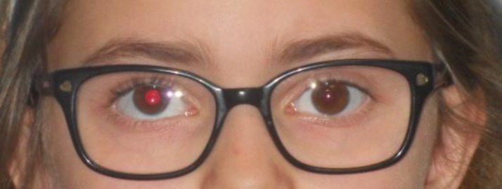
Figure 1. Patient with DVD.
Declaration of patient consent:
The authors certify that they have obtained all appropriate patient consent forms.
Author’s contribution:
A.B, M.P.G., A.B and M.A.P gave substantial contributions to the conception or design of the work in acquisition, analysis, or interpretation of data for the work. A.P and I.M. had a part in article preparing for drafting or revising it critically for important intellectual content, and I.M. gave final approval of the version to be published and agreed to be accountable for all aspects of the work in ensuring that questions related to the accuracy or integrity of any part of the work are appropriately investigated and resolved.
Conflicts of interest:
There are no conflicts of interest.
Financial support and sponsorship:
Nil.
REFERENCES
- 1.Bielschowsky A. Die einseitigen und gegensinnigen (“dissozierten”) Verikalbewegungen der Augen. Graefes Arch Ophthalmol. 1931;125(1):493–553. [Google Scholar]
- 2.Brodsky MC. Dissociated vertical divergence: a righting reflex gone wrong. Arch Ophthalmol. 1999;117(9):1216–1222. doi: 10.1001/archopht.117.9.1216. [DOI] [PubMed] [Google Scholar]
- 3.Guyton LD, Cheeseman EW, Ellis FJ, et al. Dissociated vertical deviation: An exaggerated normal eye movement used to damp cyclovertical nystagmus. Trans Am Ophthalmol Soc. 1998;96(1):389–429. [PMC free article] [PubMed] [Google Scholar]
- 4.Ten Tusscher MP, van Rijn RJ. A hypothetical mechanism for DVD: unbalanced cortical input to subcortical pathways. Strabismus. 2010;18(3):98–103. doi: 10.3109/09273972.2010.502956. [DOI] [PubMed] [Google Scholar]
- 5.Loba P, Broniarczyk-Loba A. Difficulties in diagnosis and treatment of dissociated vertical deviation (DVD) Part I. Klin Oczna. 2007;109(7):356–358. [PubMed] [Google Scholar]
- 6.Tychen L. Infantile esotropia: current neuropshysiological concepts. In: Rosenbaum AL, Santiago AP, editors. Clinical Strabismus Management. Principles and surgical techniques. Philadelphi: WB Saunders; 1999. pp. 117–138. [Google Scholar]
- 7.Esswein MB, von Noorden GK, Coburn A. Comparison of Surgical Methods in the Treatment of Dissociated Vertical Deviation. Am J of Ophthalmol. 1992;113(3):287–290. doi: 10.1016/s0002-9394(14)71580-6. [DOI] [PubMed] [Google Scholar]
- 8.Duncan LB, von Noorden GK. Surgical results in dissociated vertical deviations. J Pediatr Ophthalmol Strabismus. 1984;21(1):25–27. doi: 10.3928/0191-3913-19840101-07. [DOI] [PubMed] [Google Scholar]
- 9.Noel LP, Parks MM. Dissociated vertical deviation. Associated findings and results of surgical treatment. Can J Ophthalmol. 1982;17(1):10–13. [PubMed] [Google Scholar]
- 10.von Noorden GK, Campos EC. Theory and Management of Strabismus. 6th. St. Louis: C. V. Mosby Company; 2002. Binocular vision and ocular motility: In. [Google Scholar]
- 11.Halswanter T, Hoeranter R, Pringlinger S. Reduction of ocular muscle power by splitting of the rectus muscle I: Biomechanics. Br J Ophthalmol. 2004;88(11):1403–1408. doi: 10.1136/bjo.2004.042713. [DOI] [PMC free article] [PubMed] [Google Scholar]
- 12.Harcourt B. Faden Operation (Posterior Fixation Sutures) Eye. 1988;2(1):36–40. doi: 10.1038/eye.1988.9. [DOI] [PubMed] [Google Scholar]
- 13.de Decker W. The Faden operation. When and how to do it. Trans Ophthalmol Soc. 1981;101(2):264–270. [PubMed] [Google Scholar]
- 14.Roggenkamper P. The Faden operation. Technical aspects and indications. Bull Soc Belge Ophthalmol. 1989;232(1):25–31. [PubMed] [Google Scholar]
- 15.Hoerantner R, Pringliger S, Halswanter T. Reduction of ocular motor torque by splitting of the rectus muscle ll:Technique and results. Br J Ophthalmol. 2004;88(11):1409–1413. doi: 10.1136/bjo.2004.042721. [DOI] [PMC free article] [PubMed] [Google Scholar]
- 16.Quinn AG, Kraft SP, Day C, Taylor RS, Levin AV. A prospective evaluation of anterior transposition of the inferior oblique muscle, with and without resection in the tresatment of dissociated vertical deviation. J AAPOS. 2004;4(6):348–353. doi: 10.1067/mpa.2000.110336. [DOI] [PubMed] [Google Scholar]
- 17.Velez GA. Anteriorization of the posterior portion of the inferior oblique muscle for patients with dissociated vertical deviation. Archivos de Ophtalmologia. 1993;50(1):187–189. [Google Scholar]
- 18.Braverman DE, Scott WE. Surgical correction of dissociated vertical deviation. J Pediatr Ophthalmol Strabismus. 1977;14(6):337–341. [PubMed] [Google Scholar]
- 19.Jampolsky A. Symposium on Strabismus. Archives of Ophthalmology. 1978;97(3):565–565. [Google Scholar]
- 20.Sargent RA. Surgical correction of dissociated hyperdeviations. Am J Opthhalmol. 1976;26(1):89–93. [PubMed] [Google Scholar]
- 21.Sprague JB, Moore S, Eggers H, Knapp P. Dissociated vertical deviation treatment with the faden operation of Cuppers. Arch Ophthalmol. 1980;98(3):465–470. doi: 10.1001/archopht.1980.01020030461002. [DOI] [PubMed] [Google Scholar]
- 22.Can D, Ozkan SB, Kasim R, Duman S. Surgical treatment in highly asymmetric dissociated vertical deviations. Strabismus. 1997;5(1):21–26. doi: 10.3109/09273979709055055. [DOI] [PubMed] [Google Scholar]
- 23.Esswein MB, Von Noorden GK, Coburn A. Comparison of surgical methods in the treatment of disociated vertical deviation. Am J Ophthalmol. 1992;113(3):287–290. doi: 10.1016/s0002-9394(14)71580-6. [DOI] [PubMed] [Google Scholar]
- 24.Lorenz B, Raab I, Boergen KP. Dissociated vertical deviation: what is the most effective surgical approach? J Pediatr Ophthalmol Strabismus. 1992;29(1):21–29. doi: 10.3928/0191-3913-19920101-06. [DOI] [PubMed] [Google Scholar]
- 25.Velez G. Dissociated vertical deviation. Graefes Arch Clin Exp Ophthalmol. 1988;226(2):117–118. doi: 10.1007/BF02173296. [DOI] [PubMed] [Google Scholar]