

Abstract
We present a rare case of a patient who sustained a gunshot wound to the abdomen, injuring the aorta, IVC and right common iliac vein. After initially obtaining return of spontaneous circulation (ROSC) en route to the hospital, the patient again lost cardiac activity in the operating room during exploratory laparotomy. Resuscitative thoracotomy was performed and open cardiac massage was maintained for approximately 45 min while vessel injuries were repaired. During cardiac massage, end tidal CO2 was maintained between 15 and 31 mm Hg with 100% oxygen saturation and the patient received on-going transfusion of recycled whole blood and blood component therapy. Permissive hypotension was maintained to facilitate rapid repair of major vessels. Return of spontaneous circulation was achieved with a single 30 joule defibrillation. The patient was discharged home on hospital day 11, neurologically intact. This is the first report of survival after 45 min of open cardiac massage with aortic cross clamping, indicating that end tidal CO2 may act as an indicator of adequate end organ perfusion during protracted periods of hypotension.
Keywords: Resuscitative thoracotomy, Cardiac massage, Permissive hypotension, Penetrating injury, End tidal CO2 monitoring
Introduction
For penetrating trauma patients with loss of vital signs in the emergency department, the role of emergency resuscitative thoracotomy (ERT) with cross clamping of the aorta and open cardiac massage (OCM) may aid in getting patients to the operating room for definitive repair and source control of the hemorrhage. The expected survival of patients undergoing emergency resuscitative thoracotomy (ERT) is between 1 and 5%. Numerous factors may influence the varying degrees of success of ERT including timing, technique, and indication [1]. Multiple studies have shown that performing ERT within 15 min of Basic Life Support (BLS) for penetrating trauma improves survival [2]. ERT requires proper technique and the surgeon must be comfortable identifying the aorta above the diaphragm for cross-clamping and incising the pericardium for exposure of the heart to initiate OCM. Since the idea behind ERT is to redirect blood flow back to the brain and heart, it is indicated for witnessed cardiac arrest secondary to hemorrhage [2]. However, the maximum duration of OCM and what measurements determine adequate cardiac compressions in the face of protracted periods of hypotension remains unknown.
Case report
We present a case of a 28 year-old male who sustained a gunshot wound to the abdomen and required BLS at the scene of the injury. The patient obtained return of spontaneous circulation (ROSC) and was alerted to our facility. On arrival, he was hypotensive and tachycardic with GCS three. His airway was secured and central venous access obtained. He received four units of packed red blood cells (PRBCs) and two units of fresh frozen plasma (FFP) in the trauma bay and was transported immediately to the operating room (OR) for exploratory laparotomy.
It was evident that there was a zone I expanding hematoma at the level of the thoracolumbar spine, suspicious for either an inferior vena cava (IVC) injury or aortic injury.
The patient subsequently developed asystole. A left anterior thoracotomy was performed. The pericardium was opened, the heart was eviscerated, and OCM was initiated. The inferior pulmonary ligament was then identified and transected. The aorta was identified and dissected bluntly off the thoracic spinal column, and cross clamped. Cardiac massage was maintained by the surgical assistant with instructions to maintain end tidal CO2 (EtCO2) in the double digits. Throughout the cardiac massage, EtCO2 was between 15 and 31 mm Hg and the patient's blood pressures remained in the 70/40's with oxygen saturations at 100%. Hypotension was maintained until vascular control with repair could be achieved.
A through and through injury to the IVC and a small laceration to the side wall of the aorta were identified (Fig. 1). The right common iliac vein had a large longitudinal laceration with bullet fragments embedded into the lumbar spine slightly above the bifurcation of the IVC and aorta (Fig. 2). The small aortic injury was repaired primarily with polypropylene suture. The right common iliac vein was ligated with polypropylene suture. Proximal and distal control of the IVC was obtained by using a Rummel tourniquet technique, passing umbilical tape around the IVC. A venotomy of the anterior surface of the IVC was used to expose the back wall defect of the IVC. The back wall of the IVC was repaired intraluminally with polypropylene suture and the extended venotomy was subsequently repaired. After repair of the IVC, its diameter was more than 50% of the original size. During the vascular repair, the patient received 150 mEq of sodium bicarbonate in 50 mEq boluses, three milligrams of epinephrine, and 2 g of calcium chloride. The patient was also receiving ongoing blood component therapy of packed RBCs, FFP, and Platelets. Intra-abdominal blood was harvested with an autotransfusion device to recycle the patient's whole blood and 1 L was given.
Fig. 1.
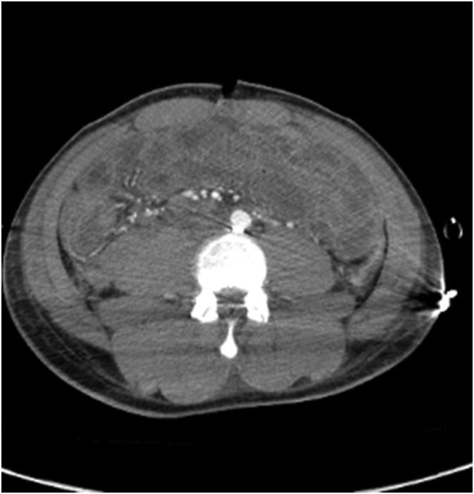
Axial CT image after surgery showing bullet fragment behind the aorta.
Fig. 2.

A coronal CT performed after surgery showing bullet fragments at the level of the IVC and aorta before their bifurcation as well as a thrombosed common iliac vein after ligation.
From the initiation of the thoracotomy to the repair of the IVC and aorta, a total of 45 min had passed. Internal cardiac defibrillation at 30 J was applied directly to the heart and the patient converted to sinus tachycardia. The patient's blood pressure went from 70/40 to 140/80 after return of spontaneous circulation. The aortic cross clamp was released and the vascular repairs were examined for their integrity. No additional repairs were required.
Extensive bleeding of the small bowel mesentery and enterotomies were noted within the ileum. The mesenteric injuries were suture ligated and approximately 15 cm of small bowel was resected without primary anastomosis. The remainder of the abdomen was explored without other injuries. The abdomen was packed with laparotomy pads and a negative pressure temporary abdominal dressing was placed. The thoracotomy was closed primarily and a chest tube was placed.
The estimated blood loss was nine liters. Four liters of auto-transfused blood were returned to the patient. Thirty-five units of PRBC, 18 units of FFP, and 20 units of apheresis platelets were also transfused. The lowest hemoglobin (Hb) during surgery was 5.4 g/dL and at the end of surgery, it was 7.8 g/dL. At the start of surgery, the patient's pH was 7.24 with a base deficit of −11, and at the end of surgery the pH was 7.44 with a base excess of 2. Total duration of surgery was 3 h and 16 min.
After surgery the patient also had a head, neck, chest, abdominal, and pelvis CT to evaluate for any additional injuries. He was found to have a small subarachnoid hemorrhage in the fronto-parietal region. The patient returned to the OR on hospital day two for enteroenterostomy of the small bowel with primary closure of his fascia.
By hospital day five, the patient was extubated, following commands, and was out of bed with physical therapy. He was found on subsequent exam to have an ulnar nerve palsy secondary to a through and through gunshot wound to the right hand requiring outpatient follow up. The patient's vertebral injury and SAH were both deemed non-operative by the neurosurgeon. The patient was discharged home on hospital day 11; alert and oriented, tolerating a regular diet, and ambulating without assistance. The patient was followed up to one year post discharge and was living on his own and fully independent.
This is a very rare case of survival and full recovery after a protracted course of permissive hypotension with cardiac massage to repair complex vascular injuries that would have otherwise rapidly bled out. Perfusion to the essential organs was guided by the presence of EtCO2 during cardiac massage. Since EtCO2 is a by-product of cellular respiration, maintenance of EtCO2 suggests that active metabolism of carbohydrates at the cellular level is present, and therefore oxygen delivery is adequate. Oxygen delivery is the product of cardiac output and the oxygen carrying content. The oxygen carrying content is dependent on the patient's oxygen saturation and the hemoglobin, both of which were maintained throughout the case. While the patient did not have return of spontaneous circulation until after internal cardioversion, our manual compression provided enough cardiac output to deliver oxygen and maintain cellular metabolism [3,4]. This allowed the patient's vital organs to remain viable while repairing the vena cava and aorta. Thus our explanation of the patient's survival is that OCM when guided by EtCO2 can provide enough cardiac output for a meaningful recovery.
References
- 1.Pust G.D., Namias N. Resuscitative thoracotomy. Int. J. Surg. 2016;33:202–208. doi: 10.1016/j.ijsu.2016.04.006. Sep 1. [DOI] [PubMed] [Google Scholar]
- 2.Seamon M.J., Haut E.R., Van Arendonk K. An evidence-based approach to patient selection for emergency department thoracotomy: a practice management guideline from the Eastern Association for the Surgery of Trauma. J. Trauma Acute Care Surg. 2015;79(1):159–173. doi: 10.1097/TA.0000000000000648. [DOI] [PubMed] [Google Scholar]
- 3.Garnett A.R., Ornato J.P., Gonzalez E.R., Johnson E.B. End-tidal carbon dioxide monitoring during cardiopulmonary resuscitation. JAMA. 1987;257(4):512–515. Jan 23-30. [PubMed] [Google Scholar]
- 4.Jin X., Weil M.H., Tang W. End-tidal carbon dioxide as a noninvasive indicator of cardiac index during circulatory shock. Crit. Care Med. 2000;28(7):2415–2419. doi: 10.1097/00003246-200007000-00037. Jul 1. [DOI] [PubMed] [Google Scholar]