Abstract
Arthroscopy-assisted partial wrist-fusion techniques are becoming more popular nowadays. It became clearer that avoiding the violation of important ligament and tendinous structures – which is impossible when using the classic open techniques – enables a more biological approach, which is essential for faster healing and improvement in function. We describe the use of the triquetrum-hamate (TH) portal, which is seldomly applied in routine arthroscopic techniques for hand and wrist surgery, as an accessory portal to better perform anterior midcarpal debridement in four-corner fusion. This trick enables an almost complete anterior resection of the capitate and hamate chondral surfaces, increasing the subchondral osseous contact in the midcarpal joint after fixation, thus leading to higher consolidation rates.
Keywords: wrist joint, wrist injuries, osteoarthritis, arthroscopy
Introduction
Arthroscopy-assisted partial wrist-fusion techniques are becoming more popular nowadays. It became clearer that avoiding the violation of important ligament and tendinous structures – which is impossible when using the classic open techniques – enables a more biological approach, which is essential for faster healing and improvement in function.
We describe the use of the triquetrum-hamate (TH) portal, which is seldomly applied in routine arthroscopic techniques for hand and wrist surgery, as an accessory portal to better perform anterior midcarpal debridement in four-corner fusion. 1 This trick enables an almost complete anterior resection of the capitate and hamate chondral surfaces, increasing the subchondral osseous contact in the midcarpal joint after fixation, thus leading to higher consolidation rates.
Surgical Technique
The standard technique is performed using the 3/4, 6R, midcarpal radial (MCR) and midcarpal ulnar (MCU) arthroscopic portals. The scaphoid is removed with burrs or with a small open (most often volar) approach. The midcarpal articular surfacesare removed with burrs. Cancellous bone grafting is necessary in some cases. The lunate is aligned and temporarily fixed to the radius, and the midcarpal joint is reduced. Bone alignment is secured with cannulated compression screws inserted percutaneously under fluoroscopic visualization.
The TH portal is made just ulnarly to the ECU tendon at the level of the triquetrum-hamate joint, distally to the 6U radiocarpal portal. Rarely used, it was described for visualization and debridement of the TH joint and as an inflow/outflow portal. Damage to the dorsal cutaneous branch of the sensory nerve should be avoided. 2
In our only case in which this trick was performed, after the debridement of all chondral surfaces, as in the aforementioned standard technique, the TH portal was created, and an excellent access to the anterior chondral surfaces of the hamate and capitate was obtained. This enabled the resection of all cartilage down to the subchondral bone, considerably increasing the contact surface between the lunate and the head of the capitate, and between the distal part of the triquetrum and the proximal surface of the hamate ( Figs. 1A, 1B , 2A, 2B, 2C and 2D ). Afterwards, the surgical steps are similar to those of the standard approach for a four-corner fusion. 3 4
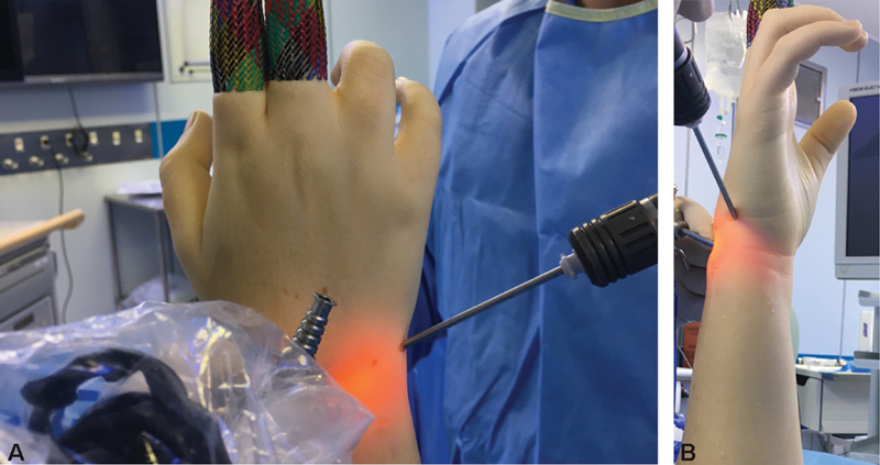
Fig. 1.

( A ) Dorsal view of the wrist after the creation of the triquetrum-hamate (TH) portal distal to the 6U portal. ( B ) Ulnar view of the wrist after the creation of the TH portal distal to the 6U portal.
Fig. 2.

( A ) Schematics for visualization through the midcarpal ulnar (MCU) portal, which is usually applied in the standard four-corner fusion technique. ( B ) Schematics for visualization through the TH portal. This portal provides much better visualization of the dorsal chondral surface of the capitate. ( C ) Schematicsfor the visualizartion and instrumentation through standard dorsal portals, as in the standard technique, in a lateral wrist perspective (as would have been seen through the TH portal). ( D ) Schematics for instrumentation through the TH portal: the shaver reaches much better the anterior aspect of the chondral surface of the capitate, as seen in our modification of the standard technique.
Conclusion
We found this a useful tip to improve the postoperative quality and consolidation rates, and we expect to have better clinical outcomes as well as faster healing and function recovery postoperatively.
Bone fusion was achieved six weeks postoperatively. The patient maintained 50% of the contralateral range of motion, with minimal pain.
Conflito de Interesses Os autores declaram não ter conflito de interesses.
Ética
Não foi solicitada aprovação ética para este estudo, pois nenhum dado do paciente é ou pode ser identificado e/ou divulgado com a publicação de nossa nota técnica. Este estudo foi realizado de acordo com a Declaração de Helsinque, revisada em 2013.
O registro do estudo e o consentimento livre e esclarecido não são aplicáveis pelos motivos descritos anteriormente.
Ethics
Ethical approval was not sought for the present study because no patient data will or can be identified and/or released in publishing our technical note. The present study was completed in accordance with the Helsinki Declaration, as revised in 2013.
Trial registration and informed consent are not applicable for the aforementioned reasons.
Suporte Financeiro
Não houve suporte financeiro de fontes públicas, comerciais, ou sem fins lucrativos.
Financial Support
The present study received no financial support from public, commercial, or not-for-profit sources.
Estudo desenvolvido no Departamento de Ortopedia e Traumatologia, Serviço de Cirurgia da Mão e Microcirurgia, Santa Casa de Misericórdia, São Paulo, Brasil.
Study developed at the Departament of Orthopedics Traumatology, Hand Surgery Microsurgery Service, Santa Casa de Misericórdia, São Paulo, Brazil.
Referências
- 1.Viegas S F. Midcarpal arthroscopy: anatomy and technique. Arthroscopy. 1992;8(03):385–390. doi: 10.1016/0749-8063(92)90075-m. [DOI] [PubMed] [Google Scholar]
- 2.Wolf J M, Dukas A, Pensak M. Advances in wrist arthroscopy. J Am Acad Orthop Surg. 2012;20(11):725–734. doi: 10.5435/JAAOS-20-11-725. [DOI] [PubMed] [Google Scholar]
- 3.Vihanto A, Kotkansalo T, Pääkkönen M. The Learning Curve and Pitfalls of Arthroscopic Four-Corner Arthrodesis. J Wrist Surg. 2019;8(03):202–208. doi: 10.1055/s-0039-1678673. [DOI] [PMC free article] [PubMed] [Google Scholar]
- 4.Ong M T, Ho P C, Wong C W, Cheng S H, Tse W L. Wrist arthroscopy under portal site local anesthesia (PSLA) without tourniquet. J Wrist Surg. 2012;1(02):149–152. doi: 10.1055/s-0032-1326726. [DOI] [PMC free article] [PubMed] [Google Scholar]