Abstract
Introduction:
This study evaluated the propagation of dentinal microcracks and the root canal volume increase after being prepared with two endodontic instruments: ProTaper Next (PTN) and WaveOne Gold (WOG) by micro-computed tomography analysis.
Methods and Materials:
We selected 48 maxillary molars randomly distributed in two groups: PTN and WOG. The samples were scanned before and after instrumentation, and then the image analysis was performed to detect the propagation of pre-existing dentinal micro-cracks and calculate the pre- and post-instrumentation volume. The statistical analysis was performed using Student’s t-test, Fisher's exact test, and ANCOVA (P<0.05).
Results:
Dentinal microcracks were observed in 95.8% of the samples, both PTN and WOG instruments propagated microcracks after instrumentation, but there was no significant difference between the instruments (P=0.538). In relation to the root canal volume there was no statistic difference between PTN and WOG systems for the mesiobuccal (P=0.426) and distobuccal root canals (P=0.523).
Conclusion:
We can conclude that both ProTaper Next and WOG systems propagate dentinal microcracks after root canal preparation in this in vitro study, without statistical significance. The root canal volume prepared also showed no statistically significant difference between the two groups. This in vitro study requires further studies for more concrete conclusions.
Key Words: Endodontics, Microcracks, Microtomography, Root Canal Preparation
Introduction
Biomechanical preparation of root canals is an important step in the endodontic treatment because it eliminates bacteria, removes debris, organic and necrotic tissues, and enlarges the root canal favoring the filling [1-3]. Some complications related to endodontic therapy such as vertical root fracture (VRF) may occur; despite the endodontically treated or untreated teeth, these complications can mainly occur due to the propagation of dentin microcracks caused by root canal biomechanical preparation, leading to tooth loss [4-7].
Several rotary and reciprocating instruments have been introduced in the endodontic field during the last decade to improve the effectiveness of endodontic treatment [8-10]. Due to the high elasticity of the nickel-titanium alloy (NiTi), endodontic instruments have greater flexibility, allowing them to fulfill the original root canal trajectory, making the fundamental tools in endodontic therapy [11, 12].
Among these new systems generations, ProTaper Next (PTN) (Dentsply, Sirona, Ballaigues, Switzerland) and WaveOne Gold (WOG) instruments (Dentsply, Sirona, Ballaigues, Switzerland) can be cited. The PTN instruments consist of a NiTi alloy that receives a heat treatment called M-Wire. It works in a continuous rotary motion and presents a sequence of five instruments: X1, X2, X3, X4 and X5, which comprises the following diameters, respectively: 17/0.04, 25/0.06, 30/0.07, 40/0.06 and 50/0.06 [4, 13, 14]. On the other hand, the WOG is composed of a gold NiTi alloy with heat treatment. Its kinematics is represented by a reciprocating motion of 150º counterclockwise and 30º clockwise, with 120º difference between the two movements and comes in four single-use sizes: Small (20/0.07), Primary (25/0.07), Medium (35/0.06) and Large (45/0.05) [4, 12-14].
In recent years, studies have focused on dentinal defects that occur during root canal preparation, with different instruments NiTi [2, 10, 15, 16].
Considering the importance of correlating the characteristics of the instrument and the root canal morphology with the propagation of existing dentinal microcracks, a study using a non-destructive evaluation method is necessary. Thus, this study proposed to evaluate the dentinal microcracks propagation associated with the endodontic treatment, as well as the volume of the root canal prepared with different systems (PTN and WOG) by means of micro-computed tomography (micro-CT).
Methods and Materials
Sample size calculation
The sample size calculation was determined as described in the previous studies, observing the variation of 18.3% to 51.6% of the presence of complete and incomplete dentistry defects [7, 16-18]. For this study, to observe the same frequency of the defects produced by the instrumentation systems evaluated in the root canal dentin, the Chi-square test and variance statistical test (G*Power 3.1 for Macintosh; Heinrich Heine, Universität Düsseldorf, Düsseldorf, Germany) with α=0.05 and β=0.95 showed a minimum number of 8 samples per group.
Sample selection
The study protocol was approved by the institutional research ethics committee (Protocol no.: 2.817.257). A total of 48 human first and second maxillary molars teeth were collected. All selected teeth, healthy and/or restored, had complete fully formed apex, with a root length between 19 and 21 mm. Teeth with previous endodontic treatment, internal/external resorption or root caries were excluded from the study. The teeth were extracted for reasons unrelated to this study and donated by patients, who signed the donating form. The teeth were stored in distilled water until use.
Initial microcomputed tomography scans
The teeth were embedded in wax, to ensure no movement during the scan. Firstly, the micro-CT pre-instrumentation was performed. Sample scans were performed using a Skyscan 1174 micro-CT device (Bruker, Micro-CT, Kontich, Belgium) setting the parameters for image acquisition: 50 kV, 800 mA, 57 min of exposure time and 17.5 μm of an isotropic resolution. The images reconstructions were performed using the NRecon software v.1.6.9.8 (Bruker, Micro-CT, Kontich, Belgium).
Root canal preparation
Samples were randomly divided into two groups (n=24), according to the instrument used. PTN group: preparation with ProTaper Next (n=24), and WOG group: preparation with WaveOne Gold (n=24). Both systems used 25 mm-length instruments. The working length (WL) was established by inserting a K#10 file (Dentsply Sirona, Ballaigues, Switzerland) until its tip was visible in the apical foramen. This length was measured, and the WL set at 1 mm below. All canals were prepared with K#10 and K#15 files (Dentsply Sirona, Ballaigues, Switzerland) prior to mechanized instrumentation. After these procedures, the mesiobuccal (MB) and distobuccal (DB) canals of the PTN group were prepared, following the sequence X1 (17/0.04), X2 (25/0.06) and X3 (30/0.07) in continuous rotary motion, and each instrument was used to prepare 3 teeth (six root canals) and then discarded. In the WOG group, the preparation was performed with WOG primary instrument (25/0.07) in reciprocating motion, using one instrument for each tooth (two canals). A single operator, specialist in endodontics with enough experience in mechanical instrumentation, carried out all the root canal preparations with X-Smart IQ motor (Sirona Dentsply, Ballaigues, Switzerland) according to the manufacturer's instructions. During the biomechanical preparation, the canals were irrigated with 2 mL of 2.5% sodium hypochlorite (NaOCl) at each 3 mm advance of the instrument within the root canal. The final irrigation was performed using 6 mL of 17% ethylenediaminetetraacetic acid (EDTA) for 3 min, followed by 6 mL of 2.5% NaOCl. For the removal of the NaOCl residues, the canals were flushed with 6 mL of 0.9% saline solution.
Afterwards, the teeth were scanned and the images were reconstructed again, using the same parameters previously described, in order to evaluate the propagation of dentinal microcracks and to calculate the volume after root canal preparation.
Image analysis
The cross-section images from the pre- and post-instrumentation were registered, aligned and superimposed by an automatic process through the DataViewer v.1.5.6 software (Bruker, Micro-CT, Kontich, Belgium). Then, the cross-section images of the mesiobuccal and distobuccal canals were analyzed with the CT Analyser v.1.14.4.1 software (Bruker, Micro-CT, Kontich, Belgium) from the furcation junction to the root apex by two pre-calibrated observers (n=48000), to identify the presence of dentinal microcracks. Initially, the pre-instrumentation images were analyzed and the cross-sectional position corresponding to the observer microcrack was registered.
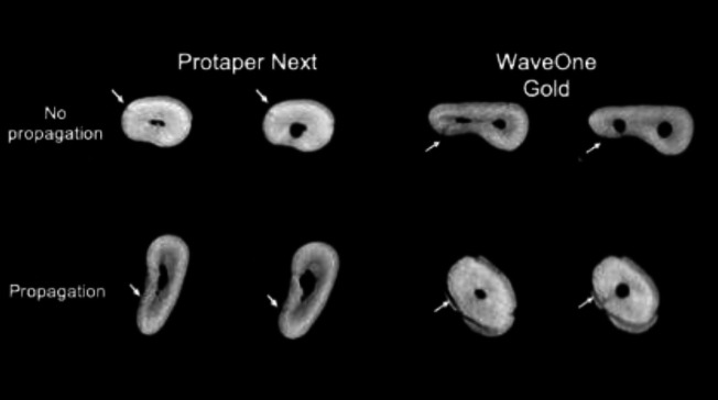
Subsequently, the images of the corresponding cross-sections were also examined after post-instrumentation to verify the microcrack propagation observed in the pre-instrumentation image (Figure 1). In case of divergence, the observers examined the images together until an agreement was reached. The samples were divided into two groups: without propagation and with propagation of microcracks, and the results were expressed as a percentage. Then, the volume calculations of the MB and DB canals after pre- and post-preparation were performed with the CT analyzer software as described previously.
Statistical analysis
The results of the volume measurements were described by means, SD, medians, minimums and maximums. For the group’s comparison defined by the instruments, in the initial evaluation, the Student's t test was used for independent samples. The analysis of the final evaluation and the difference between pre- and post-instrumentation, was performed considering the covariance analysis model (ANCOVA) adjusting for the initial evaluation. For the comparison between the two assessments within each group, the Student's t test for paired samples was used. The instrument comparison for categorical variables was made using Fisher's exact test. To compare the root canals, the binomial test was used. The P<0.05 values was used to indicate statistical significance. The data were analyzed using the Stata/SE v.14.1 software (StataCorpLP, College Station, TX, USA).
Table 1.
Comparison of the MB and DB root canals between instruments (PTN vs WOG) regarding to the microcracks propagation
| Root canal | Variable | Classification | Instruments [N (%)] | P -value * (PTN vs WOG) | |
|---|---|---|---|---|---|
| PTN | WOG | ||||
| MB | Microcracks | No | 1 (4.2%) | 1 (4.2%) | 1 |
| Yes | 23 (95.8%) | 23 (95.8%) | |||
| Propagated | No | 15 (65.2%) | 15 (65.2%) | 1 | |
| Yes | 8 (34.8%) | 8 (34.8%) | |||
| DB | Microcracks | No | 3 (12.5%) | 4 (16.7%) | 1 |
| Yes | 21 (87.5%) | 20 (83.3%) | |||
| Propagated | No | 12 (57.1%) | 9 (45.0%) | 0.538 | |
| Yes | 9 (42.9%) | 11 (55.0%) | |||
MB: mesiobuccal canal; DB: distobuccal canal; PTN: ProTaper Next; WOG: WaveOne Gold; *Fisher’s exact test (P<0.05)
Results
Dentinal microcracks were observed in 95.8% of the samples, both PTN and WOG instruments propagated dentinal microcracks after root canal instrumentation (P=0.538). The null hypothesis tested that the probabilities of propagation are equal for the two instruments (PTN and WOG), versus the alternative hypothesis of different probabilities, for each canal (MB and DB), restricted to teeth that showed microcracks (Figure 1).
Table 1 shows the frequencies and percentages of teeth and the P-values of the statistical tests. For each instrument (PTN and WOG), the null hypothesis was tested that the proportions of teeth with microcracks are equal for the two canals (MB and DB), versus the alternative hypothesis of different proportions. Table 2 presented frequencies and percentages of the combined MB and DB result and the P-values of the statistical tests.
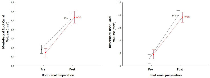
In relation to the root canal volume, there was no statistic difference between PTN and WOG systems for the MB (P=0.426) and DB root canals (P=0.523) (Figure 2). Tables 3 (MB root canal) and 4 (DB root canal) show the descriptive statistics of the pre- and post-instrumentation volume. The P-values of the statistical tests are also showed.
Discussion
This study compared the propagation of dentinal microcracks and the initial and final volume after root canal preparation of mesiobuccal and distobuccal canals of maxillary molars with PTN and WOG systems. Our results showed that both of the instrumentation systems used, reciprocating and rotary motion, increased the number of microcracks at the root, with a considerable increase in volume and removal of dentin after the biomechanical preparation of the root canal, however without statistical difference between the instrumentation systems (PTN and WOG) nor between the root canals (MB and DB).
VRF is a complication which could occur in endodontically treated or untreated teeth that often results in tooth loss. This situation may occur because of microcracks which propagate with the generation of stress caused by occlusal forces [1, 5, 6, 9]. Pop et al. [19] reported that the biomechanical preparation of the root canal may induce compressive stresses in the dentinal walls and may increase the propagation of existing dentinal microcracks. The microcracks are often detected in extracted teeth, being observed in this study in 95.8% of pre-instrumentation root canals.
Table 2.
Comparison of PTN and WOG instruments between root canals (MB vs DB) in relation to microcracks propagation
| Instruments | Variable | Classification | Root canal [N (%)] | P -value* (MB vs DB) | |
|---|---|---|---|---|---|
| MB [N (%)] | DB | ||||
| PTN | Microcracks | No | 1 (4.2%) | 3 (12.5%) | 0.625 |
| Yes | 23 (95.8%) | 21 (87.5%) | |||
| Propagated** | No | 12 (60%) | 12 (60%) | 1 | |
| Yes | 8 (40%) | 8 (40%) | |||
| WOG | Microcracks | No | 1 (4.2%) | 4 (16.7%) | 0.250 |
| Yes | 23 (95.8%) | 20 (83.3%) | |||
| Propagated** | No | 14 (70%) | 9 (45%) | 0.227 | |
| Yes | 6 (30%) | 11 (55%) | |||
MB: mesiobuccal canal; DB: distobuccal canal; PTN: ProTaper Next; WOG: WaveOne Gold; *Binomial test (P<0.05); **Restricted to teeth that had micro cracks on both root canals
Table 3.
Mean (SD) values for the volume of the mesiobuccal (MB) root canal (n=24)
| Time evaluation | Instruments | P -value* (PTN vs WOG) | |
|---|---|---|---|
| PTN | WOG | ||
| Initial | 1.92 (1.16) | 1.72 (1.25) | 0.563 |
| Final | 3.58 (1.77) | 3.68 (1.49) | 0.426 |
| Dif (final-initial) | 1.66 (1.25) | 1.96 (1.19) | 0.426 |
| P-value ** (initial vs final) | <0.001 | <0.001 | |
PTN: ProTaper Next; WOG: WaveOne Gold; Results described by Mean ± standard deviation values; * Student’s t-test for independent samples (initial); ANOVA adjusted for initial evaluation (final and difference between final and initial) (P<0.05); ** Student's t-test for paired samples (P<0.05)
Table 4.
Mean (SD) values for the volume of the distobuccal (DB) root canal (n=24)
| Time evaluation | Instruments | P -value* (PTN vs WOG) | |
|---|---|---|---|
| PTN | WOG | ||
| Initial | 1.27 (0.75) | 1.45 (1.00) | 0.483 |
| Final | 2.98 (1.09) | 2.93 (0.90) | 0.523 |
| Dif (final-initial) | 1.71 (0.95) | 1.47 (0.92) | 0.523 |
| P-value ** (initial vs final) | <0.001 | <0.001 | |
PTN: ProTaper Next; WOG: WaveOne Gold; Results described by Mean (SD) values; * Student’s t-test for independent samples (initial); ANOVA adjusted for initial evaluation (final and difference between final and initial) (P<0.05); ** Student's t-test for paired samples (P<0.05)
Studies using root-section methods and direct evaluation by optical microscopy reported a relationship between the root canal preparation with continuous rotary and/or reciprocating instruments and the formation of new dentinal microcracks [2, 4, 20, 21]. This method is easy to perform because it uses low-cost instruments, and produces immediate results with simple data analysis. However, it presents some limitations, such as the need for samples destruction, and the evaluation of only few sections per tooth (between three and six) [2, 4, 16, 22]. The root sectioning method presents a significant disadvantage related to their destructive nature, which, in turn, may influence the results reported in the literature [15].
In contrast to these microscopic observation studies, dentinal microcracks could be analyzed by micro-CT, with highly accurate results without the sample destruction and examine in detail hundreds of pre- and/or post-treatment cross-sectional images to determine the location of microcracks [23-25]. Studies using micro-CT method have reported no causal relationship between root canal biomechanical preparation with rotary or reciprocating systems in the formation of dentinal microcracks [15, 23].
The literature has been reported a high percentage rate of dentinal defects after instrumentation with NiTi systems [2, 20, 21, 26]. Liu et al. [25] reported that continuous rotary systems with multiple instruments caused more dentinal defects on the root surface than continuous rotary and reciprocating systems developed by single use. Ashwinkumar et al. [27] also observed that the root canal preparation with ProTaper instrument was associated with a significantly higher number of microcracks compared to the WO system. This differs from the results obtained in the present study, in which there was no statistical difference between ProTaper and WaveOne in the propagation of dentinal microcracks, since both systems caused the propagation of existing microcracks. Regarding the difference between the results of the two studies De-Deus et al. [15] and De-Deus et al. [16] reported that the root sectioning method presents a disadvantage related to its own destructive nature, which is probably the main cause of these results reported in the literature. The present study proposed to analyze the dentinal microcracks propagation using micro-CT. This method is highly accurate as it does not destroy the sample and allows the three-dimensional visualization of hundreds of images before and after instrumentation, increasing the internal consistence and validity of the analysis, since each sample serves as its own control.
Studies on the formation of dentinal microcracks after root canal preparation with NiTi systems using the micro-CT method, have reported that there is no relationship between instrumentation with continuous rotary or reciprocating systems in the formation of dentinal microcracks [3, 7, 15, 16]. However, to date, no studies have been published to evaluate the microcracks propagation, only the formation of these dentin defects after biomechanical preparation. In the present study, no new cracks were observed after the instrumentation. The cracks present in the images evaluated after instrumentation were in correspondence with pre-instrumentation images.
According to some authors, several factors may influence the formation of dentinal microcracks, such as different heat surface treatment, design, cross-sectional shape, diameter and kinematics of the instrument [1, 28, 29]. However, De Deus et al. [16] and Karatas et al. [24] stated that there is no difference between systems with different heat treatments. In the present study, the studied groups presented the thermal treatment with M-Wire alloy for PTN system and gold alloy for the WOG system; both groups showed great flexibility and decrease cyclic fatigue, which supposedly did not influence the formation of dentinal defects.
The instrument cross-section may be influenced due to the number of touches caused in the root walls, promoting different degrees of stress. The PTN system has a decentralized rectangular cross-section with two cutting blades, minimizing contact with the dentin [8, 13, 29, 30]. On the other hand, the WOG instrument has a parallelogram-shaped cross-section alternating touches in the dentine with one and two cutting blades during 360° of rotation [12, 14]. Although the cross-sections are different, this seems not significantly affect the propagation of dentinal microcracks because their sections are optimized and almost do not touch the dentin walls during the root canal biomechanical preparation. Pédulla et al. [31] evaluated six instruments with different geometric characteristics and concluded that this parameter does not significantly affect the incidence of dentin microcracks.
The diameter of the instrument may be a contributing factor in the generation of dentinal defects. According to Tamse et al. [32] the more radicular dentin is removed, the greater the risk of initiating root fractures. However, we found that the PTN group, even with a larger diameter, showed no statistically significant difference compared to WOG group in relation to the propagation of dentinal microcracks.
The instrumentation of the root canal with continuous rotary systems forms a variable degree of rotating force in the root canal walls, being able to generate the formation of dentinal defects [24]. In contrast, the reciprocating motion decreases the continuous rotational force and the constant torque applied to the root canal wall, resulting in less dental damage [24]. In the present study, the difference between the kinematics of the PTN and WOG instruments supposedly did not affect the dentinal microcracks propagation, which is in agreement with studies that compared the continuous rotary and reciprocating motion, and report that there is no relation between the microcracks formation with root canal biomechanical preparation [15, 23, 33].
Some studies use only root canals without anatomical complexity to evaluate the propagation of dentinal microcracks. This anatomical homogeneity may not reproduce real clinical situations. For this reason, in this study we used MB and DB roots of maxillary molars, which present the main clinical difficulty. Although the MB root canals show a higher curvature degree compared to DB canals, our results showed no statistically significant difference was observed when compared to the propagation of microcracks among these root canals.
In addition to the evaluation of the dentinal microcracks, micro-CT allows detailed assessment of the volume and geometry of the root canal through two- and three- dimensional images [12]. The root canal volume was compared separately for each canal. Although the diameter of the PTN system selected was higher in comparison to the WOG group, there was no significant difference in the initial and final volume between these instruments, which is in accordance with Dioguardi et al. [34]; they compared Protaper F2 with WaveOne primary and found no statistical difference between groups. Although in the present study no root canal volume difference was observed between the different diameter instruments (30.07×25.07), a greater preparation is important as it allows a higher enlargement in the apical third of the root canal, improving the efficiency of irrigation and, consequently, a better disinfection of the root canal [28].
Within the limitations of this study, the presented results could guide the choice of the root canal instrumentation system. The dentinal microcracks were observed in 95.8% of the teeth, both PTN and WOG instruments propagated dentinal microcracks, but there was no statistical difference between these instruments. In addition, in comparison with the volume, both systems had a similar amount of dentin removal.
Conclusion
Based on our in vitro study, we could confidently conclude that both PTN and WOG systems propagate microcracks in the dentinal wall after root canal preparation, with no statistical difference between the two (PTN and WOG) nor between the type of root canal prepared (MB and DB). The root canal volume also showed no statistically significant difference between the PTN and WOG preparation, regardless of the difference in diameter and taper between the instruments.
Conflict of Interest:
‘None declared’.
Acknowledgment
This research did not receive any specific grant from funding agencies in the public, commercial or not-for-profit sectors.
References
- 1.Yoldas O, Yilmaz S, Atakan G, Kuden C, Kasan Z. Dentinal microcrack formation during root canal preparations by different NiTi rotary instruments and the self-adjusting file. J Endod. 2012;38(2):232–5. doi: 10.1016/j.joen.2011.10.011. [DOI] [PubMed] [Google Scholar]
- 2.Katanec T, Miletić I, Baršić G, Kqiku-Bliblikaj L, Žižak M, Krmek SJ. Incidence of Dentinal Micro Cracks during Root Canal Preparation with Self Adjusting File, Reciproc Blue, and ProTaper Next. Iranian endodontic journal. 2020;15(1):6–11. doi: 10.22037/iej.v15i1.26667. [DOI] [PMC free article] [PubMed] [Google Scholar]
- 3.Bayram HM, Bayram E, Ocak M, Uygun AD, Celik HH. Effect of ProTaper Gold, Self-Adjusting File, and XP-endo Shaper instruments on dentinal microcrack formation: a micro–computed tomographic study. Journal of endodontics. 2017;43(7):1166–9. doi: 10.1016/j.joen.2017.02.005. [DOI] [PubMed] [Google Scholar]
- 4.Cassimiro M, Romeiro K, Gominho L, de Almeida A, Silva L, Albuquerque D. Effects of Reciproc, ProTaper Next and WaveOne Gold on Root Canal Walls: A Stereomicroscope Analysis. Iran Endod J. 2018;13(2):228–33. doi: 10.22037/iej.v13i2.16327. [DOI] [PMC free article] [PubMed] [Google Scholar]
- 5.Chan CP, Lin CP, Tseng SC, Jeng JH. Vertical root fracture in endodontically versus nonendodontically treated teeth: a survey of 315 cases in Chinese patients. Oral Surg Oral Med Oral Pathol Oral Radiol Endod. 1999;87(4):504–7. doi: 10.1016/s1079-2104(99)70252-0. [DOI] [PubMed] [Google Scholar]
- 6.Tsesis I, Rosen E, Tamse A, Taschieri S, Kfir A. Diagnosis of vertical root fractures in endodontically treated teeth based on clinical and radiographic indices: a systematic review. J Endod. 2010;36(9):1455–8. doi: 10.1016/j.joen.2010.05.003. [DOI] [PubMed] [Google Scholar]
- 7.Zuolo ML, De-Deus G, Belladonna FG, Silva EJ, Lopes RT, Souza EM, Versiani MA, Zaia AA. Micro-computed Tomography Assessment of Dentinal Micro-cracks after Root Canal Preparation with TRUShape and Self-adjusting File Systems. J Endod. 2017;43(4):619–22. doi: 10.1016/j.joen.2016.11.013. [DOI] [PubMed] [Google Scholar]
- 8.Haapasalo M, Shen Y. Evolution of nickel–titanium instruments: from past to future. Endodontic Topics. 2013;29(1):3–17. [Google Scholar]
- 9.Khoshbin E, Donyavi Z, Abbasi Atibeh E, Roshanaei G, Amani F. The Effect of Canal Preparation with Four Different Rotary Systems on Formation of Dentinal Cracks: An In Vitro Evaluation. Iran Endod J. 2018;13(2):163–8. doi: 10.22037/iej.v13i2.16416. [DOI] [PMC free article] [PubMed] [Google Scholar]
- 10.Harandi A, Mirzaeerad S, Mehrabani M, Mahmoudi E, Bijani A. Incidence of Dentinal Crack after Root Canal Preparation by ProTaper Universal, Neolix and SafeSider Systems. Iran Endod J. 2017;12(4):432–8. doi: 10.22037/iej.v12i4.17597. [DOI] [PMC free article] [PubMed] [Google Scholar]
- 11.Paqué F, Ganahl D, Peters OA. Effects of root canal preparation on apical geometry assessed by micro-computed tomography. J Endod. 2009;35(7):1056–9. doi: 10.1016/j.joen.2009.04.020. [DOI] [PubMed] [Google Scholar]
- 12.van der Vyver PJ, Paleker F, Vorster M, de Wet FA. Root Canal Shaping Using Nickel Titanium, M-Wire, and Gold Wire: A Micro-computed Tomographic Comparative Study of One Shape, ProTaper Next, and WaveOne Gold Instruments in Maxillary First Molars. J Endod. 2019;45(1):62–7. doi: 10.1016/j.joen.2018.09.013. [DOI] [PubMed] [Google Scholar]
- 13.Yamamura B, Cox TC, Heddaya B, Flake NM, Johnson JD, Paranjpe A. Comparing canal transportation and centering ability of endosequence and vortex rotary files by using micro-computed tomography. J Endod. 2012;38(8):1121–5. doi: 10.1016/j.joen.2012.04.019. [DOI] [PubMed] [Google Scholar]
- 14.Gündoğar M, Özyürek T. Cyclic Fatigue Resistance of OneShape, HyFlex EDM, WaveOne Gold, and Reciproc Blue Nickel-titanium Instruments. J Endod. 2017;43(7):1192–6. doi: 10.1016/j.joen.2017.03.009. [DOI] [PubMed] [Google Scholar]
- 15.De-Deus G, Belladonna FG, Souza EM, Silva EJ, Neves Ade A, Alves H, Lopes RT, Versiani MA. Micro-computed Tomographic Assessment on the Effect of ProTaper Next and Twisted File Adaptive Systems on Dentinal Cracks. J Endod. 2015;41(7):1116–9. doi: 10.1016/j.joen.2015.02.012. [DOI] [PubMed] [Google Scholar]
- 16.De-Deus G, César de Azevedo Carvalhal J, Belladonna FG, Silva E, Lopes RT, Moreira Filho RE, Souza EM, Provenzano JC, Versiani MA. Dentinal Microcrack Development after Canal Preparation: A Longitudinal in Situ Micro-computed Tomography Study Using a Cadaver Model. J Endod. 2017;43(9):1553–8. doi: 10.1016/j.joen.2017.04.027. [DOI] [PubMed] [Google Scholar]
- 17.De-Deus G, Belladonna FG, Marins JR, Silva EJ, Neves AA, Souza EM, Machado AC, Lopes RT, Versiani MA. On the Causality Between Dentinal Defects and Root Canal Preparation: A Micro-CT Assessment. Braz Dent J. 2016;27(6):664–9. doi: 10.1590/0103-6440201601002. [DOI] [PubMed] [Google Scholar]
- 18.De-Deus G, Belladonna FG, Silva EJ, Marins JR, Souza EM, Perez R, Lopes RT, Versiani MA, Paciornik S, Neves Ade A. Micro-CT Evaluation of Non-instrumented Canal Areas with Different Enlargements Performed by NiTi Systems. Braz Dent J. 2015;26(6):624–9. doi: 10.1590/0103-6440201300116. [DOI] [PubMed] [Google Scholar]
- 19.Pop I, Manoharan A, Zanini F, Tromba G, Patel S, Foschi F. Synchrotron light-based μCT to analyse the presence of dentinal microcracks post-rotary and reciprocating NiTi instrumentation. Clin Oral Investig. 2015;19(1):11–6. doi: 10.1007/s00784-014-1206-5. [DOI] [PubMed] [Google Scholar]
- 20.Arias A, Lee YH, Peters CI, Gluskin AH, Peters OA. Comparison of 2 canal preparation techniques in the induction of microcracks: a pilot study with cadaver mandibles. J Endod. 2014;40(7):982–5. doi: 10.1016/j.joen.2013.12.003. [DOI] [PubMed] [Google Scholar]
- 21.Saber SE, Schäfer E. Incidence of dentinal defects after preparation of severely curved root canals using the Reciproc single-file system with and without prior creation of a glide path. Int Endod J. 2016;49(11):1057–64. doi: 10.1111/iej.12555. [DOI] [PubMed] [Google Scholar]
- 22.Coelho MS, Card SJ, Tawil PZ. Light-emitting Diode Assessment of Dentinal Defects after Root Canal Preparation with Profile, TRUShape, and WaveOne Gold Systems. J Endod. 2016;42(9):1393–6. doi: 10.1016/j.joen.2016.06.003. [DOI] [PubMed] [Google Scholar]
- 23.De-Deus G, Silva EJ, Marins J, Souza E, Neves Ade A, Gonçalves Belladonna F, Alves H, Lopes RT, Versiani MA. Lack of causal relationship between dentinal microcracks and root canal preparation with reciprocation systems. J Endod. 2014;40(9):1447–50. doi: 10.1016/j.joen.2014.02.019. [DOI] [PubMed] [Google Scholar]
- 24.Karataş E, Gündüz HA, Kırıcı D, Arslan H, Topçu M, Yeter KY. Dentinal crack formation during root canal preparations by the twisted file adaptive, ProTaper Next, ProTaper Universal, and WaveOne instruments. J Endod. 2015;41(2):261–4. doi: 10.1016/j.joen.2014.10.019. [DOI] [PubMed] [Google Scholar]
- 25.Liu R, Hou BX, Wesselink PR, Wu MK, Shemesh H. The incidence of root microcracks caused by 3 different single-file systems versus the ProTaper system. J Endod. 2013;39(8):1054–6. doi: 10.1016/j.joen.2013.04.013. [DOI] [PubMed] [Google Scholar]
- 26.De-Deus G, Belladonna FG, Silva E, Souza EM, Carvalhal JCA, Perez R, Lopes RT, Versiani MA. Micro-CT assessment of dentinal micro-cracks after root canal filling procedures. Int Endod J. 2017;50(9):895–901. doi: 10.1111/iej.12706. [DOI] [PubMed] [Google Scholar]
- 27.Ashwinkumar V, Krithikadatta J, Surendran S, Velmurugan N. Effect of reciprocating file motion on microcrack formation in root canals: an SEM study. Int Endod J. 2014;47(7):622–7. doi: 10.1111/iej.12197. [DOI] [PubMed] [Google Scholar]
- 28.Kim HC, Lee MH, Yum J, Versluis A, Lee CJ, Kim BM. Potential relationship between design of nickel-titanium rotary instruments and vertical root fracture. J Endod. 2010;36(7):1195–9. doi: 10.1016/j.joen.2010.02.010. [DOI] [PubMed] [Google Scholar]
- 29.Capar ID, Arslan H, Akcay M, Uysal B. Effects of ProTaper Universal, ProTaper Next, and HyFlex instruments on crack formation in dentin. J Endod. 2014;40(9):1482–4. doi: 10.1016/j.joen.2014.02.026. [DOI] [PubMed] [Google Scholar]
- 30.Huang Z, Quan J, Liu J, Zhang W, Zhang X, Hu X. A microcomputed tomography evaluation of the shaping ability of three thermally-treated nickel-titanium rotary file systems in curved canals. J Int Med Res. 2019;47(1):325–34. doi: 10.1177/0300060518801451. [DOI] [PMC free article] [PubMed] [Google Scholar]
- 31.Pedullà E, Genovesi F, Rapisarda S, La Rosa GR, Grande NM, Plotino G, Adorno CG. Effects of 6 Single-File Systems on Dentinal Crack Formation. J Endod. 2017;43(3):456–61. doi: 10.1016/j.joen.2016.10.038. [DOI] [PubMed] [Google Scholar]
- 32.Tamse A, Fuss Z, Lustig J, Kaplavi J. An evaluation of endodontically treated vertically fractured teeth. J Endod. 1999;25(7):506–8. doi: 10.1016/S0099-2399(99)80292-1. [DOI] [PubMed] [Google Scholar]
- 33.Rose E, Svec T. An Evaluation of Apical Cracks in Teeth Undergoing Orthograde Root Canal Instrumentation. J Endod. 2015;41(12):2021–4. doi: 10.1016/j.joen.2015.08.023. [DOI] [PubMed] [Google Scholar]
- 34.Dioguardi M, Troiano G, Laino L, Lo Russo L, Giannatempo G, Lauritano F, Cicciù M, Lo Muzio L. ProTaper and WaveOne systems three-dimensional comparison of device parameters after the shaping technique A micro-CT study on simulated root canals. Int J Clin Exp Med. 2015;8(10):17830–4. [PMC free article] [PubMed] [Google Scholar]


