

Abstract
Ruptures of the annular pulleys of the finger flexor tendons are not common in the general population. In sport climbing, these structures can be abnormally stressed, mainly because of the so-called crimped position, an extreme flexion of the proximal interphalangeal joint, levering an abnormal tension by flexor tendons. Complete pulley tears manifest with explicit pain and an inability, but strains or minor tears might only be perceived by individuals like professional climbers, since they can bring total disability to crucial grip positions. Complete tears of one or more pulleys have already been characterized by ultrasound and magnetic resonance, but no imaging features were described for strains or smaller partial tears. We describe the case of a climber with symptoms of an A2-pulley injury, in whom ultrasound imaging revealed reversible features of fusiform thickening and hypoechogenicity, which resemble the strains that we find in similar structures like tendons and other ligaments.
Keywords: Ultrasound, Pulley, Strain, Climbing, Rehabilitation
Introduction
Ruptures of the annular pulleys of the finger flexor tendons are not common in the general population [1]. Besides trauma and surgical section, sport climbing is the paradigmatic activity in which these structures can be abnormally stressed, mainly because of a specific holding type: the crimped position (Fig. 1). The crimped position is used to hold small edges (supporting only the distal phalanges) while the proximal interphalangeal (PIP) joints are flexed at 90 degrees or more [2]. The flexion of the PIP joint enables the additional recruitment of the flexor digitorum profundus (FDP) muscle and this maneuver tends to be intuitive while climbing. The more it flexes the stronger the climber can pull, since the contracting FDP muscle gets shortened, increasing the number of actin-myosin cross-bridges, and thus maximal force [3]. Some climbers can hold their entire body weight on one hand while crimping on small holds. The tension that the flexor tendons apply on annular pulleys on a crimped position while they try to keep them close to the bone is unusually high (lever mechanism).
Fig. 1.
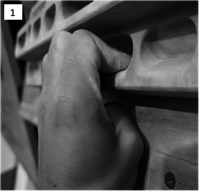
The crimped position, a climbing holding type that consists of grabbing a small edge with a flexed proximal interphalangeal joint
Complete tears of one or more pulleys manifest with explicit pain and an inability for daily activities, but strains or minor tears might only be of interest to individuals like professional climbers since they can bring total disability to crucial grip positions. The distance measured by ultrasound between the bone and the finger flexor tendons can help diagnose complete tears of one or more pulleys [2, 4, 5], and magnetic resonance can display similar features [6]. To date, no characteristic imaging features were described for strains or smaller partial tears of finger flexor tendon annular pulleys.
Case report
We describe the case of a 29 years-old male with more than eight years of sustained climbing activity, presenting to the clinic with a two-week history of pain on the radial side of the proximal phalanx of his fourth left finger, after a full crimp maximal effort. He could climb open-handed (with slight or no flexion of the PIP joints) without pain or diminished strength but couldn’t hold small edges on a crimped position free of pain. On observation there was no bowstring of the flexor tendons, but anteroposterior pressure of the referred proximal phalanx elicited pain. An ultrasound examination was requested. Using a high-frequency (12–22 MHz) hockey-stick probe of a high-end ultrasound machine, the distance between flexor tendons and bone at the mid part of the A2-pulley was null (Fig. 2), even after a flexion dynamic maneuver, ruling out a complete tear of the A2-pulley. A more detailed exploration of the painful area in a short-axis view, which corresponded to the radial and distal part of the A2-pulley close to its bony insertion, showed a fusiform and disproportionate thickening of this area, when compared with the asymptomatic contralateral A2-pulley at the same location (1.81 mm versus 1.05 mm), alongside with hypoechogenicity (Fig. 3a and b). Findings were confirmed in a coronal long-axis view (Fig. 3c and d), where a clear thickening of the distal part of the A2-pulley was evident when compared to proximal levels bilaterally, but disproportionately thicker on the left symptomatic one (1.50 mm on the left versus 0.98 mm on the right distally, and 0.40 mm on the left versus 0.49 mm on the right more proximally). There was no Doppler signal, and a sagittal long-axis view showed no ultrasound changes on the pulley or flexor tendons (Fig. 2). After six weeks of complete avoidance of the crimped position, a reassessment was made, with significant regression of the assumed pathological findings: thickening was reduced to 1.31 mm in short-axis view and to 0.96 mm in coronal long-axis view (Fig. 4a and b). By this time, the athlete started climbing without any hand-positioning restrictions and no pain at all.
Fig. 2.

Ultrasound image of the A2-pulley and finger flexor tendons of the fourth left finger in a long-axis sagittal view. Legend: arrows—A2-pulley; asterisk—finger flexor tendons; cardinal—proximal phalanx bone cortex
Fig. 3.

Ultrasound images of the A2-pulleys of the fourth finger, comparing the injured left side with the uninjured right one, either in short axis view (a and b) and long axis view (c and d). Figures a and b display a short axis view of the A2-pulley at its radial and distal part, the first being the injured left side, depicting a fusiform and disproportionate thickening on the left side (1.81 mm at the left versus 1.05 mm at the right), alongside with relative hypoechogenicity. Figures c and d display a long-axis coronal view of the A2-pulley at its radial part, the first being the injured left side, both showing a thickening of the more distal part, but disproportionately bigger on the injured left side (1.50 mm at the left versus 0.98 mm at the right), with proximal segments being equally thick. Legend: arrows—A2-pulley; asterisk—finger flexor tendons; cardinal—proximal phalanx bone cortex
Fig. 4.

Ultrasound images of the A2-pulley of the injured left fourth finger, six weeks after avoidance of the crimped position while maintaining remaining climbing activities, with a short-axis view of the radial and distal part (a) and long-axis coronal view of the radial part (b) showing a significant decrease of the previous pathological findings, namely a reduction in thickness of the same A2-pulley from 1.81 to 1.31 mm, in short axis view, and from 1.50 to 0.96 mm, in long axis coronal view. Legend: arrows—A2-pulley; asterisk—finger flexor tendons; cardinal—proximal phalanx bone cortex
Discussion
The increasing safety and popularity of sport climbing turned it into a relevant and fast-growing sport, being an Olympic discipline since Tokyo 2020. Considering recreational and professional athletes, the number of overall practitioners is nowadays significant [7]. Pulley injuries are among the most common injuries in climbers [8]. Other than clinical examination, data concerning imaging characterization of minor injuries like pulley strains is lacking. We describe the case of a climber with symptoms of an A2-pulley injury, in whom ultrasound imaging revealed reversible features of fusiform thickening and hypoechogenicity, which resemble the strains that we find in similar structures like tendons and other ligaments. The contralateral and asymptomatic pulley also revealed a mild and more homogeneous (but not fusiform) thickening of the same distal segment, when compared to more proximal levels of the same pulley, which could be interpreted as an adaptational change on this distal area of the pulley, more stressed in climbers, requiring validation in controlled studies.
Until very recently, imaging documentation of this kind of lesion was only provided by magnetic resonance. Now, using high-end ultrasound machines, a more widespread, available, fast and cheap imaging method, the physician has the opportunity to assess and approach these patients more conveniently, provided that the ultrasound operator has the required training to perform the exam.
Conclusion
We describe the case of a recreational sport climbing young male with a two-week history of pain on the radial side of the proximal phalanx of his fourth left finger after a full crimp maximal effort in the practice of sport climbing. Ultrasound examination with a high-end machine allowed the characterization of a flexor tendon A2 pulley strain.
In conclusion, ultrasound features of pulley strains and other non-pathologic adaptational changes are yet to be established, especially in climbers. We hope that its crescent clinical relevance encourages future investigation in this area.
Acknowledgements
We thank to the Imaging and Techniques Unit of the Rheumatology Department of the Centro Hospitalar Universitário Lisboa Norte / Centro Académico de Lisboa for the complete availability and full interest in this investigation.
Declarations
Conflict of interest
None declared.
Funding
No funding was used in this work.
Patient and public involvement
Patients and/or the public were not involved in the design, or conduct, or reporting, or dissemination plans of this work.
Consent to publish
The authors affirm that the patient involved in this case report provided informed consent for publication of the images in Figs. 1, 2, 3 and 4.
Author contributions
TF, MS and FS contributed equally to this work. TF and FS participated in the design of the study. TF and MS contributed to clinical data collection. TF and FS revised the technical content of the manuscript. TF, MS and FS prepared the manuscript.
Footnotes
Publisher's Note
Springer Nature remains neutral with regard to jurisdictional claims in published maps and institutional affiliations.
References
- 1.Zafonte B, Rendulic D, Szabo RM. Flexor pulley system: anatomy, injury, and management. J Hand Surg Am. 2014;39(12):2525–2532. doi: 10.1016/j.jhsa.2014.06.005. [DOI] [PubMed] [Google Scholar]
- 2.Schoffl VR, Schoffl I. Injuries to the finger flexor pulley system in rock climbers: current concepts. J Hand Surg Am. 2006;31(4):647–654. doi: 10.1016/j.jhsa.2006.02.011. [DOI] [PubMed] [Google Scholar]
- 3.Fitts RH, McDonald KS, Schluter JM. The determinants of skeletal muscle force and power: their adaptability with changes in activity pattern. J Biomech. 1991;24(Suppl 1):111–122. doi: 10.1016/0021-9290(91)90382-W. [DOI] [PubMed] [Google Scholar]
- 4.King EA, Lien JR. Flexor tendon pulley injuries in rock climbers. Hand Clin. 2017;33(1):141–148. doi: 10.1016/j.hcl.2016.08.006. [DOI] [PubMed] [Google Scholar]
- 5.Miro PH, vanSonnenberg E, Sabb DM, Schoffl V. Finger flexor pulley injuries in rock climbers. Wilderness Environ Med. 2021;32(2):247–258. doi: 10.1016/j.wem.2021.01.011. [DOI] [PubMed] [Google Scholar]
- 6.Hoff MN, Greenberg TD. MRI sport-specific pulley imaging. Skeletal Radiol. 2018;47(7):989–992. doi: 10.1007/s00256-017-2786-3. [DOI] [PubMed] [Google Scholar]
- 7.Kuelthau W (2022) Rock climbing statistics: accidents, injuries, deaths and demographics. https://www.99boulders.com/the-growth-of-climbing
- 8.Schoffl V, Simon M, Lutter C. Finger and shoulder injuries in rock climbing. Orthopade. 2019;48(12):1005–1012. doi: 10.1007/s00132-019-03825-3. [DOI] [PubMed] [Google Scholar]