

Abstract
Background
Auricular avulsion remains one of the most common acquired ear deformities. Due to the complexity of each case, vast details of treatment approaches were to be considered, including in the case of partial auricular avulsion. Here we presented a case report with factors affecting the efficacy of simple reattachment as a treatment choice for a partial auricular avulsion.
Case report
A 28-year-old male with partial auricular avulsion was treated with simple reattachment, achieving satisfactory results and without complications.
Conclusion
The presence of an inferior pedicle, shorter injury to surgery time, and careful considerations of technical steps in conducting the simple reattachment and postoperative management are to increase the outcomes in partial auricular avulsions.
Keywords: Partial auricular avulsion, Simple reattachment, Prognostic factors
Introduction
Auricular avulsion is one of the most common types of acquired ear deformity and might result in a wide kind of deformity. Several traumatic injuries could cause auricular avulsion, with bite injuries and traffic accidents as the leading causes [1]. Injury to the external ear may affect a vast aspect of human life, including cosmetic or even psychological consequences. Although avulsion to the external ear does not directly affect the hearing function, the extent of traumatic injury may also injure the hearing pathway. Due to the complexity of its anatomical structure and unique vascularization, reconstruction of ear avulsion remains a substantial challenge. Although multiple surgical approach was established, there is currently no proper consensus or guidelines for partial auricular avulsion. Most common surgical methods such as: reattachment as composite graft, reconstruction using local flaps, pocket principle, Baudet method and microvascular repair. Microvascular repair appears to be the best aesthetic outcome, however the need for blood transfusion and higher operative time should considered [2]. Surgical approach for auricular avulsion broadly classified from the total of operation needed defined as ‘stage’, such as ‘one-stage’ for a single operation or ‘two-stage’ (or more) when prior surgery was needed to create the framework for the avulsed ear [3]. Here we presented a partial auricular avulsion receiving ‘one-stage’ direct re-attachment.
Case report
The patient was admitted to emergency department with a laceration wound, 2 h prior to the admission. The patient was attacked from behind by a group of unknown people and slashed the left ear. No history of fall, loss of consciousness, loss of hearing, seizure, vomit, or other ear, nose, and throat complaints after the accident. Vital signs and functions are normal, with the clinical examination of the head showed no other injury than the auricular avulsion. Local examination of the left ear showed a partial auricular avulsion from a sharp-cutting injury on the ascending helix, ranging from retro auricular to the conchae side (Fig. 1). Small pedicle from the antitragus to the lobule was still intact. There was also active bleeding, blood clotting, and cartilage exposure. There is no injury to the ear canal and the tympanic membrane was intact.
Fig. 1.
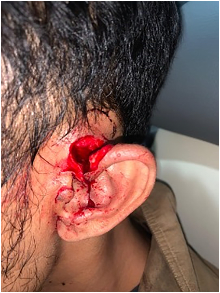
Clinical Documentation of the partial auricular avulsion before the surgery.
The patient immediately received tetanus vaccine for tetanus prevention, tranexamic acid for the active bleeding, and ketorolac for the pain then underwent wound debridement and simple reattachment using a 5–0 monofilament suture. The simple reattachment was done with the help of local anesthesia using lidocaine. There was no bleeding and complications after the treatment (Fig. 2). Upon discharge, the patient received a controlled dose of oral clindamycin, oral tranexamic acid, and oral diclofenac. The patient was followed-up after 3 months with no deformities, cauliflower ear and defect by necrosis (Fig. 3, Fig. 4).
Fig. 2.

Clinical Documentation of the auricle after the surgery.
Fig. 3.

Clinical Documentation of the ear following 3 months after the simple reattachment (anterior).
Fig. 4.

Clinical Documentation of the ear following 3 months after the simple reattachment (posterior).
Discussion
Simple reattachment is one of the most straightforward approaches, with a short operation time and usually brief bedside management [4]. It is heavily considered in the case of incomplete ear separation presented with adequate vascular supply, usually shown in the presence of active bleeding from the avulsed part. Heavy venous congestion, contamination, or less neat, crushing injuries should be avoided when considering simple reattachment. On the other hand, a complete avulsion of the ear should be approached with a more complex treatment, either with a secondary, two-stage reconstruction or additional microsurgery [5] The main reason for simple reattachment as the main approach in partial auricular avulsions is that the small pedicle remaining might provide an adequate vascular supply, regardless of the position of the pedicle. The confirmed presence of the helical arcade supplying even the outermost part of the helix gives one surgeon the option of more simple form of treatment without the need of a more complex surgery, as with the simple reattachment [6,7]. The location and width of the pedicle left in partial auricular avulsion might affect the outcomes after simple reattachment. In two cases in which pedicles were only 5 mm, the patient experienced venous congestion postoperatively. Both were attached to the superior helix [7,8]. Moreover, one case with a pedicle site of 7 mm at the superior site of the base of the helix, had a complication of the necrotic area of the lobule [4]. Four cases in the D'Arcangelo et al. had pedicles in the inferior site, ranging from 6 mm to 25 mm. All of them did not experience any complication [4]. Anatomically, the superior temporal artery (mainly supplying from the superior part) and the posterior auricular artery (mainly supplying from the inferior part) both contribute to the helical arcade [9]. However, pedicle in the inferior site and wider pedicle width, as also shown in our study, might have better prognosis.
Injury to surgery time might also play a role in the outcomes in partial auricular avulsion, especially receiving simple reattachment. In the included studies, all cases experiencing complications were admitted to the hospital five hours or more (5 to 22 h) [7,4,8,10]. A recent review by Al-Ali et al. explained the non-essential role of injury to surgery time due to the low metabolic demand of the ear, however, had increased rate of venous congestion [3]. Several detailed technical aspects might be considered in conducting the simple reattachment technique. Preoperative intravenous broad-spectrum antibiotics, which had not been done in our case, might be considered [4]. Although both local and general anesthesia might be applied, local anesthesia should be used with caution due to the aggravation of edema and the risk of vascular damage [4]. As our study demonstrated, a 5-0 suture for the cartilage and the skin is sufficient to give adequate result [4]. Despite our patient showing no complication without using an anticoagulant, a postoperative adjuvant of anticoagulant might be crucial and should always be considered. Previous studies showed the absence of venous congestion with the use of postoperative anticoagulant, while it presented in some cases not mentioning any use of immediate anticoagulant treatmen [4]. Several complications might occur in partial auricular avulsion receiving simple reattachment. In the retrospective study by D'Arcangelo et al., one patient experienced a postoperative necrotic lobule area. This patient received the longest injury-to-surgery time (5 h), with only a tiny pedicle width (7 mm) in the superior location that might barely supply the lobule area [10]. Both of the studies by Albdour et al. and Erdmann et al. showed venous congestion as a postoperative complication, showing complete resolution through different approaches. Proper wound care, aggressive rewarming, and local low-molecular-weight heparin (LMWH) might be used effectively against severe venous congestion [8]. Another option to tackle venous congestion is through leech therapy, advantaging the vasodilating, increasing vascular permeability, and anticoagulating properties of their saliva. Intermittent leech therapy for three days showed an improvement of post-simple reattachment congestion [7].
Conclusion
A multiple operation might provide a better result, single operation requires lesser operative time and minimal in-patient admission. Preserved pedicles, especially the inferior site, shorter injury to surgery time, and careful considerations in conducting the simple reattachment and the postoperative treatments might improve the outcomes in partial auricular avulsion. As our study suggested the efficacy of simple reattachment for partial auricular avulsions, further study with higher quality of evidence is currently needed.
Declaration of competing interest
The authors declare the following financial interests/personal relationships which may be considered as potential competing interests: We wish to confirm that there are no known conflicts of interest associated with this publication and there has been no significant financial support for this work that could have influenced its outcome.
References
- 1.Kolodzynski M.N., Kon M., Egger S., Breugem C.C. Mechanisms of ear trauma and reconstructive techniques in 105 consecutive patients. Eur. Arch. Otorhinolaryngol. 2017;274:723–728. doi: 10.1007/s00405-016-4299-4. [DOI] [PMC free article] [PubMed] [Google Scholar]
- 2.Gailey A.D., Farquhar D., Clark J.M., Shockley W.W. Auricular avulsion injuries and reattachment techniques: a systematic review. Laryngoscope Investig. Otolaryngol. 2020;5:381–389. doi: 10.1002/lio2.372. [DOI] [PMC free article] [PubMed] [Google Scholar]
- 3.Al-Ali M.A., Abu-Zidan F.M. Auricular avulsion injuries: literature review and management algorithm. Turk. J. Emerg. Med. 2022;22:59–66. doi: 10.4103/2452-2473.342811. [DOI] [PMC free article] [PubMed] [Google Scholar]
- 4.D’Arcangelo M., Al-Ali M.A., Abu-Zidan F.M. Primary reattachment of near-complete ear amputation: a successful outcome. Ear Nose Throat J. 2022;101:NP436–NP440. doi: 10.1177/0145561320982170. [DOI] [PubMed] [Google Scholar]
- 5.Zhang C., Teng L., Xu J.-J., Lu J.-J., Xie F., Yang L.-Y., et al. Incomplete ear amputation. J. Craniofac. Surg. 2018;29:2231–2233. doi: 10.1097/SCS.0000000000005054. [DOI] [PubMed] [Google Scholar]
- 6.Zilinsky I., Erdmann D., Weissman O., Hammer N., Sora M.-C., Schenck T.L., et al. Reevaluation of the arterial blood supply of the auricle. J. Anat. 2017;230:315–324. doi: 10.1111/joa.12550. [DOI] [PMC free article] [PubMed] [Google Scholar]
- 7.Erdmann D., Bruno A.D., Follmar K.E., Stokes T.H., Gonyon D.L., Marcus J.R. The helical arcade: anatomic basis for survival in near-total ear avulsion. J. Craniofac. Surg. 2009;20:245–248. doi: 10.1097/SCS.0b013e318184343a. [DOI] [PubMed] [Google Scholar]
- 8.Albdour M., Ammar H.M., Alnaser M.M.S., Alzaben F.S., Malek S. Non-microvascular successful Management of Near-total ear Avulsion. Plast. Reconstr. Surg. - Glob. Open. 2021;9 doi: 10.1097/GOX.0000000000003386. [DOI] [PMC free article] [PubMed] [Google Scholar]
- 9.Armin B.B., Ruder R.O., Azizadeh B. Partial auricular reconstruction. Semin. Plast. Surg. 2011;25:249–256. doi: 10.1055/s-0031-1288916. [DOI] [PMC free article] [PubMed] [Google Scholar]
- 10.Zhu J., Zhao H., Wu K., Lv C., Bi H., Sun M., et al. Reconstruction of auricular conchal defects with local flaps. Medicine. 2016;95 doi: 10.1097/MD.0000000000005282. [DOI] [PMC free article] [PubMed] [Google Scholar]