Abstract
目的
通过系统评价及Meta分析的方法评估在后牙区骨量不足植入4 mm超短种植体(ESI)的安全性和效用。
方法
电子检索2010年1月1日—2022年8月31日PubMed、Embase、Cochrane Library、Web of Science、CNKI、万方数据库有关ESI与标准种植体(SI)(≥8 mm)的随机对照试验或临床对照试验,并进行引文检索。应用RevMan 5.4版软件进行Meta分析。
结果
共11篇研究符合纳入标准,其中随机对照试验6篇,临床对照试验5篇。Meta分析结果:在后牙区种植时,ESI与SI的存留率差异无统计学意义[RR=1.23,95%CI(0.66,2.27),P=0.52];ESI表现出更稳定的边缘骨水平[MD=−0.16,95%CI(−0.25,−0.07),P=0.000 7],更少的生物并发症[RR=0.34,95%CI(0.19,0.62),P=0.000 4],然而机械并发症更多[RR=2.89,95%CI(1.05,7.92),P=0.04]。
结论
基于有限的证据,在后牙牙槽嵴高度低于5 mm时应用ESI可以获得媲美SI的临床效果,且具备技术敏感性低、术后生物并发症少等优势。由于ESI长期随访证据不足、样本量有限,仍需要更多临床试验评估ESI的长期效用。
Keywords: 超短种植体, 标准种植体, 存留率, 边缘骨吸收, 并发症, Meta分析
Abstract
Objective
This study aimed to systematically evaluate the safety and clinical efficacy of 4 mm-extra-short implant (ESI) placement in severely atrophic posterior areas.
Methods
Databases of PubMed, Embase, Cochrane Library, Web of Science, CNKI, and Wanfang from January 1, 2010, until August 31, 2022, were searched to identify randomized controlled trials or controlled clinical trials related to ESI and standard implants (SI). An additional hand search of the references of included articles was also conducted. Meta-analyses were carried out with RevMan 5.4 software.
Results
A total of 11 studies were included, involving six randomized controlled trials and five controlled clinical trials. The meta-analyses indicated that when implants were placed in the posterior area, the implant survival rate between ESI and SI did not significantly differ [RR=1.23, 95%CI (0.66, 2.27), P=0.52]. ESI resulted in significantly stable marginal bone level [MD=−0.16, 95%CI (−0.25, −0.07), P=0.000 7] and less biological complications [RR=0.34, 95%CI (0.19, 0.62), P=0.000 4] but more mechanical complications [RR=2.89, 95%CI (1.05, 7.92), P=0.04].
Conclusion
Based on the limited evidence, ESI could achieve clinical outcomes similar to those of SI when the height of the posterior alveolar bone is less than 5 mm, with lower technical sensitivity and fewer postoperative clinical complications than SI. Due to insufficient evidence and limited sample size, further clinical trials are needed to verify the long-term efficacy of ESI.
Keywords: extra-short implant, standard implant, survival rate, marginal bone resorption, complications, Meta-analysis
种植体的植入需要牙槽嵴具备足够的三维骨量,然而缺牙后上颌窦气化、废用性萎缩吸收等常导致牙槽嵴垂直骨量不足。临床上通过上颌窦底提升、下颌神经移位术、块状骨移植术等[1]–[3]扩增垂直骨量以满足标准种植体(standard implant,SI)放置,但存在术后并发症多、技术敏感性高、额外费用等缺陷[4]。如何在牙槽嵴高度不足以植入SI的条件下,简化种植程序、提高舒适度且获得预期的种植成功率一直是如今研究的热点。
已有大量学者[5]–[7]提出在后牙区植入6~8 mm的短种植体可以获得与SI相当的表现,然而在不足5 mm的牙槽嵴高度下仍存在局限性。得益于种植体设计和表面处理的改良和进步,4 mm超短种植体(extra-short implant,ESI)被逐渐应用于临床并取得了一定疗效。Leighton等[8]的一项前瞻性队列研究报道,在18名患者的下颌后牙区植入18颗ESI,种植体3年存活率为100%。Barausse等[9]的一项随机对照试验报道,在80名患者的萎缩后牙区植入80颗ESI,种植体5年存活率为92%。然而,ESI的冠根比、修复方式等一直存在争议[10]。此外,ESI在种植成功率、骨结合稳定性、术后并发症等方面是否与SI具有可比性仍有待商榷。本研究旨在比较ESI与SI在种植体存活率、边缘骨吸收以及机械和生物并发症,综合评价ESI的中长期效用,为ESI的临床应用提供理论依据。
1. 材料和方法
1.1. 文献纳入与排除标准
文献纳入标准遵循PICOS框架:1)研究对象(participant):上颌或下颌后牙缺失允许ESI的植入或附加骨增量手术后可植入SI的患者;2)干预(intervention):后牙区植入ESI(L=4 mm);3)对照(comparison):后牙区植入SI(L≥8 mm);4)结果(outcomes):种植体存留率、边缘骨吸收、机械和生物并发症。5)研究设计(study design):随机对照试验(randomized controlled trial,RCT)和临床对照试验(controlled clinical trial,CCT)。
文献排除标准:1)综述、动物试验、摘要;2)研究未报道所需结局指标;3)纳入研究的样本量过少(<5)、随访时间不足(<1年)。
1.2. 文献检索
电子检索2010年1月1日—2022年8月31日PubMed、Embase、Cochrane Library、Web of Science、CNKI、万方数据库有关ESI的RCT或CCT,并进行引文检索。中文数据库以“短种植体”、“超短种植体”、“边缘骨吸收”、“存留率”主题词进行检索。英文数据库检索策略如下(以PubMed为例):(“implant dental”[Title/Abstract] OR “implants dental”[Title/Abstract] OR “dental implant”[Title/Abstract] OR “Dental Implants”[MeSH Terms]) AND (“ultra-short”[Title/Abstract] OR “ultrashort”[Title/Abstract] OR “extra-short”[Title/Abstract] OR “4-mm”[Title/Abstract] OR “super-short”[Title/Abstract]) AND “clinical trial”[Publication Type]) AND (2010:2022[pdat]。
1.3. 文献筛选和资料提取
2名研究员(GJM和YJY)独立筛选并交叉核对,如遇到分歧通过协商解决,若无法达成一致,则由第三方(ZRM)参与讨论裁定。第一步审阅标题和摘要,对检索结果进行初筛,第二步阅读全文确定纳入的文献,同时对纳入文献的关键数据和相关信息进行汇总。使用Kappa系数计算研究员间关于研究纳入的一致程度。提取的关键信息包括:作者、发表年份、研究类型、种植区、术后修复时机、骨增量程序、结局指标、样本量。主要结局指标:种植牙存留率、边缘骨吸收;次要结局指标:机械并发症(修复体松动、折断、崩裂和脱落等)和生物并发症(疼痛、渗出、种植体周黏膜炎和种植体周围炎等)。
1.4. 文献质量评估
采用Newcastle-Ottawa Scale(NOS)评估CCT的偏倚风险,主要项目包括:研究对象选择(4分),组间可比性(2分)和结果测量(3分),该量表满分为9分,超过6分属于高质量证据,3~6分存在中等偏倚风险,低于3分存在高偏倚风险。
用Cochrane协作工具评估纳入RCT的偏倚风险。主要评估6个指标:随机序列产生(选择性偏倚)、分配隐藏(选择性偏倚)、盲法(患者盲法、结果评估者盲法)、不完整结果数据(失访偏倚)、选择性结果报道(报告偏倚)和其他偏倚。满足所有指标或一个指标不明确/不满足属于高质量证据;2个指标不明确/不满足存在中等偏倚风险;2个以上指标不明确/不满足或者存在1个及以上高风险指标表示高偏倚风险。
1.5. 统计学方法
所有统计分析使用RevMan 5.4软件进行。二分类变量数据采用相对危险度(risk ratio,RR)作为效应指标,连续变量数据选用均数差(mean difference,MD),并计算相应的95%可信区间(confidence interval,CI),设定P<0.05为差异有统计学意义。采用Q检验、I2检验来分析研究间的异质性,当各研究间异质性不明显时(P>0.05,I2≤50%),采用固定效应模型;当各研究间异质性明显时(P<0.05,I2≥50%),采用随机效应模型。异质性明显的结果采用敏感性分析、亚组分析。
2. 结果
2.1. 文章检索及偏倚风险评估结果
共检索相关文献1 494篇,其中PubMed 97篇、Embase 357篇、The Cochrane Library 444篇、Web of Science 564篇、CNKI 23篇、万方9篇。
剔除重复文献后剩余1 372篇,阅读文题和摘要后初筛得到52篇,审阅全文复筛得到20篇,最终纳入Meta分析11篇[9],[11]–[20](表1)。研究员间一致性K值为0.865(标题和摘要初筛)和0.894(全文复筛),表明研究员间结果一致性良好。偏倚风险评估结果:6项RCT[9],[12]–[13],[16],[19]–[20]属于中等偏倚风险;3项CCT[14],[17]–[18]属于高质量证据,2项CCT[11],[15]被评估为中等偏倚风险。
表 1. 纳入研究的基本特征.
Tab 1 Characteristics of the included studies
| 作者/年份 | 研究类型 | 种植区 | 样本量 | 种植体尺寸(直径×长度/mm) | 术后修复时机/月 | 骨增量程序(对照组) | 随访/年 | 结局指标 |
| Barausse2019[20] | RCT | 下颌 | ESI:124 | ESI:4.0×4.0 | 4 | — | 3 | ①②③④ |
| SI:116 | SI:4.0×8.5,10,11.5,13 | |||||||
| Barausse2022[9] | RCT | 全口 | ESI:80 | ESI:4,4.5×4 | 4 | 骨块移植术; | 5 | ①②③④ |
| SI:87 | SI:4×10,11.5,13 | 上颌窦底提升术 | ||||||
| Bolle2018[19] | RCT | 全口 | ESI:43 | ESI:4.0,4.5×4.0 | 4 | 骨块移植术; | 1 | ①②③④ |
| SI:46 | SI:4.0×8.5,10,11.5,13 | 上颌窦底提升术 | ||||||
| Calvo-Guirado2016[18] | CCT | 下颌 | ESI:40 | ESI:4.1×4.0 | 3 | — | 1 | ①②③④ |
| SI:20 | SI:4.1×10 | |||||||
| Estévez-Pérez2020[17] | CCT | 上颌 | ESI:16 | ESI:4.8×4.0 | 3 | — | 3 | ①③④ |
| SI:16 | SI:4.8×8,10,12 | |||||||
| Felice2016[16] | RCT | 全口 | ESI:124 | ESI:4.0×4.0 | 4 | — | 1 | ①②③④ |
| SI:116 | SI:4.0×8.5,10,11.5,13 | |||||||
| Gašperšič2021[15] | CCT | 上颌 | ESI:17 | ESI:4.1×4.0 | 6 | — | 1 | ①③④ |
| SI:11 | SI:4.1×10 | |||||||
| Karcı2021[14] | CCT | 下颌 | ESI:30 | ESI:4.1×4.0 | — | — | 3 | ①③④ |
| SI:30 | SI:4.1×8,10,12 | |||||||
| Rokn2018[13] | RCT | 全口 | ESI:25 | ESI:4.1×4.0 | 2 | 骨块移植术 | 1 | ①②③④ |
| SI:22 | SI:4.1×8.0,10 | |||||||
| Rossi2021[12] | RCT | 上颌 | ESI:12 | ESI:4.1×4.0 | 1.5 | 上颌窦底提升术 | 2 | ①②③④ |
| SI:10 | SI:3.3,4.1×10 | |||||||
| Torassa2020[11] | CCT | 上颌 | ESI:11 | ESI:4.1×4.0 | 6 | — | 2 | ①②③④ |
| SI:11 | SI:3.3,4.1×10 |
注:①:种植体存留率;②:边缘骨吸收;③:机械并发症;④:生物并发症。
2.2. Meta分析结果
2.2.1. 种植体存留率
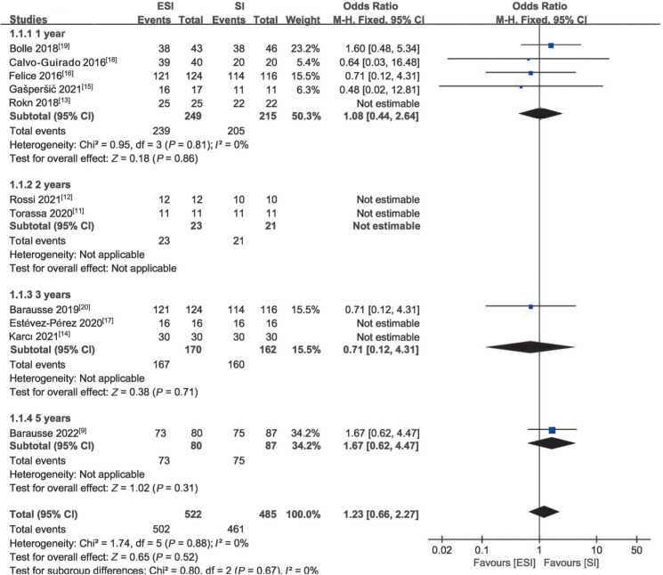
所有纳入文献均报道了这一结局指标。各研究间同质性好(P=0.88,I2=0%),采用固定效应模型进行合并分析。考虑到随访时间不同可能造成结果偏倚,故根据随访时间进行亚组分析。结果表明:ESI的种植体存留率与SI之间差异无统计学意义[RR=1.23,95%CI(0.66,2.27),P=0.52](图1)。
图 1. 种植体存留率的Meta分析.
Fig 1 Meta-analysis of implant survival rate
2.2.2. 种植体周边缘骨吸收
8篇文献报告这一指标,其中6篇RCT,2篇CCT。由于各研究间高度异质性(P=0.000 3,I2=74%),采用随机效应模型。Meta分析表明,ESI的边缘骨吸收量少于SI,差异具有统计学意义[MD=−0.16,95%CI(−0.25,−0.07),P=0.000 7](表2)。
表 2. 种植体边缘骨吸收、并发症的Meta分析.
Tab 2 Meta-analysis of marginal bone lossand complications
| 结局指标 | 亚组 | 纳入研究数 | 效应模型 | MD/RR | 95%CI | P值 |
| 边缘骨吸收 | 1年 | 4[13],[16],[18]–[19] | 随机 | −0.13 | −0.22~−0.05 | 0.003 |
| 2年 | 2[11]–[12] | 随机 | −0.14 | −0.62~0.34 | 0.57 | |
| 3年 | 1[20] | 随机 | −0.06 | −0.14~0.02 | 0.14 | |
| 5年 | 1[9] | 随机 | −0.41 | −0.56~−0.26 | <0.000 01 | |
| 合并效应值 | −0.16 | −0.25~−0.07 | <0.000 7 | |||
| 机械并发症 | 1年 | 5[13],[15]–[16],[18]–[19] | 固定 | 1.65 | 0.26~10.39 | 0.59 |
| 2年 | 2[11]–[12] | 固定 | − | − | − | |
| 3年 | 3[14],[17],[20] | 固定 | 10.72 | 0.59~196.13 | 0.11 | |
| 5年 | 1[9] | 固定 | 2.27 | 0.55~9.40 | 0.26 | |
| 合并效应值 | 2.89 | 1.05~7.92 | 0.04 | |||
| 生物并发症 | 1年 | 5[13],[15]–[16],[18]–[19] | 固定 | 0.19 | 0.07~0.53 | 0.002 |
| 2年 | 2[11]–[12] | 固定 | 3.00 | 0.26~34.57 | 0.38 | |
| 3年 | 3[14],[17],[20] | 固定 | 0.93 | 0.23~3.82 | 0.92 | |
| 5年 | 1[9] | 固定 | 0.26 | 0.09~0.73 | 0.01 | |
| 合并效应值 | 0.34 | 0.19~0.62 | 0.000 4 |
注:种植体周边缘骨吸收采用MD;机械并发症和生物并发症采用RR。
2.2.3. 并发症
1)机械并发症11篇纳入文献报道了机械并发症。对2组机械并发症进行Meta分析,各研究间同质性好(P=0.54,I2=0%),选择固定效应模型。合并结果表明与SI相比,ESI的机械并发症发生率较高[RR=2.89,95%CI(1.05,7.92),P=0.04](表2)。
2)生物并发症11篇纳入文献报道了生物并发症。由于各研究间同质性较好(P=0.10,I2=47%),采用固定效应模型。合并结果表明与SI相比,ESI的生物并发症发生率较低[RR=0.34,95%CI(0.19,0.62),P=0.000 4](表2)。
2.3. 敏感性分析
仔细阅读11篇文献全文,核对研究数据是否正确,以及纳入标准、研究设计是否存在问题,核查结果显示无误,各文献质量较高,研究的可信度高。依次剔除纳入研究的各个文献,发现各组结果均无明显变化,说明Meta分析结果稳定。
2.4. 发表偏倚分析
以种植体存留率为例,对术后种植体存留率结果进行发表偏倚风险分析,结果显示漏斗图无明显不对称,表明发表偏倚对最终结果几乎无影响,总体纳入研究中高样本量的研究居多,Meta分析的结果较为可靠。
3. 讨论
以往涉及ESI的Meta分析[21]仅纳入4项研究,评估了301名患者及321颗植体,随访时间为1年,有限的样本量和随访时间使得结果的可靠性略不足。本研究共纳入11项研究,6项为RCT,包括560名患者,共植入1 007颗种植体,随访时间为1~5年,提高了统计学效能。此外,考虑到随访时间不同可能造成结果偏倚,故本研究根据随访时间进行亚组分析,提高了结果可靠性。
3.1. 存留率
Meta分析结果表明ESI与SI在治疗牙列缺失的患者中成功率无差异,这一结果与几项研究[9],[11],[14]的结果一致。有学者[22]–[23]认为,种植体成功率与种植体长度的相关性有限,短种植体的应用也佐证了这一观点。Naguib等[24]研究发现,种植体的长度每增加3 mm,其表面积可增加10%;种植体直径每增加0.25 mm,其表面积可增加10%,认为扩增种植体直径较仅扩增种植体长度能获得更多的骨-种植体接触面积。Gümrükçü等[25]通过有限元分析方法评估了种植体承受负荷时的应力分布,发现种植体颈部3 mm区域是应力集中处,3 mm之外的区域的应力分布明显衰减。这被概括为“有效骨-种植体表面”并逐渐被认可[26]。简单地说,维持种植体颈部足够的有效骨-种植体表面,能够更有效地适应种植体所受的负荷,这侧面印证了ESI的设计理念。
需要指出的是,文献报告的种植体失败绝大多数发生在早期,失败原因可能与初期稳定性不足和早期愈合欠佳有关。有学者[27]提出,即刻/早期负重种植技术易导致种植体与周围骨组织之间形成纤维性愈合,本研究纳入的文献均采用延期负荷,在一定程度上降低即刻/早期负荷带来的种植体失败风险,从而提高了ESI存留率,因此建议慎重选择负载时机。
3.2. 种植体周边缘骨吸收
种植体边缘骨吸收程度是衡量种植体长期成功的重要参考。本研究显示,ESI修复完成1~5年内边缘骨吸收都小于1 mm且低于SI。基于2018年ITI共识[28]标准,种植修复后第一年内骨吸收<1 mm,以后每年的吸收不超过0.2 mm,表明ESI应用于后牙的疗效可以达到预期。造成这一结果主要有两个方面:一方面,ESI的宽直径增加了与颈部皮质骨接触的功能表面积,减少了颈部单位面积的应力分布,有助于提高初始稳定性并减少边缘骨吸收[29]–[30];另一方面,可能与在修复结构上,往往注意使用联冠修复、减少修复冠的颊舌径及冠高度、降低牙尖斜度等措施有关[6]–[7]。
3.3. 并发症
并发症包括机械并发症和生物并发症,前者是对种植体及其相关部件机械损伤的统称,后者是指种植术后出现疼痛、感染等不适症状[31]–[32]。基于3项研究[9],[19]–[20]报道的机械并发症和6项研究[9],[12]–[13],[16],[19]–[20]报道的生物并发症证据,Meta分析结果显示与SI相比,ESI的机械并发症发生率较高,而生物并发症的发生率较低。
种植修复常见的机械并发症有:冠松动/折裂、螺丝松动/折断、基台松动/折裂、种植体折裂等。本文3项研究[9],[19]–[20]共报道ESI组522个种植体或修复冠中14个(2.7%)出现机械并发症,包括7个种植体螺丝松动、1个螺丝断裂、2个冠松动、4个冠折裂。与机械并发症直接相关的是种植义齿修复部件所受咬合应力的大小。根据有限元分析[22],轴向负荷下,应力集中在种植体螺丝及基台与种植体界面之间,不同冠种植体比率(crown‑implant ratio,C/I)对其影响不大;但在非轴向负荷下,C/I增大,则会增大杠杆臂而显著影响以上两个界面的应力分布。临床上往往通过联冠修复、减少修复冠颊舌径、降低牙尖斜度等方式减少轴向与非轴向负荷咬合应力,尽可能减少机械并发症的发生。da Rocha等[33]认为当冠高大于15 mm时会增加机械并发症的发生率,在牙槽骨吸收严重、上下咬合间距增大的病例中,选择ESI必然导致C/I的过分增大,这时就需要考虑是否选择非种植的修复方式效果更佳。尽管部分学者[23],[34]认为C/I与机械并发症的发生并无显著相关性,为安全起见,仍建议在选择ESI时根据C/I制定个性化修复方案,尽可能减少非轴向咬合力诱发ESI机械并发症的风险。
本研究在种植体水平评估了每一类生物并发症,ESI组522个种植体中发生生物并发症16个(3.1%),其中包括疼痛10个、种植体周黏膜炎6个;SI组485个种植体发生生物并发症43个(8.9%),其中包括疼痛31个、渗出1个、种植体周黏膜炎6个、膜暴露5个。不少临床研究[23],[34]–[35]表明C/I增加似乎与种植体生物并发症无显著性关系,与本研究结果一致。SI组生物并发症数量更多可能与纳入研究中颌骨严重萎缩病例需要附加骨增量手术相关,另外,单冠常规修复相较于联冠修复可能具有更高的种植体并发症风险[6],[36]。就修复方式对SI和ESI组生物并发症的影响而言,需要设计更加完善的临床试验,其一,规范修复方式以便于定性和定量分析;其二,将修复方式作为重要混杂因素进行分层分析,确定其对不同种植体长度临床效果的影响。
在严格把握适应证的情况下,ESI更大限度地避免了上颌窦底提升、引导性骨再生、下颌神经管移位术等创伤较大的术式,将复杂手术程序转化为简单程序,进一步降低技术敏感性,减轻患者术后反应,并在一定程度上扩大了后牙区种植修复的适应证。但要注意以下临床要点:上颌后牙区骨质通常较为疏松,皮质骨少,使用ESI的风险较高;在可用骨宽度允许下应尽可能选择较宽的种植体;在修复结构上,使用联冠修复、减少修复冠的颊舌径及冠高度、降低牙尖斜度等有助于减少并发症。
虽然本研究纳入了较高质量的RCT,但存在一定的局限性:1)各研究间的结局指标不尽相同,部分评估指标无法进行Meta分析;2)部分研究的样本量较少、随访时间相对较短。因此,仍需要更多高质量、随访时间长的临床研究进一步评估ESI临床效用。
综上所述,基于有限的证据,在后牙牙槽嵴高度低于5 mm时应用ESI可以获得媲美SI的临床效果,且具备技术敏感性低、术后生物并发症少等优势。由于ESI长期随访证据不足、样本量有限,仍需要更多临床试验评估ESI的长期效用。
Funding Statement
[基金项目] 兰州大学口腔医院科研基金(lzukqky-2020-t07,lzukqky-2022-t04)
Supported by: Hospital of Stomatology Lanzhou University Scientific Research Project (lzukqky-2020-t07, lzukqky-2022-t04).
Footnotes
利益冲突声明:作者声明本文无利益冲突。
References
- 1.史 远, 杨 国利. 外斜线取骨自体骨移植及其他牙槽嵴骨增量法的研究进展[J] 口腔医学. 2021;41(6):557–560, 571. [Google Scholar]; Shi Y, Yang GL. Research progress of autogenous bone graft from mandibular lateral oblique line and other alveolar ridge augmentation methods[J] Stomatology. 2021;41(6):557–560, 571. [Google Scholar]
- 2.范 震, 刘 月, 王 佐林. 牙列缺失倾斜种植设计[J] 华西口腔医学杂志. 2021;39(4):377–385. doi: 10.7518/hxkq.2021.04.002. [DOI] [PMC free article] [PubMed] [Google Scholar]; Fan Z, Liu Y, Wang ZL. Tilted implantation technique for edentulous patients[J] West China J Stomatol. 2021;39(4):377–385. doi: 10.7518/hxkq.2021.04.002. [DOI] [PMC free article] [PubMed] [Google Scholar]
- 3.Romanos GE. Severe atrophy of the posterior mandible and inferior alveolar nerve transposition[J] Int J Periodontics Restorative Dent. 2021;41(5):e199–e204. doi: 10.11607/prd.5782. [DOI] [PubMed] [Google Scholar]
- 4.张 富贵, 宿 玉成, 邱 立新, et al. 牙槽骨缺损骨增量手术方案的专家共识[J] 口腔疾病防治. 2022;30(4):229–236. [Google Scholar]; Zhang FG, Su YC, Qiu LX, et al. Expert consensus on the bone augmentation surgery for alveolar bone defects[J] J Dent Prevent Treat. 2022;30(4):229–236. [Google Scholar]
- 5.Lozano-Carrascal N, Anglada-Bosqued A, Salomó-Coll O, et al. Short implants (<8 mm) versus longer implants (≥8 mm) with lateral sinus floor augmentation in posterior atrophic maxilla: a meta-analysis of RCT s in humans[J] Med Oral Patol Oral Cir Bucal. 2020;25(2):e168–e179. doi: 10.4317/medoral.23248. [DOI] [PMC free article] [PubMed] [Google Scholar]
- 6.Li QL, Yao MF, Cao RY, et al. Survival rates of splinted and nonsplinted prostheses supported by short dental implants (≤8.5 mm): a systematic review and meta-analysis [J] J Prosthodont. 2022;31(1):9–21. doi: 10.1111/jopr.13402. [DOI] [PubMed] [Google Scholar]
- 7.Afrashtehfar KI, Katsoulis J, Koka S, et al. Single versus splinted short implants at sinus augmented sites: a systematic review and meta-analysis[J] J Stomatol Oral Maxillofac Surg. 2021;122(3):303–310. doi: 10.1016/j.jormas.2020.08.013. [DOI] [PubMed] [Google Scholar]
- 8.Leighton Y, Carpio L, Weber B, et al. Clinical evaluation of single 4-mm implants in the posterior mandible: a 3-year follow-up pilot study[J] J Prosthet Dent. 2022;127(1):80–85. doi: 10.1016/j.prosdent.2020.06.039. [DOI] [PubMed] [Google Scholar]
- 9.Barausse C, Pistilli R, Canullo L, et al. A 5-year randomized controlled clinical trial comparing 4-mm ultrashort to longer implants placed in regenerated bone in the posterior atrophic jaw[J] Clin Implant Dent Relat Res. 2022;24(1):4–12. doi: 10.1111/cid.13061. [DOI] [PubMed] [Google Scholar]
- 10.路 泊遥, 杨 大维, 刘 蔚晴, et al. 超短种植体临床应用效果的影响因素[J] 国际口腔医学杂志. 2021;48(3):329–333. [Google Scholar]; Lu BY, Yang DW, Liu WQ, et al. Influencing factors of the clinical application effect of ultrashort implant[J] Int J Stomatol. 2021;48(3):329–333. [Google Scholar]
- 11.Torassa D, Naldini P, Calvo-Guirado JL, et al. Prospective, clinical pilot study with eleven 4-mm extra-short implants splinted to longer implants for posterior maxilla rehabilitation[J] J Clin Med. 2020;9(2):357. doi: 10.3390/jcm9020357. [DOI] [PMC free article] [PubMed] [Google Scholar]
- 12.Rossi F, Tuci L, Ferraioli L, et al. Two-year follow-up of 4-mm-long implants used as distal support of full-arch FDPs compared to 10-mm implants installed after sinus floor elevation. A randomized clinical trial[J] Int J Environ Res Public Health. 2021;18(7):3846. doi: 10.3390/ijerph18073846. [DOI] [PMC free article] [PubMed] [Google Scholar]
- 13.Rokn AR, Monzavi A, Panjnoush M, et al. Comparing 4-mm dental implants to longer implants placed in augmented bones in the atrophic posterior mandibles: one-year results of a randomized controlled trial[J] Clin Implant Dent Relat Res. 2018;20(6):997–1002. doi: 10.1111/cid.12672. [DOI] [PubMed] [Google Scholar]
- 14.Karcı BL, Oncu E. Comparison of osteoimmunological and microbiological parameters of extra short and longer implants loaded in the posterior mandible: a split mouth randomized clinical study[J] Acta Stomatol Croat. 2021;55(3):238–247. doi: 10.15644/asc55/3/1. [DOI] [PMC free article] [PubMed] [Google Scholar]
- 15.Gašperšič R, Dard M, Linder S, et al. One-year results assessing the performance of prosthetic rehabilitations in the posterior maxilla supported by 4-mm extrashort implants splinted to 10-mm implants: a prospective case series[J] Int J Oral Maxillofac Implants. 2021;36(2):371–378. doi: 10.11607/jomi.8645. [DOI] [PubMed] [Google Scholar]
- 16.Felice P, Checchi L, Barausse C, et al. Posterior jaws rehabilitated with partial prostheses supported by 4.0×4.0 mm or by longer implants: one-year post-loading results from a multicenter randomised controlled trial[J] Eur J Oral Implantol. 2016;9(1):35–45. [PubMed] [Google Scholar]
- 17.Estévez-Pérez D, Bustamante-Hernández N, Labaig-Rueda C, et al. Comparative analysis of peri-implant bone loss in extra-short, short, and conventional implants. a 3-year retrospective study[J] Int J Environ Res Public Health. 2020;17(24):9278. doi: 10.3390/ijerph17249278. [DOI] [PMC free article] [PubMed] [Google Scholar]
- 18.Calvo-Guirado JL, López Torres JA, Dard M, et al. Evaluation of extrashort 4-mm implants in mandibular edentulous patients with reduced bone height in comparison with standard implants: a 12-month results [J] Clin Oral Implants Res. 2016;27(7):867–874. doi: 10.1111/clr.12704. [DOI] [PubMed] [Google Scholar]
- 19.Bolle C, Felice P, Barausse C, et al. 4 mm long vs longer implants in augmented bone in posterior atrophic jaws: 1-year post-loading results from a multicentre randomised controlled trial[J] Eur J Oral Implantol. 2018;11(1):31–47. [PubMed] [Google Scholar]
- 20.Barausse C, Felice P, Pistilli R, et al. Posterior jaw rehabilitation using partial prostheses supported by implants 4.0×4.0 mm or longer: three-year postloading results of a multicentrerandomised controlled trial[J] Clin Trials Dent. 2019;1:25–36. [Google Scholar]
- 21.Moraschini V, Mourão CFAB, Montemezzi P, et al. Clinical comparation of extra-short (4 mm) and long (>8 mm) dental implants placed in mandibular bone: a systematic review and metanalysis[J] Healthcare (Basel) 2021;9(3):315. doi: 10.3390/healthcare9030315. [DOI] [PMC free article] [PubMed] [Google Scholar]
- 22.Elias D, Valerio CS, de Oliveira DD, et al. Evaluation of different heights of prosthetic crowns supported by an ultra-short implant using three-dimensional finite element analysis[J] Int J Prosthodont. 2020;33(1):81–90. doi: 10.11607/ijp.6247. [DOI] [PubMed] [Google Scholar]
- 23.Ercal P, Taysi AE, Ayvalioglu DC, et al. Impact of peri-implant bone resorption, prosthetic materials, and crown to implant ratio on the stress distribution of short implants: a finite element analysis[J] Med Biol Eng Comput. 2021;59(4):813–824. doi: 10.1007/s11517-021-02342-w. [DOI] [PubMed] [Google Scholar]
- 24.Naguib GH, Hashem AB, Natto Z, et al. The effect of Implant length and diameter on stress distribution of tooth-implant and implant supported prostheses:an in-vitro finite element study[J] J Oral Implantol. 2021 Dec 22; doi: 10.1563/aaid-joi-D-21-00023. [DOI] [PubMed] [Google Scholar]
- 25.Gümrükçü Z, Korkmaz YT. Influence of implant number, length, and tilting degree on stress distribution in atrophic maxilla: a finite element study[J] Med Biol Eng Comput. 2018;56(6):979–989. doi: 10.1007/s11517-017-1737-4. [DOI] [PubMed] [Google Scholar]
- 26.Wang M, Liu F, Ulm C, et al. Short implants versus longer implants with sinus floor elevation: a systemic review and meta-analysis of randomized controlled trials with a post-loading follow-up duration of 5 years[J] Materials (Basel) 2022;15(13):4722. doi: 10.3390/ma15134722. [DOI] [PMC free article] [PubMed] [Google Scholar]
- 27.Leles CR, de Paula MS, Curado TFF, et al. Flapped versus flapless surgery and delayed versus immediate loading for a four mini implant mandibular overdenture: a RCT on post-surgical symptoms and short-term clinical outcomes[J] Clin Oral Implants Res. 2022;33(9):953–964. doi: 10.1111/clr.13974. [DOI] [PubMed] [Google Scholar]
- 28.Jung RE, Al-Nawas B, Araujo M, et al. Group 1 ITI consensus report: the influence of implant length and design and medications on clinical and patient-reported outcomes[J] Clin Oral Implants Res. 2018;29:69–77. doi: 10.1111/clr.13342. [DOI] [PubMed] [Google Scholar]
- 29.Kim S, Jung UW, Cho KS, et al. Retrospective radiographic observational study of 1 692 Straumann tissue-level dental implants over 10 years: I. Implant survival and loss pattern[J] Clin Implant Dent Relat Res. 2018;20(5):860–866. doi: 10.1111/cid.12659. [DOI] [PubMed] [Google Scholar]
- 30.Lin G, Ye S, Liu F, et al. A retrospective study of 30,959 implants: risk factors associated with early and late implant loss[J] J Clin Periodontol. 2018;45(6):733–743. doi: 10.1111/jcpe.12898. [DOI] [PubMed] [Google Scholar]
- 31.De Angelis P, Gasparini G, Camodeca F, et al. Technical and biological complications of screw-retained (CAD/CAM) monolithic and partial veneer zirconia for fixed dental prostheses on posterior implants using a digital workflow: a 3-year cross-sectional retrospective study[J] Biomed Res Int. 2021;2021:5581435. doi: 10.1155/2021/5581435. [DOI] [PMC free article] [PubMed] [Google Scholar]
- 32.施 斌, 吴 涛. 种植修复体机械并发症的原因、预防及处理[J] 口腔疾病防治. 2018;26(7):415–421. [Google Scholar]; Shi B, Wu T. Technical complications associated with implant prostheses in terms of reason, prevention, and management[J] J Dent Prevent Treat. 2018;26(7):415–421. [Google Scholar]
- 33.da Rocha Ferreira JJ, Machado LFM, Oliveira JM, et al. Effect of crown-to-implant ratio and crown height space on marginal bone stress: a finite element analysis[J] Int J Implant Dent. 2021;7(1):81. doi: 10.1186/s40729-021-00368-1. [DOI] [PMC free article] [PubMed] [Google Scholar]
- 34.Malchiodi L, Ricciardi G, Salandini A, et al. Influence of crown-implant ratio on implant success rate of ultra-short dental implants: results of a 8- to 10-year retrospective study[J] Clin Oral Investig. 2020;24(9):3213–3222. doi: 10.1007/s00784-020-03195-7. [DOI] [PubMed] [Google Scholar]
- 35.Ravidà A, Barootchi S, Alkanderi A, et al. The effect of crown-to-implant ratio on the clinical outcomes of dental implants: a systematic review[J] Int J Oral Maxillofac Implants. 2019;34(5):1121–1131. doi: 10.11607/jomi.7355. [DOI] [PubMed] [Google Scholar]
- 36.de Souza Batista VE, Verri FR, Lemos CAA, et al. Should the restoration of adjacent implants be splinted or nonsplinted? A systematic review and meta-analysis[J] J Prosthet Dent. 2019;121(1):41–51. doi: 10.1016/j.prosdent.2018.03.004. [DOI] [PubMed] [Google Scholar]


