

Abstract
Anthrax is a rare zoonotic disease in humans caused by Bacillus anthracis. The most common form of this disease is cutaneous anthrax. Rarely, eye involvement may occur.
In this case, a nine-year-old male patient with anthrax on his left eyelids is presented. From the patient's history, it was learned that a slight papular reaction occurred on the left side of the eye, then the lesion enlarged within three days, and edema developed around the eye. On the fifth day of the patient's preseptal cellulitis diagnosis, progress in eye lesions and necrosis and eschar formation around the eyes were detected, while Bacillus anthracis polymerase chain reaction (PCR) positivity was detected on the fifth day of the patient's complaints. The patient was treated with ciprofloxacin and clindamycin and a clinical response was achieved.
Anthrax should be kept in mind in the differential diagnosis of preseptal and orbital cellulitis, especially in patients who have close contact with animals. If palpebral anthrax is not treated effectively on time, it can leave scars on the eyelids and cause permanent deformities and loss of function. Early diagnosis and initiation of antibiotic therapy significantly reduce the occurrence of complications. In this case report, a pediatric case with eyelid anthrax, which is rarely seen in anthrax disease, is presented.
Keywords: Anthrax, child, eyelid
Anthrax is a zoonotic disease, which is caused by Bacillus anthracis, whose incidence is decreasing gradually in the world and is more common in underdeveloped countries. Anthrax is transmitted to humans through the cutaneous route by trauma that disrupts skin integrity, the lungs by inhalation, and the gastrointestinal tract by ingesting infected meats. Anthrax occurs in three clinical forms, depending on the route of infection: cutaneous, pulmonary, and gastrointestinal anthrax. Cutaneous anthrax accounts for 95% of anthrax cases.[1] Cutaneous anthrax is most commonly seen in the head, neck, and upper extremities, and although palpebral involvement is rare, it has an important place in eyelid complications.[2] Palpebral anthrax accounts for only 4% of anthrax cases. In cases of palpebral anthrax, complications such as cicatricial ectropion, eschar formation, and lagophthalmia may develop despite treatment. It has been reported that the prevalence of complications decreases with early diagnosis and appropriate antibiotic treatment.[2,3]
This report presents a pediatric case diagnosed and treated with palpebral anthrax. Written informed consent was obtained from the patient's mother to present this case study.
Case Report
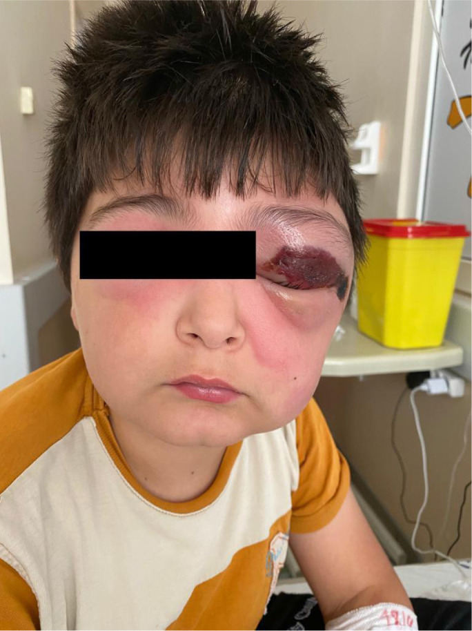
A 9-year-old male patient applied to the doctor on the third day of the formation of an itchy papular lesion that gradually enlarged on the left side of the eye. He was hospitalized after being diagnosed with preseptal cellulitis. Anthrax was considered on the fifth day of antibiotic treatment because of the progression and necrosis of the lesion. It was learned that the patient's father was a butcher, the patient had contact with raw meat, and his brother had anthrax on his finger before. In the patient's physical examination, there was redness and swelling around both eyes and on the nose, a 2x4 cm black eschar on the left upper eyelid, and a 1.5 cm area of intense necrosis on the left side of the eye. No additional pathological finding was found in the eye examination. Other system examinations were normal. The photograph of the case at the beginning of the treatment is shown in Figure 1.
Figure 1.
Pigment incontinence in papillary dermis (black arrow); prominent, nodular type fibrosis in deep dermis (red arrows) (H&E, 100X, 200X).
There was no microorganism upon gram staining of the sample collected from the edge of the patient’s lesion, who was considered to have palpebral anthrax. Blood agar was cultivated with the sample, yet there was no reproduction. The patient's polymerase chain reaction (PCR) test was positive for B. anthracis. The treatment of the patient was arranged as ciprofloxacin and clindamycin. Oedema in the right eyelid and face decreased on Day 3 of the treatment, and the typical eschar completely covered the lesion on the left eyelid on Day 6. The photo of the patient on Day 6 of the treatment is given in Figure 2.
Figure 2.

Patient's left eyelid during treatment.
After 28 days of antibiotic treatment was completed, the patient was discharged with eschar in his left eye. It was learned that the eschar fell spontaneously on the 43rd day. After the eschar fell, ophthalmologic examination revealed minimal ptosis in the left eye, and improvement in eyelid structure and contour. The photograph of the patient after the eschar has fallen is shown in Figure 3.
Figure 3.

Patient's left eyelid after treatment.
Discussion
Anthrax is still a significant public health problem, especially in developing countries where livestock breeding is widespread.[2] Our case was from the eastern Anatolia region in Turkey, where anthrax was endemic. In cutaneous anthrax, a small papule occurs, usually accompanied by a burning and itching sensation on the skin 2-3 days after the cutaneous inoculation of anthrax spores through minor cuts on the skin. In most cases, the incubation period varies between 1 and 7 days. The lesion expands within 1-2 days, with vesicle formation on the lesion. The middle of the vesicle is sunken and filled with fluid. The circumference of the said lesion is edematous and erythematous, yet painless. The liquid inside the vesicle becomes cloudy within a few days and turns blue-black. Finally, the vesicle explodes, followed by a black-colored ulcer with a significant dent. This lesion is called the anthrax pustule. Typical eschar occurs in 7 to 10 days.[1,4]
In our case, it was learned that a small itchy papular lesion first appeared on the left side of the eye, enlarged within three days and became vesicular and necrosis occurred on the lesion. On the sixth day of the follow-up, it was observed that the typical eschar completely covered the lesion on the left eyelid. With these findings, a diagnosis of cutaneous anthrax was considered. Suspecting anthrax is important in the diagnosis, and taking a social history plays a key role in the diagnosis. Studies in the literature have reported that almost all of the patients live in rural areas and have a history of close contact with animals and animal products.[5,6] It was reported that the father of the patient was a butcher and the patient had a history of contact with raw meat after slaughter.
The disease is diagnosed by basil growth in culture, detection on direct smear, or positive PCR test for bacillus. Recently, PCR testing has been increasingly used in the diagnosis of anthrax.[4] The diagnosis was confirmed with a positive PCR test for B. anthracis in the swab sample taken from the lesion margin. All three clinical forms of anthrax can be fatal if left untreated. Therefore, early diagnosis and treatment are essential in anthrax cases. Sepsis occurs in 10-20% of untreated cases, resulting in death.[7] However, the mortality rate is below 1%, thanks to appropriate antibiotic therapy, keeping the airways open, and steroid therapy when necessary.[4] High-dose parenteral penicillin is the antibiotic of choice in the treatment of anthrax. In severe cases, a second agent capable of penetrating the central nervous system may be included in the treatment.[7] In systemic anthrax cases where meningitis cannot be excluded, triple combination therapy including quinolone antibiotics, β-lactams (carbapenem, penicillin, ampicillin), and a protein synthesis inhibitor (linezolid, clindamycin) is recommended.[8,9] In a study by Engin et al.,[9] 39 patients with anthrax were examined, and it was reported that most patients with cutaneous anthrax were treated with penicillin, and three patients received steroid therapy for diffuse edema. In addition to ciprofloxacin and clindamycin treatment, we applied steroid treatment to our patient for 2 days due to rapidly progressive edema around the eyes and face.
Surgical incision in skin anthrax is not recommended as it may cause worsening symptoms and enlargement of the lesion. Topical antibiotics are not effective. The cutaneous lesion's local and gauze dressing are sufficient.[10] In cases of cutaneous anthrax with palpebral involvement, systemic symptoms and eschar progression are stopped with antibiotics.[4]
Bracham et al.[10] suggested that surgical intervention should be avoided in anthrax lesions. Otherwise, it may cause the infection to spread elsewhere and exacerbate symptoms. Çelebi et al.[6] reported that cicatricial ectropion occurred in the lower eyelid in a patient with anthrax treated with high-dose penicillin, and they obtained satisfactory results after transconjunctival resection and full-thickness skin grafting in their patients. However, despite early and effective treatment in patients with palpebral anthrax, local complications are frequently reported in parallel with the extent of the lesion. Gilliland et al.[11] reported that %47 of patients with palpebral anthrax developed ectropion, %19 lagophthalmus, and %9.5 corneal scarring.. It has been reported in the literature that eschars usually fall off spontaneously within 2-3 weeks.[5] In this case, eschar tissue regressed spontaneously in the sixth week and minimal ptosis was detected on the left eyelid after the eschar tissue was removed, but reconstruction was not required.
Anthrax should be kept in mind in the differential diagnosis of preseptal and orbital cellulitis in regions where anthrax is common, especially in patients who have close contact with animals. With early diagnosis and appropriate treatment of rare palpebral anthrax, it is possible to prevent serious complications, and our case is valuable in this regard.
Footnotes
Please cite this article as ”Teleke Kaymaz S, Cetin FT, Ozgur Gundeslioglu O, Kaplan F, Ulas B, Ozcan AA. A Rare Presentation of Anthrax: A Pediatric Patient with Palpebral Anthrax. Med Bull Sisli Etfal Hosp 2024;58(1):127–130”.
Disclosures
Informed consent
Written informed consent was obtained from the patient for the publication of the case report and the accompanying images.
Peer-review
Externally peer-reviewed.
Conflict of Interest
None declared.
Authorship Contributions
Concept – S.T.K., F.T.C., O.O.G., F.K., B.U., A.A.O.; Design – S.T.K., F.T.C., O.O.G., F.K., B.U., A.A.O.; Supervision – S.T.K., F.T.C., O.O.G., F.K., B.U., A.A.O.; Materials – S.T.K., F.T.C., O.O.G., F.K., B.U.; Data collection &/or processing – S.T.K., F.T.C., O.O.G., F.K., B.U., A.A.O.; Analysis and/or interpretation – S.T.K., F.T.C., O.O.G., F.K., B.U., A.A.O.; Literature search – S.T.K., F.T.C., O.O.G., F.K.; Writing – S.T.K., F.T.C., O.O.G., F.K.; Critical review – S.T.K., F.T.C., O.O.G., F.K., B.U., A.A.O.
References
- 1.Kimberlin DW. Anthrax. In: Kimberlin DW, Barnett ED, Lynfield R, Sawyer MH, editors. Red Book: 2021 Report of the Committee on Infectious Diseases. 30th ed. Grove Village, IL: American Academy of Pediatrics; 2021. pp. 214–20. [Google Scholar]
- 2.Eshraghi B, Zarrin Y, Fazel M. Palpebral anthrax, a rare though important condition in villagers: a case report and literature review. Int J Infect Dis. 2020;99:260–2. doi: 10.1016/j.ijid.2020.07.083. [DOI] [PubMed] [Google Scholar]
- 3.Bozpolat A, Atici D, Tekerek NU, Arslan D. Palpebral anthrax. Pediatr Infect Dis J. 2017;36:1216–7. doi: 10.1097/INF.0000000000001674. [DOI] [PubMed] [Google Scholar]
- 4.Çaça İ, Çakmak S, Ünlü K, Sakalar YB, Kadiroglu AK. Cutaneous anthrax on eyelids. Jpn J Ophthalmol. 2004;48:268–71. doi: 10.1007/s10384-003-0047-6. [DOI] [PubMed] [Google Scholar]
- 5.Tekin R, Ari Ş, Dal T, Kaya Ş, Kortak MZ, Dursun B, et al. Evaluation of cutaneous palpebral anthrax. Cutan Ocul Toxicol. 2013;32:294–8. doi: 10.3109/15569527.2013.781620. [DOI] [PubMed] [Google Scholar]
- 6.Çelebi S, Çelebi H, Çeliker ÜÖ, Kandemir B, Alagöz G, Esmerligil S. Anthrax as the cause of preseptal cellulitis. Acta Ophthalmol Scand. 1997;75:462–3. doi: 10.1111/j.1600-0420.1997.tb00414.x. [DOI] [PubMed] [Google Scholar]
- 7.Turnbull P, editor. 4th ed. Geneva: World Health Organization; 2008. Anthrax in Humans and Animals; pp. 70–88. [PubMed] [Google Scholar]
- 8.Hendricks KA, Wright ME, Shadomy SV, Bradley JS, Morrow MG, Pavia AT, et al. Workgroup on Anthrax Clinical Guidelines Centers for disease control and prevention expert panel meetings on prevention and treatment of anthrax in adults. Emerg Infect Dis. 2014;20:e130687. doi: 10.3201/eid2002.130687. [DOI] [PMC free article] [PubMed] [Google Scholar]
- 9.Engin A, Elaldı N, Dökmetaş İ, Bakıcı MZ, Kaya Ş, Bakır M. Cutaneous anthrax in the central Anatolia region of Turkey: a review of 39 adults cases. Turkiye Klinikleri J Med Sci. 2010;30:1032–8. [Google Scholar]
- 10.Brachman PS, Evans AS, Brachman PS, editors. Bacterial Infections of Humans, Epidemiology and Control. 3th ed. New York: Plenum Medical; 1991. Anthrax; p. 75. [Google Scholar]
- 11.Gilliland G, Starks V, Vrcek I, Gilliland C. Periorbital cellulitis due to cutaneous anthrax. Int Ophthalmol. 2015;35:843–5. doi: 10.1007/s10792-015-0057-7. [DOI] [PubMed] [Google Scholar]