Abstract
Aim
Root canal treatment (RCT) is a common procedure practiced daily by dentists worldwide. The current systematic review aimed to evaluate and compare clinical studies on the quality of root canal fillings (RCFs) carried out by dentists with different levels of experience conducted worldwide with those conducted specifically in Saudi Arabia (SA).
Materials and Methods
A full literature search was conducted in Clarivate Analytics’ Web of Science, Elsevier’s Scopus, Embase, CINHAL, and PubMed, without a restriction to studies published before January 2015. Also, a manual search was carried out by checking papers that may have been missed during the electronic search. The following keywords were used: [(quality of root canal filling(s)) OR (quality of root canal obturation)) and dental practitioners as (general dental practitioners; final year students; endodontist; specialist) AND (root canal obturation) OR (endodontic treatment)]. Parameters of the quality of RCFs, such as length, density, and taper, were assessed and counted.
Results
A total of 13 worldwide and nine SA studies were included in this review, published between 2015 and 2023. Molars were the most treated teeth, at 42.3% and 40.2% for the worldwide and SA studies, respectively. Cases treated by final year students had the highest percentage, at 60.0% for both study groups. The percentages of acceptable quality, with regard to the length, density, and taper of RCFs, were 70.9%, 77.6%, and 84.3%, and 73.2%, 64.6%, and 67.8% for the worldwide and SA studies, respectively.
Conclusion
The overall acceptable quality of RCFs was marginally higher in worldwide studies than in SA studies. Both prevalences can be considered as good, which indicates that the quality of RCFs is moving in the right direction.
Keywords: root canal filling, quality, obturation, Saudi Arabia, clinical studies, procedural errors
Background
The global prevalence of teeth with root canal fillings (RCFs) provides an indication of the total of number and activity of dentists dedicated to treating teeth endodontically and removing pulpal diseases. Root canal treatment (RCT) is a very common treatment all over the world. More than 50% of people investigated in one analysis had a minimum of one RCF.1 The international rate of RCT is, on average, greater than 8%. When studies from the 20th century are compared with those of the 21st century, a decline in the incidence of RCT is detected, which may reflect a change in the therapeutic attitudes of dentists in the management of endodontic diseases.1
The purposes of RCT are to preserve the role of a tooth, treat diseases of the pulp, and minimize, cure, and prevent the diseases of periapical tissue. Apical periodontitis is primarily triggered by microbial occupation due to dental caries or trauma, or contact of the pulp material with the oral microbiota during routine dental procedures.2 RCT also reduces the microbial residents within the root canal structure. This is achieved through full chemomechanical preparation and ultimately a sealed obturation of the root canal space to prevent reinfection.3 Therefore, it is essential to evaluate the quality of RCFs through radiographic examination. This evaluation involves assessing the radiographic appearance of the RCFs, specifically looking for well-obturated canals where the filling extends close to the apical constriction of every canal.4
The American Association of Endodontists employs radiographic evaluation of three criteria to estimate the methodical achievement of obturation: the length, shape, and density of the RCF.5 The length of the RCF is considered acceptable if the root filling ends ≤2 mm from the radiographic apex; uniform density of the root filling without voids, and with no visible canal space, is an indication of good density; and the taper of the RCF, in the form of a proper, consistent taper from the coronal to the apical part of the filling, is a good reflection of the canal shape.5,6 The guidelines of the European Society of Endodontology for the radiographic evaluation of RCFs recommend that the root canal should be entirely filled, with no space or voids among the filling and canal wall, and placed 0.5–2 mm beneath the radiographic apex.6 Overfilling or inadequate filling of a root canal obturation will determine the success rate of RCT.7
Several studies have reported that the outcome of RCFs is influenced by the length of RCF in relation to the apex.8–13 In addition to the length of the RCF, other studies assessed the density and taper of RCFs, and recorded different percentages of those parameters among studies conducted internationally8–10,14–17 and in Saudi Arabia (SA).12,13,18–20
Furthermore, procedural inaccuracies, such as the development of ledges, zip and elbow developments, separated instruments, and perforations, have the potential to arise during RCT procedures. Such errors could lead to cleaning and shaping difficulties, root filling leakage, and infection of periradicular tissues, thereby posing a threat to the overall success of RCT outcomes.21 Achieving optimal clinical outcomes in endodontic work necessitates a combination of up-to-date knowledge, comprehensive training, and the use of modern technology. Continuous education and training can contribute toward improving the skills required for difficult procedures, enhancing the practitioner’s ability to navigate complex anatomical variations and challenging cases.22 Several studies have recorded varying percentages of procedural errors worldwide8,10,16,23 and in SA.12,24–26
The quality of non-surgical RCT may vary in dentistry, as it can be performed by final year students (FYSs), general dental practitioners (GDPs), and specialists. These differences are accompanied by dissimilar stages of knowledge, years of clinical practice, and dexterity. Some studies have revealed that GDPs do not follow the guidelines on which they were trained during their basic education.27,28 In addition, Bajawi et al and Yusufoğlu and Sariçam reported that canal treatment completed by endodontists was better than that performed by GDPs in terms of the quality of RCFs.19,29 Furthermore, Pietrzycka et al reported a higher percentage of overfilled root canal being accomplished by specialists than by GDP, with probable explanations being the endodontists’ efforts to reach the root canal apex and their better preparation.30
Quite a few systematic reviews have been performed to evaluate the quality of RCFs. León-López et al evaluated the incidence and frequency of RCF globally, and concluded that more than 50% of people have at least one canal with an RCF.1 The procedural quality of RCFs accomplished by students during their study program is low, which may indicate that root canal education is inadequate.31 Burns et al documented that the outcomes of primary RCT remain high, and, overall, it is a consistent and effective way of preserving normal teeth, no matter which outcome criteria are used.32 This review was designed to assess and compare the studies conducted worldwide to those carried out in SA, in relation to the quality of RCFs accomplished by FYSs, GDPs, and endodontists, regarding the parameters of density, taper, and length.
Materials and Methods
Review Question
This review was undertaken following the procedures of the Preferred Reporting Items for Systematic Reviews and Meta-Analyses (PRISMA; www.prisma-statement.org).31,33,34 The Population, Intervention, Comparison, Outcome (PICO) set-up was utilized to articulate the focused question, based on Ribeiro et al,31 ie, whether the quality of RCFs achieved by FYSs, GDPs, and endodontists, in the form of density, length, and taper (Intervention), used to restore anterior, premolar, and molar teeth (Population), is the same in all teeth (Outcome), in studies conducted worldwide compared to those carried out in SA (Comparison).
Selection Criteria
Most RCFs are performed by students during their final year of study and GDPs, but a smaller number of more difficult cases are carried out by endodontists. The inclusion and eligibility measures were as follows: (i) in vivo surveys evaluating the quality of RCT (obturations) of all maxillary and mandibular teeth; (ii) surveys judging the quality, in terms of acceptable and unacceptable quality, performed by FYSs, GDPs, and endodontists; (iii) studies using periapical x-rays; (iv) studies measuring at least two of the three parameters (length, taper, density); and (v) papers available in English. In vitro research involving RCT teeth treated for preclinical teeth in the preclinical setting or using finite element analysis were excluded. Systematic reviews, pilot studies, editorials, case documents or series, and published papers that were only available in languages other than English were similarly excluded from this review.
Literature Search
The search strategy used a combination of Medical Subject Headings (MeSH) terms and free keywords (root canal treatment, root canal filling, general dental practitioners, final year students, endodontist, worldwide studies, Saudi Arabia studies), along with Boolean operators (AND, OR, NOT) in relation to the PICO question. An electronic literature search of the databases was conducted between September and December 2023 by two operators, including a senior librarian specializing in dental sciences, using the following databases: Clarivate Analytics’ Web of Science, Elsevier’s Scopus, Embase, CINHAL, and PubMed (MEDLINE), without restricting studies published before January 2015. In addition, a manual search was performed by checking the bibliographies of all initially selected papers to identify studies that may have been missed in the electronic search. Studies published before 2015 were excluded because of the introduction of more recent materials and equipment used in RCT globally.
Study Selection
The paper collection steps passed through several phases, based on: (i) title importance, (ii) abstract significance, and (iii) analysis of the whole paper. The collected papers saved by both the electronic and manual searches were grouped and evaluated for inclusion according to the eligibility criteria.
Data Extraction and Analysis
Microsoft Office Excel software was used to extract data from the included articles, which were separated into those conducted worldwide and those conducted in SA. These data included (i) study characteristics, which included researcher(s), publication years, and country in which the study was conducted, number of canals treated, study design and type, percentage of tooth types, and the dental setting and operators’ level or status; (ii) quality of the RCF treatment obturations in relation to length, density, and tapers, with all of these parameters being assessed as acceptable or unacceptable, and registered as a total percentage; (iii) procedural errors that arose during RCT and obturation of the canals, in the form of ledges, apical transportation, fractured instruments, apical perforation, root perforation, etc; and (iv) the outcome of the assessed parameters and presence of significant differences, if any.
Quality of Evidence
The value of evidence for each study was reviewed according to the Grading of Recommendations, Assessment, Development and Evaluation (GRADE) Handbook.35 According to GRADE, aspects that can decrease the quality of evidence comprise things such as outcomes in study approach or accomplishment (risk of bias), variation of outcomes, indirectness of evidence, imprecision, and publication bias. The quality of the assessed studies was gauged using the restrictions of earlier published systematic reviews conducting in vivo surveys.32,36,37 GRADE’s quality of evidence levels are categorized from low to high: very low, low, moderate, and high.35
Results
Literature Search and Study Selection
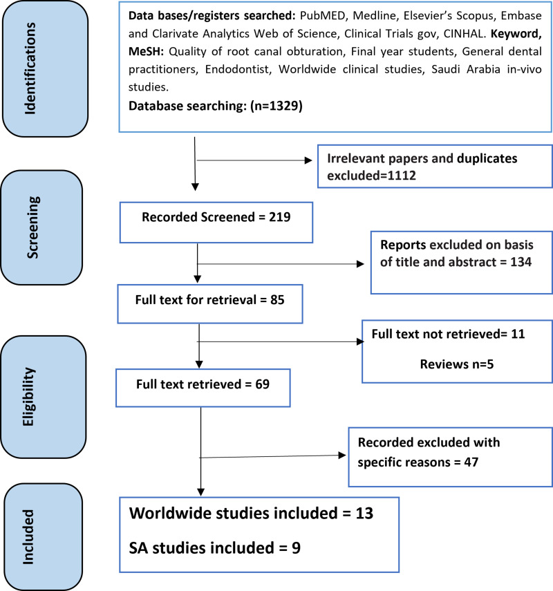
The principal search resulted in 1329 studies. After eliminating 1112 unrelated and duplicate papers and titles, the authors went through the abstracts of 219 papers to eliminate ineligible research. A total of 134, 16 (11+5), and 47 published articles were omitted for specific reasons (title and abstract contents, studies measuring only one of the three RCF parameters, unretrieved papers, reviews, or studies using other types of x-ray), and 22 papers were nominated for whole paper retrieval. Finally, 13 worldwide8–11,14–17,23,29,30,38,39 and nine SA articles 12,13,18–20,24–26,40 were included in the current review. These papers measured the quality of clinical RCF or obturation achieved by FYSs, GDPs, and endodontists (specialists and consultants) for anterior (incisors and canines), premolar, and molar teeth. No disagreements were raised between the evaluators. The PRISMA flowchart of the literature search method is shown in Figure 1. Table 1 summarizes the in vivo papers evaluating the quality of RCT conducted worldwide and in SA.
Figure 1.
PRISMA flowchart of the selection process of clinical studies for this review.
Table 1.
Summary of In Vivo Studies Evaluating the Quality of Root Canal Treatment Conducted Worldwide (n=13) and in Saudi Arabia (n=9)
| Study Characteristics | Quality of Obturation | Procedural Errors | Assessed Outcome Parameters | |||||||||
|---|---|---|---|---|---|---|---|---|---|---|---|---|
| Length (%) | Density (%) | Taper (%) | ||||||||||
| Author(s), Year, Country | Number of Canals | Study Design and Type | Tooth Type (%) | Dental Setting; Operators | Acceptable | Unacceptable | Acceptable | Unacceptable | Acceptable | Unacceptable | ||
| Worldwide Studies | ||||||||||||
| Gavini et al,8 2022, Brazil | 2213 | Cross-sectional retrospective |
Anterior 25.8% Premolar 32.2% Molar 42.0% |
Dental school; FYSs | 72.9% | 27.1% | 87.3% | 22.7% | 91.6% | 8.4% | Instrument fractures 0.81%. In last 5 mm of apical tip 77.8% | Better results in maxillary teeth |
| Silnovic et al,9 2023, Sweden |
60 | Retrospective | Anterior 27.1% Premolar 31.8% Molar 41.8% |
Polyclinics, governmental; GDP |
28.7% | 71.3% | NM | NM | NM | NM | NM | Poor quality in anterior and molars |
| Ameen et al,14 2024, United Arab Emirates | 601 | Cross-sectional retrospective |
Anterior 48.4% Premolar 51.6% |
Dental school, private; FYSs |
93.5% | 6.5% | 96.5% | 3.5% | 98.2% | 1.8% | NM | SD ↔ anterior and premolars regarding taper, density, and overall quality |
| Al Shehadat et al,10 2023, United Arab Emirates | 124 | Cross-sectional retrospective |
Anterior 32.9% Premolar 45.6% Molar 21.5% |
Private dental school; FYSs | 73.5% | 26.5% | 57.5% | 42.3% | 66.2% | 33.8% | Ledge 5.4%, apical transportation 3.5%, fractured instrument 1% |
SD ↔ quality parameters SD ↔ ledge formation and apical transportation |
| Laukkanen et al,11 2021, Finland |
426 | Cross-sectional retrospective |
Anterior 34.2% | Governmental; GDP | 71.0% | 29.0% | NM | NM | NM | NM | NM | SD ↔ teeth, poorer in molars |
| Premolar 40.7% | 57.0% | 43.0% | NM | NM | NM | NM | ||||||
| Molar 25.1% | 43.0% | 57.0% | NM | NM | NM | NM | ||||||
| Ribeiro et al,15 2019, Brazil | 274 | Retrospective | Anterior 39% | Governmental; FYSs | 71.7% | 28.9% | 99.7% | 0.3% | 96.6% | 3.7% | NM | 80% unsatisfactory quality |
| Premolar 61% | 67.3% | 32.7% | 98.1% | 1.9% | 96.0% | 4.0% | ||||||
| Saatchi et al,16 2018, Iran | 1674 | Cross-sectional | Anterior 9.7% | Governmental; FYSs | 57.7% | 42.3% | NM | NM | 67.5% | 32.5% | Ledge 12.8%, foramen perforation 2%, root perforation 2.4% |
SD ↔ procedural errors, higher molars |
| Premolar 21.9% | 61.3% | 38.7% | NM | NM | 69.5% | 30.5% | ||||||
| Molar 68.4% | 51.3% | 48.7% | NM | NM | 63.7% | 36.3% | ||||||
| Fritz et al,17 2021, Brazil | 442 | Prospective | Anterior 38.2% | FYSs | 94.5% | 5.5% | NM | NM | 96.8% | 3.2% | NM | SD ↔ anterior and premolars |
| Premolar 45.0% | 96.5% | 3.5% | NM | NM | 96.5% | 3.5% | ||||||
| Molar 16.8% | 92.1% | 7.9% | NM | NM | 92.4% | 7.6% | ||||||
| Pietrzycka et al,30 2022, Poland | 219 | Retrospective randomized double-blind comparison | Anterior 43.7% Premolar 42.2% Molar 14.1% |
GDP | 85.8% | 14.2% | 99.5% | 0.5% | NM | NM | NM | NSD ↔ GDP and specialist |
| 257 | Anterior 26.5% Premolar 18.4% Molar 45.1% |
Specialist | 74.5% | 25.5% | 99.3% | 0.7% | NM | NM | ||||
| Wong et al,23 2016, Malaysia |
75 | Retrospective clinical audit | Anterior 26.7% | FYSs | 75.8% | 24.2% | 75.8% | 24.2% | NM | NM | Ledge 9.3%, perforation 11.4%, instrument separation 0.7% | SD ↔ misshape |
| Premolar 29.3% | 78.3% | 21.7% | 65.3% | 34.85 | NM | NM | ||||||
| Molar 44.0% | 61.5% | 38.5% | 57.7% | 42.3% | NM | NM | ||||||
| Yusufoğlu et al,29 2021, Turkey | 3115 | Retrospective | Max & Mand Molars | GDP | 76.7% | 23.3% | 37.3% | 62.7% | NM | NM | Separated instrument 2.6%, ledges 0.4%, lateral perforation 0.1% | SD ↔ GP and endodontist in obturation quality NSD iatrogenic |
| Endodontist | 82.3% | 17.7% | 62.7% | 37.3% | NM | NM | Separated instrument 4.6%, ledges 0.5%, lateral perforation 0.1% | |||||
| Elemam et al,38 2015, Libya | 284 | Retrospective | Anterior 9.7% Premolar 15.5% Molar 73.3 |
Governmental; FYSs | 48.6% | 51.35% | 75.8% | 24.2% | 68.8% | 31.2% | NM | SD ↔ overall quality between tooth types |
| Awooda et al,39 2016, Sudan | 173 | Retrospective, cross-sectional | Anterior 35.3% Premolar 27.2% Molar 37.5% |
Private college; FYSs | 71.7% | 28.3% | 72.8% | 27.2% | 94.8% | 5.2% | Separated instrument 3.5% |
|
| Saudi Studies | ||||||||||||
| Alshehri et al,12 2023 | 278 | Retrospective | Anterior 100% | Governmental; FYSs | 85.6% | 14.4% | 65.1% | 34.9% | 71.9% | 28.1% | Ledge 4.7%, root perforation 0.4%, foramen perforation 0.7% |
SD ↔ 4th, 5th, 6th SD ↔ Max and Man teeth NSD ↔ Male and female |
| Al-Obaida et al,13 2020 | 200 | Cross-sectional prospective | Private hospital: Anterior 24.0% Premolar 33.0% Molar 43.0% |
Private; GDP | 48.0% | 52.0% | 60.5% | 39.5% | 56.5% | 43.5% | NM | Tooth type: SD ↔ length and tapering Hospital type: SD ↔ length, tapering, density |
| Governmental; GDP | 60.0% | 40.0% | 71.5% | 29.5% | 71.5% | 29.5% | ||||||
| 200 | Government hospital: Anterior 32.5% Premolar 28.5% Molar 39.0% |
Private; GDP | 41.7% | 58.8% | 46.0% | 58.1% | 42.8% | 62.0% | ||||
| Governmental; GDP | 58.3% | 41.2% | 54.0% | 41.9% | 57.0% | 38.0% | ||||||
| Habib et al,18 2018 | 390 | Cross-sectional retrospective | Anterior 27.4% Premolar 27.7% Molar 42.9% |
Private college; FYSs | 59.5% | 40.5% | 50.8% | 49.2% | 57.4% | 42.6% | NM | SD ↔ length and density NSD ↔ tapering |
| Kader et al,20 2016 | 352 | Retrospective observational | NM | Governmental; FYSs | 61.7% | 38.3% | 54.0% | 46.0% | 53.1% | 46.9% | Ledge formation and gauging | NM |
| Bajawi et al,19 2018 | 209 | Retrospective cross-sectional | Max 58.2% Mand 41.8% |
Governmental ; Dental center consultant |
69.6% | 30.4% | 100.0% | 00.0% | 100.0% | 00.0% | NM | SD ↔ arch, canal position, level of experience |
| Specialist | 62.5% | 37.5% | 81.3% | 18.8% | 87.5% | 12.5% | ||||||
| GDP | 46.4% | 53.6% | 75.8% | 24.2% | 77.7% | 22.3% | ||||||
| Abumostafa et al,24 2015 | 450 | Retrospective | Max 48.9% Mand 51.1% Anterior 14.9% Posterior 85.1% |
Private college; FYSs | 77.6% | 22.5% | 46.4% | 53.6% | 78.8% | 26.2% | Ledge 2.4%, transportation 3.1%, apical perforation 1.1%, root perforation 0.2%, stripping perforation and fractured instrument 1.1% | NM |
| Akbar,25 2015 | 130 | Cross-sectional | Anterior 12.3% Premolar 13.8% Molar 74.6% |
Governmental; FYSs | 76.5% | 23.3% | NM | NM | NM | NM | Separated instrument 3.1%, stripping perforation 2.3%, furcal perforation 0.8%, coronal leakage 0.8% |
SD ↔ under-filling and poor filling and apical radiolucency |
| Smadi et al,26 2015 | 66 | Prospective | Anterior 40.8% Premolar 40.4% Molar 18.8% |
Governmental; FYSs | 61.5% | 38.5% | 50.5% | 49.5% | 56.1% | 43.9% | Present in 85.3%. Man molars were highest | SD ↔ errors among teeth and obturation parameters |
| Mustafa,40 2022 | 400 | Retrospective clinical study | Anterior 36.0% Premolar 17.0% Molar 47.0% |
Governmental; FYSs | 67.3% | 32.7% | 51.7% | 48.3% | 74.9% | 25.1% | NM | SD ↔ length |
Abbreviations: NM, none mentioned; SD, significant difference; ↔, between; GDP, general dental practitioner; NSD, non-significant difference; FYS, final year student; Max, maxillary; Man, mandibular.
Characteristics of the Included Clinical Studies
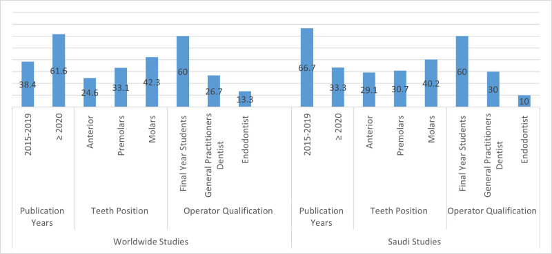
Figure 2 shows the features of all clinical studies included in this review. Thirteen of the studies were conducted worldwide,8–11,14–17,23,29,30,38,39 and the others were carried out in SA.12,13,18–20,24–26,40 In relation to publication year, the highest percentage of worldwide studies, 61.6%, was published in 2020 and later, whereas the highest percentage of SA studies, 66.7%, was conducted between 2015 and 2019. Teeth with three RCFs were the most treated teeth, at 42.3% and 40.2% of worldwide and SA studies, respectively. Cases treated by FYSs were the highest percentages, at 60.0% for both study groups, while those treated by specialists saw the lowest percentages, at 13.3% and 10.0% for the worldwide and SA studies.
Figure 2.
Characteristics of the worldwide and Saudi clinical studies included in this review.
Root Canal Filling Outcomes
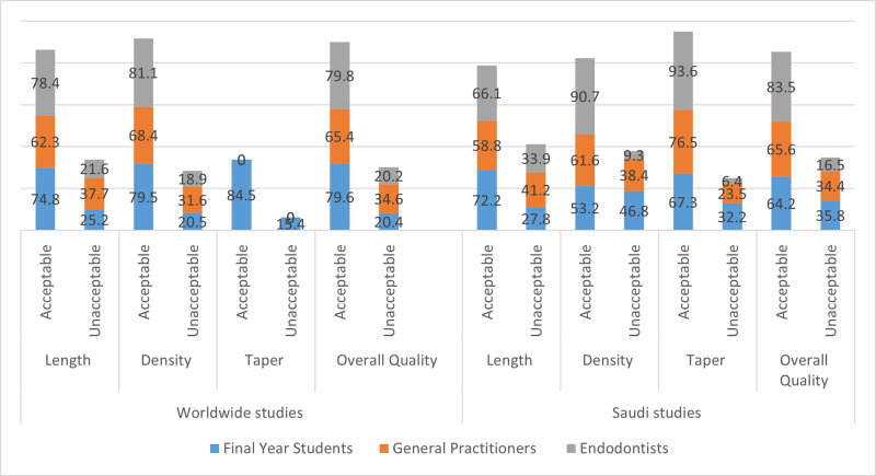
Figure 3 shows the ratios of the different quality of RCF parameters, in relation to different operator levels and experience in the worldwide and SA studies. Endodontists recorded the highest acceptable quality percentages in all RCF quality parameters, except for taper, for which FYSs recorded a higher percentage as there were no worldwide studies dealing with taper parameters. The overall acceptable RCF quality was slightly higher among specialists in the SA studies in comparison to the worldwide studies, at 83.5% and 79.8%, respectively.
Figure 3.
Ratio of acceptable and unacceptable parameters of root canal quality in relation to operator level and study type.
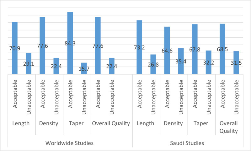
Figure 4 shows that the percentage of acceptable quality was much higher than that of unacceptable quality of the RCF parameters (density, length, and taper) in both study groups. The percentages of acceptable quality in relation to the taper, density, and length of RCF canals were 70.9%, 77.6%, and 84.3%, and 73.2%, 64.6%, and 67.8% for the worldwide and SA studies, respectively. The overall adequate quality of RCFs was slightly higher among studies documented worldwide (77.6%), in comparison with 68.5% for SA studies.
Figure 4.
Percentage of acceptable and unacceptable parameters of quality of root canal filling in worldwide and Saudi studies.
Procedural Errors During Treatment
Only eight articles, divided equally between worldwide8,10,16,23 and SA studies,12,24–26 assessed the procedural errors associated with the RCT steps. Fractured and separated instruments, followed by ledge and perforation, were the most commonly recorded procedural errors. Molars were the most commonly affected teeth.26
Results of Bias Assessment
Overall, most of the assessed studies analyzed in the present review carried a moderate to high risk of bias, in both the worldwide studies and those papers carried out among SA populations, except for those studies recording a low-grade risk bias.8,11,18,24
Discussion
In endodontics, steps are planned to preserve the health of periapical tissues in cases of disease or damage to the dental pulp, or to re-establish periapical health when apical periodontitis has previously been established.41 These aims may be accomplished by non-surgical RCT, retreatments and, sometimes, surgery at the end of the roots. The purpose of RCT is, primarily, to sterilize the root canal structure and keep it clean, and, subsequently, to seal the pulp cavity acceptably and achieve a suitable coronal restoration.6 This systematic review was designed to assess and compare the studies conducted worldwide to those carried out in Saudi Arabia in relation to the quality of RCFs (in the form of density, taper, and length) accomplished by FYSs, GDPs, and endodontists. This information can help to identify areas where improvement is needed and guide educational initiatives for graduate students and practitioners to ensure that they are producing RCFs with good quality in terms of adequate length, good density, and proper taper.
The results revealed a high frequency (77.6% and 68.5%) of overall acceptable technical quality of RCFs worldwide and in SA, respectively. A much lower percentage of 48.75% for the overall acceptable value was reported by Ribeiro et al, but their report was limited to studies conducted on undergraduate students and papers carried out before 2015.31 The reason for the higher value in this review is that the included papers were more recent, having been published between 2015 and 2023, and thus took into account the recent advances in RCT instruments and materials. Moreover, this study included not only FYSs but also GDPs and endodontists.
It is challenging to exactly estimate the quality of RCF using only radiographic assessment. Periapical x-rays, while the most frequently used method, do not replicate the three-dimensional system of the RCF.42 It is probable that areas of obturation and lost canals, along with additional assessment principles, were unobserved, resulting in an overvaluation of RCT quality.43 Radiographic measures for the quality of RCFs were gauged in agreement with the American Association of Endodontists, European procedures, and earlier research on the outcome of the quality of the RCF.5,6,8–12 The restrictions of procedural quality that were detailed, such as root filling taper, length, and density, were possibly essential to assess the success of the treatment. Regarding achievement percentage and ratios, it would be more suitable to choose experimental or clinical research with a follow-up appointment after RCT, assessing curative percentage, signs and radiographic characteristics.
Only studies that concentrated on the taper, density, and length of RCFs were included. Most of the earlier published papers measured the apical end of the root filling at ≤2 mm from the x-ray apex as the acceptable position.5,6,18,20 The length of the RCF is a significant assessment parameter, and is determined by gauging the apical boundary of the obturation from the end of the tooth on postoperative x-rays.44 The rates of acceptable length for the quality of the RCFs performed by FYSs, GDPs, and specialists were, respectively, 72.2%, 58.8%, and 66.1% for the SA studies and 74.8%, 62.8%, and 78.4% for the worldwide studies. Both study groups showed high percentages of acceptable quality for cases performed by FYSs, which could be related to their clinical work being carried out under direct supervision of staff. This could take the form of checking a step before proceeding to the next one.
The taper parameter of the RCF is demarcated, as even and reliable tapers from the coronal zone to the apical point are similar to the unique outline of the original canal.24 The tapering of RCFs for SA studies was 67.3%, 76.5%, and 93.6% for FYSs, GDPs, and specialists, respectively. However, acceptable quality of RCFs performed by FYSs ranging from 10.9% to 85.1% was reported by Ribeiro et al,31 with the most common reason for an unacceptable RCF being related to difficulties with RCF density. The relationship between density and treatment outcome is not clear. Kirkevang et al found that the incidence of voids in RCFs had a significant influence on the rate of apical periodontitis.45 Hommez et al stated that the prevalence of apical periodontitis was 47.1% in cases of non-homogeneous RCF, and 27.7% in clinical cases of homogeneous RCF.46 So, the less dense and non-homogeneous an RCF, the greater the chance of a negative influence on the treatment outcome.
Frontal and single-rooted teeth had meaningfully better RCF values than posterior teeth and those with two or three canals.47 Molars have a higher occurrence of narrow, curved pulp chambers, and posterior teeth are difficult access, making treatment more challenging.48 In contrast, anterior teeth usually have a single canal with less curvature, which may contribute to better outcomes.11,13,15,17,23 However, molar teeth were the most commonly treated teeth, recorded at 42.3% and 40.2% in the worldwide and SA studies, respectively. Molars are often more susceptible to carious lesion because of early eruption, their larger surface area, and grooves providing more spaces for plaque and food particles to accumulate. In addition, molars are harder to clean thoroughly, making them more prone to caries.49 Finally, most of the SA studies did not divide the type of teeth into anterior teeth, premolars, and molars, and therefore we did not compare this parameter with the worldwide studies.
Procedural errors are mistakes that may occur during the diagnosis or procedure of RCT. This review found that fractured and separated instruments, followed by ledge and perforation, were the most frequently recorded procedural errors.8,10,12,19,23–25 These can happen owing to a lack of familiarity, the practitioner neglecting to pay due attention, or unexpected and unstable environments that may be encountered throughout the treatment. Procedural errors are an important factor in the long-term success rates of endodontically treated teeth. Therefore, errors such as tool fractures, ledge-zip creations, and perforations may be a source of failure in non-surgical RCT.50 Moreover, mistakes made during the cleaning and disinfecting of the canal system have an adverse effect on the accomplishment of the RCT, by producing insufficient chemomechanical preparation and obturation of the root canals.51 The occurrence of procedural errors may be partly due to the more complex canal structure of teeth with narrow and curved pulpal canals, which necessitate more time and patience with regard to suitably cleaning, shaping, and obturation.26,52
Advanced education and the greater quantity of cases completed by endodontists compared to GDPs may be the reasons for the endodontists’ higher achievement of treatment excellence. It is not surprising that specialists achieved the highest percentage of acceptable quality RCF cases; this may be associated with their level of experience, and also with the limited number of worldwide studies,29,30 as well as the lack of international studies assessing the taper parameters. The percentages of the quality of RCFs were slightly lower among GDPs in both groups of studies; this could be explained by GDPs usually accepting most of the cases in their clinics before any referral to a specialist. Also, FYSs recorded acceptable rates in all RCF parameters, and this can be attributed to students operating under the direct supervision of staff during their clinical work. Similar results were recorded in both study groups in relation to overall RCF quality, which indicates that no great differences in satisfactory outcome were found between the included worldwide and SA studies. Pietrzycka et al and Bajawi et al described higher percentages of overfilled teeth performed by root canal specialists than by GDPs, and more underfilling performed by GDPs than by specialists and consultant endodontists; possible explanations include the efforts made by endodontists to reach the end of the root canal and improved preparations.19,30
By considering the importance of radiographic evaluation in RCF, dental practitioners can strive to achieve higher standards of care, leading to better patient satisfaction and oral health outcomes.53 As such, adopting a culture of quality assessment and continuous improvement will be helpful in shaping the future of endodontic practice in SA as well as in other countries. It is vital for dental educators and professionals to prioritize the incorporation of radiographic evaluation as a standard practice in assessing the quality of RCFs.42,48 By emphasizing the significance of radiographic examination in endodontic care, the dental community can work toward achieving uniformity and consistency in the quality of RCFs.54 This will not only contribute to the improvement of patient care but also elevate standards of endodontic practice.
A recent systematic review stated that the use of rotary instruments for evaluation of the quality of RCF, as well as for the instrumentation and obturation steps, is strongly recommended. Between different clinical settings and practices, the outcome of the rotary systems was in the range of excellent and good, and resulted in the acceptable quality of the RCF parameters and overall quality performed by these instruments, and few procedural errors.55
Further systematic reviews could be conducted including more articles published in different languages, and using more recent materials and equipment, such as cone-beam computed tomography (CBCT) scanners. The present review, which assessed clinical research, has several important drawbacks. The methodologies used were highly heterogeneous between the involved studies. Further, most of the studies were categorized as having “high to moderate” quality bias. Finally, discrepancies in the length, width, curvature, and number of roots of the anterior, premolar, and molar teeth among the papers in this review may have resulted in differences in acceptable and unacceptable RCF quality. Any of these restrictions could affect the overall RCF quality of the countries in which these studies were conducted.
Conclusions
From the present systematic review, the following conclusions can be drawn. The acceptable quality rates of the RCFs in the three parameters (taper, density, and length) were higher for the worldwide studies, ranging between 70.9% and 84.3%, than for the SA studies, between 64.6% and 73.2%. The overall acceptable quality rate of RCFs was marginally higher in the worldwide studies, at 77.6%, in comparison to 68.5% in the SA studies. Both percentages can be considered as good, which indicates that the quality of treatment is moving in the right direction.
Disclosure
The authors report no conflicts of interest in this work.
References
- 1.León-López M, Cabanillas-Balsera D, Martín-González J, Montero-Miralles P, Saúco-Márquez JJ, Segura-Egea JJ. Prevalence of root canal treatment worldwide: a systematic review and meta-analysis. Int Endod J. 2022;55(11):1105–1127. doi: 10.1111/iej.13822 [DOI] [PMC free article] [PubMed] [Google Scholar]
- 2.Siqueira JF, Rôças IN. Clinical implications and microbiology of bacterial persistence after treatment procedures. J Endod. 2008;34(11):1291–1301.e3. doi: 10.1016/j.joen.2008.07.028 [DOI] [PubMed] [Google Scholar]
- 3.Kharouf N, Arntz Y, Eid A, et al. Physicochemical and antibacterial properties of novel, premixed calcium silicate-based sealer compared to powder-liquid bioceramic sealer. J Clin Med. 2020;9(10):3096. doi: 10.3390/jcm9103096 [DOI] [PMC free article] [PubMed] [Google Scholar]
- 4.Gound TG, Sather JP, Kong TS, Makkawy HA, Marx DB. Graduating dental students’ ability to produce quality root canal fillings using single- or multiple-cone obturation techniques. J Dent Educ. 2009;73(6):696–705. PMID: 19491347. doi: 10.1002/j.0022-0337.2009.73.6.tb04749.x [DOI] [PubMed] [Google Scholar]
- 5.American Association of Endodontists. Endodontics: colleagues for Excellence. Available from: https://bestendoglenview.com/wp-content/uploads/2012/04/Obturation-of-root-canal-systems.pdf. Accessed September 03, 2024.
- 6.European Society of Endodontology. Quality guidelines for endodontic treatment: consensus report of the European Society of Endodontology. Int Endod J. 2006;39(12):921–930. doi: 10.1111/j.1365-2591.2006.01180.x [DOI] [PubMed] [Google Scholar]
- 7.Jenkins SM, Hayes SJ, Dummer PM. A study of endodontic treatment carried out in dental practice within the UK. Int Endod J. 2001;34(1):16–22. doi: 10.1046/j.1365-2591.2001.00341.x [DOI] [PubMed] [Google Scholar]
- 8.Gavini G, Candeiro GTM, Potgornik Ferreira F, et al. Retrospective study of endodontic treatment performed by undergraduate students using reciprocating instrumentation and single-cone obturation. J Dent Educ. 2022;86(6):751–758. doi: 10.1002/jdd.12884 [DOI] [PubMed] [Google Scholar]
- 9.Silnovic Z, Kvist T, Frisk F. Periapical status and technical quality in root canal filled teeth in a cross sectional study in Jönköping, Sweden. Acta Odontol Scand. 2023;81(3):249–254. doi: 10.1080/00016357.2022.2121322 [DOI] [PubMed] [Google Scholar]
- 10.Al Shehadat S, El-Kishawi M, AlMudalal A, et al. An audit of the technical quality and iatrogenic errors of root canal treatment by undergraduate dental students at the University of Sharjah. Eur J Dent. 2023;17(1):191–199. doi: 10.1055/s-0042-1743150 [DOI] [PMC free article] [PubMed] [Google Scholar]
- 11.Laukkanen E, Vehkalahti MM, Kotiranta AK. Radiographic outcome of root canal treatment in general dental practice: tooth type and quality of root filling as prognostic factors. Acta Odontol Scand. 2021;79(1):37–42. doi: 10.1080/00016357.2020.1773531 [DOI] [PubMed] [Google Scholar]
- 12.Alshehri TA, Aljami A, Alzayer H, et al. Assessment of modality and accuracy of single root canal treatment performed by undergraduate students in Saudi Arabia: a retrospective study. Cureus. 2023;15(1):e33483. doi: 10.7759/cureus.33483 [DOI] [PMC free article] [PubMed] [Google Scholar]
- 13.Al-Obaida MI, Alwehaiby KM, Al-Hindi OH, Merdad K, Al-Madi EM. Radiographic evaluation of the technical quality of root canal filling in Riyadh government and private hospitals. Saudi Endod J. 2020;10:194–198. doi: 10.4103/sej.sej_156_19 [DOI] [Google Scholar]
- 14.Ameen M, Alhadi D, Almaslamani A, Saleh AR. Impact of using single-file reciprocating system on the quality of root canal treatment treated by undergraduate students. Saudi Dental J. 2024;2024:1. doi: 10.21203/rs.3.rs-3112598/v1 [DOI] [Google Scholar]
- 15.Ribeiro DM, Henckel MD, Mello FD, Felippe MCS, Felippe WT. Radiographic analysis the obturation’s quality in root canal treatment performed by a South Brazilian sample of undergraduate students. RGO Rev Gaúch Odontol. 2019;67:e20190040. doi: 10.1590/1981-863720190004020180038 [DOI] [Google Scholar]
- 16.Saatchi M, Mohammadi G, Vali Sichani A, Moshkforoush S. Technical quality of root canal treatment performed by undergraduate clinical students of Isfahan dental school. Iran Endod J. 2018;13(1):88–93. doi: 10.22037/iej.v13i1.18517 [DOI] [PMC free article] [PubMed] [Google Scholar]
- 17.Fritz ALC, Ribeiro FC, Xavier JMB, Claudia Mendonça Reis CM, Demuner C. Evaluation of quality of root canal fillings performed by undergraduate students of a Brazilian university. Brazilian J Health Rev. 2021;4(5):18964–18975. doi: 10.34119/bjhrv4n5-039 [DOI] [Google Scholar]
- 18.Habib AA, Doumani MD, Nassani MZ, et al. Radiographic assessment of the quality of root canal fillings performed by senior dental students. Eur Endod J. 2018;3(2):101–106. doi: 10.14744/eej.2018.69775 [DOI] [PMC free article] [PubMed] [Google Scholar]
- 19.Bajawi AM, AL Sagoor SA, Alhadi AA, et al. Radiographic assessment of the quality of root canal treatments performed by practitioners with different levels of experience. Biomed Pharmacol J. 2018;11(3):1609–1616. doi: 10.13005/bpj/1528 [DOI] [Google Scholar]
- 20.Kader MA, Almagtaf AS, Babiker A, Nasim VS, Latheef AA, Shaik SA. Assessment of the quality of root canal treatment performed by undergraduates in college of dentistry, King Khalid University, Saudi Arabia: a Radiographic Analysis. J Int Oral Health. 2016;8(5):575–578. doi: 10.2047/jioh-08-05-09 [DOI] [Google Scholar]
- 21.Peters OA. Current challenges and concepts in the preparation of root canal systems: a review. J Endod. 2004;30(8):559–567. doi: 10.1097/01.don.0000129039.59003.9d [DOI] [PubMed] [Google Scholar]
- 22.Färber CM, Lemos M, Said Yekta-Michael S. Effect of an endodontic e-learning application on students’ performance during their first root canal treatment on real patients: a pilot study. BMC Med Educ. 2022;22(1):394. doi: 10.1186/s12909-022-03463-y [DOI] [PMC free article] [PubMed] [Google Scholar]
- 23.Wong CY, Liaw YX, Wong JZ, Chen LC, Parolia A, Pau A. Factors associated with the technical quality of root canal fillings performed by undergraduate dental students in a Malaysian Dental School. Braz J Oral Sci. 2016;15(1):45–50. doi: 10.20396/bjos.v15i1.8647122 [DOI] [Google Scholar]
- 24.Abumostafa A, Ahmad IA, Alenzy G, AlZoman A. Quality of root canal fillings performed by undergraduate Students in Saudi Dental College. J Dent Oral Hyg. 2015;7(5):64–70. doi: 10.5897/JDOH2015.0150 [DOI] [Google Scholar]
- 25.Akbar I. Radiographic study of the problems and failures of endodontic treatment. Int J Health Sci. 2015;9(2):111–118. PMID: 26309429; PMCID: PMC4538887. [PMC free article] [PubMed] [Google Scholar]
- 26.Smadi L, Hammad M, El-Ma’aita AM. Evaluation of the quality of root canal treatments performed by dental undergraduates: is there a need to review preclinical endodontic courses. Amer J Educ Research. 2015;3(12):1554–1558. doi: 10.12691/education-3-12-11 [DOI] [Google Scholar]
- 27.Stewardson DA. Endodontic standards in general dental practice- a survey in Birmingham. Part I. European J of Prosth and Rest Dent. 2001;9(3–4):107–112. PMID: 12192945. [PubMed] [Google Scholar]
- 28.Hill EE, Rubel BS. Do dental educators need to improve their approach to teaching rubber dam use. J of Dent Edu. 2008;72:1177–1181. doi: 10.1002/j.0022-0337.2008.72.10.tb04596.x [DOI] [PubMed] [Google Scholar]
- 29.Yusufoğlu SI, Sariçam E. Comparison of endodontic treatment qualities of molar teeth performed by endodontists and practitioners: a radiographic analysis. Selcuk Dent J. 2021;8(1):127–132. doi: 10.15311/selcukdentj.597504 [DOI] [Google Scholar]
- 30.Pietrzycka K, Radwanski M, Hardan L, et al. The assessment of quality of the root canal filling and the number of visits needed for completing primary root canal treatment by operators with different experience. Bioengineering. 2022;9(9):468. doi: 10.3390/bioengineering9090468 [DOI] [PMC free article] [PubMed] [Google Scholar]
- 31.Ribeiro DM, Réus JC, Felippe WT, et al. Technical quality of root canal treatment performed by undergraduate students using hand instrumentation: a meta-analysis. Int Endod J. 2018;51(3):269–283. doi: 10.1111/iej.12853 [DOI] [PubMed] [Google Scholar]
- 32.Burns LE, Kim J, Wu Y, Alzwaideh R, McGowan R, Sigurdsson A. Outcomes of primary root canal therapy: an updated systematic review of longitudinal clinical studies published between 2003 and 2020. Int Endod J. 2022;55(7):714–731. doi: 10.1111/iej.13736 [DOI] [PMC free article] [PubMed] [Google Scholar]
- 33.AlHelal AA. Biomechanical behavior of all-ceramic endocrowns fabricated using CAD/CAM: a systematic review. J Prosthodont Res. 2023;68(1):50–62. doi: 10.2186/jpr.JPR_D_22_00296 [DOI] [PubMed] [Google Scholar]
- 34.Alwadai GS, Al Moaleem MM, Daghrery RA, et al. A comparative analysis of marginal adaptation values between lithium disilicate glass ceramics and zirconia-reinforced lithium silicate endocrowns: a systematic review of in vitro studies. Med Sci Monit. 2023;29:e942649. doi: 10.12659/MSM.942649 [DOI] [PMC free article] [PubMed] [Google Scholar]
- 35.Schünemann HJ, Oxman AD, Brozek J, et al.; GRADE Working Group. Grading quality of evidence and strength of recommendations for diagnostic tests and strategies. BMJ. 2008;336(7653):1106–1110. doi: 10.1136/bmj.39500.677199.AE [DOI] [PMC free article] [PubMed] [Google Scholar]
- 36.Wu MK, Shemesh H, Wesselink PR. Limitations of previously published systematic reviews evaluating the outcome of endodontic treatment. Int Endod J. 2009;42(8):656–666. doi: 10.1111/j.1365-2591.2009.01600.x [DOI] [PubMed] [Google Scholar]
- 37.Ng YL, Mann V, Rahbaran S, Lewsey J, Gulabivala K. Outcome of primary root canal treatment: systematic review of the literature - Part 2. Influence of clinical factors. Int Endod J. 2008;41(1):6–31. doi: 10.1111/j.1365-2591.2007.01323.x [DOI] [PubMed] [Google Scholar]
- 38.Elemam RF, Abdul Majid ZS, Groesbeck M, Azevedo ÁF. Quality of root canals performed by the inaugural class of dental students at Libyan international medical university. Int J Dent. 2015;135120. doi: 10.1155/2015/135120 [DOI] [PMC free article] [PubMed] [Google Scholar]
- 39.Awooda EM, Siddig RI, Alturki RS, Sanhouri NM. Radiographic technical quality of root canal treatment performed by undergraduate dental students at the academy dental teaching hospital, UMST, Sudan. J Int Soc Prev Community Dent. 2016;6(6):554–558. doi: 10.4103/2231-0762.195515 [DOI] [PMC free article] [PubMed] [Google Scholar]
- 40.Mustafa M. Evaluating the quality of root canal obturation performed by undergraduate dental students: a retrospective clinical study. World J Dent. 2022;13(2):133–137. doi: 10.5005/jp-journals-10015-1906 [DOI] [Google Scholar]
- 41.Karamifar K, Tondari A, Saghiri MA. Endodontic periapical lesion: an overview on the etiology, diagnosis and current treatment modalities. Eur Endod J. 2020;5(2):54–67. doi: 10.14744/eej.2020.42714 [DOI] [PMC free article] [PubMed] [Google Scholar]
- 42.Song D, Zhang L, Zhou W, et al. Comparing cone-beam computed tomography with periapical radiography for assessing root canal obturation in vivo using microsurgical findings as validation. Dentomaxillofac Radiol. 2017;46(5):20160463. doi: 10.1259/dmfr.20160463 [DOI] [PMC free article] [PubMed] [Google Scholar]
- 43.Drukteinis S, Bilvinaite G, Tusas P, Shemesh H, Peciuliene V. Porosity distribution in single cone root canal fillings performed by operators with different clinical experience: a microCT assessment. J Clin Med. 2021;10(12):2569. doi: 10.3390/jcm10122569 [DOI] [PMC free article] [PubMed] [Google Scholar]
- 44.Zhong Y, Chasen J, Yamanaka R, et al. Extension and density of root fillings and postoperative apical radiolucencies in the veterans affairs dental longitudinal study. J Endodontics. 2008;34(7):798–803. doi: 10.22037/iej.v18i4.26710 [DOI] [PMC free article] [PubMed] [Google Scholar]
- 45.Kirkevang -L-L, Ørstavik D, Hörsted‐Bindslev P, Wenzel A. Periapical status and quality of root fillings and coronal restorations in a Danish population. Int Endod J. 2000;33(6):509–515. doi: 10.1046/j.1365-2591.2000.00381.x [DOI] [PubMed] [Google Scholar]
- 46.Hommez G, Coppens C, De Moor R. Periapical health re-lated to the quality of coronal restorations and root fillings. Inte Endodontic J. 2002;35(8):680–689. doi: 10.1046/j.1365-2591.2002.00546.x [DOI] [PubMed] [Google Scholar]
- 47.Al-Anesi MS, AlKhawlani MM, Alkheraif AA, Al-Basmi AA, Alhajj MN. An audit of root canal filling quality performed by undergraduate pre-clinical dental students, Yemen. BMC Med Educ. 2019;19(1):350. doi: 10.1186/s12909-019-1798-1 [DOI] [PMC free article] [PubMed] [Google Scholar]
- 48.Nouroloyouni A, Nazi Y, Mikaieli Xiavi H, et al. Cone-beam computed tomography assessment of prevalence of procedural errors in maxillary posterior teeth. Biomed Res Int. 2023;2023:4439890. doi: 10.1155/2023/4439890 [DOI] [PMC free article] [PubMed] [Google Scholar]
- 49.Ahovuo-Saloranta A, Forss H, Walsh T, Nordblad A, Mäkelä M, Worthington HV. Pit and fissure sealants for preventing dental decay in permanent teeth. Cochrane Database Syst Rev. 2017;7(7):CD001830. doi: 10.1002/14651858.CD001830.pub5 [DOI] [PMC free article] [PubMed] [Google Scholar]
- 50.Tabassum S, Khan FR. Failure of endodontic treatment: the usual suspects. Eur J Dent. 2016;10(1):144–147. doi: 10.4103/1305-7456.175682 [DOI] [PMC free article] [PubMed] [Google Scholar]
- 51.Hülsmann M. A critical appraisal of research methods and experimental models for studies on root canal preparation. Int Endod J. 2022;55(1):95–118. doi: 10.1111/iej.13665 [DOI] [PubMed] [Google Scholar]
- 52.Ansari I, Maria R. Managing curved canals. Contemp Clin Dent. 2012;3(2):237–241. doi: 10.4103/0976-237X.96842 [DOI] [PMC free article] [PubMed] [Google Scholar]
- 53.Doğramacı EJ, Rossi-Fedele G. Patient-related outcomes and oral health-related quality of life in endodontics. Int Endod J. 2023;56(2):169–187. doi: 10.1111/iej.13830 [DOI] [PubMed] [Google Scholar]
- 54.Kalyani P, Patwa N, Gupta N, Bhatt A, Saha S, Kanjani V. Clinical and radiographic assessment of post-treatment endodontic disease by primary healthcare professionals: a hospital-based 1-year follow-up. J Family Med Prim Care. 2022;11(3):1114–1118. doi: 10.4103/jfmpc.jfmpc_2033_20 [DOI] [PMC free article] [PubMed] [Google Scholar]
- 55.Almnea RA, Al-Qahtani YM, Albinali AA, et al. Quality of root canal fillings and procedural errors for in vivo studies prepared in different clinical settings and with rotary systems: a systematic review. Med Sci Monit. 2024;30:e945225. doi: 10.12659/MSM.945225 [DOI] [PMC free article] [PubMed] [Google Scholar]


