

Abstract
A previous laboratory study using Haemaphysalis longicornis Neumann (Acari: Ixodidae) ticks of North American origin showed that larvae could acquire the Lyme disease spirochete, Borrelia burgdorferi sensu stricto (s.s.) (Spirochaetales: Spirochaetaceae) while feeding to completion on infected mice. However, the infection was lost during the molt to the nymphal stage. Nonetheless, questing H. longicornis nymphs and adults collected by drag sampling in the northeastern United States have been reported infected with B. burgdorferi s.s. DNA; occasionally these ticks appeared to be partially engorged. This raises the question of whether H. longicornis ticks can (i) acquire B. burgdorferi s.s. during an interrupted, partial blood meal on an infected host and (ii) transmit spirochetes while completing the blood meal on a second host. In this laboratory study, we demonstrated that H. longicornis nymphs could acquire B. burgdorferi s.s. from infected Mus musculus mice during a partial blood meal. Borrelia burgdorferi s.s. was detected by a multiplex polymerase chain reaction amplicon sequencing assay in 2 of 32 (6.3%) nymphs allowed to remain attached to infected mice for 48 h but, paradoxically, not in any of 25 nymphs that remained attached to infected mice for 72 h. Unfortunately, due to the low percentage of infected nymphs, we were not able to examine if such partially fed, infected nymphs were able to transmit B. burgdorferi s.s. while completing their blood meal on a second, naïve host.
Keywords: Haemaphysalis longicornis, Lyme disease, Borrelia burgdorferi sensu stricto, transmission
Introduction
The invasive tick, Haemaphysalis longicornis Neumann (Acari: Ixodidae), was first documented to infest sheep in New Jersey in 2017 (Rainey et al. 2018) and is now known to be widely established in the eastern United States, where it parasitizes a wide range of species of mammals and birds (Tufts et al. 2019, USDA 2024). In the United States, this tick species was demonstrated to be an experimental vector for, and naturally infected with, Theileria orientalis Ikeda, a causative agent of theileriosis in cattle (Thompson et al. 2020, Dinkel et al. 2021, Dye-Braumuller et al. 2023). However, this species failed to experimentally demonstrate the ability to vector Babesia bovis (Poh et al. 2024). With regard to human pathogens, H. longicornis was shown to be an experimental vector for Heartland virus, Powassan virus, and the Rocky Mountain spotted fever agent, Rickettsia rickettsii (Stanley et al., 2020, Raney et al. 2022a, 2022b), but evidence for natural infection with these disease agents is still lacking. Conversely, H. longicornis has occasionally been found naturally infected with causative agents of anaplasmosis (Anaplasma phagocytophilum) and Lyme disease (Borrelia burgdorferi sensu stricto; Spirochaetales: Spirochaetaceae) (Price et al. 2022a, 2022b, 2023, Eleftheriou et al. 2024), but experimental studies indicate this tick is either incapable or ineffective as a vector of these disease agents (Breuner et al. 2020, Levin et al. 2021). There also was a recent report of Bourbon virus RNA being detected in H. longicornis ticks collected from Virginia (Cumbie et al. 2022), but the experimental transmission of this virus has not been demonstrated.
Breuner et al. (2020) showed that H. longicornis larvae could acquire Borrelia burgdorferi s.s. while feeding to completion on infected Mus musculus mice (56% of 32 examined fed larvae were infected), but that the infection was lost during the molt to the nymphal stage (0% of 520 examined molted nymphs were infected). However, there have been subsequent reports of occasional natural infection of H. longicornis nymphs and adults with B. burgdorferi s.s. or B. burgdorferi sensu lato (Price et al. 2021, 2023, Dye-Braumuller et al. 2023). Partially engorged questing nymphs and adults have also been collected by drag sampling (Price et al. 2022b, 2023). These findings are noteworthy as previous experimental work showed that Ixodes scapularis Say (Acari: Ixodidae) larvae and nymphs having taken a partial blood meal on a B. burgdorferi s.s.-infected rodent host were able to reattach to a new host and feed to completion, and the partially fed infected nymphs were also able to transmit B. burgdorferi s.s. to the naïve hosts on which they completed their blood meal (Piesman 1991, Shih and Spielman 1993).
In this study, we sought to similarly evaluate if H. longicornis nymphs can acquire B. burgdorferi s.s. during interrupted feedings when allowed to remain attached for only 48 or 72 h on confirmed infected M. musculus mice as opposed to the 96–120 h required to feed to repletion based on previous work (Breuner et al. 2020). Assessments of the partially fed nymphs included determination of weight and engorgement index, compared to unfed nymphs, to confirm feeding, and testing by a multiplex PCR amplicon sequencing assay for the presence of B. burgdorferi s.s.
Materials and Methods
First, 10 CD-1 M. musculus mice (Charles River Laboratories, Wilmington, MA) were infected with the B. burgdorferi s.s. B31 strain (originally isolated from I. scapularis ticks collected on Shelter Island, NY). This was accomplished by exposing the mice to feeding by infected I. scapularis nymphs (B. burgdorferi s.s.-infected I. scapularis colony; Centers for Disease Control and Prevention, Fort Collins, CO): up to 20 nymphs with an estimated B. burgdorferi s.s. infection rate of 90% was introduced to each mouse. Eight mice were then confirmed infected with B. burgdorferi s.s. by culture of ear tissue biopsies that were taken 3 wk after nymphal tick infestation; cultures were grown in BSK II media (in-house) at 34°C and aliquots were examined by dark-field microscopy (400× magnification).
Next, H. longicornis nymphs were allowed to take partial blood meals from the 8 infected mice. Feeding capsules were applied to the mice, as previously described by Breuner et al. (2020), to facilitate attachment and recovery of the ticks. The specific pathogen-free H. longicornis nymphs used were obtained from a colony started with ticks from Westchester County, NY, and maintained at the Medical Entomology Laboratory of the Centers for Disease Control and Prevention, Atlanta, GA. Four mice each were assigned to the 48 h or 72 h feeding duration groups, with each mouse having 20 nymphs initially introduced into its feeding capsule. If capsules became dislodged during the feeding period and ticks were able to escape into the mouse holding cage, the mice were removed from the study. Mice were euthanized at either 48 or 72 h after the ticks were introduced, the capsules were removed, and all ticks that were not attached to the carcass and found loose in the cage were removed and discarded. Only ticks that were found attached to the mouse carcass at the time of euthanasia were included in the further steps of the study. Mouse carcasses were held for 24 h after euthanasia in a tick drop-off cage to allow ticks to detach naturally from the deceased host (Piesman 1991). Tick attachment to the deceased host was assessed at 4 and 24 h after euthanasia, and all detached ticks were collected from the water at the bottom of the drop-off cage. If ticks had not detached naturally from the host at 24 h after euthanasia, they were removed from the carcass with forceps (each mouse had 1–2 ticks manually removed after 24 h).
The partially fed H. longicornis nymphs we recovered were assessed for evidence of engorgement and infection with B. burgdorferi s.s. All the ticks that dropped from each mouse were weighed as a group as they did not weigh enough to register individually using our microbalance (Ohaus Explorer E01140, Parsippany, NJ). The total weight was divided by the number of ticks to obtain an average weight (mg) per tick. A group of unfed H. longicornis nymphs were also weighed as a comparison group. An engorgement index (the ratio between total body length and scutum length) was determined for each individual tick recovered from the mice (Fig. 1), and for unfed ticks serving as a comparison, following Price et al. (2022b) and based on the engorgement index 1 of Yeh et al. (1995). The scutal length and body length were measured using Olympus cellSens Imaging Software (Olympus, Waltham, MA). All collected partially fed ticks were assessed for B. burgdorferi s.s. infection, based on detection of the Borrelia flaB target (Hojgaard et al. 2020) and using a multiplex polymerase chain reaction (PCR) amplicon sequencing assay described by Osikowicz et al. (2024).
Fig. 1.
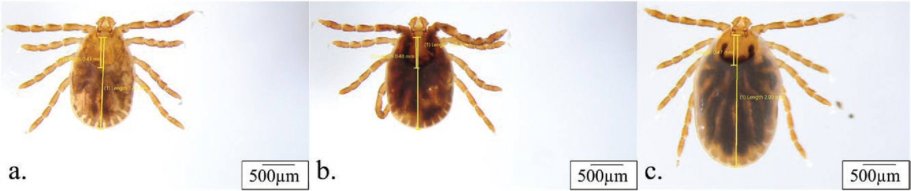
Examples of Haemaphysalis longicornis nymphs in the unfed state (A) versus having been attached to a mouse for 48 h (B) or 72 h (C). Images and measurements for scutal length and body length were taken using Olympus cellSens imaging software.
Animal use and experimental procedures were conducted in accordance with an approved protocol on file with the Centers for Disease Control and Prevention, Division of Vector-Borne Diseases, Institutional Animal Care and Use Committee.
Results and Discussion
Of the 80 H. longicornis nymphs placed on 4 mice (N01, N04, N06, and N07 were removed from the study due to their capsules becoming dislodged) 57 (71%) nymphs were found to be attached at the time of mouse euthanasia at either 48 or 72 h after the nymphs were introduced onto the mice (Table 1). The nymphal attachment rate was higher for the 48 h group at 80% for both mice, compared to 60% and 65% for the 2 mice in the 72 h group (Table 1). The median engorgement index of each group ranged from 2.98 (2.88–3.17) for unfed nymphs to 3.04 (2.94–3.20) and 3.06 (2.88–3.23) for nymphs attached for 48 h and 3.34 (3.00–3.57) and 3.79 (2.94–4.30) for nymphs fed on the 2 mice in the 72 h attachment group (Table 1 and Figure 1). The scutal indices reported by Price et al. (2022b) for unfed (2.98) and engorged (3.40) H. longicornis nymphs were similar to the engorgement indices for unfed ticks (2.98), and ticks that fed on mice for 72 h (3.34 and 3.79 for the 2 individual mice) described in this study. The mean weight was 0.22 mg for unfed nymphs, increasing to 0.33 and 0.35 mg for nymphs attached to each of 2 mice for 48 h and 0.56 and 0.60 mg for nymphs attached to each of 2 mice for 72 h group (Table 1). The mean weights of partially engorged nymphs having been attached for either 48 or 72 h in this study (0.33–0.60 mg) were much lower than reported for fully engorged and naturally detached nymphs (3.03 mg) in a previous study (Zheng et al. 2011). This is not surprising as ixodid ticks tend to ingest the majority of the blood meal in the last 12–36 h of their natural feeding period (Balashov 1972), and our study aimed to interrupt the blood meal before the nymphs had ingested enough blood to be able to molt to adults without taking in additional blood. In our study, the partially fed nymphs that had detached themselves from the deceased hosts were found on the underside of the lid, and sides of the cage. Although we cannot know if given the opportunity the partially fed ticks would have attached to a subsequent host, these ticks appeared active and presumably were seeking a second host.
Table 1.
Assessment of engorgement and Borrelia burgdorferi sensu stricto infection rate in Haemaphysalis longicornis nymphs fed on infected Mus musculus CD-1 mice for 48 or 72 h
| Data collected from H. longicornis nymphs partially fed (48 h or 72 h) on B. burgdorferi s.s.-infected CD-1 mice |
Infection with B. burgdorferi s.s. for partially engorged nymphs |
|||||
|---|---|---|---|---|---|---|
| Duration of tick feeding | Mouse ID | No. (%) of attached ticks recovered out of 20 introduced per mouseb | Median (range) of engorgement indices (body length/scutal length) | Mean weight per tick (mg)d | Percentage (no.) of infected nymphs per mousee | Percentage (no.) of infected nymphs per timepoint |
|
| ||||||
| Unfed | NAa | NAa | 2.98 (2.88–3.17)c | 0.22c | NAa | NAa |
| 48 h | N03 | 16 (80%) | 3.04 (2.94–3.20) | 0.35 | 0% (0/16) | 6.3% (2/32) |
| N05 | 16 (80%) | 3.06 (2.88–3.23) | 0.33 | 12.5% (2/16) | ||
| 72 h | N02 | 12 (60%) | 3.79 (2.94–4.30) | 0.60 | 0% (0/12) | 0% (0/25) |
| N08 | 13 (65%) | 3.34 (3.00–3.57) | 0.56 | 0% (0/13) | ||
NA, not applicable.
Number of ticks recovered after the mouse was euthanized at 48 or 72 h.
Mean engorgement index and weight calculated from 13 unfed H. longicornis nymphs of the same age as the 48 and 72 h partially engorged nymphs.
Median and range not provided; ticks were weighed collectively by mouse to obtain an average weight per tick.
Ticks were assessed for infection by detection of the Borrelia flaB target using a multiplex PCR amplicon sequencing assay as described by Osikowicz et al. (2024).
Unexpectedly, B. burgdorferi s.s. was detected in a higher number of engorged nymphs (2/32) at the 48 h attachment timepoint compared to the 72 h timepoint (0/25), despite nymphs removed at 72 h being attached for 24 h longer (Table 1). This result contrasts with the finding of Piesman (1991) for I. scapularis (I. dammini) larvae, where each increase in larval feeding time (from 8 to 16, 24, 48, and ≥72 h) on an infected mouse corresponded to an increased infection rate for B. burgdorferi s.s., and the infection rate of partially fed larvae had reached 68% (16/24 ticks) at the 48 h timepoint. Moreover, the percentages of infected partially fed H. longicornis nymphs in our study of 0–12% (0/12, 0/13, 0/16, and 2/16 ticks; Table 1) across individual mice was much lower than the 56% (18/32 ticks) reported for fully fed H. longicornis larvae (having typically completed their blood meals in 96–120 h) in a previous study with the same strain of B. burgdorferi s.s. (Breuner et al. 2020). It also is noteworthy that the nymphs acquired B. burgdorferi s.s. from only 1 of the 2 confirmed infected mice exposed to nymphal feeding for 48 h and from none of the 2 infected mice with nymphs feeding for 72 h (Table 1). We have no clear explanation for this surprising finding. Based on previous studies, H. longicornis appears to be less effective in acquiring B. burgdorferi s.s. from infected hosts compared to I. scapularis, and we cannot rule out the possibility that the mouse from which the nymphs acquired spirochetes had a higher bacteremia than the other mice at the time of nymphal feeding. The small sample size for examined partially fed nymphs (12–16 per mouse) also could have yielded stochastic results, and further investigation would be required to evaluate the relationship between feeding duration and the infection rate.
We conclude that the potential for H. longicornis nymphs to acquire B. burgdorferi s.s. via interrupted feeding in the first 72 h of a blood meal on an infected host is low. However, it remains a plausible scenario of how field-collected questing H. longicornis nymphs that have naturally detached from an infected host before completing their blood meal can be infected with B. burgdorferi s.s. DNA, without the need for transstadial transmission of spirochetes from the larval stage to have occurred. It remains unknown if H. longicornis ticks containing B. burgdorferi s.s. DNA from a partial blood meal can maintain viable spirochetes, or if they could infect another competent host while completing their blood meal.
Acknowledgments
The authors thank William Hervey, Kris Hartzer, and William Nicholson from the CDC Medical Entomology Laboratory in Atlanta, GA for providing the H. longicornis nymphs used in this work.
Footnotes
Disclaimer
The findings and conclusions of this study are those of the authors and do not necessarily represent the views of the Centers for Disease Control and Prevention.
References
- Balashov YS. 1972. Weight change dynamics during feeding. A translation of Bloodsucking ticks (Ixodoidea)—vectors of diseases of man and animals. College Park, MD: Miscellaneous Publications of the Entomological Society of America; p. 234–234. [Google Scholar]
- Breuner NE, Ford SL, Hojgaard A, et al. 2020. Failure of the Asian longhorned tick, Haemaphysalis longicornis, to serve as an experimental vector of the Lyme disease spirochete, Borrelia burgdorferi sensu stricto. Ticks Tick Borne Dis. 11:101311. 10.1016/j.ttbdis.2019.101311 [DOI] [PMC free article] [PubMed] [Google Scholar]
- Cumbie AN, Trimble RN, Eastwood G. 2022. Pathogen spillover to an invasive tick species: first detection of Bourbon virus in Haemaphysalis longicornis in the United States. Pathogens 11:454. 10.3390/pathogens11040454 [DOI] [PMC free article] [PubMed] [Google Scholar]
- Dinkel KD, Herndon DR, Noh SM, et al. 2021. Isolate of Theileria orientalis, Ikeda genotype, is transmitted to cattle by the invasive Asian longhorned tick, Haemaphysalis longicornis. Parasit Vectors 14:157. 10.1186/s13071-021-04659-9 [DOI] [PMC free article] [PubMed] [Google Scholar]
- Dye-Braumuller KC, Gual-Gonzalez L, Abiodun T, et al. 2023. Invasive Haemaphysalis longicornis (Acari: Ixodidae) investigation in South Carolina: new records of establishment, pathogen prevalence, and blood meal analyses. J. Med. Entomol. 60:1398–1405. 10.1093/jme/tjad119 [DOI] [PMC free article] [PubMed] [Google Scholar]
- Eleftheriou A, Zeiger B, Jennings J, et al. 2024. Phenology and habitat associations of the invasive Asian longhorned tick from Ohio, USA. Med. Vet. Entomol. 38:314–324. 10.1111/mve.12719 [DOI] [PubMed] [Google Scholar]
- Hojgaard A, Osikowicz LM, Eisen L, et al. 2020. Evaluation of a novel multiplex PCR amplicon sequencing assay for detection of human pathogens in Ixodes ticks. Ticks Tick Borne Dis. 11:101504. 10.1016/j.ttbdis.2020.101504 [DOI] [PMC free article] [PubMed] [Google Scholar]
- Levin ML, Stanley HM, Hartzer K, et al. 2021. Incompetence of the Asian longhorned tick (Acari: Ixodidae) in transmitting the agent of human granulocytic anaplasmosis in the United States. J. Med. Entomol. 58:1419–1423. 10.1093/jme/tjab015 [DOI] [PMC free article] [PubMed] [Google Scholar]
- Osikowicz LM, Maes SE, Eisen RJ, et al. 2024. A next generation sequencing assay combining Ixodes species identification with pathogen detection to support tick surveillance efforts in the United States. Ticks Tick Borne Dis. 15:102343. 10.1016/j.ttbdis.2024.102343 [DOI] [PMC free article] [PubMed] [Google Scholar]
- Piesman J 1991. Experimental acquisition of the Lyme disease spirochete, Borrelia burgdorferi, by larval Ixodes dammini (Acari: Ixodidae) during partial blood meals. J. Med. Entomol. 28:259–262. 10.1093/jmedent/28.2.259 [DOI] [PubMed] [Google Scholar]
- Poh KC, Aguilar M, Capelli-Peixoto J, et al. 2024. Haemaphysalis longicornis (Acari: Ixodidae) does not transmit Babesia bovis, a causative agent of cattle fever. Ticks Tick Borne Dis. 15:102374. 10.1016/j.ttbdis.2024.102374 [DOI] [PubMed] [Google Scholar]
- Price KJ, Graham CB, Witmier BJ, et al. 2021. Borrelia burgdorferi sensu stricto DNA in field-collected Haemaphysalis longicornis ticks, Pennsylvania, United States. Emerg. Infect. Dis. 27:608–611. 10.3201/eid2702.201552 [DOI] [PMC free article] [PubMed] [Google Scholar]
- Price KJ, Ayres BN, Maes SE, et al. 2022a. First detection of human pathogenic variant of Anaplasma phagocytophilum in field-collected Haemaphysalis longicornis, Pennsylvania, USA. Zoonoses Public Health. 69:143–148. 10.1111/zph.12901 [DOI] [PubMed] [Google Scholar]
- Price KJ, Witmier BJ, Eckert RA, et al. 2022b. Recovery of partially engorged Haemaphysalis longicornis (Acari: Ixodidae) ticks from active surveillance. J. Med. Entomol. 59:1842–1846. 10.1093/jme/tjac099 [DOI] [PMC free article] [PubMed] [Google Scholar]
- Price KJ, Khalil N, Witmier BJ, et al. 2023. of protozoan and bacterial infection and co-infection and partial blood feeding in the invasive tick Haemaphysalis longicornis in Pennsylvania. J. Parasitol. 109:265–273. [DOI] [PMC free article] [PubMed] [Google Scholar]
- Rainey T, Occi JL, Robbins RG, et al. 2018. Discovery of Haemaphysalis longicornis (Ixodida: Ixodidae) parasitizing a sheep in New Jersey, United States. J. Med. Entomol. 55:757–759. 10.1093/jme/tjy006 [DOI] [PubMed] [Google Scholar]
- Raney WR, Herslebs EJ, Langohr IM, et al. 2022a. Horizontal and vertical transmission of Powassan virus by the invasive Asian longhorned tick, Haemaphysalis longicornis, under laboratory conditions. Front. Cell. Infect. Microbiol. 12:923914. 10.3389/fcimb.2022.923914 [DOI] [PMC free article] [PubMed] [Google Scholar]
- Raney WR, Perry JB, Hermance ME. 2022b. Transovarial transmission of Heartland virus by invasive Asian longhorned ticks under laboratory conditions. Emerg. Infect. Dis. 28:726–729. 10.3201/eid2803.210973 [DOI] [PMC free article] [PubMed] [Google Scholar]
- Shih CM, Spielman A. 1993. Accelerated transmission of Lyme disease spirochetes by partially fed vector ticks. J. Clin. Microbiol. 31:2878–2881. 10.1128/jcm.31.11.2878-2881.1993 [DOI] [PMC free article] [PubMed] [Google Scholar]
- Stanley HM, Ford SL, Snellgrove AN, et al. 2020. The ability of the invasive Asian longhorned tick Haemaphysalis longicornis (Acari: Ixodidae) to acquire and transmit Rickettsia rickettsii (Rickettsiales: Rickettsiaceae), the agent of Rocky Mountain spotted fever, under laboratory conditions. J. Med. Entomol. 57:1635–1639. 10.1093/jme/tjaa076 [DOI] [PubMed] [Google Scholar]
- Thompson AT, White S, Shaw D, et al. 2020. Theileria orientalis Ikeda in host-seeking Haemaphysalis longicornis in Virginia, U.S.A. Ticks Tick Borne Dis. 11:101450. 10.1016/j.ttbdis.2020.101450 [DOI] [PubMed] [Google Scholar]
- Tufts DM, VanAcker MC, Fernandez MP, et al. 2019. Distribution, host-seeking phenology, and host and habitat associations of Haemaphysalis longicornis ticks, Staten Island, New York, USA. Emerg. Infect. Dis. 25:792–796. 10.3201/eid2504.181541 [DOI] [PMC free article] [PubMed] [Google Scholar]
- USDA (United States Department of Agriculture). National Haemaphysalis longicornis (Asian longhorned tick) Situation Report. [accessed 2024. Jun 6]. https://www.aphis.usda.gov/sites/default/files/longhorned-tick-sitrep.pdf.
- Yeh MT, Bak JM, Hu R, et al. 1995. Determining the duration of Ixodes scapularis (Acari: Ixodidae) attachment to tick-bite victims. J. Med. Entomol. 32:853–858. 10.1093/jmedent/32.6.853 [DOI] [PubMed] [Google Scholar]
- Zheng H, Yu Z, Chen Z, et al. 2011. Development and biological characteristics of Haemaphysalis longicornis (Acari: Ixodidae) under field conditions. Exp. Appl. Acarol. 53:377–388. 10.1007/s10493-010-9415-3 [DOI] [PubMed] [Google Scholar]