

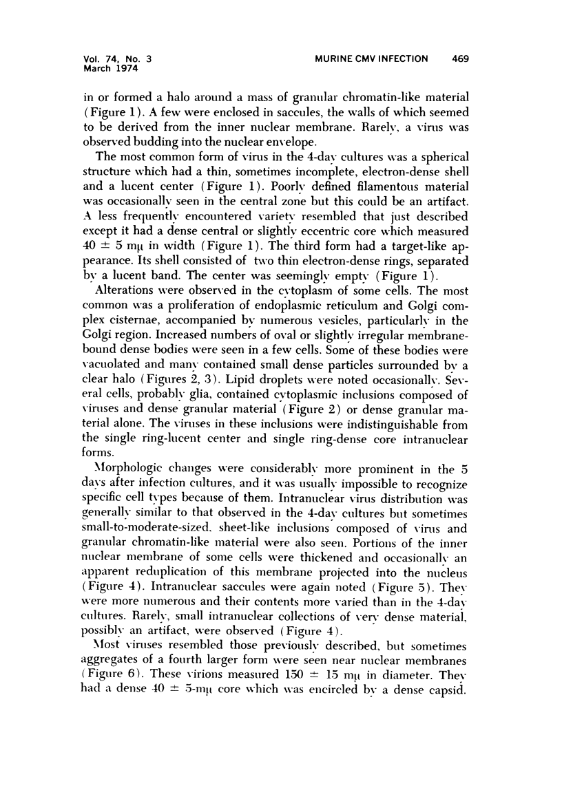
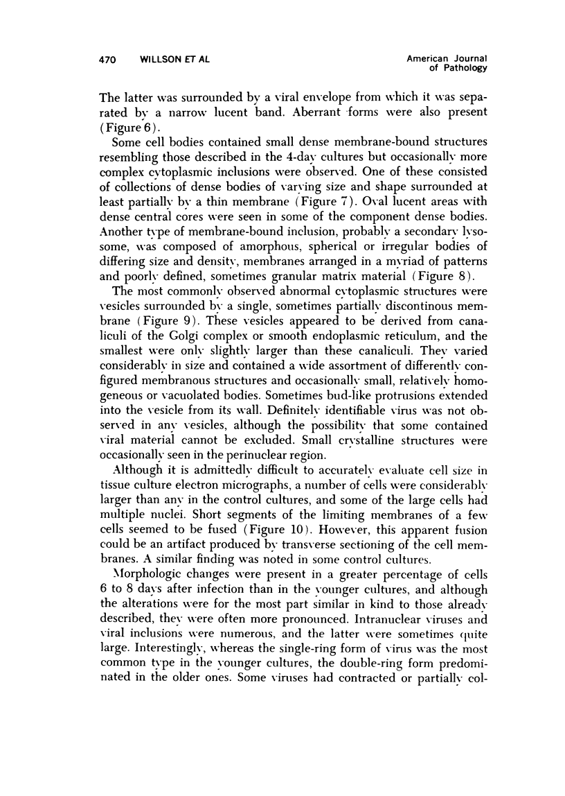
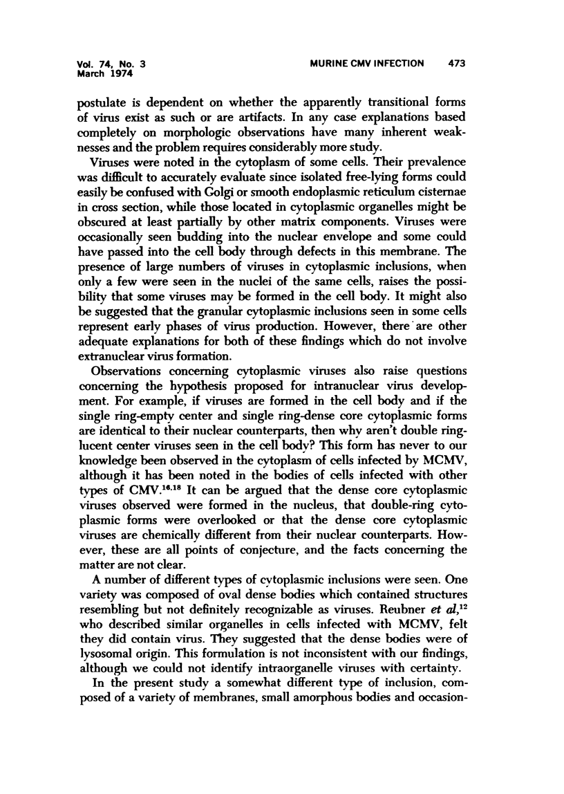
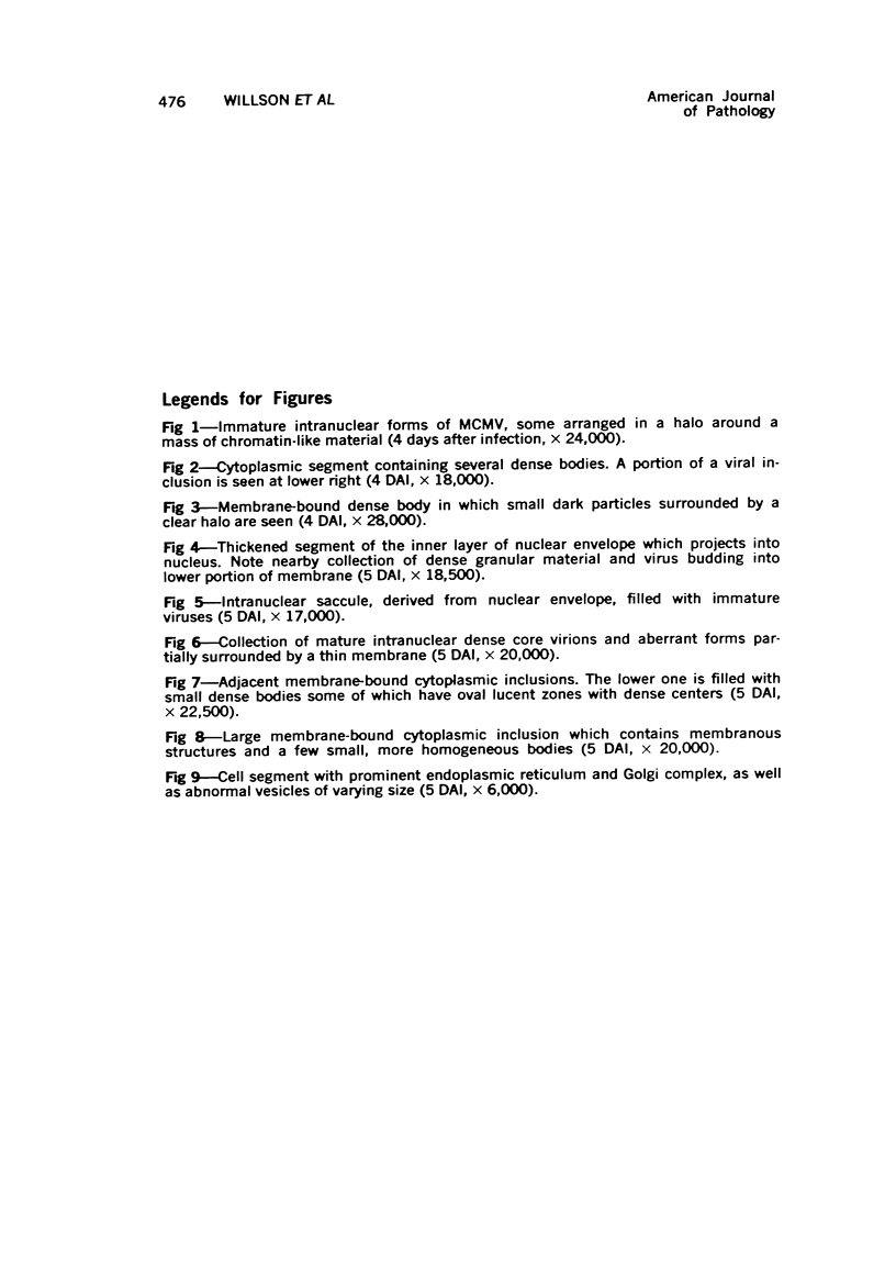
Abstract
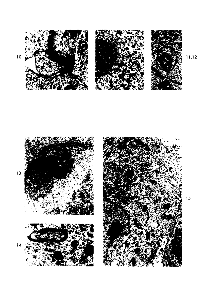
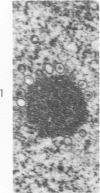
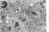
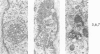
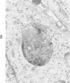
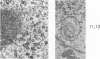
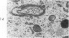
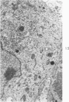
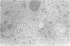
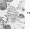
Mouse spinal cord-ganglia cultures were innoculated with murine cytomegalo-virus 14 days after explantation. Intranuclear virus was first observed 4 days after infection. The viruses, which occurred in four forms, were observed in increasing numbers during the ensuing 4 days. Differences were noted in the relative prevalence of certain of these forms in older as compared to younger cultures. This suggests that variations in virus form are related to virus maturation. Cytoplasmic viruses were occasionally observed, but their site of origin is not certain. A variety of cytoplasmic inclusions were seen, particularly in the older cultures. It seems likely that they represent specific cell responses to the presence of the virus. They were not observed in the control cultures, even though some of the latter did show severe degenerative changes.
Full text
PDF


Images in this article


Selected References
These references are in PubMed. This may not be the complete list of references from this article.
- BUNGE R. P., BUNGE M. B., PETERSON E. R. AN ELECTRON MICROSCOPE STUDY OF CULTURED RAT SPINAL CORD. J Cell Biol. 1965 Feb;24:163–191. doi: 10.1083/jcb.24.2.163. [DOI] [PMC free article] [PubMed] [Google Scholar]
- Berezesky I. K., Grimley P. M., Tyrrell S. A., Rabson A. S. Ultrastructure of a rat cytomegalovirus. Exp Mol Pathol. 1971 Jun;14(3):337–349. doi: 10.1016/0014-4800(71)90005-0. [DOI] [PubMed] [Google Scholar]
- Chopra H. C., Lloyd B. J., Jr, Ablashi D. V., Armstrong G. R. Morphologic studies of a cytomegalovirus isolated from an owl monkey. J Natl Cancer Inst. 1972 May;48(5):1333–1340. [PubMed] [Google Scholar]
- Hanshaw J. B. Congenital cytomegalovirus infection: a fifteen year perspective. J Infect Dis. 1971 May;123(5):555–561. doi: 10.1093/infdis/123.5.555. [DOI] [PubMed] [Google Scholar]
- Henson D., Strano A. J. Mouse cytomegalovirus. Necrosis of infected and morphologically normal submaxillary gland acinar cells during termination of chronic infection. Am J Pathol. 1972 Jul;68(1):183–202. [PMC free article] [PubMed] [Google Scholar]
- LUSE S. A., SMITH M. G. Electron microscopy of salivary gland viruses. J Exp Med. 1958 May 1;107(5):623–632. doi: 10.1084/jem.107.5.623. [DOI] [PMC free article] [PubMed] [Google Scholar]
- MANNINI A., MEDEARIS D. N., Jr Mouse salivary gland virus infections. Am J Hyg. 1961 May;73:329–343. doi: 10.1093/oxfordjournals.aje.a120192. [DOI] [PubMed] [Google Scholar]
- MORGAN C., ROSE H. M., HOLDEN M., JONES E. P. Electron microscopic observations on the development of herpes simplex virus. J Exp Med. 1959 Oct 1;110:643–656. doi: 10.1084/jem.110.4.643. [DOI] [PMC free article] [PubMed] [Google Scholar]
- Markham F. S., Hudson N. P. Susceptibility of the Guinea Pig Fetus to the Submaxillary Gland Virus of Guinea Pigs. Am J Pathol. 1936 Mar;12(2):175–182.1. [PMC free article] [PubMed] [Google Scholar]
- Middelkamp J. N., Patrizi G., Reed C. A. Light and electron microscopic studies of the guinea pig cytomegalovirus. J Ultrastruct Res. 1967 Apr;18(1):85–101. doi: 10.1016/s0022-5320(67)80233-8. [DOI] [PubMed] [Google Scholar]
- Nii S., Morgan C., Rose H. M. Electron microscopy of herpes simplex virus. II. Sequence of development. J Virol. 1968 May;2(5):517–536. doi: 10.1128/jvi.2.5.517-536.1968. [DOI] [PMC free article] [PubMed] [Google Scholar]
- PINKERTON H. New horizons in virus research. Proc Inst Med Chic. 1953 Feb 15;19(11):243–252. [PubMed] [Google Scholar]
- RUEBNER B. H., MIYAI K., SLUSSER R. J., WEDEMEYER P., MEDEARIS D. N., Jr MOUSE CYTOMEGALOVIRUS INFECTION. AN ELECTRON MICROSCOPIC STUDY OF HEPATIC PARENCHYMAL CELLS. Am J Pathol. 1964 May;44:799–821. [PMC free article] [PubMed] [Google Scholar]
- Reynolds D. W., Stagno S., Hosty T. S., Tiller M., Alford C. A., Jr Maternal cytomegalovirus excretion and perinatal infection. N Engl J Med. 1973 Jul 5;289(1):1–5. doi: 10.1056/NEJM197307052890101. [DOI] [PubMed] [Google Scholar]
- Ruebner B. H., Hirano T., Slusser R., Osborn J., Medearis D. N., Jr Cytomegalovirus infection. Viral ultrastructure with particular reference to the relationship of lysosomes to cytoplasmic inclusions. Am J Pathol. 1966 Jun;48(6):971–989. [PMC free article] [PubMed] [Google Scholar]
- Schneider J. F., Carp R. I., Belisle E. H., Rue C. E. The response of murine nerve tissue culture to murine cytomegalic virus. Acta Neuropathol. 1972;21(3):204–212. doi: 10.1007/BF00688499. [DOI] [PubMed] [Google Scholar]
- WYATT J. P., SAXTON J. Generalized cytomegalic inclusion disease. J Pediatr. 1950 Mar;36(3):271-94, illust. doi: 10.1016/s0022-3476(50)80097-5. [DOI] [PubMed] [Google Scholar]