Abstract
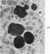
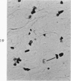
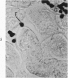
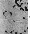
The beige mouse is a homolog of Chediak-Higashi syndrome, a disorder which is characterized by the presence of enlarged (anomalous) lysosomes in many cell types. In kidney, anomalous lysosomes are present in cells of the proximal convoluted tubules. In this study, the degradation of injected horseradish peroxidase (HRP) in lysosomes was studied in both the convoluted (S1-S2) and straight (S3) segments of the proximal tubules of beige and control (C57 B1) mice. Tissues were removed at intervals from 18 hours to 7 days after HRP injection. Peroxidase activity was visualized for light and electron microscopy by incubating sections in diaminobenzidine medium. No differences in the rate of degradation of HRP were demonstrable between anomalous lysosomes in S1-S2 cells of beige kidney and those in controls. In both animals, HRP was demonstrable in these lysosomes at 18 and 36 hours but not at 48 hours after injection. By electron microscopy, reaction product appeared as a flocculent precipitate distributed uniformly throughout the lysosome. In contrast to those of S1-S2 cells, lysosomes of beige S3 cells degraded HRP much more slowly than did those of control mice. In controls, HRP was demonstrable in S3 lysosomes at 18 hours but not at 48 hours after injection. In beige mouse kidney HRP was demonstrable in many S3 lysosomes at 48 hours, and it persisted in some lysosomes as long as 5 days after injection. These findings indicate that beige S3 lysosomes are defective in degrading protein. As reported recently, these lysosomes are also markedly enlarged and altered in content. They appear to arise as part of a renal lesion of unknown pathogenesis which is confined to the S3 segments of the proximal tubules. The slower rate of degradation of protein appears to be another manifestation of the alteration in these lysosomes.
Full text
PDF


Images in this article
Selected References
These references are in PubMed. This may not be the complete list of references from this article.
- Bennett J. M., Blume R. S., Wolff S. M. Characterization and significance of abnormal leukocyte granules in the beige mouse: a possible homologue for Chediak-Higashi Aleutian trait. J Lab Clin Med. 1969 Feb;73(2):235–243. [PubMed] [Google Scholar]
- Blume R. S., Padgett G. A., Wolff S. M., Bennett J. M. Giant neutrophil granules in the Chediak-Higashi syndrome of man, mink, cattle and mice. Can J Comp Med. 1969 Oct;33(4):271–274. [PMC free article] [PubMed] [Google Scholar]
- Davis W. C., Spicer S. S., Greene W. B., Padgett G. A. Ultrastructure of bone marrow granulocytes in normal mink and mink with the homolog of the Chediak-Higashi trait of humans. I. Origin of the abnormal granules present in the neutrophils of mink with the C-HS trait. Lab Invest. 1971 Apr;24(4):303–317. [PubMed] [Google Scholar]
- Davis W. C., Spicer S. S., Greene W. B., Padgett G. A. Ultrastructure of cells in bone marrow and peripheral blood of normal mink and mink with the homologue of the Chediak-Higashi trait of humans. II. Cytoplasmic granules in eosinophils, basophils, mononuclear cells and platelets. Am J Pathol. 1971 Jun;63(3):411–432. [PMC free article] [PubMed] [Google Scholar]
- Essner E., Oliver C. A hereditary alteration in kidneys of mice with Chediak-Higashi syndrome. Am J Pathol. 1973 Oct;73(1):217–232. [PMC free article] [PubMed] [Google Scholar]
- Gallin J. I., Bujak J. S., Patten E., Wolff S. M. Granulocyte function in the Chediak-Higashi syndrome of mice. Blood. 1974 Feb;43(2):201–206. [PubMed] [Google Scholar]
- Graham R. C., Jr, Karnovsky M. J. The early stages of absorption of injected horseradish peroxidase in the proximal tubules of mouse kidney: ultrastructural cytochemistry by a new technique. J Histochem Cytochem. 1966 Apr;14(4):291–302. doi: 10.1177/14.4.291. [DOI] [PubMed] [Google Scholar]
- LOWRY O. H., ROSEBROUGH N. J., FARR A. L., RANDALL R. J. Protein measurement with the Folin phenol reagent. J Biol Chem. 1951 Nov;193(1):265–275. [PubMed] [Google Scholar]
- Lutzner M. A., Lowrie C. T., Jordan H. W. Giant granules in leukocytes of the beige mouse. J Hered. 1967 Nov-Dec;58(6):299–300. doi: 10.1093/oxfordjournals.jhered.a107620. [DOI] [PubMed] [Google Scholar]
- Maunsbach A. B. Observations on the segmentation of the proximal tubule in the rat kidney. Comparison of results from phase contrast, fluorescence and electron microscopy. J Ultrastruct Res. 1966 Oct;16(3):239–258. doi: 10.1016/s0022-5320(66)80060-6. [DOI] [PubMed] [Google Scholar]
- Oliver C., Essner E. Distribution of anomalous lysosomes in the beige mouse: a homologue of Chediak-Higashi syndrome. J Histochem Cytochem. 1973 Mar;21(3):218–228. doi: 10.1177/21.3.218. [DOI] [PubMed] [Google Scholar]
- PIERRO L. J. PIGMENT GRANULE FORMATION IN SLATE, A COAT COLOR MUTANT IN THE MOUSE. Anat Rec. 1963 Aug;146:365–372. [PubMed] [Google Scholar]
- Prieur D. J., Davis W. C., Padgett G. A. Defective function of renal lysosomes in mice with the Chediak-Higashi syndrome. Am J Pathol. 1972 May;67(2):227–236. [PMC free article] [PubMed] [Google Scholar]
- REYNOLDS E. S. The use of lead citrate at high pH as an electron-opaque stain in electron microscopy. J Cell Biol. 1963 Apr;17:208–212. doi: 10.1083/jcb.17.1.208. [DOI] [PMC free article] [PubMed] [Google Scholar]
- Root R. K., Rosenthal A. S., Balestra D. J. Abnormal bactericidal, metabolic, and lysosomal functions of Chediak-Higashi Syndrome leukocytes. J Clin Invest. 1972 Mar;51(3):649–665. doi: 10.1172/JCI106854. [DOI] [PMC free article] [PubMed] [Google Scholar]
- SABATINI D. D., BENSCH K., BARRNETT R. J. Cytochemistry and electron microscopy. The preservation of cellular ultrastructure and enzymatic activity by aldehyde fixation. J Cell Biol. 1963 Apr;17:19–58. doi: 10.1083/jcb.17.1.19. [DOI] [PMC free article] [PubMed] [Google Scholar]
- STRAUS W. OCCURRENCE OF PHAGOSOMES AND PHAGO-LYSOSOMES IN DIFFERENT SEGMENTS OF THE NEPHRON IN RELATION TO THE REABSORPTION, TRANSPORT, DIGESTION, AND EXTRUSION OF INTRAVENOUSLY INJECTED HORSERADISH PEROXIDASE. J Cell Biol. 1964 Jun;21:295–308. doi: 10.1083/jcb.21.3.295. [DOI] [PMC free article] [PubMed] [Google Scholar]
- Spurr A. R. A low-viscosity epoxy resin embedding medium for electron microscopy. J Ultrastruct Res. 1969 Jan;26(1):31–43. doi: 10.1016/s0022-5320(69)90033-1. [DOI] [PubMed] [Google Scholar]
- White J. G. The Chediak-Higashi syndrome: a possible lysosomal disease. Blood. 1966 Aug;28(2):143–156. [PubMed] [Google Scholar]


