

Abstract
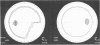
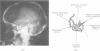
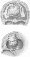
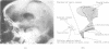
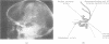
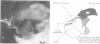
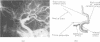
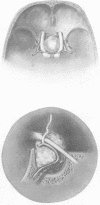
Three patients with nasal visual field defects are described. In each case it is believed that compression of the lateral fibres of the optic nerve by the anterior cerebral or internal carotid artery was the cause. Binasal hemianopia can thus be produced by a single lesion and is as much a true hemianopia as the common bitemporal one. The value of careful neuroradiological investigation to display the relationships of a tumour to the chiasma, optic nerves, and related vessels and thus explain the field defects is demonstrated.
Full text
PDF


Images in this article


Selected References
These references are in PubMed. This may not be the complete list of references from this article.
- BULL J. The normal variations in the position of the optic recess of the third ventricle. Acta radiol. 1956 Jul-Aug;46(1-2):72–80. doi: 10.3109/00016925609170814. [DOI] [PubMed] [Google Scholar]
- DILENGE D. Anatomie radiologique de la citerne opto-chiasmatique. Neurochirurgie. 1955;1(3):257–267. [PubMed] [Google Scholar]
- Gado M., Bull J. W. The carotid angiogram in suprasellar masses. Neuroradiology. 1971 Aug;2(3):136–153. doi: 10.1007/BF00335042. [DOI] [PubMed] [Google Scholar]
- RUCKER C. W., KERNOHAN J. W. Notching of the optic chiasm by overlying arteries in pituitary tumors. AMA Arch Ophthalmol. 1954 Feb;51(2):161–170. doi: 10.1001/archopht.1954.00920040163002. [DOI] [PubMed] [Google Scholar]
- Schneider R. C., Kriss F. C., Falls H. F. Prechiasmal infarction associated with intrachiasmal and suprasellar tumors. J Neurosurg. 1970 Feb;32(2):197–208. doi: 10.3171/jns.1970.32.2.0197. [DOI] [PubMed] [Google Scholar]