

Abstract
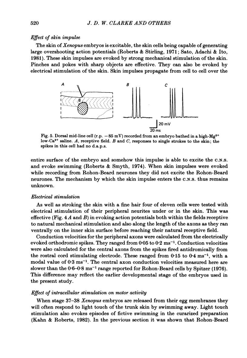
Rohon-Beard neurones show substance P-like immunoactivity in their somas and in their centrally projecting axons. Peripherally, the morphology of their free nerve endings within the trunk skin has been shown using horseradish peroxidase staining. The excitation of Rohon-Beard neurones by natural and electrical stimulation of the skin has been examined using intracellular micro-electrodes in the late embryo of Xenopus laevis. Rohon-Beard cells are sensitive to transient, local indentation of the trunk skin, responding with one or a few impulses. They adapt rapidly to repeated stimulation. They can also be excited by a brief current pulse to the skin. They are not sensitive to slow indentation of the skin, nor are they excited by epithelial action potentials. The responses to skin stimulation are not abolished by a Ringer solution containing 12 mM-Mg2+ and only 0.5 mM-Ca2+. Intracellularly evoked action potentials in single Rohon-Beard cells are sometimes sufficient to evoke sustained episodes of fictive swimming. The results indicate that Rohon-Beard cells are responsible for detecting light touch stimuli to the embryo's body and for initiating swimming in response to this stimulus.
Full text
PDF


Images in this article


Selected References
These references are in PubMed. This may not be the complete list of references from this article.
- Barber R. P., Vaughn J. E., Slemmon J. R., Salvaterra P. M., Roberts E., Leeman S. E. The origin, distribution and synaptic relationships of substance P axons in rat spinal cord. J Comp Neurol. 1979 Mar 15;184(2):331–351. doi: 10.1002/cne.901840208. [DOI] [PubMed] [Google Scholar]
- Bixby J. L., Spitzer N. C. The appearance and development of chemosensitivity in Rohon-Beard neurones of the Xenopus spinal cord. J Physiol. 1982 Sep;330:513–536. doi: 10.1113/jphysiol.1982.sp014356. [DOI] [PMC free article] [PubMed] [Google Scholar]
- Buchanan J. T., Cohen A. H. Activities of identified interneurons, motoneurons, and muscle fibers during fictive swimming in the lamprey and effects of reticulospinal and dorsal cell stimulation. J Neurophysiol. 1982 May;47(5):948–960. doi: 10.1152/jn.1982.47.5.948. [DOI] [PubMed] [Google Scholar]
- Chan-Palay V., Palay S. L. Ultrastructural identification of substance P cells and their processes in rat sensory ganglia and their terminals in the spinal cord by immunocytochemistry. Proc Natl Acad Sci U S A. 1977 Sep;74(9):4050–4054. doi: 10.1073/pnas.74.9.4050. [DOI] [PMC free article] [PubMed] [Google Scholar]
- Cuello A. C., Kanazawa I. The distribution of substance P immunoreactive fibers in the rat central nervous system. J Comp Neurol. 1978 Mar 1;178(1):129–156. doi: 10.1002/cne.901780108. [DOI] [PubMed] [Google Scholar]
- HUGHES A. The development of the primary sensory system in Xenopus laevis (Daudin). J Anat. 1957 Jul;91(3):323–338. [PMC free article] [PubMed] [Google Scholar]
- Hayes B. P., Roberts A. The anatomy of two functional types of mechanoreceptive 'free' nerve-ending in the head skin of Xenopus embryos. Proc R Soc Lond B Biol Sci. 1983 Apr 22;218(1210):61–76. doi: 10.1098/rspb.1983.0026. [DOI] [PubMed] [Google Scholar]
- Hunt S. P., Kelly J. S., Emson P. C., Kimmel J. R., Miller R. J., Wu J. Y. An immunohistochemical study of neuronal populations containing neuropeptides or gamma-aminobutyrate within the superficial layers of the rat dorsal horn. Neuroscience. 1981;6(10):1883–1898. doi: 10.1016/0306-4522(81)90029-4. [DOI] [PubMed] [Google Scholar]
- Inagaki S., Senba E., Shiosaka S., Takagi H., Kawai Y., Takatsuki K., Sakanaka M., Matsuzaki T., Tohyama M. Regional distribution of substance P-like immunoreactivity in the frog brain and spinal cord: immunohistochemical analysis. J Comp Neurol. 1981 Sep 10;201(2):243–254. doi: 10.1002/cne.902010208. [DOI] [PubMed] [Google Scholar]
- Kahn J. A., Roberts A. The central nervous origin of the swimming motor pattern in embryos of Xenopus laevis. J Exp Biol. 1982 Aug;99:185–196. doi: 10.1242/jeb.99.1.185. [DOI] [PubMed] [Google Scholar]
- Lamborghini J. E. Rohon-beard cells and other large neurons in Xenopus embryos originate during gastrulation. J Comp Neurol. 1980 Jan 15;189(2):323–333. doi: 10.1002/cne.901890208. [DOI] [PubMed] [Google Scholar]
- Ljungdahl A., Hökfelt T., Nilsson G. Distribution of substance P-like immunoreactivity in the central nervous system of the rat--I. Cell bodies and nerve terminals. Neuroscience. 1978;3(10):861–943. doi: 10.1016/0306-4522(78)90116-1. [DOI] [PubMed] [Google Scholar]
- Lorez H. P., Kemali M. Substance P-, met-enkephalin- and somatostatin-like immunoreactivity distribution in the frog spinal cord. Neurosci Lett. 1981 Oct 23;26(2):119–124. doi: 10.1016/0304-3940(81)90336-0. [DOI] [PubMed] [Google Scholar]
- Martin A. R., Wickelgren W. O. Sensory cells in the spinal cord of the sea lamprey. J Physiol. 1971 Jan;212(1):65–83. doi: 10.1113/jphysiol.1971.sp009310. [DOI] [PMC free article] [PubMed] [Google Scholar]
- Nicholls J. G., Baylor D. A. Specific modalities and receptive fields of sensory neurons in CNS of the leech. J Neurophysiol. 1968 Sep;31(5):740–756. doi: 10.1152/jn.1968.31.5.740. [DOI] [PubMed] [Google Scholar]
- Ogden T. E., Citron M. C., Pierantoni R. The jet stream microbeveler: an inexpensive way to bevel ultrafine glass micropipettes. Science. 1978 Aug 4;201(4354):469–470. doi: 10.1126/science.663670. [DOI] [PubMed] [Google Scholar]
- Ritchie T. C., Leonard R. B. Immunohistochemical studies on the distribution and origin of candidate peptidergic primary afferent neurotransmitters in the spinal cord of an elasmobranch fish, the Atlantic stingray (Dasyatis sabina). J Comp Neurol. 1983 Feb 1;213(4):414–425. doi: 10.1002/cne.902130406. [DOI] [PubMed] [Google Scholar]
- Roberts A., Clarke J. D. The neuroanatomy of an amphibian embryo spinal cord. Philos Trans R Soc Lond B Biol Sci. 1982 Jan 27;296(1081):195–212. doi: 10.1098/rstb.1982.0002. [DOI] [PubMed] [Google Scholar]
- Roberts A., Hayes B. P. The anatomy and function of 'free' nerve endings in an amphibian skin sensory system. Proc R Soc Lond B Biol Sci. 1977 Apr;196(1125):415–429. doi: 10.1098/rspb.1977.0048. [DOI] [PubMed] [Google Scholar]
- Roberts A. Neuronal growth cones in an amphibian embryo. Brain Res. 1976 Dec 24;118(3):526–530. doi: 10.1016/0006-8993(76)90326-7. [DOI] [PubMed] [Google Scholar]
- Sato E., Adachi S., Ito S. The genesis and transmission of epidermal potentials in an amphibian embryo. Dev Biol. 1981 Nov;88(1):137–146. doi: 10.1016/0012-1606(81)90225-6. [DOI] [PubMed] [Google Scholar]
- Spitzer N. C., Baccaglini P. I. Development of the action potential in embryo amphibian neurons in vivo. Brain Res. 1976 May 14;107(3):610–616. doi: 10.1016/0006-8993(76)90148-7. [DOI] [PubMed] [Google Scholar]
- Spitzer N. C. The ionic basis of the resting potential and a slow depolarizing response in Rohon-Beard neurones of Xenopus tadpoles. J Physiol. 1976 Feb;255(1):105–135. doi: 10.1113/jphysiol.1976.sp011272. [DOI] [PMC free article] [PubMed] [Google Scholar]
- Spitzer N. C. Voltage- and stage-dependent uncoupling of Rohon-Beard neurones during embryonic development of Xenopus tadpoles. J Physiol. 1982 Sep;330:145–162. doi: 10.1113/jphysiol.1982.sp014334. [DOI] [PMC free article] [PubMed] [Google Scholar]
- Teräväinen H., Rovainen C. M. Electrical activity of myotomal muscle fibers, motoneurons, and sensory dorsal cells during spinal reflexes in lampreys. J Neurophysiol. 1971 Nov;34(6):999–1009. doi: 10.1152/jn.1971.34.6.999. [DOI] [PubMed] [Google Scholar]