

Abstract
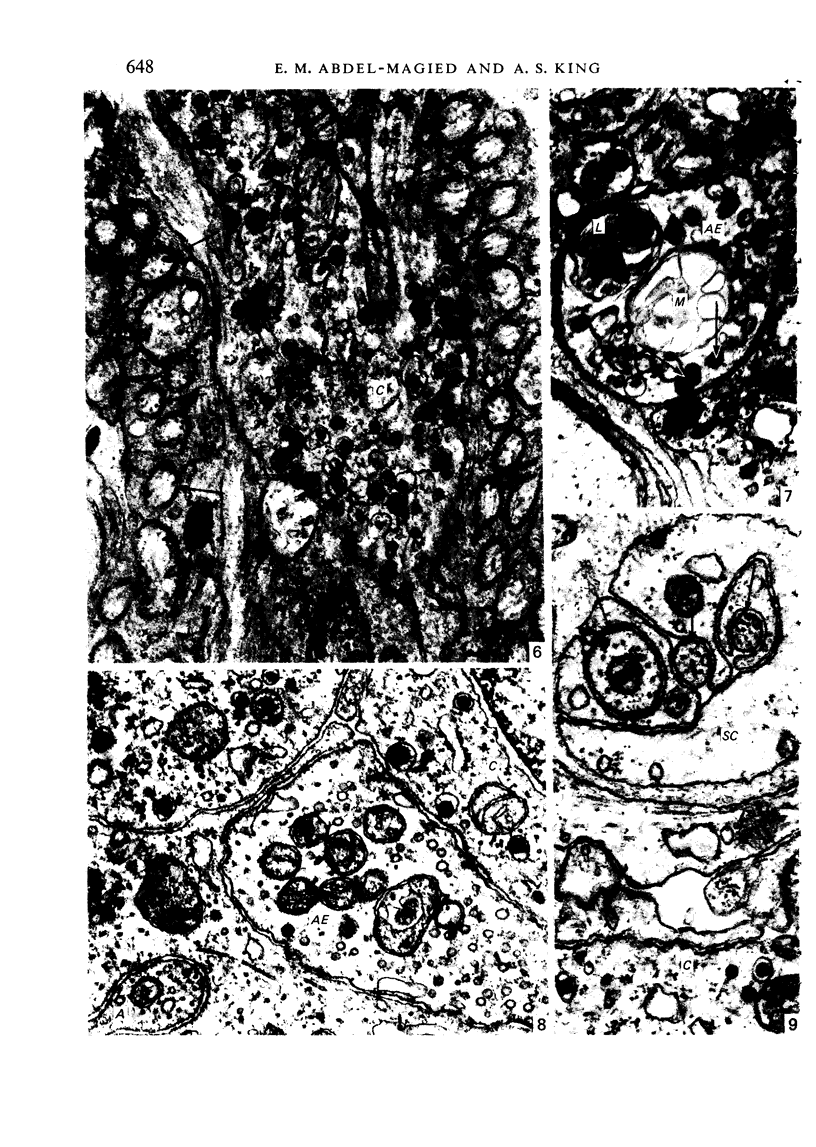
The carotid body of the domestic fowl was examined with the electron microscope after either removal of the distal vagal ganglion or midcervical vagotomy. Almost all the axonal elements of the carotid body degenerated within 5-15 days after ganglionectomy. The degeneration was considered to be due to separation of these axonal elements from their cell bodies (Wallerian degeneration) and indicated that nearly all the nerve supply of the carotid body of Gallus is derived from the vagus nerve. Degeneration of many axonal elements of the carotid body was also seen after midcervical vagotomy, but it took longer (19-41 days) to begin and had greatly increased 207-214 days after operation. It was interpreted as transganglionic degeneration, i.e. severance of the central processes of the distal vagal ganglion cells (by vagotomy) had induced slow degeneration in their peripheral processes (axonal elements in the carotid body). We conclude that the vast majority of the axonal elements in the carotid body of Gallus belong to nerve cell bodies in the distal vagal ganglion and are therefore afferent.
Full text
PDF


Images in this article


Selected References
These references are in PubMed. This may not be the complete list of references from this article.
- Abbott C. P., De Burgh Daly M., Howe A. Early ultrastructural changes in the carotid body after degenerative section of the carotid sinus nerve in the cat. Acta Anat (Basel) 1972;83(2):161–185. doi: 10.1159/000143857. [DOI] [PubMed] [Google Scholar]
- Abdel-Magied E. M., King A. S. The topographical anatomy and blood supply of the carotid body region of the domestic fowl. J Anat. 1978 Aug;126(Pt 3):535–546. [PMC free article] [PubMed] [Google Scholar]
- Arvidsson J., Grant G. Further observations on transganglionic degeneration in trigeminal primary sensory neurons. Brain Res. 1979 Feb 16;162(1):1–12. doi: 10.1016/0006-8993(79)90750-9. [DOI] [PubMed] [Google Scholar]
- Berger A. J. The distribution of the cat's carotid sinus nerve afferent and efferent cell bodies using the horseradish peroxidase technique. Brain Res. 1980 May 26;190(2):309–320. doi: 10.1016/0006-8993(80)90276-0. [DOI] [PubMed] [Google Scholar]
- Biscoe T. J. Carotid body: structure and function. Physiol Rev. 1971 Jul;51(3):437–495. doi: 10.1152/physrev.1971.51.3.437. [DOI] [PubMed] [Google Scholar]
- Biscoe T. J., Lall A., Sampson S. R. Electron microscopic and electrophysiological studies on the carotid body following intracranial section of the glossopharyngeal nerve. J Physiol. 1970 May;208(1):133–152. doi: 10.1113/jphysiol.1970.sp009110. [DOI] [PMC free article] [PubMed] [Google Scholar]
- Biscoe T. J., Pallot D. Serial reconstruction with the electron microscope of carotid body tissue. The type I cell nerve supply. Experientia. 1972 Jan 15;28(1):33–34. doi: 10.1007/BF01928247. [DOI] [PubMed] [Google Scholar]
- Butler P. J., Osborne M. P. The effect of cervical vagotomy (decentralization) on the ultrastructure of the carotid body on the duck, Anas platyrhynchos. Cell Tissue Res. 1975 Nov 19;163(4):491–502. doi: 10.1007/BF00218494. [DOI] [PubMed] [Google Scholar]
- Fidone S. J., Zapata P., Stensaas L. J. Axonal transport of labeled material into sensory nerve ending of cat carotid body. Brain Res. 1977 Mar 18;124(1):9–28. doi: 10.1016/0006-8993(77)90860-5. [DOI] [PubMed] [Google Scholar]
- Hess A. Chronically denervated rat carotid bodies. Acta Anat (Basel) 1977;97(3):307–316. doi: 10.1159/000144747. [DOI] [PubMed] [Google Scholar]
- Hess A., Zapata P. Innervation of the cat carotid body: normal and experimental studies. Fed Proc. 1972 Sep-Oct;31(5):1365–1382. [PubMed] [Google Scholar]
- Hodges R. D., King A. S., King D. Z., French E. I. The general ultrastructure of the carotid body of the domestic fowl. Cell Tissue Res. 1975 Oct 27;162(4):483–497. doi: 10.1007/BF00209348. [DOI] [PubMed] [Google Scholar]
- Kalia M., Davies R. O. A neuroanatomical search for glossopharyngeal efferents to the carotid body using the retrograde transport of horseradish peroxidase. Brain Res. 1978 Jun 30;149(2):477–481. doi: 10.1016/0006-8993(78)90489-4. [DOI] [PubMed] [Google Scholar]
- King A. S., King D. Z., Hodges R. D., Henry J. Synaptic morphology of the carotid body of the domestic fowl. Cell Tissue Res. 1975 Oct 27;162(4):459–473. doi: 10.1007/BF00209346. [DOI] [PubMed] [Google Scholar]
- Kobayashi S. Comparative cytological studies of the carotid body. 2. Ultrastructure of the synapses on the chief cell. Arch Histol Jpn. 1971 Dec;33(5):397–420. doi: 10.1679/aohc1950.33.397. [DOI] [PubMed] [Google Scholar]
- Kondo H. Innervation of the chief cells of the carotid body: an ultrastructural review. Arch Histol Jpn. 1977;40 (Suppl):221–230. doi: 10.1679/aohc1950.40.supplement_221. [DOI] [PubMed] [Google Scholar]
- Nishi K., Stensaas L. J. The ultrastructure and source of nerve endings in the carotid body. Cell Tissue Res. 1974;154(3):303–319. doi: 10.1007/BF00223728. [DOI] [PubMed] [Google Scholar]
- RUBEN R. J., HUDSON W., CHIONG A. Anatomical and physiological effects of chronic section of the eighth nerve in cat. Acta Otolaryngol. 1962 Nov-Dec;55:473–484. doi: 10.3109/00016486209127382. [DOI] [PubMed] [Google Scholar]
- Vázquez-Nin G. H., Costero I., Echeverría O. M., Aguilar R., Barroso-Moguel R. Innervation of the carotid body. An experimental quantitative study. Acta Anat (Basel) 1978;102(1):12–28. doi: 10.1159/000145613. [DOI] [PubMed] [Google Scholar]
- Wakley G. K., Bower A. J. The distal vagal ganglion of the hen (Gallus domesticus). A histological and physiological study. J Anat. 1981 Jan;132(Pt 1):95–105. [PMC free article] [PubMed] [Google Scholar]
- Wideman R. F., Jr Innervation of the parathyroid in the European starling (Sturnus vulgaris). J Morphol. 1980 Oct;166(1):65–80. doi: 10.1002/jmor.1051660106. [DOI] [PubMed] [Google Scholar]
- Zapata P., Stensaas L. J., Eyzaguirre C. Axon regeneration following a lesion of the carotid nerve: electrophysiological and ultrastructural observations. Brain Res. 1976 Aug 27;113(2):235–253. doi: 10.1016/0006-8993(76)90939-2. [DOI] [PubMed] [Google Scholar]