

Abstract
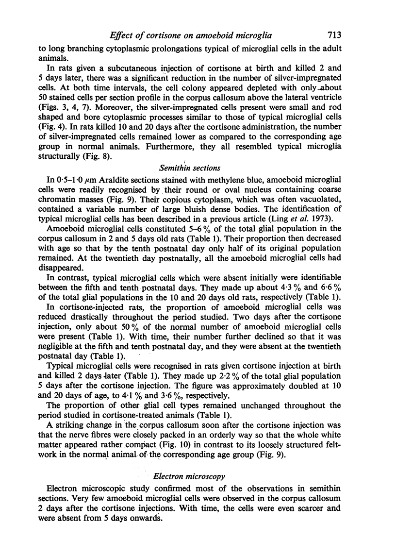
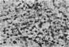
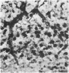
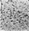
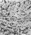
Amoeboid microglia and typical microglial cells in the corpus callosum of rats were studied following a subcutaneous injection of cortisone at birth. The rats were killed 2, 5, 10 and 20 days after the injection. The most striking change in the corpus callosum was the great reduction in the number of amoeboid microglial cells as shown by the silver carbonate stain of Rio-Hortega. The change was sustained throughout the period studied. Ramified and typical microglial cells which do not normally appear until between the fifth and tenth postnatal day, were observed on the second postnatal day after the cortisone administration. Cell enumeration in semithin sections showed that the proportion of amoboid microglial cells was reduced to 50% of their normal value soon (2 days) after the cortisone injection. This proportion was further decreased and the cells were absent from the fifth postnatal day onwards. Typical microglial cells were developed prematurely and they constituted more than 2% of the total glial population 2 days after the cortisone injection. Another striking change noted in the semithin section was the increase in the compactness of the axons in the corpus callosum in the experimental animals. Electron microscopic observations were in general agreement with the light microscopy. The amoeboid microglial cells appeared less active; they contained small Golgi apparatus, accumulations of lipid droplets and sparse lysosomes. The cell outline was regular. The reduction in the number of amoeboid microglia after the cortisone injection was explained by the fact that the drug suppressed the production of their precursor cells, i.e. circulating monocytes, Moreover, it was suggested that cortisone probably interfered with the phagocytic activity of the amoeboid microglial cells which would normally undergo structural changes to become the quiescent microglia which were observed in the early postnatal animals.
Full text
PDF


Images in this article


Selected References
These references are in PubMed. This may not be the complete list of references from this article.
- BARBER M., DELAUNAY A. Effect of plasma of cortisone-treated guinea-pigs on the growth of fibroblasts and macrophages in tissue cultures in vitro. J Pathol Bacteriol. 1951 Jul;63(3):549–551. [PubMed] [Google Scholar]
- Craddock C. G., Winkelstein A., Matsuyuki Y., Lawrence J. S. The immune response to foreign red blood cells and the participation of short-lived lymphocytes. J Exp Med. 1967 Jun 1;125(6):1149–1172. doi: 10.1084/jem.125.6.1149. [DOI] [PMC free article] [PubMed] [Google Scholar]
- DOUGHERTY T. F., SCHNEEBELI G. L. Role of cortisone in regulation of inflammation. Proc Soc Exp Biol Med. 1950 Dec;75(3):854–859. doi: 10.3181/00379727-75-18368. [DOI] [PubMed] [Google Scholar]
- FIELD E. J. Effect of cortisone on the neonatal rat. Nature. 1954 Jul 24;174(4421):182–182. doi: 10.1038/174182a0. [DOI] [PubMed] [Google Scholar]
- FIELD E. J. Observations on the development of microglia together with a note on the influence of cortisone. J Anat. 1955 Apr;89(2):201–208. [PMC free article] [PubMed] [Google Scholar]
- FURNESS G. Effect of cortisone on the macrophages of different species of animal. J Bacteriol. 1959 Apr;77(4):461–464. doi: 10.1128/jb.77.4.461-464.1959. [DOI] [PMC free article] [PubMed] [Google Scholar]
- Ling E. A. Cytochemical localization of peroxidase in amoeboid cells in the corpus callosum in postnatal rats. Arch Histol Jpn. 1980 Oct;43(4):305–310. doi: 10.1679/aohc1950.43.305. [DOI] [PubMed] [Google Scholar]
- Ling E. A. Light and electron microscopic demonstration of some lysosomal enzymes in the amoeboid microglia in neonatal rat brain. J Anat. 1977 Jul;123(Pt 3):637–648. [PMC free article] [PubMed] [Google Scholar]
- Ling E. A., Paterson J. A., Privat A., Mori S., Leblond C. P. Investigation of glial cells in semithin sections. I. Identification of glial cells in the brain of young rats. J Comp Neurol. 1973 May 1;149(1):43–71. doi: 10.1002/cne.901490104. [DOI] [PubMed] [Google Scholar]
- Ling E. A., Penney D., Leblond C. P. Use of carbon labeling to demonstrate the role of blood monocytes as precursors of the 'ameboid cells' present in the corpus callosum of postnatal rats. J Comp Neurol. 1980 Oct 1;193(3):631–657. doi: 10.1002/cne.901930304. [DOI] [PubMed] [Google Scholar]
- Ling E. A. Some aspects of amoeboid microglia in the corpus callosum and neighbouring regions of neonatal rats. J Anat. 1976 Feb;121(Pt 1):29–45. [PMC free article] [PubMed] [Google Scholar]
- Ling E. A., Tan C. K. Amoeboid microglial cells in the corpus callosum of neonatal rats. Arch Histol Jpn. 1974 Mar;36(4):265–280. doi: 10.1679/aohc1950.36.265. [DOI] [PubMed] [Google Scholar]
- Ling E. A. Transformation of monocytes into amoeboid microglia in the corpus callosum of postnatal rats, as shown by labelling monocytes by carbon particles. J Anat. 1979 Jun;128(Pt 4):847–858. [PMC free article] [PubMed] [Google Scholar]
- PAFF G. H., STEWART R. Free wandering cells and cortisone. Proc Soc Exp Biol Med. 1953 Jul;83(3):591–592. doi: 10.3181/00379727-83-20428. [DOI] [PubMed] [Google Scholar]
- Slonecker C. E., Lim W. C. Effects of hydrocortisone on the cells in an acute inflammatory exudate. Lab Invest. 1972 Jul;27(1):123–128. [PubMed] [Google Scholar]
- Thompson J., van Furth R. The effect of glucocorticosteroids on the kinetics of mononuclear phagocytes. J Exp Med. 1970 Mar 1;131(3):429–442. doi: 10.1084/jem.131.3.429. [DOI] [PMC free article] [PubMed] [Google Scholar]
- Thompson J., van Furth R. The effect of glucocorticosteroids on the proliferation and kinetics of promonocytes and monocytes of the bone marrow. J Exp Med. 1973 Jan 1;137(1):10–21. doi: 10.1084/jem.137.1.10. [DOI] [PMC free article] [PubMed] [Google Scholar]