

Abstract
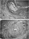
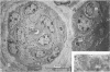
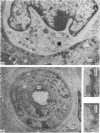
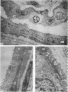
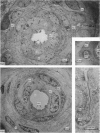
The duct of the cow, sheep and goat can be divided into two main parts, the intrafollicular region and the intradermal region. Three sub-regions, namely the perifollicular zone, the duct body and the duct/fundus transition zone, can be distinguished within the latter. In the outer portion of the hair follicle, the luminal surface was keratinized but the presence, in the cow and sheep, of surface microvilli with closely associated vesicles deeper within this layer suggested the possibility of a reabsorptive role. The intradermal duct had junctional complexes at the luminal extremity throughout its length. Gap junctions were observed between basal cells only in the lower part of the duct body. Dovetailing between luminal cells in the intradermal duct was barely noticeable in the cow but prominent in the goat. In the duct/fundus transition zone of all three species there was evidence of cell differentiation, the potential significance of which is discussed. There was little change in the ultrastructure of the duct in any of the three species, other than an increase in lumen size, as a result of heat exposure and sweating activity.
Full text
PDF


Images in this article

Selected References
These references are in PubMed. This may not be the complete list of references from this article.
- Allen T. E., Bligh J. A comparative study of the temporal patterns of cutaneous water vapour loss from some domesticated mammals with epitrichial sweat glands. Comp Biochem Physiol. 1969 Oct 15;31(2):347–363. doi: 10.1016/0010-406x(69)91659-4. [DOI] [PubMed] [Google Scholar]
- Christophers E., Plewig G. Formation of the acrosyringium. Arch Dermatol. 1973 Mar;107(3):378–382. [PubMed] [Google Scholar]
- Hashimoto K. Demonstration of the intercellular spaces of the human eccrine sweat gland by lanthanum. II. The duct. J Ultrastruct Res. 1971 Dec;37(5):504–520. doi: 10.1016/s0022-5320(71)80021-7. [DOI] [PubMed] [Google Scholar]
- Hashimoto K., Gross B. G., Lever W. F. The ultrastructure of the skin of human embryos. I. The intraepidermal eccrine sweat duct. J Invest Dermatol. 1965 Sep;45(3):139–151. doi: 10.1038/jid.1965.110. [DOI] [PubMed] [Google Scholar]
- Jenkinson D. M., Mabon R. M. The effect of temperature and humidity on skin surface pH and the ionic composition of skin secretions in Ayrshire cattle. Br Vet J. 1973 May-Jun;129(3):282–295. doi: 10.1016/s0007-1935(17)36482-5. [DOI] [PubMed] [Google Scholar]
- Jenkinson D. M., Montgomery I., Elder H. Y. Studies on the nature of the peripheral sudomotor control mechanism. J Anat. 1978 Mar;125(Pt 3):625–639. [PMC free article] [PubMed] [Google Scholar]
- Jenkinson D. M., Montgomery I., Elder H. Y. The ultrastructure of the sweat glands of the ox, sheep and goat during sweating and recovery. J Anat. 1979 Aug;129(Pt 1):117–140. [PMC free article] [PubMed] [Google Scholar]
- Jimbow K., Sato S., Kukita A. Langerhans' cells of the normal human pilosebaceous system. An electron microscopic investigation. J Invest Dermatol. 1969 Feb;52(2):177–180. doi: 10.1038/jid.1969.26. [DOI] [PubMed] [Google Scholar]
- Kurosumi K. Fine structure of the human sweat ducts of eccrine and apocrine types. Arch Histol Jpn. 1977 Jun;40(3):203–224. doi: 10.1679/aohc1950.40.203. [DOI] [PubMed] [Google Scholar]
- LOBITZ W. C., Jr, DOBSON R. L. Responses of the secretory coil of the human eccrine sweat gland to controlled injury. J Invest Dermatol. 1957 Feb;28(2):105-19; discussion, 119-20. doi: 10.1038/jid.1957.12. [DOI] [PubMed] [Google Scholar]
- LOBITZ W. C., Jr, HOLYOKE J. B., MONTAGNA W. Responses of the human eccrine sweat duct to controlled injury: growth center of the epidermal sweat duct unit. J Invest Dermatol. 1954 Nov;23(5):329–344. doi: 10.1038/jid.1954.116. [DOI] [PubMed] [Google Scholar]
- Sato K. The physiology, pharmacology, and biochemistry of the eccrine sweat gland. Rev Physiol Biochem Pharmacol. 1977;79:51–131. doi: 10.1007/BFb0037089. [DOI] [PubMed] [Google Scholar]
- Shibasaki S., Ito T. Electron microscopic study on the duct of the human eccrine sweat gland with special remarks on the transitional portion epithelium. Arch Histol Jpn. 1967 Jun;28(3):285–312. doi: 10.1679/aohc1950.28.285. [DOI] [PubMed] [Google Scholar]
- Stingl G., Katz S. I., Green I., Shevach E. M. The functional role of Langerhans cells. J Invest Dermatol. 1980 May;74(5):315–318. doi: 10.1111/1523-1747.ep12543548. [DOI] [PubMed] [Google Scholar]
- Tani M., Yamamoto K., Mishima Y. Apocrine acrosyringeal complex in the human skin. J Invest Dermatol. 1980 Nov;75(5):431–435. doi: 10.1111/1523-1747.ep12524083. [DOI] [PubMed] [Google Scholar]