



Abstract
Background:
The literature regarding clinical olfaction, olfactory loss, and olfactory dysfunction has expanded rapidly over the past two decades, with an exponential rise in the past year. There is substantial variability in the quality of this literature and a need to consolidate and critically review the evidence. It is with that aim that we have gathered experts from around the world to produce this International Consensus on Allergy and Rhinology: Olfaction (ICAR:O).
Methods:
Using previously described methodology, specific topics were developed relating to olfaction. Each topic was assigned a literature review, evidence-based review, or evidence-based review with recommendations format as dictated by available evidence and scope within the ICAR:O document. Following iterative reviews of each topic, the ICAR:O document was integrated and reviewed by all authors for final consensus.
Results:
The ICAR:O document reviews nearly 100 separate topics within the realm of olfaction, including diagnosis, epidemiology, disease burden, diagnosis, testing, etiology, treatment, and associated pathologies.
Conclusion:
This critical review of the existing clinical olfaction literature provides much needed insight and clarity into the evaluation, diagnosis, and treatment of patients with olfactory dysfunction, while also clearly delineating gaps in our knowledge and evidence base that we should investigate further.
Keywords: anosmia, evidence-based medicine, hyposmia, loss of smell, olfaction, olfactory dysfunction, olfactory loss, parosmia, phantosmia, systematic review
I |. INTRODUCTION
The field of olfaction is a relatively young one. Detailed knowledge of the mechanisms of the olfactory system were only discovered in the second half of the 21st century, with Richard Axel and Linda Buck awarded the 2004 Nobel Prize for their landmark description of odorant receptors and the organization of the olfactory epithelium (OE), olfactory bulb (OB), and olfactory cortex.1 An explosion of investigation followed in both the basic science research realm as well as clinical study, steadily growing in number of publications and complexity of study design over the 2 decades that have followed, peaking within the past year as the COVID-19 pandemic brought loss of smell and taste to the forefront of international importance and recognition.2,3
In the many decades before Axel and Buck’s publication, articles listed in PubMed under “olfaction” totaled less than 5000. In the decade that followed, publications matched this number and over the next decade continued to accelerate until, in the decade between 2011 and 2021, there were 13,618 publications, with 2325 in the year 2020 alone.
Although basic science research is integral to our understanding of the system and invaluable in creating the foundation for any translational or clinical study, with the vast amount of literature to evaluate, we decided to limit this document to the existing clinical knowledge in the field of olfaction. Similar to other International Consensus in Allergy and Rhinology (ICAR) documents on chronic rhinosinusitis (CRS) and allergic rhinitis (AR),4–6 our goal with producing this document is to summarize the best external evidence to provide practitioners the means to practice evidence-based medicine when diagnosing and treating these patients. As is the case among many fields of medicine, especially those that affect patients less commonly, the quality of the existing clinical literature published on olfactory loss and dysfunction is highly variable, with studies ranging from well-designed randomized controlled clinical trials to summaries of expert opinion and conjecture. The goal of this International Consensus of Allergy and Rhinology: Olfaction (ICAR:O) was to critically review the literature for olfaction-related epidemiology, psychological and social burden, pathophysiology, evaluation and diagnosis, and management.
With the management of olfactory loss or dysfunction being an inherently multidisciplinary field, we endeavored to include authors from a wide array of expertise to ensure the highest and most insightful coverage of the subject. More than 50 international authors undertook a structured review of the literature in nearly 100 topic areas related to olfaction. Although highly dependent on the quality of the existing literature, wherever possible recommendations based on the evidence were made, with benefit, harm, and cost considerations reported. However, as noted in prior ICAR documents, this document is not a clinical practice guideline and not a meta-analysis. In fact, because of the wide heterogeneity of the data and reporting measures found in the literature in this field, a meta-analysis would not be appropriate or possible. Many of our current treatment paradigms are based on relatively weak external evidence, illustrated by the wide variation in treatment methodology that exists around the globe for these patients. When we do not have high-level evidence on which to base our practice decisions, it is in our best interest as clinicians and scientists to identify the gaps in our current knowledge and attempt to design and perform studies that can help fill those gaps and therefore better help our patients.
As stated in all prior ICAR documents, this document should not be considered as determining a standard of care or medical necessity and cannot be thought of as dictating care for any individual patient. Each patient has their own unique history, background, demographic, and clinical circumstances that may affect the evaluation and treatment of their specific olfactory loss or dysfunction. Finally, the idea of creating a document such as this, which strives to gather and review all of the existing clinical evidence on olfactory loss and dysfunction, is that by identifying the areas that need more research, more research will then be performed, and thus the evidence and recommendations made herein will change over time and revisions will be made to them appropriately.
II |. METHODS
A |. Topic Development
All ICAR documents follow the formula of literature review described in 2011 by Rudmik and Smith,7 utilizing their method of iterative evidence-based review (EBR) with recommendations (EBRRs). The literature was analyzed, assessed for level of evidence (LOE), and, when appropriate, recommendations were given.
The subject matter of clinical olfaction was divided into 75 topics. Each topic area was assigned a senior author, recognized as an expert in the field. Authors were selected based on prior authorship of significant contributions to the olfactory literature and were selected from the fields of rhinology, neurology, and chemosensory science. Depending on the type of topic and the quality of evidence available in each topic, the section author was assigned either a simple literature review, an EBR, or an EBRR.
To provide the content for each topic, a systematic review of the literature for each topic using Ovid MEDLINE (1947 to July 2020), Embase (1974 to July 2020), and Cochrane Library databases was performed using the Preferred Reporting Items for Systematic Reviews and Meta-Analyses (PRISMA) standardized guidelines. The search began by identifying any previously published systematic reviews or guidelines pertaining to the assigned topic. Because clinical recommendations are best supported by randomized controlled trials (RCTs), the search focused on identifying these studies to provide the strongest LOE. When these did not exist, observational studies were then identified. Reference lists of all identified studies were examined to ensure that all relevant studies were captured. If the authors felt as though a non–English language study should be included in the review, the article was appropriately translated to minimize the risk of missing important data during the development of recommendations.8 One major exception to the search window was made for the section on COVID-19–related olfactory dysfunction (OD). The evidence for this topic was rapidly evolving during the time of the writing and editing of this document, and we felt it would do the readership a disservice if we left out pertinent information that was only realized after the literature search window had closed.
To optimize transparency of the evidence, all included studies in EBR and EBRR topic sections are presented in a standardized table format and the quality of each study was evaluated to receive a level based on the Oxford Levels of Evidence (Level 1a to 5) (Table II.A-1).9 At the completion of the systematic review and research quality evaluation for each clinical topic, an aggregate grade of evidence was produced for the topic based on the guidelines from the American Academy of Pediatrics (AAP) Steering Committee on Quality Improvement and Management (SCQIM)10 (Table II.A-2). After providing an aggregate grade of evidence for each EBRR topic (A to D), a recommendation using the AAP SCQIM guidelines was produced (Table II.A-3). The recommendation was based on the aggregate grade of evidence as well as the balance of benefit, harm, and costs. A summary of the EBRR development process is provided in Figure II.A-1.
TABLE II.A.1.
Levels of evidence
| Level | Diagnosis | Therapy/prevention/Etiology |
|---|---|---|
| 1 | Systematic review of cross-sectional studies with consistently applied reference standards and blinding | Systematic review of randomized trials or n-of-1 trials |
| 2 | Individual cross-sectional studies with consistently applied reference standards and blinding | Randomized trial or observational study with dramatic effect |
| 3 | Cohort study or control arm of randomized triala | Nonrandomized controlled cohort/follow-up studyb |
| 4 | Case series or case-control studies, or poor-quality prognostic cohort studyb | Case series or case-control studies, or historically controlled studiesb |
| 5 | Not applicable | Mechanism-based reasoning |
The level may be graded down on the basis of study design, inconsistency between studies, indirectness of evidence, or imprecision, or because the absolute effect size was very small; the level may be graded up if there is a large or very large effect size or if a significant dose-response relationship is demonstrated.
A systematic review is generally better than an individual study.
TABLE II.A.2.
Aggregate grade of evidence
| Grade | Research quality |
|---|---|
| A | Well-designed RCTs |
| B | RCTs with minor limitations |
| Overwhelming consistent evidence from observational studies | |
| C | Observational studies (case-control and cohort design) |
| D | Expert opinion |
| Case reports | |
| Reasoning from first principles |
RCT = randomized controlled trial.
TABLE II.A.3.
AAP-defined strategy for recommendation development
| Evidence quality | Preponderance of benefit over harm | Balance of benefit and harm | Preponderance of harm over benefit |
|---|---|---|---|
| A. Well-designed RCTs | Strong recommendation | Option | Strong recommendation against |
| B. RCTs with minor limitations; overwhelmingly consistent evidence from observational studies | Recommendation | ||
| C. Observational studies (case-control and cohort design) | Recommendation against | ||
| D. Expert opinion, case reports, reasoning from first principles | Option | No recommendation | |
AAP = American Academy of Pediatrics; RCT = randomized controlled trial.
FIGURE II.A-1.
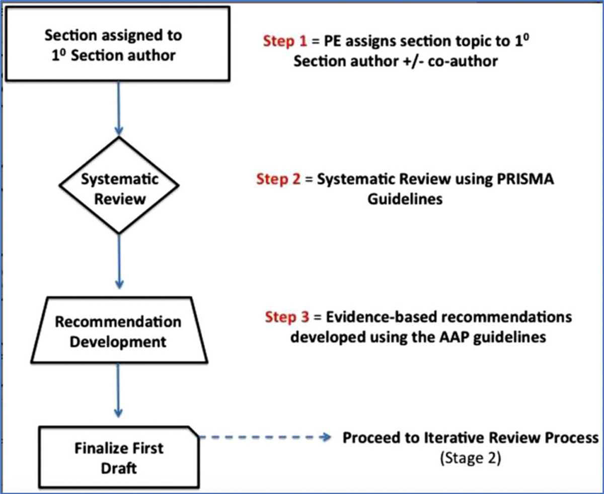
Stage I of Iterative Review Process
B |. Iterative Review
Each topic was written with appropriate tables and potential recommendations by the initial author assigned. Each section then underwent an online iterative review process using two independent reviewers (Figure II.A-2). Each iterative reviewer evaluated the completeness of the identified literature and evaluated whether EBRRs were appropriate. If any content changes were suggested by the first iterative reviewer, these were sent back to the initial author to revise the section until all changes were agreed on by the initial author and the first reviewer. The revised topic was then subsequently reviewed by a second reviewer. Both initial and first and second iterative authors of the topic agreed on all changes before each section was allowed to proceed into the final ICAR statement stage.
FIGURE II.A-2.

Stage II of Iterative Review Process
For topics with more limited evidence, the EBR process was completed with the evidence table. For those topics with sufficient evidence to produce a recommendation (ie, an EBRR), a recommendation using the AAP guidelines was produced. It is important to note that each evidence-based recommendation took into account the aggregate grade of evidence along with the balance of benefit, harm, and costs (Table II.A-3).
C |. ICAR:O Statement
After the review and completion of all topic sections, the principal editor (Z.M.P.) compiled them into one ICAR:O statement. This draft document was then reviewed by all contributing authors who submitted suggestions and edits. Once consensus among all authors had been reached regarding the literature and final recommendations, the final ICAR:O article was produced.
III |. DEFINITIONS
A |. Anosmia and Hyposmia
Anosmia is defined as an absence of olfaction with an inability to detect and correctly identify odors, as measured by a validated, standardized olfactory test.11–13 While anosmia, by definition, describes complete smell loss, functional anosmia refers to the possible existence of trace olfactory function (OF) but at a level not considered to be useful or noticeable in daily life.12,14 Hyposmia or microsmia is defined as partial smell loss.12,14 Specific anosmia is an inability to detect one or more specific odorants while olfaction of other odorants is intact.15
As self-assessment of olfactory loss can be unreliable, the diagnosis of anosmia is traditionally confirmed based on the absolute number of correct answers on psychophysical olfactory testing, with the threshold established from patients with complete loss of smell.12,13,16 Normosmia (normal OF) for most olfactory tests is based on normative data from healthy 16- to 35-year-old patients, although normative data have been collected for all age groups on certain tests, such as the University of Pennsylvania Smell Identification Test (UPSIT®).13 Hyposmia is an absolute score below the 10th percentile of that normosmic group.12,16,17 Hyposmia can be further delineated into mild, moderate, or severe hyposmia.13 OF should be assessed by validated tests of odor threshold and either odor identification or discrimination. Composite scores may be more reliable than tests of only one component of olfactory ability.14
B |. Parosmia
Parosmia is defined as a qualitative dysfunction from a distorted perception of smell in the presence of an odor object.14 These distorted smells are frequently reported to be disgusting or disagreeable and only very rarely would be considered pleasant. Common descriptors include “burned,” “foul,” “disgusting,” and “fecal.”18–22 Patients often report difficulty in characterizing these odors, and, therefore, these terms should be considered as shorthand for their unpleasantness, rather than definitively accurate descriptions. Parosmic experiences can range from simply “strange” to inducing powerful feelings, such as nausea, and preventing normal food intake.
C |. Phantosmia
Phantosmia is defined as a qualitative dysfunction of smell in the absence of an odor object.14 Here, perception of an odor occurs without an external stimulus. Descriptors for phantom odors may be similar in some ways to those used for parosmia: “burned,” “chemical,” and “like cigarette smoke.”18,20,21,23 It is often difficult for the individual to accept that there is no external source for these perceptions, and they often search their homes or work environments exhaustively seeking the source. Unlike the qualitative changes experienced with parosmia, phantosmic perceptions can occur at any time. Sometimes, both parosmia and phantosmia can occur together in the same patient.20,23
IV |. INDIVIDUAL BURDEN OF OD
A |. Psychological sequelae: Potential effects on interpersonal relationships and emotional state
The sense of smell serves three core purposes: prevention of close encounters with environmental hazards, monitoring and guidance of nutrition, and mediation of interpersonal communication.24 OD hence disturbs functioning of all of those domains. As a consequence, a substantial number of affected individuals state that they experience a poorer overall quality of life (QOL),25 which particularly affects emotional well-being and interpersonal relationships. Evidence of the costs of smell impairment are summarized below with regard to both aspects (Table IV.1 and Tables IV.2–IV.4).
TABLE IV.1.
Section evidence summary: Emotional state
| Study | Year | LOE | Study design | Population | Outcome | Conclusions |
|---|---|---|---|---|---|---|
| Stevenson24 | 2010 | 4 | Literature review | Animal studies, olfactory loss in patients, human studies on evidence of that function | Identification and categorization of the main functions of human olfaction | Identification of three major classes of functions: ingestion, avoiding environmental hazards, and social communication with specific subfunctions |
| Croy, Nordin, and Hummel25 | 2014 | 4 | Literature review | Quantitative and qualitative, and patients with congenital olfactory disorder | Links between olfactory impairment and general QOL/depression | Olfactory impairment associated with disturbances in various life areas (food, harmful event detection, social situations); majority of patients with olfactory disorder deal well but a limited proportion experience reduced QOL and increased depression scores |
| Croy and Hummel26 | 2017 | 4 | Literature review | Healthy individuals, depressed patients, patients with olfactory disorder | Links between olfaction and depression | Interaction between olfaction and depression by two suggested pathways: (1) impaired OF as a consequence of reduced olfactory attention and input; and (2) OD as a marker for enhanced vulnerability to depression |
| Kohli et al27 | 2016 | 4 | Literature review | Primary depression patients or primary patients with OD | Links between OD and depression | Reciprocal relationship: depressive patients show reduced olfactory performance, patients with OD exhibit depressive symptoms |
| Schablitzky and Pause28 | 2014 | 4 | Literature review | Healthy individuals, distinct groups of major depressive disorder, bipolar disorder, seasonal affective disorder | Olfactory performance (odor sensitivity, identification, discrimination, and odor ratings) in depressed patients and in healthy individuals experiencing only some depressive symptoms or a transient state of sad mood | Major depressive disorder relates to reduced olfactory sensitivity but not to odor identification/discrimination, no associations in bipolar disorder/seasonal affective disorder but in healthy individuals exhibiting subclinical depressive states |
| Rochet et al29 | 2018 | 4 | Literature review | Healthy individuals, depressed and clinically improved patients, patients with OD | Links between olfaction and depression, olfactory markers of depression | Olfactory impairment affects QOL/daily life, associations with depression; (heterogenous findings regarding olfactory markers of depression Reciprocal relationship between OD, depression/QOL |
| Erskine and Philpott30 | 2019 | 4 | Case series, qualitative research | Patients with smell disorder | Subjective experiences of patients with smell disorder | Identified themes: negative emotional impact, feelings of isolation, impaired relationships and daily functioning, impact on physical health, and the difficulty in and financial burden of seeking help |
| Philpott and Boak31 | 2014 | 3 | Cohort | Patients with OD | Consequences of smell disorder on patients’ daily life and affected areas | OD associated with psychological impairment and reduced life quality: 43% of the patients reported depression, 45% anxiety, 92% impairment of eating, 57% isolation, and 54% relationship difficulties; women were more affected then men |
| Frasnelli and Hummel32 | 2005 | 2 | Cross-sectional controlled | Patients with OD (quantitative and qualitative) and HCs | Qualitative and quantitative OD and impact on daily life | Patients with parosmia as well as quantitative OD show higher rates of daily life complaints when compared with patients experiencing quantitative olfactory impairment only; patients with quantitative olfactory impairment exhibited more complaints than HCs |
| Desiato et al33 | 2020 | 1 | Systematic review and meta-analysis | Study cohorts recruited from the general population | Prevalence of OD in the healthy general population | Overall prevalence of OD of 22.2%; reported higher prevalences when measured with expanded identification tests <8 items and in patients aged >55 years |
| Murr et al34 | 2018 | 2 | Prospective controlled | Patients with OD and HCs | Importance of olfaction | Highest importance of olfaction in young, healthy women (aged ≤25 years); patients with OD reported decreased importance of olfaction; possible coping mechanism |
| Kollndorfer et al35 | 2017 | 2 | Prospective controlled | Patients with anosmia and HCs | Link between self-esteem and QOL in OD | Decreased life quality and reduced body-related self-esteem in patients with anosmia; low life quality and self-esteem related to depressive symptoms |
| Keller and Malaspina20 | 2013 | 4 | Patient-report series | Patients with OD | Subjective experiences with olfactory loss | Impaired life quality, in particular reflected by reported social isolation and anhedonia |
| Blomqvist et al36 | 2004 | 3 | Cohort | Patients with OD | Well-being and coping in patients with olfactory loss | Impaired life quality (eg, physical health, financial security, social relations, leisure, and emotional stability) and negative effects on well-being; patients use problem-and emotion-focused coping |
| Oleszkiewicz et al37 | 2020 | 2 | Cohort | Individuals declaring normal sense of smell | Undetected olfactory loss and relationship to cognitive performance and well-being | 59 of 203 individuals with impaired olfaction; differences between affected and nonaffected individuals in cognitive functioning but not in well-being and chemosensory communication |
| Schafer, Schriever, and Croy38 | 2021 | 4 | Literature review | Patients with OD and healthy individuals | Causes and consequences related to the main functions of olfaction | Impaired enjoyment of food, worries about hazards, and social insecurities lead to decreased life quality; recommendation to focus medical and psychological treatment options on patients with concomitant impairment caused by smell loss; provide treatment and coping strategies |
LOE = level of evidence; OD = olfactory dysfunction; OF = olfactory function; QOL = quality of life.
TABLE IV.2.
Section evidence summary: Interpersonal relationships
| Study | Year | LOE | Study design | Population | Outcome | Conclusions |
|---|---|---|---|---|---|---|
| Lobmaier et al39 | 2018 | 2 | Cross-sectional experimental | Healthy individuals, men rating women’s body odor samples | Relation between body odor attractiveness and reproductive hormones | Men agreed on body odor attractiveness ratings, which were higher in women with higher estradiol and progesterone levels |
| de Groot et al40 | 2015 | 2 | Cross-sectional experimental | Healthy individuals | Relation between chemosignals (body odors sampled in a happy emotional state) and emotional reaction of the receiver | Exposure to body odor collected from senders of chemosignals in a happy state induced a facial expression and perceptual-processing style indicative of happiness in the receivers |
| Gelstein et al41 | 2011 | 2 | Cross-sectional experimental | Healthy individuals, men sniffing women’s tears | Relation between chemosignals (women’s tears) and emotional reaction of the receiver | Sniffing of tears related to reduced sexual appeal evaluation of women’s faces, reduced self-related arousal, reduced testosterone levels, as well as reduced brain activity related to sexual arousal |
| Prehn-Kristensen et al42 | 2009 | 2 | Cross-sectional experimental | Healthy individuals | Neural reactions in response to perception of chemosignals (body odors sampled in anxiety vs sport state) | Anxiety body odors activate brain areas related to processing of social emotional stimuli (fusiform gyrus) and regulation of empathy (insula, precuneus, cingulate cortex) |
| Sorokowska, Sorokowski, and Szmajke43 | 2012 | 2 | Cross-sectional experimental | Healthy individuals | Link between body odor, personality traits, and dominance | Correlation between self-rated odor donor personality traits and external judgments based on odor alone for extraversion, neuroticism, and dominance |
| Wedekind et al44 | 1995 | 2 | Cross-sectional experimental | Healthy individuals, women rating men’s body odors | Link between major histocompatibility complex, body odor, and attractiveness | More pleasant perception of body odors when major histocompatibility complex dissimilar; preference erased in women taking oral contraception |
| Rattaz et al45 | 2005 | 2 | Cross-sectional experimental | Full-term newborns | Effectiveness of familiar and unfamiliar odors in soothing during routine heel-stick | Familiar odor (maternal milk/vanilla) associated with reduced stress response |
| Granqvist et al46 | 2019 | 2 | Cross-sectional experimental | Healthy individuals | Effect of exposure to partner’s body odor on discomfort and psychophysiological stress | Partner body odor decreased subjective discomfort during a stressful event; reduced skin conductance in highly secure individuals |
| Lundström and Jones-Gotman47 | 2009 | 2 | Cross-sectional experimental | Healthy individuals | Links between olfactory identification ability and degree of romantic love in partnership | Negative correlation between degree of romantic love and ability to identify body odor of an opposite-sex friend but not of their same-sex friend |
| Okamoto et al48 | 2016 | 3 | Cohort | Healthy individuals, parents | Links between child-rearing and olfaction | Parents actively seek their child’s odor in daily rearing; the child’s head is the most frequent source of affective experiences and the child’s bottom of practical |
| Croy, Nordin, and Hummel25 | 2014 | 4 | Literature review | Quantitative, qualitative, and congenital OD | Links between olfactory impairment and general QOL/depression | Olfactory impairment associated with disturbances in various life areas (food, harmful event detection, social situations); majority of patients with OD deal well but a limited proportion experience reduced QOL and increased depression scores |
| Drummond, Douglas, and Olver49 | 2013 | 4 | Case series, qualitative research design | Patients with severe TBI and olfactory loss | Impact of olfactory impairment on daily activities and social participation | OD has a significant impact on various activities and social role |
| Keller and Malaspina20 | 2013 | 4 | Patient-report series | Patients with OD | Subjective experiences with olfactory loss | Impaired life quality, in particular reflected by reported social isolation and anhedonia |
| Brämerson, Nordin, and Bende50 | 2007 | 3 | Prospective cohort | Patients with OD | Description of how quantitative and qualitative olfactory disorders are diagnosed, what the causes are, and how QOL is compromised in patients | Patients with reduced sense of smell, often combined with qualitative disorders, exhibit significantly reduced QOL, particularly in paid employment, household work, and social and family life |
| Erskine and Philpott30 | 2019 | 4 | Case series, qualitative research design | Patients with smell disorder | Subjective experiences of patients with smell disorder | Identified themes: negative emotional impact, feelings of isolation, impaired relationships and daily functioning, impact on physical health, and the difficulty in and financial burden of seeking help |
| Lundström et al51 | 2013 | 2 | Cross-sectional | Healthy individuals, comparing mothers and nulliparae | Neural responses to unfamiliar infant body odors | Infant body odors elicit reward-related activations, maternal status-dependent activity in neostriatal areas |
| Schäfer, Michael, and Croy52 | 2019 | 2 | Cross-sectional | Healthy individuals, mothers | Neural responses to body odor of their own and unfamiliar infant | Infant body odors elicit regions of pleasure and reward independent from familiarity (own vs unfamiliar baby) |
| Mahmut and Croy53 | 2019 | 4 | Literature review | Healthy individuals, patients with OD | Links of olfactory ability and romantic relationships | Body odor perception moderates mate choice and provides a source of comfort in existing relationships, and alteration of preference may signal the breakdown ofa relationship |
| Herz and Inlicht54 | 2002 | 3 | Cohort | Healthy individuals | Importance of social and physical traits in heterosexual attraction | Women ranked body odor as more important for attraction than looks, natural body odor as the most influential olfactory variable for sexual interest in men and women; men rated good looks as most important |
| Sorokowska et al55 | 2018 | 2 | Cross-sectional | Healthy individuals | Body odor attractiveness and human leukocyte antigen similarity | Women not using hormonal contraception rated human leukocyte antigen-similar body odors as less attractive; no influence of human leukocyte antigen similarity was observed for women using hormonal contraception and men |
| Bendas, Hummel, and Croy56 | 2018 | 2 | Cross-sectional | Healthy individuals | Link between odor threshold and sexual desire, sexual experience, and sexual performance | High olfactory sensitivity relates to higher pleasantness of sexual activities and higher frequency of orgasms in women |
| Croy et al57 | 2012 | 2 | Cross-sectional | Congenital anosmic patients and HCs | Link between olfactory impairment and functions of daily life | Patients differed only slightly from controls in terms of enhanced social insecurity, increased risk for depressive symptoms, and household accidents |
| Schäfer et al58 | 2019 | 2 | Cross-sectional | Patients with smell disorder and healthy individuals | Link between olfactory impairment and sexual desire | 29% of patients reported decreased sexual desire after olfactory loss, predicted by depressive symptoms and OF; no differences in standardized questionnaire |
| Oleszkiewicz et al37 | 2020 | 2 | Cohort | Individuals declaring normal sense of smell | Undetected olfactory loss and relationship to cognitive performance and well-being | 59 of 203 individuals with impaired olfaction; differences between affected and nonaffected individuals in cognitive functioning but not in well-being and chemosensory communication |
HC = healthy control; OD = olfactory dysfunction; OF =olfactory function; QOL = quality of life; TBI = traumatic brain injury.
TABLE IV.4.
Section evidence summary: Increased mortality
| Study | Year | LOE | Study design | Population | Outcome | Conclusions |
|---|---|---|---|---|---|---|
| Wilson et al73 | 2011 | 2 | Longitudinal cohort | Retired Chicago-area adults, mean age 79.7 years | All-cause mortality; mean 4.2 years | Difficulty with odor identification is associated with increased risk of death |
| Gopinath et al74 | 2012 | 2 | Longitudinal cohort | Australian adults aged ≥60 years | All-cause mortality; 5 years | The relationship between olfaction and mortality may be largely mediated by cognitive impairment |
| Pinto et al75 | 2014 | 2 | Longitudinal cohort | US adults aged ≥57 years | All-cause mortality; 5 years | OF is one of the strongest predictors of 5-year mortality in a nationally representative sample of older US adults |
| Devanand et al76 | 2015 | 2 | Longitudinal cohort | New York City adults, Medicare beneficiaries aged ≥65 years | All-cause mortality; mean 4.1 years | Anosmia is a particularly strong predictor of dementia |
| Schubert et al77 | 2017 | 2 | Longitudinal cohort | Beaver Dam, WI, adults aged 53–97 years | All-cause mortality; mean 12.8 years | Olfactory impairment, but not hearing or visual impairment, is associated with increased mortality |
| Ekstrom et al78 | 2017 | 2 | Longitudinal cohort | Swedish adults aged 40–90 years | All-cause mortality; 10 years | Presence or absence of dementia does not attenuate the association between olfactory loss and mortality |
| Leschak and Eisenberger79 | 2018 | 2 | Longitudinal cohort | Older US adults aged ≥57 years | All-cause mortality; 5 years | Social network size partially mediated the olfactory-mortality link in women (nationally representative samples of older US adults) |
| Laudisio et al80 | 2019 | 2 | Longitudinal cohort | Italian adults aged ≥65 years | All-cause mortality; 9 years | The relationship between olfaction and mortality may be mediated through frailty, possibly via inflammation |
| Liu et al81 | 2019 | 2 | Longitudinal cohort | Pittsburgh, PA, and Memphis, TN, adults aged 70–79 years | All-cause and cause-specific mortality; 3, 5, 10, and 13 years | Neurodegenerative diseases and weight loss explain only part of the increased mortality |
| Choi et al82 | 2021 | 2 | Cohort study with National Death Index follow-up | US adults aged >40 years | All-cause mortality; 5 years | Objective (but not subjective) OD is associated with increased mortality among older (≥65 years) but not middle-aged (40–64 years) US adults |
LOE= level of evidence; OD = olfactory dysfunction; OF = olfactory function.
Emotional state
Previous research has repeatedly demonstrated associations between decreased olfaction and anhedonia or depression.26–28 Because of largely shared neural pathways (eg, amygdala, hippocampus, insula, and orbitofrontal cortex [OFC]),29 this link is not surprising. Croy and Hummel26 suggest that possible mechanisms behind this association might include that: (1) dysfunction of the OB (as the initial station of olfactory processing) results in decreased neural signaling into subsequent cortices; or (2) the consequence of depressive behavior (eg, withdrawal) leads to diminished olfactory input and consecutive diminished OF. Regardless of the mechanisms involved, negative feelings such as anhedonia, sadness, fear, or frustration are reported by about one third of patients with olfactory loss,25,30,31 with varying prevalence attributable to individual patient characteristics. For example, higher prevalence has been reported in patients with hyposmia versus those with anosmia,32 while evidence regarding sex effects is mixed,32,33 but with women reporting particular effects in social domains.31 The latter may be explained by the generally higher value placed on the sense of smell and importance of olfaction in women, in particular young women, compared with other demographic groups.34 Individuals with reduced self-esteem have been shown to be prone to the emergence of depressive symptoms from olfactory losses.35 Single reports disclose disturbances in a wide array of life areas, including hygiene behavior, domestic life,31 or the enjoyment of simple pleasures, such as the smell of flowers, perfumes, or nature.20 In view of these reports, the low general QOL measured in these populations is not surprising. However, not every patient with an olfactory disorder is bothered to a substantial degree. It has to be considered that most reported data are obtained from patients seeking help, thus suggesting selection bias.25,36 In contrast, Oleszkiewicz et al37 revealed that people with unnoticed olfactory loss do not differ from controls in terms of their well-being. However, within the group of patients disturbed by their sensory loss, concomitant psychological burden should be carefully assessed and diagnosed. Practitioners should be especially aware of the demographic groups most affected.38 For such predisposed populations, suitable interventions, eg, consultation with a psychologist or psychiatrist, should be provided in order to prevent manifestation and exacerbation of long-term side effects such as social isolation or anxiety.
Interpersonal relationships
Human chemosensory signals, such as those released from body odor, convey various data points of information about the individual, which inform sensory social communication. This information reflects hormonal39 or emotional states,40–42 personality traits,43 and the genetic constitution44 of the releaser. Familiar body odors can signal comfort,45,46 and may be associated with affectionate feelings.47,48 OD is thus likely to be associated with deficits in receiving, processing, and interpretation of such interpersonal sensory information. Patients with olfactory disorders frequently complain about impairment in social situations, isolation, or feelings of social insecurity.20,25,49 This is of significant relevance in the context of intimate relationships, such as relationships between parent and child or between romantic partners.30,50 Regarding the former, parents report the body odor of their child as an affective and instrumental cue,48 as infant odor is associated with neural correlates of reward in the maternal brain.51,52 The latter was studied by Mahmut and Croy53 who reported evidence for the involvement of olfaction in the “initiation, maintenance, and breakdown of romantic relationships.” As body odors signal attractiveness54,55 or mediate sexual experience56 in normosmic individuals, dysosmic patients exhibit a reduced number of sexual partners and experience enhanced partnership insecurity,57 as well as reduced sexual desire, which can affect intimacy and pleasure.58 The reduced self-confidence in social domains may hamper both the quality of established relationships and also the development of new relationships, thus increasing risk of social isolation,59,60 which, in turn, might be a predictor for depressive symptoms. However, again, this association has only been found for individuals troubled enough by their olfactory impairment to seek professional help, and not by those who are unaware and unaffected by their deficit.37
OD can affect interpersonal relationships and emotional state.
Aggregate grade of evidence:
B (Level 1: one study; Level 2: 20 studies; Level 3: five studies; Level 4: 12 studies).
B |. Safety
Chemosensation plays a critical role for all organisms, from single-celled amoebas to higher-level organisms such as humans, to respond to their environments. In humans, while much attention is directed toward the impact of OD on feeding behaviors and QOL,24,61,62 the critical importance of olfaction on personal safety—most notably the avoidance of injury from fires, ingestion of spoiled food, and inhalation of noxious chemicals—cannot be disregarded.24 Objective data directly linking smell loss to such potential harms are lacking. An early study attempted to explore causes of the disproportionate number of deaths in persons aged >60 years in England caused by “coal-gas poisoning,” demonstrating that 33% of those aged >65 years, compared with 7% aged <65 years, were unable to recognize the odor of “town gas.”63 Another study reporting on the demographics of fire victims in New Jersey showed an overrepresentation of the very young and elderly among fire victims, when compared with state demographics, arguing that this might be explained, in part, by reduced olfaction in the latter group.64 Studies employing patient reports of having experienced OD-related safety events showed significant differences between anosmic, hyposmic, and normosmic populations for both acquired65–67 and congenital57 olfactory deficits. The odds ratio of experiencing “hazardous events” compared with controls was 2.94 for anosmics and 1.30 to 2.18 for hyposmics of varying degrees, while increased risk was also noted in patients aged <65 years and women, potentially related to differing risks of exposure during work and home activities.67 However, difficulties exist in normalizing data for frequency of exposure to such events, as well as length or nature (quantitative versus qualitative) of OD. Many studies have explored the QOL impact of OD. Those including safety-related issues have indicated increased incidence of fear or concern for gas leaks (49%–60%20,61,62,68), smoke/fires (30%–50%20,36,62,69,70), chemical exposures (6%–40%62,70), and eating spoiled foods (15%–71%20,36,61,68–70). However, only two of these studies employed some form of olfactory-intact control population, with one relying on patient-report of function,62 and the other using objective testing.61 Most authors advocate the importance of counseling olfactory-impaired patients on these hazards and compensatory strategies for risk mitigation. The Individual Importance of Olfaction Questionnaire has been used to compare the importance of olfaction in daily life, showing lower scores in anosmic compared with hyposmic or control patients,71 suggesting compensation among afflicted individuals. However, research does not support cross-modality compensation among sensory-impaired individuals. Thresholds for detection of rotten food odor showed no differences between blind or deaf individuals or unimpaired controls.72
Limited primarily subjective data suggest an increased risk of personal safety events, as well as deficits in QOL associated with fear of such events, in patients with impaired olfaction. Although appropriate intervention studies are lacking, most authors suggest counseling impaired patients on risk-mitigation strategies as a low-cost risk intervention.
OD affects personal safety.
Aggregate grade of evidence:
C (Level 4: 14 studies; Level 5: one study).
C |. Increased Mortality
Olfaction has been linked to a number of conditions, most notably neurodegenerative disease and the ultimate health outcome: mortality.
The first paper to connect impairment in odor identification (using the Brief Smell Identification Test, or B-SIT) with increased, adjusted risk of death was published by Wilson et al73 in 2011 in the Rush Memory and Aging Project, a prospective, longitudinal study of the development of Alzheimer disease (AD). Consequently, Gopinath et al74 examined this question in the Blue Mountains Eye Study in Australia. Although they found a relationship between the San Diego Odor Identification Test (SDOIT) score and increased risk of all-cause mortality, the association was not significant after adjustment for cognition. Pinto et al75 demonstrated a robust relationship between poor odor identification (5-item Sniffin’ Sticks [SS] test) and odds of mortality in the National Social Life, Health, and Aging Project (NSHAP), a nationally representative data set. Using the 40-item UPSITR®, Devanand et al76 showed increased hazard of death for patients in the lower quartiles of function compared with those in the highest quartile in a multiethnic community cohort from New York City, using the Washington Heights/Inwood Columbia Aging Project. Schubert et al77 examined data from EHLS (Epidemiology of Hearing Loss Study), a population-based longitudinal study of sensory function and aging in Beaver Dam, WI, and found that sensory dysfunction predicted mortality but was specific to olfaction (8-item SDOIT) and not hearing or vision. Ekström78 expanded on these findings using data from the Betula project, a Swedish population–based longitudinal study of aging, memory, and health, and determined that the relationship between decreased odor identification (13-item Scandinavian Odor-Identification Test [SOIT]) was not mediated by conversion to dementia before death, suggesting that the mechanism was not solely via the development of neurodegenerative disease. Similarly, examining underlying mechanisms, Leschak et al79 found that social network size partially mediated the olfactory-mortality link in women in a reanalysis of NSHAP data, implicating social context. Laudisio et al80 found that OD (self-reported inability to detect at least two of three common odors) was associated with reduced survival, an association that varied according to frailty and systemic inflammation (serum increased interleukin [IL] 6 levels) in a prospective population-based study of the development of late-life disability in Tuscany, Italy, (InChianti [Invecchiare in Chianti] study). Recently, Liu et al81 found a close connection between decreased odor identification (B-SIT) and death in the Health, Aging, and Body Composition study, which examined older adults from Pittsburgh, PA, and Memphis, TN. Interestingly, they identified neurodegenerative and cardiovascular diseases as key outcomes and showed that neurodegenerative diseases explained only 22% and weight loss explained only 6% of the higher 10-year mortality among participants with poor olfaction. This study had the longest follow-up. Finally, Choi et al82 linked 2013–2014 National Health and Nutrition Examination Survey (NHANES) participants to the National Death Index and found that objective olfactory impairment predicted 5-year mortality in patients aged ≥65 years but not in middle-aged patients after adjusted analyses.
These studies are all of sizable cohorts and include diverse older adult participants in a variety of populations across the world, with specific inclusion and exclusion criteria. All (excepting the InChianti study) objectively assessed odor identification. We note that they do so in completely different ways using different forms of testing, both long and short. All studies controlled for key confounding factors and all include objective measures. The analysis strategy varies among the studies (eg, logistic regression, cox analyses, and hazard ratios). Nevertheless, almost all of these studies found robust (excepting the Blue Mountains Eye study) and consistent relationships between poor olfaction and subsequent mortality (time to follow-up ranged from 4.1 to 13 years). Several provide dose- response analyses. Thus, the aggregate LOE supporting a connection between olfaction and death is B (overwhelming consistent evidence from 9 observational studies, all Level 2). These conclusions are viewed as extremely strong given the inability to perform randomized trials for this question.
Decrease in olfaction is associated with increased mortality.
Aggregate grade of evidence:
B (Level 2: 10 studies).
V |. ANATOMY AND PHYSIOLOGY
A |. Olfactory Epithelium to Olfactory Bulb
The peripheral olfactory organ is the OE, a true neuroepithelium that lines the olfactory cleft (OC) of the nasal cavity, including the ventral cribriform plate, the medial vertical lamellae of the superior turbinates as well as variable portions of the middle turbinates, and the superior portion of the nasal septum.83–86 While the remainder of the nasal cavity and paranasal sinuses are lined by respiratory mucosa, the specialized olfactory neuroepithelium is composed of several distinct cell types: olfactory sensory neurons (OSNs), basal cells, sustentacular cells, microvillar cells, and ducts from Bowman glands. Deep to the OE lies a lamina propria containing olfactory nerve fascicles with nonmyelinating ensheathing glia, blood vessels, and Bowman glands. Immune cell populations may be abundant within the olfactory mucosa. Inspired odors selectively activate OSNs, whose axons form cranial nerve I and project to the PBs, terminating on specific glomeruli.87 Odor molecules reaching the OC are detected by olfactory receptors (ORs), G-protein–coupled receptors expressed on neuronal immotile cilia embedded in the mucus layer at the OE surface.1,88 Odorant molecules use the mucus layer to bind to these receptors, and binding triggers OSN depolarization. The OR family in humans contains ≈350 genes, and evidence suggests that a given OSN generally expresses a single OR.88,89 Distinct ORs are activated by specific sets of odors and may be broadly or narrowly tuned.90 Each OB glomerulus receives input from a subset of OSNs expressing the same OR proteins.91 In this way, the pattern of glomerular activation in the OB maps the neural response to different odorants.
An important feature of the OE is its reparative capacity. OSNs, exposed to the nasal airspace, are vulnerable to injury, and neuronal lifespan is variable and regulated by multiple factors.92–94 Like other self-renewing epithelia, basal stem and progenitor cells in the OE divide and produce new cells as needed to maintain epithelial homeostasis under typical conditions.95,96 In animal models, OE basal cells can produce OSNs, sustentacular cells, and microvillar cells.97,98 Olfactory injury and repair has been well studied in rodent models,100,101,99 and evidence suggests that similar repair mechanisms are active in adult humans.89 Nonetheless, acquired olfactory disorders in humans and the potential recovery from them—or lack thereof—remain incompletely understood.
B |. Olfactory Bulb to olfactory cortical structures
The axonal projections from the sensory neurons of the OE are conveyed by the olfactory nerve (cranial nerve I) to the OB. The bulb is a laminated structure consisting, from superficial to deep, of (1) an outermost olfactory nerve layer; (2) a glomerular layer encompassing over a thousand regions of neuropil, each termed a glomerulus, wherein olfactory axons synapse with the interneurons that surround the glomeruli and with the deeper relay neurons; (3) an external plexiform layer that contains one type of relay neuron, the tufted (T) cells, and several other interneuronal cell types; (4) the mitral (M) cell layer, the other type of projection neuron; (5) an internal plexiform layer with multiple additional interneuronal types; (6) an internal granular layer with its massive population of axonless granule cells that sharpen the patterns of M/T cell activity; and (7) a vestigial ependymal layer derived from the olfactory ventricle that serves as the migratory pathway for newly born periglomerular neurons and granule cells throughout life.103 Projections from the M/T cells in the lateral olfactory tract sweep over the surface of the three-layered paleocortex of the ventral forebrain before synapsing in cortical layer I.104 Multiple distinct areas are innervated by the OB and are collectively categorized as the primary olfactory cortex (POC), including the anterior olfactory nucleus, olfactory tubercle, piriform cortex, cortical amygdala, and lateral entorhinal area. These cortical areas are extensively interconnected ipsilaterally and contralaterally with each other.103 Smell information encoded by the POC is carried from the lateral entorhinal area to the hippocampus via the lateral perforant path, to deep portions of the amygdala and the lateral hypothalamus by the projection of the endopiriform nucleus deep to the POC, and to the OFC both directly and via the mediodorsal nucleus of the thalamus.103
The receptotopic organization of the projections from the OE to the OB converts odorant stimuli into a spatial map of activity across the glomerular layer of the OB, with different patterns produced by different odorants.105 The spatial map of activity is sharpened by the circuitry of the bulb. The neural processing by the bulb is also modulated on the basis of sensory experience; parts of the OB that respond to odorants that are behaviorally associated with positive or negative reinforcement incorporate a larger number of newly born interneurons.106 In contrast, the projection of the bulb onto the piriform cortex is spatially diffuse104; the axons of M/T cells receiving synaptic input from a single glomerulus disperse among the piriform cortex, and the projections from functionally disparate glomeruli are largely indistinguishable from each other.107 An exception is the projection to the cortical amygdala where the M/T cells of individual glomeruli also project broadly but innervate distinct patches that differ from one glomerulus to the next.108 In terms of odorant representation in the piriform cortex, spiking activity is sparse and likewise distributed.108,109 The olfactory tubercle apparently encodes odorant valency (whether a smell is considered pleasant or unpleasant) and is considered a part of the ventral striatum with a dense innervation by midbrain dopaminergic neurons.110 At the higher cortical level, the OFC also seems to integrate odorant and reward information to help guide motivated behavior.111
VI |. INCIDENCE AND PREVALENCE
The absolute precise incidence and prevalence of olfactory disorders are still unknown. Despite increasing efforts to characterize and diagnose OD and its numerous causes, prevalence rates range widely from ≈1.5% to 25% worldwide. The wide range of published epidemiologic data is largely secondary to heterogeneity in olfactory testing methodology and study populations. There is at least concordance that OD increases in prevalence with age and is more common in men relative to women.14,112
The methods of olfactory assessment used in epidemiologic studies vary widely. Although a multitude of dedicated olfactory assessment tools are available worldwide, self-reported OD is a commonly used metric.14,113 While self-report measures are valuable, these assessments typically lack sensitivity and underestimate the degree of OD as compared with psychophysical instruments.114,115 Nonetheless, the lack of an accepted, universal psychophysical instrument, coupled with wide variation in patient demographics, exposures, and cultural differences among studies, makes determination of prevalence rates challenging.112
Self-reported prevalence rates have been explored in several large, population-based studies. A survey of ≈80,000 US adults aged >18 years, utilizing national adjustment estimates, extrapolated that 1.4% of the US adult population experienced olfactory impairment. This prevalence rate markedly increased in older individuals, with 40% of individuals aged >65 years reporting OD.116 Meanwhile, olfactory questionnaires from a nationally representative Korean database reported a prevalence rate of OD of 4.5%.117 Two additional studies in Europe and the United States, using questionnaires aimed primarily at determining the prevalence of CRS, reported prevalence rates of OD of 7.6% and 9.4%, respectively.118,119
Between 2011 and 2014, the US nationally representative NHANES database queried participants regarding the presence and frequency of olfactory disturbances. The estimated prevalence of olfactory disturbances was 10.6%±1.0% when patients were asked whether they experienced a smell disturbance in the preceding 12 months; however, when considering participants with self-reported changes in OF “since age 25,” prevalence rates increased to ≈23%.120,121 Meanwhile, psychophysical assessment using the Pocket Smell Test (PST) demonstrated rates of 12.4% and 13.5% from the 2011 to 2012 and 2013 to 2014 interview cycles, respectively.122,123 In the same database, 6.5% of participants experienced phantom odor perception.124
Several additional large population-based studies have included psychophysical measures of OF. Utilizing the SOIT in a nationally representative population from Sweden, the prevalence of OD was 19.1%, with nearly 6% of participants designated as anosmic.125 Notably, self-reports of “worse-than-normal” olfaction was 15.3% in the same population.126 An Australian investigation of participants from in and around Sydney, using the SDOIT, identified impaired olfaction in 27% of participants.127 In a Spanish study, participants were given four microencapsulated odorants and asked to correctly detect, recognize, and identify each odorant. Prevalence of impaired detection was 19.4%, with 0.3% of the population reported as anosmic. Meanwhile, 43.5% (0.2% anosmic) and 48.8% (0.8% anosmic) of the population were designated as having impaired olfactory recognition and identification, respectively.128
Multiple US-based studies have utilized both self-reporting and psychophysical testing. In a large cohort of participants from Wisconsin, OD was identified in 24.5% of all participants and 62.5% of participants aged >80 years, as defined by the SDOIT.114 Additional US-based studies examining aged populations with various psychometric olfactory instruments have reported rates of OD from 2.7% to 100%, with significant variation regarding the definitions of dysfunction, study size, participant demographics and age.129–134,76
Overall, OD is a common condition, with a wide range of prevalence among population-based studies. Accurate population-level incidence and prevalence rates are challenging to fully elucidate but appear to be higher in more elderly persons and men.
VII |. PATHOPHYSIOLOGY
A |. Sinonasal Inflammatory Disease
1 |. Basic underlying mechanisms
Sinonasal inflammatory disease is the most common cause of olfactory loss.135–137 Olfaction relies on conduction of odorants from the air to the OE and subsequent sensorineural signaling to the brain. Clinical and basic science research suggests that disruption of both of these mechanisms contributes to OD in the setting of sinonasal inflammation.
Sinonasal mucosal inflammation, and especially nasal polyposis, results in a conductive olfactory loss from physical obstruction of airflow and anterograde restriction of odorants from accessing the OC.138,139 Increased resistance to airflow has been associated with decreased perception of odor strength140 that improves with nasal valve dilation.141 Computational fluid dynamics in patients with CRS with nasal polyps (CRSwNP) has shown variation in airflow disruption based on polyp location that correlates to the degree of OD, with the greatest dysfunction in patients with OC polyps and the least dysfunction with polyps confined to the middle meatus.142,143 Similarly, OC opacification on computed tomography (CT), reflective of OC patency, has been shown to correlate with OD differentially by CRS type.144,145 Removal of obstruction either through surgical146–148 or anti-inflammatory149,150 treatment results in similar levels of improvement in olfaction. Additionally, chronic inflammation has been speculated to alter olfactory mucus composition, impeding conduction of odorants.151
While airflow patency plays an important role, it does not fully correlate with the degree of olfactory loss in sinonasal inflammatory disease,152–154 suggesting the contribution of other mechanisms. In contrast to conductive loss, sensorineural OD involves disruption of OSN signaling and processing. The pseudostratified OE is composed of multiple neuronal and non-neuronal cell types that may be affected by inflammation. Its location in the nasal airway makes it vulnerable both to direct injury from exogenous inflammatory stimuli, as well as secondary injury from endogenous antimicrobial defenses of the adjacent respiratory mucosa. Although this damage disrupts OE integrity and function, the OE has a remarkable ability to regenerate, with mitotically active globose basal cells continuously replacing OSNs and maintaining the apical non-neuronal barrier.155–157 Horizontal basal cells provide a secondary, quiescent stem cell pool that is activated after severe injury.100,90 The signaling pathways that guide regeneration are incompletely understood, but include p63 and Notch158–160 in mice, and appear to be modulated by inflammatory mediators such as tumor necrosis factor (TNF)161–167 and nuclear factor-κB–mediated cross-talk between horizontal basal cells and immune cells.168,169 In animal models, exposure of the OE to bacteria or allergens produces an influx of inflammatory cells associated with neuronal loss and decreased renewal of immature olfactory neurons,169–173 with similar findings noted in specimens from anosmic patients.174–177 Markers of inflammation, such as tissue eosinophilia177,178 and the presence of type 2 cytokines in mucus obtained from the OC,153,179–183 have been reported to correlate with olfactory loss in patients with CRSwNP.
The OE is impacted by, and likely participates in, sinonasal inflammatory disease, with varying contributions of conductive and sensorineural mechanisms on OF and OE structure. Medical therapy that targets inflammation likely improves olfaction both by increasing airflow and by reducing local inflammatory cells and mediators.137,146,150,184 The expression of steroid receptors on OE cells 185,186 in animal models and the attenuation of OE lesions after topical administration of steroids may suggest additional direct effects of corticosteroids on OE function.187 Irreversible olfactory loss after longstanding sinonasal inflammatory disease may be a result of neurogenic exhaustion or metaplastic changes to the OE. While reduction of sinonasal inflammation remains the primary treatment strategy, future therapies may target neuroprotective mechanisms or activation of progenitor cell–mediated regeneration.188,189
Sinonasal inflammatory disease as a cause of olfactory dysfunction.
Aggregate grade of evidence:
B (Level 1: one study; Level 2: one study; Level 3: nine studies; Level 4: one study).
Decreased odorant conduction as a mechanism of inflammation-associated OD.
Aggregate grade of evidence:
B (Level 1: one study; Level 2: three studies; Level 3: three studies; Level 4: five studies).
Sensorineural mechanisms as an underlying cause of inflammation-associated OD.
Aggregate grade of evidence:
C (Level 3: three studies; Level 5: four studies).
2 |. Related to CRS
a. In relation to phenotype (nasal polyps or no nasal polyps)
The degree of OD commonly varies by CRS phenotype, with patients with CRSwNP usually demonstrating a higher prevalence and severity of olfactory impairment than patients with CRSsNP.14,147,190–193 The factors contributing to olfactory loss in patients with CRS are complex and likely a consequence of multiple pathophysiological mechanisms that may differ depending on phenotype. Mechanical obstruction of odorant transmission to the OC neuroepithelium can be a result of mucus, edema, and/or nasal polyps (NPs) and is usually more severe in patients with CRSwNP.194,195 As noted in the prior section, in this mechanism, the polyps and edema characteristic of the CRSwNP phenotype block odorants from reaching the OC. Among patients with CRSwNP, OC opacification on CT scan correlates with the severity of OD.196 Differences in orthonasal versus retronasal OF have been demonstrated, with retronasal OF better preserved compared with orthonasal function among patients with CRSwNP.139,196 Patients with CRSsNP tend to have less OC opacification on CT scan, suggesting less disruption of odorant delivery as compared with CRSwNP.196 Direct inflammation at the level of the neuroepithelium is another possible mechanism of CRS-related olfactory loss.197 In this mechanism, odorants may reach the OC but inflammatory changes of the neuroepithelium disrupt transduction. In CRSsNP animal models where inflammatory mediators such as TNF-α were directly induced in olfactory inflammation, neuronal cell death and inhibition of OE proliferation were observed.194,195 This neuroepithelial inflammation was temporary and resulted in reversible interference in odorant transduction. In patients with CRSwNP, mucosal inflammation and tissue eosinophilia (>5 eosinophils per high-power field) have been associated with worse objective OF at baseline.197 Following sinus surgery, improvements have been reported among patients with nasal polyposis and eosinophilia.198–200 In the section to follow on endotyping, studies have also found a correlation between olfaction and the level of inflammatory proteins found in OC mucus, including IL-5, IL-13, and IgE, among others. Although these inflammatory proteins are most commonly seen in patients with CRSwNP, they may also be elevated in patients with CRSsNP, suggesting that phentoypes are not always reflective of underlying endotype.197 OC neuroepithelium remodeling represents another potential mechanism for CRS olfactory loss.201 Biopsy of the OC in patients with chronic inflammation has shown changes to the neuroepithelium, with resulting squamous metaplasia, fibrosis, or replacement of the OE with respiratory epithelium.177,202,203 Several studies have also found associations between olfaction and OB remodeling.177,204 When examining objective disease burden among patients with CRSsNP, higher severity of sinonasal inflammation has been associated with smaller OB volumes (OBVs) and decreased retronasal OF.204 Inflammatory-related changes in the olfactory neuroepithelium, as previously described, are postulated to result in decreased sensory input to the OB resulting in a decrease in OBV. Additionally, among patients with CRSwNP, changes in OBVs have been examined in response to medical and surgical treatment with a correlation observed between improvement in OF and increase in OBV.205
b. In relation to endotype
CRS has been traditionally classified based on clinically observed phenotype,208 eg, the presence (CRSwNP) or absence (CRSsNP) of NPs, the presence of aspirin-sensitivity (aspirin-related respiratory disease [AERD]), or the presence of fungal elements in allergic fungal sinusitis.209–212 The CRSwNP and AERD phenotypes have significantly higher prevalence of OD, as previously discussed. However, in recent years, there has been a research push toward classifying CRS into endotypes unified by common pathobiological or molecular mechanism rather than clinically observed characteristics. These efforts are motivated, in part, by the new availability of precision biologic drugs that target specific mechanisms of inflammation in CRS. Additionally, there is evidence that certain phenotypes such as CRSwNP may have significant endotypic heterogeneity in different parts of the world.213–215 Of particular interest to olfactory outcomes in CRS has been the ability of monoclonal antibodies against type 2 inflammation (previously known as Th2 inflammation) to improve OF in CRSwNP. Clinical trials studying these medications allow insight into mechanisms driving CRS-associated olfactory loss. This section will summarize endotyping studies in CRS that have specifically evaluated olfaction with mention of randomized controlled studies of precision biologics that report olfactory outcomes.
A number of studies have examined tissue and mucus biomarkers from the OC of patients with CRS, mostly in a cross-sectional fashion (Table VII-3). In terms of endotyping, several studies have reported measurement of individual cytokines, chemokines, and or cellular products and their relationship to olfaction,177,179,180,183 whereas one study utilized supervised or unsupervised mechanisms to dimensionally reduce inflammatory mediators and classify patients into clusters organized by commonalities in their inflammatory profile.216 The latter method of analysis, while commonly thought of as endotyping, does not always produce pathogenically unifying clusters, as a single cluster can be identified by multiple mechanisms. From these studies, type 2 cytokines such as IL-5 and IL-13, as well as markers of eosinophilia, measured in olfactory tissue or in secretions in the OC appear consistently related with OD as measured using both the UPSITR® and SS measurements. In the studies that have utilized larger panels of inflammatory mediators, IL-6 and IL-10 cytokines in olfactory mucus, which are not traditionally considered type 2 cytokines, have also been associated with OD in more than one independent study.179,183
TABLE VII.3.
Section evidence summary: CRS related olfactory loss in relation to endotypic factors
| Study | Year | LOE | Study design | Study groups | Sample studied and olfactory testing method | Endotypic factors associated with olfactory findings |
|---|---|---|---|---|---|---|
| Schlosser[180] | 2016 | 2 | Cross-sectional | 34 patients: 19 with CRSsNP and with 15 CRSwNP | OC mucus SS-TDI | IL-5 was associated with worse overall SS-TDI score and identification |
| Lavin[177] | 2017 | 2 | Cross-sectional | 30 patients: 7 controls, 10 with CRSsNP, and 13 with CRSwNP | Superior turbinate tissue UPSIT® and SS-T before ESS | CLC protein gene expression was associated with worse UPSIT® and threshold scores |
| Wu[179] | 2018 | 2 | Cross-sectional | 67 patients: 31 CRSsNP, 36 CRSwNP | OC mucus UPSIT® before ESS | IL-2, IL-5, IL-6, IL-10 and IL-13 were significantly associated with SIT scores |
| Morse[216] | 2019 | 2 | Cross-sectional | 110 patients: 49 with CRSsNP and 61 with CRSwNP | Middle meatal mucus UPSIT® before ESS | A cluster characterized by high IL-5 and IL-13 levels had significantly higher objective olfactory deficit; however, IL-5 and IL-13 alone were not independently associated when AERD status and CT score were modeled |
| Wu[217] | 2020 | 2 | Longitudinal after sinus surgery | 76 patients: 36 with CRSsNP and 30 with CRSwNP | Superior turbinate mucosa SS-TDI | Preoperative eosinophilia was associated with objective olfactory decline |
| Soler[183] | 2020 | 2 | Cross-sectional | 62 patients: 25 with CRSsNP and 37 with CRSwNP | OC mucus SS-TDI | IL-5, IL-6, IL-13, IL-9, IL-10, IL-23, CCL2, CCL3, and IgE were associated with TDI score Correlations between inflammatory mediators and olfaction only were observed among patients with CRSwNP |
AERD = aspirin-related respiratory disease; CCL = chemokine (C-C motif) ligand; CLC = Charcot-Leyden crystal; CRS = chronic rhinosinusitis; CRSwNP = chronic rhinosinusitis with nasal polyps; CRSsNP = chronic rhinosinusitis without nasal polyps; ESS = endoscopic sinus surgery; CT = computed tomography; IL = interleukin; LOE = level of evidence; OC = olfactory cleft; SIT= Smell Identification Test; SS-T = Sniffin’Sticks threshold only; SS-TDI = Sniffin’ Sticks threshold, discrimination, identification; TDI = threshold, discrimination, identification combination; UPSIT® = University of Pennsylvania Smell Identification Test.
While these studies do elucidate inflammatory mediators present in the OC among patients with OD, they do not provide a mechanistic understanding of how type 2 inflammation causes olfactory loss. Evidence does suggest that at least some of the olfactory loss is conductive in nature, with the identified inflammatory factors also correlated with edema in the narrow OC, as measured by radiographic opacification.183 Interestingly, in the studies that have separated analyses out by CRSsNP and CRSwNP phenotypes, the associations between endotype and OF appear significant primarily among patients with CRSwNP, suggesting that the effects of type 2 inflammation explain a greater portion of the variance in OF among these patients.183 Currently, there are no studies that have utilized endotyping approaches to predict olfactory outcomes after surgery; however, a recent study found that eosinophilic inflammation in the superior turbinate was predictive of olfactory deficit after 3 months of sinus surgery.217
The two biologic medications specifically targeting aspects of type 2 inflammation in CRSwNP included objectively measured olfaction as an end point.218–220 These will not be discussed in detail here, but the improvements observed relative to placebo nonetheless provide definitive evidence that type 2 inflammation is mechanistically important to olfactory deficit. Dupilumab, which targets the common receptor of IL-4 and IL-13, is known to inhibit lymphocyte differentiation and lineage commitment and plays a role in Th0 to Th2 differentiation, B-cell isotype switching to IgE, and antibody secretion and differentiation of epithelial cells into mucus-secreting cells.221,222 Omalizumab, in contrast, targets soluble and cell-bound IgE. Evidence that both of these precision biological medications improved olfactory outcomes in patients with CRSwNP relative to placebo provides evidence that these inflammatory effects directly or indirectly cause olfactory deficit and provide the impetus for endotyping-based approaches to study CRS-associated olfactory loss.
CRS endotyping is associated with OF.
Aggregate grade of evidence:
C (Level 4: five studies).
3 |. Related to AR or CRS
Extensive evidence supports the association between rhinosinusitis and OD, although the prevalence of OD in patients with rhinitis varies significantly in the literature. In a large population study in Sweden, subjective hyposmia was reported by ≈30% of patients with non-AR, 13% with AR, and 12% of healthy individuals.223 In South Korea, a diagnosis of OD was strongly associated with AR compared with healthy individuals (odds ratio, 4.88).224 In a systematic review of 36 studies, OD was observed in 10% to 90% of patients with AR, with most studies reporting between 20% to 40%.225 This finding is corroborated among pediatric populations; one study identified a significant increase in OD only for pediatric patients whose symptoms exceeded 3 years.226–229 One explanation for the wide range of OD in this population is that some studies have included patients with comorbid CRS.
A variety of subjective and objective metrics have been used to assess OF in patients with rhinitis. The severity of OD is typically within the mild to moderate range; true anosmia is rare.225,230,231 Patients with perennial AR or non-AR exhibit symptoms of OD year-round. On the other hand, patients with seasonal AR exhibit hyposmia during allergy season, with normalization of odor discrimination and identification extra-seasonally, but they appear to demonstrate persistently depressed odor thresholds.153,225,232 Suzuki et al233 demonstrated that patients with seasonal AR for ≥10 years, in particular, experience extra-seasonal OD.
Fewer studies have specifically investigated the effects of non-AR on olfaction. Some evidence suggests higher rates and more severe OD in patients with non-AR compared with patients with AR, but this finding is inconsistent among the published literature.223,229,234,235
Two primary mechanisms have been proposed to explain the OD observed in patients with rhinitis. OD may be secondary to an obstructive phenomenon leading to reduced airflow through the OC.236 However, the literature more strongly supports the notion that inflammatory cytokines detrimentally affect the function of the olfactory mucosa.153,225,235–237 Murine models of AR have demonstrated OD secondary to infiltration of eosinophils, mast cells, plasma cells, macrophages, and neutrophils in the OE.238–240 A study by Kim et al239 demonstrated that mice with AR exhibited higher rates of olfactory stem cell apoptosis induced by TNF-α with a synergistic effect from IL-5.
The literature strongly supports the association between rhinitis and OD with variable incidence and severity depending on the subtype of rhinitis and selection of the study population.
OD is associated with rhinitis.
Aggregate grade of evidence:
C (Level 2: seven studies; Level 3: three studies; Level 4: nine studies).
B |. Postviral Loss
1 |. Non–COVID-19 related
Although COVID-19 is the most well-known viral cause of olfactory loss to the general public, olfactory experts have been treating postviral OD (PVOD) for years before the pandemic. The pathophysiology of PVOD following an infectious illness has not been clearly delineated.241 As noted above, olfaction is a complicated process that includes many cellular and signaling pathways. As a result, there is a difference in the pathophysiology between olfactory loss in acute infectious processes and the more chronic PVOD. Nonetheless, studies have shown several key elements that may play a vital role in understanding how OD occurs following a viral infection.
There are a multitude of viruses that have been shown to be present in the nasal respiratory epithelium of hyposmic/anosmic patients following a viral respiratory infection. These viruses include, but likely are not limited to, parainfluenza, Epstein-Barr virus, coronavirus, rhinovirus, influenza virus, respiratory syncytial virus, adenovirus, coxsackievirus, enterovirus, poliovirus, and herpes virus.241–244 One recent study has shown rhinovirus and coronavirus to be the most commonly identified viruses in PVOD.245 Viruses have been shown to damage a variety of cells within the olfactory system including OR neurons, which detect odorants and odorant-binding proteins.242 Other studies have shown that the olfactory neuroepithelium undergoes cellular changes caused by viral insult.241 These changes include the replacement of the neuroepithelium with respiratory-like epithelium, a highly disorganized OE compared with patients without OD, and occasionally metaplastic squamous epithelium.201,246 Other studies show that there is an increase in neurogenesis in response to the viral insult.247 This results in a larger proportion of immature neurons compared with mature neurons, which may impact overall olfactory ability. Additionally, dendrites in the epithelium of patients with postinfectious olfactory disorders have been shown to be truncated and not able to reach the surface layer as would be seen in healthy tissue.201,246,247 This may result in the inability of the neuroepithelium to detect odorants. Recent translational studies have shown that viruses may also cause indirect damage to olfactory cell function. These studies demonstrate that olfactory cells may clear viral elements without destroying them, and that viral elements can persist in nerve tissue.248,249 The immune response and persistence of viral elements do not fully explain the observed changes to olfactory neuroepithelium nor the presence of PVOD in some patients compared with others. These studies suggest that viral infections drive OD in varying ways depending on the host’s genetic makeup, immune response, and environment, so that there is not a clearly defined pathophysiological pathway at this time for all viral causes.
In addition to the previously mentioned viral effects on OE in relation to PVOD, there is also the acute onset of nasal congestion that hinders OF and often accompanies a viral infection.250 Nasal congestion limits the airflow among the OE, and without proper airflow, odorants are unable to be detected by the OE. This process is acute and short-lived, and the sense of smell would theoretically return once the inflammation subsides. Unfortunately for some patients, OD persists, likely because of neuroepithelial injury after this acute stage. The exact percentage of patients with persistent OD is not well-defined because the total incidence of postviral olfactory loss (PVOL) is not known, although this group makes up ≈20% to 30% of most series accounting for the etiology of OD in patients presenting for treatment.251 Nonetheless, 35% to 46% of patients with PVOD will gain clinically significant improvement.252 For those who do not recover, the pathophysiology of OD may be the result of several underlying factors.
Postinfectious changes can extend further along the olfactory pathways. PVOD decreases the size of the OB on imaging studies.253 The volume of the OB negatively correlates with the level of OD.254 It is unclear whether the OB is decreasing in size because of the lack of neural input caused by damage in the OE or whether the OB is decreasing as a direct impact from viral damage in the bulb itself.255 Viral inoculation in the nostrils of mice have shown spongiotic damage to the OB likely related to the infiltration of the bulb by lymphocytes and neutrophils. The OBs in the inoculated mice were still decreased 5 months after injection.247 Another study also showed direct cellular damage at the level of the OB in mice when inoculated with the influenza virus.256 This appears to be consistent with human imaging studies in patients with hyposmia/anosmia.
Another possible influence on PVOD is the host immune response to viruses. One study using a viral analog to induce an immune response showed that the neutrophil-mediated innate immune response damages neuroepithelial cells.257 Another study found IL-6 to be significantly elevated in the plasma, saliva, and nasal mucosa of patients with hyposmia. IL-6 is a known proinflammatory cytokine that is present in other chronic diseases.181 Although there is much work to be done to elucidate the contributions of the immune response, there appears to be a correlation between the immune response and PVOD.
Ultimately, more studies need to be performed to identify the exact underlying mechanisms of chronic OD following viral infections, and whether this is consistent or varies depending on the infecting virus. The complexity of olfaction allows for many possible pathways. Nonetheless, current data suggest that the changes to the neuroepithelium and OB may be the key areas in the pathophysiology of postinfectious OD (PIOD).
OD can occur after viral infection.
Aggregate grade of evidence:
C (Level 2b: two studies; Level 3c: two studies; Level 4c: eight studies).
2 |. COVID-19 related
Otolaryngologists were the first to draw attention to COVID-19–related smell loss and champion its role as an early, and often only, sign of COVID-19 infection.2,3,258 Despite the rapidly growing evidence base, the exact mechanisms underpinning the pathophysiologic basis for OD related to this viral process are still under investigation, and our understanding is likely to continue to evolve as evidence accrues. Three mechanisms have been proposed and likely coexist: conductive loss caused by OC obstruction, injury to the OE, and injury to the OB.
Conductive anosmia
Impairment of nasal airflow caused by nasal obstruction will restrict delivery of odorants to the OE, a common cause of short-term olfactory impairment associated with the “common cold” caused by endemic coronaviruses.259,260 However, although nasal congestion is sometimes reported by patients with COVID-19, it is less frequently reported than with other coronavirus-associated upper respiratory infections (URIs),262 suggesting that an alternative or additional mechanism may be responsible.
Nevertheless, localized obstruction caused by edema within the OC has been proposed as one potential mechanism, and one study has shown a high prevalence of complete obstruction of the OC in MRI scans performed within 15 days of onset of COVID-19 OD,262 which had resolved in more than half of cases at 1-month follow-up, accompanied by improvement in OF. In contrast, other radiological studies of patients with more persistent loss have found this to be an uncommon persistent finding.263 Whether obstruction of the OC contributes to the severity of early OD by preventing access of odorants to the OSNs or reflects a consequence of epithelial injury is unclear at this time.
Injury to the OE
Olfactory epithelial injury has been demonstrated in prior cases of postviral loss and could account for the transient edema noted in the OC discussed above. Histological studies in prior non–COVID-19 cases of postviral loss have demonstrated damage to the OE including OSNs and consequent scarring and atrophy, with correlation found between the severity of epithelial destruction and OD.264 A postmortem study of two patients with COVID-19 reporting anosmia showed focal atrophy of the OE, leukocytic infiltration of the lamina propria, and evidence of axonal damage in the olfactory nerve fibers.265 Similarly, animal models of SARS-CoV-2266 have demonstrated massive destruction of the OE after nasal inoculation and loss of cilia, with evidence of recovery observed as early as day 4 after exposure, although incomplete by day 14.
Angiotensin-converting enzyme 2 (ACE2), a receptor on the cell surface required for SARS-CoV-2 viral entry, has been shown to be expressed by the sustentacular supporting cells and basal cells of the OE, but not on the OSNs themselves.267,268 Staining from a preclinical study showed that SARS-CoV-2 infected the sustentacular cells but not OSNs, and the virus was not found in the OB or central nervous system (CNS).269 The sustentacular cells support olfactory receptor neuron function in a number of ways, including endocytosing odorant-binding proteins, removing toxic volatiles, and supplying glucose to the cilia of the olfactory receptor neuron. Therefore, damage to these cells may precipitate reduced sensitivity and the loss of cilia from the OSNs, resulting in OD even though the OSNs do not themselves express ACE2 or become directly infected. Injury to the supporting cells as the predominant mechanism causing OD seems consistent with the rapid pattern of recovery reported in the majority of patients, with many reporting resolution within the first 7 to 14 days,270–272 faster than would be expected for immediate OSN replacement and maturation but in keeping with the faster recovery of sustentacular cells.273 In more severe cases, loss of the supporting cells could lead to an eventual secondary loss of the OSNs, as their role in supporting the normal inherent regenerative turnover of OSNs is consistent with the presentation of many of these patients with initial recovery from their COVID-19–related loss who then present 3 to 4 months later with a secondary hyposmia, often accompanied by parosmia.
In addition, the immune response may play a role in COVID-19–related OD. Large increases in macrophages are found in the OE and lamina propria of animal models after SARS-CoV-2 infection.267 Persistence of inflammation may prevent recovery of the OE and restoration of the OSNs. Induction of inflammation in a murine model of CRS-associated anosmia demonstrated inhibition of basal cell differentiation and neuronal depletion.169 Results of olfactory epithelial biopsies from 3 deceased COVID-19 patients showed significantly higher levels of the proinflammatory cytokine TNF-α than biopsies taken from non-infected living controls,274 although postmortem artifact cannot be excluded. Some of the most recent studies, currently only available in preprint and therefore to be interpreted with caution, propose an inflammatory-mediated loss of odorant receptor expression on otherwise intact OSNs; this is supported by animal models275 and olfactory epithelial biopsies harvested from patients with COVID-19 postmortem.276
Clinical studies have found that the severity of OD is inversely correlated with recovery rates,270,271 and may also reflect the severity of epithelial injury. Results of an in vivo biopsy of a patient with anosmia persisting 3 months after diagnosis showed extensive destruction of the OE consistent with mucosal biopsies harvested early in the course of infection in animal models.277
OB infection and propagation to the CNS
Propagation of viruses by retrograde axonal transport to the OB and beyond to the CNS is well described278 and has been shown to be associated with anosmia in herpetic encephalitis279 in murine models. Animal models of OC43 coronavirus infection have demonstrated viral particles within the OB 3 days after inoculation280 and through the cortex by day 7. ACE2 transgenic mice inoculated with SARS-CoV-1 similarly supported a route of viral entry through the OB with rapid invasion of the CNS.281
A series of 37 MRI scans performed in hospitalized patients with COVID-19 reported signal abnormalities of the OB in 19% of cases.282 Several case reports documented hyperintensity in the OB, which resolved on repeat imaging 1 month later with subsequent loss of OBV283–285; however, it was unclear whether this reflected transient initial edema or subsequent atrophy. Patients with PVOL have previously been found to have reduced volume in the OB and olfactory cortex.254 One patient with persistent COVID-19–induced OD had MRI performed before COVID-19 infection, which provided baseline volumes of her OB and confirmed significant atrophy of the OB in images performed 2 months after onset.286 Positron emission tomography imaging found hypometabolism in the gyrus rectus in two patients with persistent COVID-19 OD.287 While these studies have reported evidence of neurotropism, atrophy, and hypometabolism, this may be an indirect consequence of loss of function at the level of the OE, and they do not provide direct proof of retrograde transport of SARS-CoV-2 into the OB.
One of the first postmortem studies in a patient with severe respiratory COVID-19 disease and anosmia found extensive tissue damage within the olfactory nerve and intracytoplasmic viral inclusion bodies in the OB.288 A larger postmortem series in preprint demonstrated that three of 32 OB samples were positive for SARS-CoV-2 RNA.289 In contrast, a series of four postmortem studies failed to demonstrate injury to either the OE or OB, although it was not reported whether these patients reported olfactory deficits.290
We are slowly gaining better understanding of how SARS-CoV-2 gains entry into the OSNs and the OB in the absence of ACE2 expression. SARS-CoV-2 may utilize basigin (CD147) and neuropilin-1 as docking receptors on intracerebral vascular endothelial cells in order to cross the blood-brain barrier, while a range of proteases including TMPRSS11A/B, cathepsin B and L, and furin have been shown to facilitate viral cell entry and replication.291 Alternatively, the virus may gain entry through cerebrospinal fluid (CSF)–filled spaces in perineural nerve sheaths and then into the ventricular system.292
Anosmia as a protective mechanism?
The destruction of the OE is thought to be an unwanted consequence of direct infection of epithelial cells and injury caused by associated inflammation. The prevalence of olfactory loss appears to be higher in patients reporting a milder course of COVID-19 infection.293,294 Although this may simply reflect recall bias in patients with more severe symptoms,295 one study utilizing psychophysical testing found a higher prevalence of OD 30 days after infection in patients with mild or moderate disease when compared with those with severe COVID-19.296 It has been hypothesized that the damage to the olfactory pathway may be protective in preventing viral entry to the CNS.297 There is some support from animal models for this theory; destruction of the OE before inoculation has been shown to protect against intracranial invasion in murine studies.278 Similarly, ablation of the OB can prevent CNS infection after nasal inoculation with a neurotropic coronavirus.298
It is possible that post-COVID OD may be caused by disruption at many levels of the olfactory pathway; however, current evidence supports viral-mediated injury to the sustentacular cells, resulting in indirect injury to the OSNs or downregulation of receptors as the most likely mechanism in COVID-19–related anosmia. While recovery may occur quickly in most patients, ongoing disruption of the OE or persistent inflammation may account for more long-lasting loss. There is less evidence to support a neurotropic pathway as playing a major role. The mechanism underlying parosmia, a prevalent symptom developing in the months after SARS-CoV-2 infection, is likely intimately related to the underlying mechanism of olfactory loss and is an area where further research is needed.
C |. Head Trauma
Olfactory impairment associated with traumatic injury (head trauma or brain injury) can be attributed to several mechanisms: (1) injury to the nasal cavity resulting in a conductive loss (blockage of airflow to the ORs); (2) injury to the olfactory nerves preventing olfactory signals from reaching cortical regions for odor processing (discrimination, identification); and (3) brain injuries including cortical contusion and hemorrhage resulting from coup or contrecoup injuries or displacement of the brain within the cranial vault. In moderate to severe head injuries, severing of the olfactory nerves at the level of the cribriform plate may result in a total loss of smell function (complete anosmia).
Head injury is one of the most common causes of post traumatic olfactory loss. In a US national study of 1281 adults, OF was found to be impaired in patients aged ≥40 years in 10.1% who reported loss of consciousness caused by head injury (n = 178) and 10.0% of those reporting serious injury to the face or skull (n = 203).122 In a study of 114 children with head injuries, olfactory impairment was present in 12% of the cases.299 Multiple studies have examined the overall occurrence of olfactory impairment following head injury, with reports ranging between 7% and 22%.69,122,300–303
Trauma to the nasal passages and conductive pathways can block airflow and impair OF. Biopsy findings of patients with trauma-related anosmia have revealed injury to the OR cells and cilia.304 Fractures including fronto-orbital and Le Fort fractures have been associated with posttraumatic smell loss. In a study of 5000 patients with injuries to the head or face,305 olfactory impairment was found in 44.8% of those with facial or skull fractures and 11.3% of those with fractures of the nasal bones.
A common sequela of head injury is damage to the olfactory nerves, even in mild cases of head injury.306 Back and forth movement of the brain (coup-contrecoup forces) generated in blows to the head can tear or cause injury to the delicate olfactory nerve fibers as they pass through the cribriform plate and connect with the OBs.307,308
Cortical injuries resulting from head trauma, including contusions and bleeding, may result in anosmia, hyposmia, parosmia, or phantosmia. The type of smell loss depends on the brain regions involved.309 Yousem et al310 studied primary sites of injury in patients with posttraumatic anosmia and hyposmia. Using MRI they found the highest incidence of posttraumatic encephalomalacia was in the OB and olfactory tracts, subfrontal lobes, and temporal lobes. In a study of 176 combat-blast injuries, 35% of patients with olfactory loss had abnormal findings on brain imaging.311 Skull base fractures are likely to injure the olfactory nerves and result in complete anosmia.301 Blows to the back of the head are more likely to result in olfactory loss than blows to the front.305,312,313 Sports injuries also play a role in olfactory loss. In a study comparing American football players and controls, 17% of the football players had olfactory losses attributed to either a single traumatic brain injury or multiple traumatic brain injuries.314 Olfactory loss increases with severity of injury, defined by posttraumatic amnesia,313 Glasgow Coma Scale (GCS), or mild, moderate or severe head injury.302,315,316 Children with mild head trauma were found to have lower OF scores than an age matched control group.317 Lower GCS scores in children also correlate with poor performance on olfactory tests.318
OD can be caused by head trauma.
Aggregate grade of evidence:
C (Level 3: one study; Level 4: 10 studies).
D |. Related to toxin exposure: environmental or work-related
The true prevalence of olfactory impairment related to occupational exposure to chemicals is unknown, with a likely frequency of 0.5% to 5% of all OD.319 There is high likelihood that occupational exposure is underdiagnosed for patients presenting with idiopathic smell disorders.319 Agents that have been associated with OD include metals (cadmium, manganese, chromium, arsenic, lead, mercury, aluminum, and nickel), organic compounds (butyl acetate, benzene, and benzyl acetate), industrial agents (paint solvents, styrene, and toluene), dusts (cement and hardwood), and nonmetal inorganic compounds (methyl-bromide, hydrogen sulfide, and chlorine).320
Metal exposure occurs in the form of metal dust or vapors.321 Of the metals, cadmium is the most commonly known to cause olfactory impairment, as this metal targets the first olfactory neuron.320,322 Cadmium is used in the production of storage batteries and can be present in the environment through waste incineration, sewage, and fertilizers.323 Previous studies have found a higher prevalence of smell loss and higher olfactory thresholds in cadmium-exposed workers compared with controls, which is directly related with the years of exposure.322,324–330
Exposure to manganese, another metal, is also associated with OD.331–335 Inhaled manganese is absorbed by the olfactory neurons and transported from the OB to the olfactory cortex.336 In manganese-exposed ferroalloy plant workers, high urinary manganese was associated with worsened odor detection thresholds.335 However, in professional welders exposed to manganese, workers with the highest manganese blood levels exhibited better OF than those with the lowest levels.326 Whether this effect is transitory before decompensation of the OF is unknown.333
Styrene is a solvent used in the plastic industry that has been associated with atrophy of the OE in mice.337 However, in humans, a study of chronically exposed workers to styrene showed no differences in the phenylethyl alcohol detection threshold and odor identification compared with controls.338 Interestingly, the exposed workers did have exposure-induced olfactory adaptation with elevated thresholds to the exposed odor, which is known as “industrial anosmia.”
A variety of industrial solvents and solvent mixtures that contain hydrocarbons have been associated with olfactory impairment. Hydrocarbons can be present in cleaning products, paints, and in printing and plastic manufacturing, among other products.339–344 In a cross-sectional study, respondents with exposure to vapors such as paints, cleaning products, glues, solvents, acids, and welding/soldering fumes were more likely to have experienced olfactory disturbance in the previous 12 months.333 In past studies, workers in plastic manufacturing had decreased olfactory threshold scores but not in odor identification scores.345 In a cross-sectional study of Korean workers in automobile repair, printing, shoemaking, and plating industries, all had a higher prevalence of OD compared with office workers.346
Ambient air pollution may also impact OF by contacting the OE, translocating to the OB and migrating to the olfactory cortex causing direct damage of the tissue or inducing local inflammation.347 In older US adults, exposure to nitrogen dioxide was associated with OD.348 Residents of cities exposed to severe air pollution have OD demonstrated by worse smell scores than those living in nonpolluted regions. Moreover, the OB showed endothelial hyperplasia and neuronal accumulation of particles.349
The available evidence shows that the association of multiple environmental, toxin, and work factors are related to olfaction impairment; however, no direct causality can be concluded.
Toxin exposure, environmental pollution, and exposure to particulate matter is associated with smell disorders.
Aggregate grade of evidence:
C (Level 4: 30 studies).
E |. Related to medications
Numerous medications from a broad range of therapeutic classes have been associated with changes in OF. Despite the commonality of medication-related changes in olfaction, there is a paucity of research on both the implicated medications and underlying pathophysiology of OD. The lack of such data are both caused by the wide range of incidence of medication-related olfactory changes, and also because the patient population that most commonly experiences medication-related changes in olfaction often has many risk factors for baseline OD including advanced age, medical comorbidities, and polypharmacy.358,359 Additionally, the complexity of the olfactory system further complicates this mechanistic investigation, as many of the hundreds of receptors and interacting molecular signaling pathways that make up the olfactory system are potential targets of an exponential number of indiscriminate drug interactions.360
The body of literature dedicated to medication-related changes in olfaction is of low quality and summarized in Table VII.8. Although many reports of olfactory loss following administration of medications are anecdotally described in large pharmaceutical databases,361 there is increasing use of psychophysical olfactory testing used to describe the perturbations in olfaction. The drugs with the strongest data supporting the associations of decreased olfaction include zinc, tetrahydrocannabinol, remifentanil, and sildenafil.362–364 Furthermore, it has long been recognized that chemotherapeutic agents may also impair the regenerative ability of the olfactory system, leading to transient or more lasting effects.358,365 Numerous other drugs are associated with reports of OD and include commonplace medications such as propofol, duloxetine, midodrine, metoprolol, local anesthetics, and oral antibiotics.359,366–372 Meanwhile, there is some evidence that thyroid hormone modulation and α1A-adrenoceptor antagonism may lead to olfactory improvements, although the clinical significance and mechanism of these findings are unknown.373
TABLE VII.8.
Section evidence summary: Related to RT
| Study | Year | LOE | Study design | Study groups | Clinical end point | Conclusions |
|---|---|---|---|---|---|---|
| Álvarez-Camacho et al388 | 2017 | 2* | Systematic review | 23 studies N = 13 to 1411 patients |
OD as a side effect of RT | Odor detection, identification, and discrimination are impaired after RT for HNC A dose relationship exists between RT and odor identification and discrimination |
| Brämerson et al385 | 2013 | 3 | Cohort | 14 patients with HNC whose treatment included high-dose RT to the OE 56 patients with HNC whose treatment included RT sparing the OE |
SOIT | 20 months after RT, patients with HNC treated with high doses to the OE had worsened odor thresholds and identification scores than those treated with low-dose RT |
| Galletti et al389 | 2016 | 4 | Case-control | 9 patients with NPC treated with RT and chemotherapy 9HCs |
Olfactory ERPs Hyposmia Rating Scale Olfactory VAS |
Significant differences in the latency and amplitude of olfactory ERPs between patients and controls, correlating with subjective olfactory assessments |
| Gurushekar et al382 | 2020 | 3 | Cohort | 13 patients with HNC undergoing RT | CCCRC olfactory test Mucociliary clearance AHSP questionnaire |
Decrease in objective OF during RT with improvement after 3 months, but persistent mucociliary dysfunction |
| Hölscher et al384 | 2005 | 3 | Cohort | 22 patients undergoing head and neck RT with a high dose to the OE 22 patients undergoing H&N RT with a low dose to the OE |
SS-ID, SS-D, SS-T | During RT, there was no significant difference in odor threshold or identification between groups, but discrimination was significantly lower in those receiving a higher dose of RT Odor identification was lower in patients with higher dose to the OE ≥6 months post-RT |
| Jalali et al393 | 2014 | 3 | Cohort study | 54 patients with HNC or brain malignancy | n-Butanol threshold In vivo dosimetry | Reduced olfactory thresholds scores (elevated thresholds) 6 months after RT, with a dose-dependent response |
| Riva et al383 | 2019 | 3 | Cohort | 10 patients undergoing RT for HNC, excluding nasal tumors | SS-ID, SS-D, SS-T Nasal obstruction symptom score |
Decrease in odor TDI scores during RT with recovery after 3 months; however, 40% with subjective persistent hyposmia |
| Riva et al387 | 2015 | 3 | Cohort | 30 HCs 30 patients with NPC treated with RT and chemotherapy |
SS-ID, SS-D, SS-T Symptom survey | ≥2 years post-RT, patients exhibited worsened odor threshold and TDI scores as compared with HCs No difference based on type of RT |
| Veyseller et al390 | 2014 | 4 | Case-control | 24 patients with NPC treated with RT ≥12 months ago 14 HCs |
CCCRC olfactory test Olfactory bulb volume (MRI) |
OF and OB size were significantly lower in patients following RT as compared with controls |
| Wang et al386 | 2015 | 3 | Cohort | 41 patients with NPC treated with intensity-modulated RT | UPSIT® TWSNOT-22 |
One year after completion of IMRT, mild OD still existed |
AHSP = Appetite, Hunger and Sensory Perception; CCCRC = Connecticut Chemosensory Clinical Research Center; ERP = event-related potential; HC = healthy control; HNC = head and neck cancer; MRI = magnetic resonance imaging; NPC = nasopharyngeal cancer; OB = olfactory bulb; OD = olfactory dysfunction; OE = olfactory epithelium; OF = olfactory function; RT = radiation therapy; SOIT = Scandinavian Odor-Identification Test; SS-ID = Sniffin’ Sticks identification only; SS-D, Sniffin’ Sticks discrimination only; SS-T = Sniffin’ Sticks thresholds only; TDI = thresholds, discrimination, and identification; TWSNOT-22 = Taiwanese version of the 22-item Sino-Nasal Outcome Test; UPSIT® = University of Pennsylvania Smell Identification Test; VAS = visual analog scale.
Level of evidence (LOE) downgraded because of heterogeneity of results and lack of randomized controlled trials.
Although several studies have investigated the use of oral zinc supplementation to treat olfactory loss without overall convincing evidence that it can,374 it has been widely recognized that topical administration of zinc ions is associated with olfactory loss. Initially, during the 1930s, it was demonstrated that topical administration of zinc sulfate could result in OD, and ≈70 years later the topical administration of zinc gluconate was found to have similar effects.375–379 In vitro animal studies suggest that topical administration of zinc contributes to cell death of olfactory neurons and direct loss of the olfactory neuroepithelium.380,381
Although the quality of evidence for each individual medication is of low quality and pathophysiologic mechanisms are poorly understood, there is substantial evidence that medication usage of a wide array of both prescription and nonprescription medications may result in deficits of OF. Importantly, for otolaryngologists who routinely use topical tetracaine, lidocaine, and phenylephrine in their offices, although tetracaine and lidocaine do cause a transient increase in olfactory threshold during the visit, these medications appear safe and without long-term effect on the olfactory system.370–372
Multiple medications can have detrimental effects on olfaction.
Aggregate grade of evidence:
C (Level 2: three studies; Level 3: two studies; Level 4: seven studies; Level 5: two studies).
F |. Postradiation Therapy
OD is a potential sequela of radiation therapy (RT) for head and neck tumors, and various mechanisms of injury have been proposed. Several prospective cohort studies have demonstrated that patients treated with RT experience impaired olfaction during and immediately following completion of treatment, as measured by both subjective and objective metrics.382–384 A systematic review of 23 studies demonstrated impairment in odor detection, discrimination, and identification after RT.385 The majority of patients in these cohorts were treated for head and neck cancer, although some patients with brain tumors or cutaneous malignancies have been studied as well. Following the completion of RT, some patients may experience a partial or even complete recovery of OF.382,383 In a study of 70 patients, Bramerson et al385 demonstrated that radiation dosage was significantly related to OD, while age, sex, and concurrent chemotherapy administration were not.385 In a series of 56 patients, Hölscher et al384 demonstrated that higher radiation doses to the OE were associated with lower odor discrimination scores 2 weeks after initiating RT, but no dose-dependent difference was observed for odor identification and threshold scores.384
Several investigators have demonstrated persistent objective OD over 1 year following the completion of RT. Such studies have utilized a variety of outcome metrics, including the UPSITR®, Connecticut Chemosensory Clinical Research Center (CCCRC) olfactory test, SS, and measurement of event-related potentials (ERPs) to assess odor threshold, discrimination, and identification (TDI), suggesting that RT-induced OD is both qualitative and quantitative.384–390
Various mechanisms have been proposed regarding the pathophysiology of these observed changes, although there is limited evidence in their validation. Proposed mechanisms include direct cytotoxic damage to the OE, OB, or its supporting cells; impaired neurogenesis; treatment-induced obstruction of the OC; and decreased vascular perfusion to the OC. Murine models have demonstrated that ionizing radiation affects olfactory neurogenesis and OB plasticity.391,392 Patients with nasopharyngeal cancer treated with RT have been shown to exhibit reductions in OBV on posttreatment MRI, measured ≥1 year after completion of therapy.390
Regarding prognosis, there appears to be a radiation dose-dependent effect on long-term OD.384,385,393 However, individual outcomes may be unpredictable, as Jilali et al393 demonstrated that the actual dose delivered to the nasal mucosa and OC is variable despite similar total radiation doses. This finding may explain some of the inconsistency in published outcomes of olfaction following RT.
Radiation to the olfactory system can lead to OD that is sometimes temporary but can be permanent in some patients.
Aggregate grade of evidence:
C (Level 2: one study; Level 3: seven studies; Level 4: two studies).
G |. Related to underlying systemic disease
1 |. Autoimmune
Our systematic literature review identified that olfactory impairment is observed in many autoimmune diseases that have different underlying pathophysiology (Table VII.9). We identified studies in primary Sjögren syndrome,394–401 systemic sclerosis,402,403 multiple sclerosis,404–431 granulomatosis with polyangiitis,432–436 systemic lupus erythematosus (SLE),403,437–439 rheumatoid arthritis,440 myasthenia gravis,441–443 neuromyelitis optica,444 Behçet disease,445–447 and Mikulicz disease.448 Studies have used different methodologies but associations with age, sex,398,409,415,421 and mood disorders397,402,403,408,409 have been observed. Association with disease activity,395,403,405,409,410,412,413,415–418,420,421,425–428,430,435,436 438,439,441,442 neurological manifestations,403,405–411,413,415,418 420,421,425–428,430,431,437 magnetic resonance imaging (MRI) abnormalities,403,406,411,414,418,419,423,429,444,447 and autoantibodies403,441,444,445 have been found in different autoimmune diseases. There are only four longitudinal studies, and therefore results regarding worsening or stabilization of OD are controversial.403,425,428,430
TABLE VII.9.
Section evidence summary: Related to autoimmune disease
| Disease | Study | Year | LOE | Study design | Study groups | Clinical end point | Conclusions |
|---|---|---|---|---|---|---|---|
| SS | Al-Ezzi et al394 | 2017 | 2 | Systematic review with meta-analysis | 378 patients with primary SS compared with HCs | Standard mean deviation of olfcactory ability from normal | The impact of primary SS on patients vs HCs was: smell standard mean deviation 0.78 (95% CI, 1.29–0.27) |
| Henkin et al395 | 1972 | 4 | Cross-sectional | 29 patients with SS and 10 patients with various other diseases of the parotid glands | Detection and recognition thresholds for pyridine, nitrobenzene, and thiophene | 45% with hyposmia Cyclophosphamide improved smell function | |
| Jones et al396 | 1974 | 4 | Case-control | 14 female patients with SS and 16 controls | Forced-choice three-stimulus sniff technique | All patients with SS had hyposmia, inflammatory changes in the nasal mucous membrane, and nasal accumulation of 99mtechnetium pertechnetate | |
| Weiffenbach and Fox397 | 1993 | 4 | Case-control | 30 patients with SS and 16 HCs | UPSIT® | Patients with SS scored worse than controls. The lower score of the patients showed a significant depression of olfactory sensitivity | |
| Kamel et al398 | 2009 | 4 | Case-control | 28 patients with SS and 37 HCs | UPSIT® | SS patients scored worse than HCs Taste and smell thresholds were correlated Association with reduced QOL |
|
| Midilli et al399 | 2013 | 4 | Case-control | 77 patients with SS and 77 HCs | 5 component smell discrimination test | SS patients scored the same as controls Smell disorder was associated with nasal polyposis |
|
| Su et al400 | 2015 | 4 | Case-control | 15 patients with SS and 32 patients with burning mouth syndrome used as controls | SS-TDI | Olfactory scores were the same between SS and burning mouth syndrome groups | |
| Rasmussen et al401 | 1986 | 4 | Case-control | 36 patients with SS and 36 controls | Elsberg olfactometer | No difference between groups was shown No correlation with mucociliary clearance |
|
| SSc | |||||||
| Amital et al402 | 2014 | 4 | Case-control | 20 patients with SSc and 21 controls | SS-TDI | 3 of 20 (15%) patients had SSc hyposmia TDI SSc < controls TDI scores correlate inversely with BDI-II |
|
| Bombini et al403 | 2018 | 4 | Case-control | 143 patients with SLE, 57 with SSc, and 166 HCs | SS-TDI, MoCA, BAI, BDI, MRI, (anti-P) antibodies | OD was seen in 54.5% of patients with SLE, 59.3% with SSc, and and 14.45% of HCs SLE and SSc TDI < HCs. OD was associated with age, inflammation, and hippocampus and amygdala volume In patients with SLE, there was an association with anti-P, anxiety, and depression symptoms |
|
| MS | Ansari et al404 | 1976 | 4 | Case-control | 40 patients with MS and 24 controls | Amyl acetate and nitrobenzene double-blind threshold tests | Patients with MS patients had no detectable olfactory deficit compared with controls No correlation was seen between visual and olfactory involvement |
| Samkoff et al405 | 1996 | 4 | Case-control | 16 patients with MS and 14 controls | UPSIT® | Patients with MS scored the same as controls Negative correlation between UPSIT® scores and EDSS |
|
| Doty et al406 | 1997 | 4 | Case series | 26 patients with MS | UPSIT® MRI with gadolinium |
38.5% of patients with MS had olfactory loss Negative correlation with lesion load |
|
| Hawkes et al407 | 1997 | 4 | Case-control | 72 patients with MS and 96 controls | UPSIT® Olfactory-evoked potentials |
15% patients had abnormal UPSIT® 25% patients had abnormal olfactory-evoked potentials UPSIT® scores correlated with EDSS UPSIT® scores with the H2S-evoked response |
|
| Zivadinov et al408 | 1999 | 4 | Case-control | 73 patients with MS and 40 controls | B-SIT and clinical variables | 12.5% patients with MS had an absolute loss of smell Borderline normal in 10% and abnormal in 12.5% Correlations between smell identification score and symptoms of anxiety, depression, and severity of neurological impairment |
|
| Zorzon et al409 | 2000 | 4 | Case-control | 40 patients with MS and 40 controls | B-SIT | 12.5% of patients had abnormal olfactory B-SIT MS score was worse than in controls Sex, age, disease duration, disability, anxiety, depression, lesion load Correlation between B-SIT score and olfactory brain lesion load, and negative correlation EDSS |
|
| Fleiner et al410 | 2010 | 4 | Case-control | 16 patients with MS and 16 controls | SS-TDI | MS: 50% hyposmia EDSS score was inversely correlated with the identification subtest |
|
| Goektas et al411 | 2011 | 4 | Cross-sectional, case-control | 36 patients with MS and 36 controls | SS-TDI | 44.4% of patients with MS had olfactory alteration OBV correlated with OF Identification scores correlated with neurological scores |
|
| Lutterotti et al412 | 2011 | 4 | Case-control | 50 patients with MS and 30 controls | SS-TDI | Patients with MS scored worse than controls on TDI, threshold, and identification Worsened smell threshold earlier in disease and then impaired identification with widespread chronic disease |
|
| Dahlslett et al413 | 2012 | 4 | Case-control | 30 patients with MS and 30 controls | Olfactory ERP SS-TDI |
Patients with MS scored worse on TDI Olfactory ERP 23.8% hyposmia TDI 40% hyposmia TDI score inversely correlated with EDSS score Identification inversely correlated with disease duration and EDSS |
|
| Erb et al414 | 2012 | 4 | Case-control | 30 patients with MS and 30 controls | SS-TDI | Threshold and discrimination scores were similar between patients with MS and controls, whereas total TDI and identification values were poorer in patients with MS No correlation between fractional anisotropy reduction in lesions and the EDSS or the TDI score Identification: correlation with fractional anisotropy values of lesions in the olfactory brain |
|
| Silva et al415 | 2012 | 4 | Case-control | 153 patients with MS and 165 controls | B-SIT | Patients with MS scored worse on B-SIT compared with controls Age, disease duration, education, EDSS, depression, and MMSE |
|
| Rolet et al416 | 2013 | 4 | Case series | 50 patients with MS | SS-TDI | OD was 40% threshold, 18% discrimination, and 10% identification Identification: correlation positivity with EDSS and negatively with medical record TDI was inversely correlated with disease progression |
|
| Caminiti et al417 | 2014 | 4 | Case-control | 30 patients with MS and 30 controls | Olfactory ERP | 7 of 30 patients did not show Olfactory ERP 16 of 23 patients had amplitude significantly lower than in the control group |
|
| Erb-Eigner et al418 | 2014 | 4 | Case-control | 30 patients with MS and 12 controls | SS-TDI | Patients with MS scored worse than controls TDI score increased with decreased fractional anisotropy, increased mean diffusivity, and increased radial diffusivity Fractional anisotropy decreased in olfactory structures TDI correlated with EDSS |
|
| Holinski et al419 | 2014 | 4 | Case series | 20 patients with MS | Olfactometer | 25% hyposmic. Negative correlation of OBV and hydrogen sulfide latencies Hyposmic patients had smaller OBVs and higher volume of lesions in the OB |
|
| Caglayan et al420 | 2016 | 4 | Case-control | 29 patients with MS and30 controls | SS-TDI | Patients with MS had worse thresholds compared with controls Threshold, identification, and TDI correlated with age TDI correlated with MMSE and EDSS |
|
| Jordy et al421 | 2016 | 4 | Case-control | 100 patients with MS and 100 controls | CCCRC olfactory test | Olfactory alteration was seen in 32% of patients with MS compared with 3% controls | |
| Kandemir et al422 | 2016 | 4 | Case-control | 20 patients with MS and 20 controls | B-SIT | No difference in total smell scores and disease duration or relapse | |
| Li et al423 | 2016 | 4 | Case-control | 26 patients with MS and 26 controls | T&T olfactometer | 42.3% had olfactory impairment but there was no difference between MS and control groups T&T correlated with EDSS OB was smaller in patients with OD |
|
| Good et al424 | 2017 | 4 | Case-control | 73 patients with MS and 73 controls | UPSIT® ODT |
Patients with MS scored worse than controls on the UPSIT® ODT correlation with lesion volume |
|
| Uecker et al425 | 2017 | 4 | Case series | 20 patients with MS | SS-TDI | 50% hyposmia No significant change during follow-up Discrimination correlated negatively with number of relapses VAS correlated with the TDI score of the longitudinally tested patients |
|
| Atalar et al426 | 2018 | 4 | Case-control | 31 patients with MS and 24 controls | CCCRC olfactory test | Smell identification, smell threshold, and mean olfactory scores were all worse compared with controls Disease duration and and number of MS attacks and CCCRC scores were inversely correlated MOCA test scores and CCCRC scores/subscores were positively correlated |
|
| Bsteh et al427 | 2018 | 4 | Case-control | Relapse group: 28 patients with MS Stable group: 27 patients with MS as controls |
SS test (only threshold) | Olfactory threshold was impaired in patients with acute MS relapse Relapse group MS EDSS < controls |
|
| Ciurleo et al428 | 2018 | 4 | Case series | 30 RRMS | CCCRC olfactory test | MS olfactory alterations were related to disability progression and disease activity | |
| Li et al429 | 2018 | 4 | Case-control | 37 patients with NMO and 37 patients with MS | T&T olfactometer, gray matter voxel-based morphometryand MRI | Olfactory deficits: 51.4% in patients with NMO and 40.5% in patients with MS Patients with NMO with OD had OBs larger than patients with MS with OD |
|
| Bsteh et al430 | 2017 | 4 | Case-control | 128 patients with relapsing remitting MS and 9 patients with progressiveMS | SS-TDI | Discrimination and identification worsened over 3 years Threshold impairment was transient and predicted inflammatory disease activity, while identification and discrimination were associated with disability progression |
|
| Carotenuto et al431 | 2019 | 4 | Cross-sectional, case-control | 55 patients with MS and 20 controls | UPSIT® | Worsened score compared with controls Scores on the SDMT, CVLT-II, BVMT, and COWAT were related to olfactory test score |
|
| GPA | Göktas et al432 | 2010 | 4 | Case series | 9 patients with GPA | SS-TDI | Patients with GPA had OD |
| Laudien et al433 | 2009 | 4 | Case series | 76 patients with GPA | SS-TDI | 14 (18.4%) with OD | |
| Fasunla et al434 | 2012 | 4 | Case-control | 16 patients with GPA and 16 controls | SS-TDI | Patients with GPA scored worse than controls | |
| Proft et al435 | 2014 | 4 | Case-control | 44 patients with GPA and 44 controls | SS-TDI | Patients with GPA scored worse in all domains compared with controls, with 75% hyposmia Discriminationn: lower scores with azathioprin |
|
| Zycinska et al436 | 2016 | 4 | Case series | 43 patients with GPA | SS-TDI | 74% of patients with GPA had OD, scoring below normal on TDI and all domains | |
| SLE | Cavaco et al437 | 2012 | 4 | Case-control | 85 patients with SLE and 85 controls | B-SIT | Patients with SLE and neuropsychiatric SLE scored worse on B-SIT compared with controls Greater OD in patients with neuropsychiatric SLE than controls or nonneuropsychiatric SLE patients |
| Chen et al438 | 2019 | 4 | Case-control | 65 patients with SLE and 50 controls | CCCRC olfactory test | OD was correlated with SLE disease activity and presence of anti-P antibodies | |
| Bombini et al403 | 2018 | 4 | Longitudinal case-control | 143 patients with SLE, 57 patients with SSc, and 166 HCs | SS-TDI, MoCA, BAI, BDI, MRI, (anti-P) antibodies | OD was seen in 54.5% of patients with SLE, 59.3% of patients with SSc, and 14.45% of HCs OD was associated with age, inflammation, and smaller hippocampi and amygdalae volumes In patients with SLE, OD was associated with anti-P antibodies and anxiety and depression symptoms |
|
| Shoenfeld et al439 | 2009 | 4 | Case-control | 50 patients with SLE and 50 controls | SS-TDI | Patients with SLE scored worse on TDI than controls | |
| Rheumatoid arthritis | Steinbach et al440 | 2011 | 4 | Cross-sectional, case-control | 111 patients | SS-TDI | Patients with rheumatoid arthritis scored worse on overall TDI and threshold compared with controls No correlation with disease activity, severity, extra-articular manifestations, or autoantibodies |
| Myasthenia gravis | Leon-Sarmiento et al441 | 2012 | 4 | Cross-sectional, case-control | 27 patients with myasthenia gravis, 11 patients with polymiositis,and 27 HCs | UPSIT® | Patients with myasthenia gravis UPSIT® < HCs Patients with polymiositis UPSIT® < HCs |
| Tekeli et al442 | 2015 | 4 | Case-control | 30 patients with myasthenia gravis and 30 controls | SS-TDI | Patients with myasthenia gravis showed significantly lower olfactory and gustatory scores than controls Olfactory loss correlated with severity of the disease |
|
| Leon-Sarmiento et al443 | 2013 | 4 | Literature review January 1950 through December 2012 |
Case reports | NA | Myasthenia gravis was associated with olfactory impairment | |
| Neuromyelitis optica | Zhang et al444 | 2015 | 4 | Case-control | 49 patients with neuromyelitis optica and 26 controls | T&T olfactometer | Neuromyelitis optica spectrum disorders: 53% of patients with OD had smaller OBVs than patients without it or controls Both detection and recognition thresholds for olfaction were negatively correlated with OBV |
| BD | Veyseller et al445 | 2014 | 4 | Case-control | 30 patients with BD and 30 controls | CCCRC olfactory test | Patients with BD scored worse than controls |
| Akyol et al446 | 2016 | 4 | Case-control | 50 patients with BD and 46 controls | SS-TDI | Patients with BD scored worse on TDI and identification domains compared with controls | |
| Doğan et al447 | 2017 | 4 | Case-control | 16 patients with BD and 16 controls | CCCRC olfactory test | Patients with BD scored worse than controls Parenchymal involvement led to worse scores |
|
| Mikuliczs disease | Takano et al448 | 2011 | 4 | Case series | 44 patients with Mikuliczs disease | T&T olfactometer | 45% of patients had olfactory abnormalities Association of IgG4-positive plasmacytes in the nasal mucosa with olfactory abnormalities |
B-SIT® = Brief Smell Identifiation Test; BAI = Beck Anxiety Inventory; BD = Behçet disease; BDI = Beck Depression Inventory; BVMT = Brief Visuospatial Memory Test; CCCRC = Connecticut Chemosensory Clinical Research Center; CI = confidence interval; COWAT = Controlled Oral Word Association Test; CVLT-II = California Verbal Learning Test-II; EDSS = Expanded Disability Status Scale; ERP = event-related potential; GPA = granulomatosis with polyangiitis; HC = healthy control; LOE = level of evidence; MMSE = Mini-Mental Status Examination; MRI = magnetic resonance imaging; MoCA = Montreal Cognitive Assessment; MS = multiple sclerosis; NA = not available; NMO = neuromyelitis optica; OB = olfactory bulb; OBV = olfactory bulb volume; OD = olfactory dysfunction; ODT = odor detection threshold; OF = olfactory function; PMS = progressive multiple sclerosis; SDMT = Symbol Digit Modalities Test; SLE = systemic lupus erythematosus; SS-TDI = Sniffin’ Sticks threshold, discrimination, identification combination; QOL = quality of life; RRMS = relapsing-remitting multiple sclerosis; SS = Sniffin’ Sticks; SSc = systemic sclerosis; T&T = Toyoda and Takagi; TDI = threshold, discrimination, identification; UPSIT® = University of Pennsylvania Smell Identification Test; VAS = visual analog scale.
Autoimmune diseases are a potential cause of OD.
Aggregate grade of evidence:
C (Level 2: one study; Level 4: 55 studies).
2 |. Vitamin-mineral deficiency
Vitamins and minerals play a crucial role in healthy maintenance of the olfactory mucosa, neuronal pathway, and repair mechanisms, and disorders involving them can therefore derange the system.
Zinc is widely known to be a trace metal involved in the enzyme activity of cell proliferation.449 As a result, it has been considered an important element when maintaining OF. Deficiency in this trace metal has been linked with anosmia, but excess has also been associated with toxic effects on the olfactory system.449,450 Mechanisms for the latter include inhibition of glutathione reductase, induction of necrosis, impairment of the electron transport chain, and dysregulation of copper or calcium homeostasis.450–452 Furthermore, deficiencies in copper and nickel can produce similar smell alterations when assessing receptor response profiles.449
The OR neurons primarily use glutamate, a neurotransmitter, during the excitation phase. Concentration variations can cause oxidative stress, as shown in AD, and can occur secondarily to low vitamin E levels. These alterations in concentrations can ultimately lead to shifts in smell sensation.453–455
The mechanism for regeneration of the OE is not entirely clear, although specific pathways have been noted. Of these, vitamin A and its metabolites play an important role in tissue development and regeneration, with deficiencies implicated during olfactory embryogenesis and adult regeneration.449,456,457
B vitamins, including vitamin B6 and B12, play a crucial role in growth and development, specifically in nerve perseveration of the smell sensation. Vitamin B12 can affect nerve function in multiple locations, including the spinal cord, brain, optic nerve, and peripheral nerves. With regards to olfaction, the mechanism of action is similar and can produce clinically symptomatic patients through deficiencies, although no difference in treatment.449,458
As shown through the importance of multiple vitamins and minerals, ultimately malnutrition can have a significant negative effect on the olfactory organ. This can occur through protein and calorie deficits, total parenteral nutrition without adequate replacement, specific vitamin or mineral insufficiency, or other dietary deficiencies. Although it would be mechanistically reasonable to consider vitamin and mineral deficiencies to cause OD, there is no high-level data to currently prove this.
3 |. Endocrine related
There are multiple endocrine disorders that can potentially affect olfactory mechanisms. Endocrine dysfunction can produce changes within the mucosal lining of the nose, the olfactory neural pathway, or the olfactory repair mechanisms.
Disorders involving the hypothalamus can include hypothalamic dysfunction, which can lead to primary amenorrhoea and occasionally anosmia. In the same vein, patients with Froehlich syndrome, or adiposogenital dystrophy, experience smell deviations following damage to the arcuate nucleus and ventromedial nuclei of the hypothalamus.459 Subsequent lack of hormone secretion from the anterior pituitary causes delay in normal puberty and its associated features.459,460
The pituitary gland itself, while crucial in various homeostatic functions, also plays an important role in olfaction. Endocrinologic manifestations of Cushing syndrome can include inappropriate antidiuretic hormone secretion, catecholamine secretion, hyperprolactinemia, and adrenocorticotropic hormone secretion. There is the potential for the subsequent symptoms associated with these derangements to include anosmia.460 On the other hand, patients with adrenocortical insufficiency (at times secondary to a pituitary cause), also called Addison disease, have a decreased ability to recognize odors. This is primarily related to the effects of hormonal reduction on smell function, but also attributed to the actions of those hormones on stem cells in the OE, which induce maturation and differentiation.461,462 Acromegaly and gigantism, secondary to hypersecretion of growth hormone and, in turn, insulinlike growth factor 1, are chronic, progressive, multisystem diseases. Part of the spectrum of clinical features can include hyposmia or anosmia. It is also worth noting that patients with de Morsier syndrome, septo-optic dysplasia, can have symptoms of anosmia secondary to pituitary variability.459,463
Patients with hypothyroidism have similar impairments in smell recognition secondary to deficient hormonal effects on the olfactory organ, and, by treating primary hypothyroidism, olfactory ability can improve.464,471
Other deviations resulting in olfactory variations can affect the OB and receptor environment. Kallman syndrome, otherwise known as hypogandotrophic hypogonadism, is an X-linked neuronal migrational disorder that causes anosmia secondary to aplasia of the OB.465,466 Turner syndrome shares some parallel symptomology to Kallman syndrome, including OD, but with markedly different etiology.467 As noted in the section above, patients with Sjögren syndrome can experience excessive dryness of the nasal mucosa, as evidenced in atrophic rhinitis, with resultant OD secondary to loss of moisture within the receptor environment. This ultimately leads to diminished chemoreception and transduction and effects on the hypothalamic-pituitary-adrenal axis.398,459 Interestingly, normal changes during pregnancy can result in notable alterations in perception of smells secondary to hormonal changes in the mucosa. These changes can be responsible or manifest as either hyperosmia, hyposmia, or anosmia, with most cases only temporary until the time of delivery.468
Finally, a combination of the secondary neurodegeneration and microvascular disease associated with diabetes mellitus (DM) results in a significant proportion of patients with DM experiencing diminished smell sensation.469,470 Although this can be gradual in onset, and often undetected, there seems to be no correlation between DM duration and prevalence of OD.
Underlying endocrine disorders can affect the functionality of the olfactory system.
Aggregate grade of evidence:
C (Level 3: four studies; Level 4: four studies; Level 5: three studies).
4 |. Renal failure
Our systematic literature review identified that patients with chronic kidney disease (CKD) and end-stage kidney disease (ESKD) commonly experience olfactory impairment—a finding consistent with narrative reviews by Raff et al,472 Landis et al,473 and recently by Robles-Osorio et al.474 Controversies persist, regarding which aspects of olfaction are affected in renal patients, or whether undergoing dialysis alleviates olfactory impairment.
Kidney disease affects odor identification capacity,472,473,475 and OD correlates with the severity of kidney disease.476,477 Odor discrimination is also diminished in renal patients.475,478–480 Results concerning odor detection threshold in these patients are conflicting, describing either no change475,481,482 or significant impairment.477 Most early studies, however, had sample size limitations.473,478–480
Recently, Koseoglu et al483 reported impaired odor identification, discrimination, and threshold in non-DM patients with renal failure versus control participants. This study found that ≈80% of renal patients experience olfactory impairment and suggested that dialysis may improve olfaction.
In the largest study to date (n = 161), Nigwekar et al484 reported odor identification impairment in most patients with CKD (≈70%) and ESKD (≈90%). Detection threshold was comparable between patients with CKD and control participants, but higher in patients with ESKD.
Proposed explanations for olfactory impairment in renal patients477 range from accumulation of uremic toxins impairing olfaction485,486 or inducing polyneuropathy,487 to nutrient removal by dialysis impairing regeneration and renewal of olfactory cells.480 Despite uremia being a previously accepted widespread explanation,486 Raff et al472 found no correlation between accumulated uremic toxins and impaired olfaction in patients with ESKD. Notably, this olfactory impairment appears to be physiologically reversible.488 Improving olfaction in kidney transplant recipients also attests to the reversibility of ESKD-associated olfactory losses.477
Earlier studies reported that kidney patients are unaware of their disease-associated olfactory decline.473,475,489 Self-assessments of smell and taste are similar in controls and patients with CKD or ESKD, despite significant differences on formal testing in identification among them and in threshold between CKD and ESKD13—not surprisingly for mild hyposmia.490,491 However, many patients do complain that the smell and taste of food are less pleasant than before renal impairment.480,481,492,493
Reports on the effect of dialysis on olfactory losses are inconsistent,486 ranging from improvement after hemodialysis,482 or no change,477 to a slight worsening of olfaction.478,479 Further assessments in larger numbers of patients are required.
H |. Related to sinonasal or intracranial tumor
Sinonasal or intracranial neoplasms may lead to OD via anatomic obstruction, direct tumor involvement, or iatrogenically from tumor resection. Within this setting, smell loss can occur from either a conductive or neurosensory mechanism. Conductive olfactory loss results from anatomical obstruction of nasal airflow to the OC and neuroepithelium.494 Neurosensory deficits reflect damage or dysfunction to the olfactory neural pathway, typically from tumor involvement of the OE or OB or higher processing centers such as the prefrontal or temporal lobe.495–498
Sinonasal tumors, such as squamous cell carcinoma, inverted papillomas, and esthesioneuroblastomas, often present with unilateral more than bilateral symptoms.496,499–502 Esthesioneuroblastomas, also known as olfactory neuroblastomas, which originate from the basal progenitor cells within the olfactory neuroepithelium, can present with nasal airway obstruction, epistaxis, and/or olfactory disturbances.499,500 Similarly, intracranial neoplasms within the anterior cranial fossa, such as olfactory groove meningiomas, supratentorial meningiomas, frontal lobe gliomas, craniopharyngiomas, and pituitary neoplasms with suprasellar spread, can present with smell disturbances caused by their compression or invasion of the olfactory nerves.502–505
Iatrogenic interventions within the nose for sinonasal or intracranial tumor extirpation can cause both transient and permanent olfactory loss.506,507 The disturbance in OF from surgery can occur through four means: mechanical injury, airflow modification, vascular/neural injury, and other.494,501 Mechanical injuries reflect direct trauma to the olfactory neuroepithelium, such as traction or thermal injury to the olfactory filia or direct resection for tumor extirpation. Airflow modifiers represent any anatomical changes, such as scarring, which prevent airflow to the OC and mucosa. Additionally, transient hyposmia may occur as a result of postoperative edema or packing. Vascular injury arises from iatrogenic ischemia to the OE, while neural compromise may stem from a postoperative infection. Other mechanisms include medications and general anesthesia.494,501,508
While minimally invasive endoscopic skull base approaches have allowed reduction in morbidities associated with traditional open approaches, they require maximal exposure of the skull base, endangering significant portions of the peripheral olfactory structures.494,509,510 Contemporary endoscopic approaches have been shown to preserve OF when compared with traditional transseptal microscopic approaches.511,512 However, expanded endonasal approaches may have a higher risk of olfactory injury when compared with limited transsphenoidal approaches.494
Olfactory-preserving techniques have been described to curtail the risk of olfactory disturbance. These include preservation of the septal olfactory strip, avoidance of electrocautery during nasoseptal harvest, limiting the elevation of a pedicled nasoseptal mucosal flap, and preservation of the middle turbinates and upper 2/3 of the superior turbinates.509,510,513–517 For select intracranial tumors that are unilateral and amenable to access via only one nostril, a unilateral endoscopic transnasal approach with preservation of the contralateral OC and OB has been proposed to assist with smell preservation.518,519
I |. Related to increasing age
OD has a well-established association with advancing age. A systematic review and meta-analysis of 25 individual studies, including 175,073 healthy individuals with a mean age of 63.5 years (range, 18–101 years), cites an overall population prevalence of 22.2%.33 This rate rises to 34.5% in studies with a mean age >55 years compared with 7.5% in studies with a mean age <55 years. Another meta-analysis using effect size identifies that the most significant decrease in olfaction begins in the fifth decade of life.520 The odds ratio for hyposmia ranged from 1.06 to 1.79 for every 5-year increment in age.123,333,521 Individual cross-sectional studies have found rates of hyposmia in 13.9% to 50% of individuals >65 years and up to 80% in those >80 years.114,122,125,133,522–524 Longitudinal studies have supported the findings of cross-sectional studies with one citing an overall 5-year incidence of developing OD in 12.5% of previously normosmic older adults, ranging from 4.1% in those aged 53 to 59 years and up to 47.1% in those aged 80 to 97 years.521 Specific risk factors appear to be involved in decreased olfaction, including male sex, concurrent sinonasal disorders, smoking, alcohol abuse, obesity, low socioeconomic status, minority status, and caregiver dependency, while other factors appear protective, such as regular exercise.521,525–528
Initial improvement in olfactory ability through childhood is followed by deterioration in later adulthood, possibly because odor identification requires both detection and cognitive processing with associated discrimination, recognition, and name retrieval. Odor identification in children <10 years is worse than teenagers and adults, likely related to either underdeveloped cognitive processing or difficulty in testing methodology in this age group, and improves through the second decade of life.529,530 While some studies have suggested that odor detection thresholds and overall olfactory ability remain relatively stable from childhood through late adulthood, partly as a result of increased odor familiarity over time, most research has identified age as the most consistently proven risk factor for smell loss, with optimal olfactory performance in the third to fourth decade of life followed by slow steady deterioration that accelerates after age 60 years and becomes particularly severe after age 70 to 80 years.125,128,130,134,33,333,524,529–535 Notably, 5-year mortality rates in these hyposmic elderly individuals has been found to be as much as 36% higher compared with their normosmic counterparts, highlighting clinical significance.73,122,522
Several underlying pathophysiologic mechanisms have been proposed to explain the association between age and olfaction. Odor identification requires both peripheral sensory perception as well as central cognitive processing, and insults at any point along the pathway may compromise olfaction. Possible mechanisms associated with the olfactory neuroepithelium include age-related atrophy; cumulative exposure to pollution, toxins, and bacteria; decrease in mucosal blood flow; chronic inflammation; impaired mucociliary function; decreased regenerative capacity; replacement with respiratory epithelium; decrease in the number and specificity of ORs; reductionin the size and number of patent foramina in the cribriform plate; impairment of immunologic and enzymatic defense mechanisms; and cellular accumulation of amyloid and tau filaments.522,525,536–538 The OB may demonstrate atrophy, loss of neuronal elements, and decreased laminae and glomeruli with age, as well as accumulation of tau and α-synuclein.522,525,539,540 At higher-level processing centers, olfactory loss may be associated with age-related cortical degeneration, specifically reduction in the volume or function of the hippocampus, amygdala, piriform cortex, OFC, anterior olfactory sulcus, and cholinergic system.522,525,527,541 Some studies suggest a decline in the trigeminal contribution to olfaction may play a role, although this is unconfirmed.527 Genetic predispositions exist for age-related hyposmia, including the val66met polymorphism of brain-derived neurotrophic factor and the ε4-allele of human apolipoprotein E gene.522 Despite the contribution of genetics, which has been shown to influence the intensity and perception of olfaction, twin studies suggest that environmental factors likely contribute to a greater degree than genetic factors with increased age.538,542
While broad age-related trends are well-established, significant heterogeneity exists between study findings because of variation in study populations, olfactory instruments, and classification of dysfunction. Studies sometimes designate dysfunction based on normative age-specific cutoffs rather than ideal levels, limiting comparison.134 Subjective self-assessment yields a much lower prevalence than objective testing, indicating a significant lack of sensitivity in relying on patient report alone, with up to 75% of patients not recognizing their own smell loss.11,33,122,125,128,522,527,543 Sensitivity can be improved by querying specifically about age-related changes in smell function.121
Given the risks associated with smell loss and the wide prevalence despite lack of recognition, consideration may be given for brief testing to screen for severe dysfunction in aging individuals. Consensus in standardized objective olfactory instruments and definitions of dysfunction should be sought to more effectively compare outcomes and share knowledge of this common and important problem.
Increasing age after the fourth decade is associated with decreasing OF.
Aggregate grade of evidence:
B (Level 1: two studies; Level 2: 27 studies).
J |. Related to neurodegenerative disease
Over the past decade, multiple studies have demonstrated that OD may be the earliest sign of neurodegeneration, affecting those with subjective cognitive decline, mild cognitive impairment (MCI), AD, and Parkinson disease (PD).
In preclinical AD, patients can experience subjective cognitive decline that causes them concern, although classic neuropsychological tests are not able to detect any change in cognition at this time.13,132,406–416,418,419,421–426,429–431,544–850 A meta-analysis of five studies evaluating OF in individuals with subjective cognitive decline and in healthy older adults found that there was a significant difference, with slight relative impairment in patients with subjective cognitive decline.668
In the Mayo Clinic Study of Aging, participants were classified as having normal cognition, amnestic MCI, nonamnestic MCI, or dementia. This population-based prospective cohort study found that olfactory impairment is associated with amnestic MCI and the progression of amnestic MCI to AD dementia.638
A quantitative meta-analysis was performed on 31 previous studies including the one above comparing OF in patients with MCI and healthy older adults. This also found that olfactory deficits are present and robust in patients with MCI compared with healthy older adults, and that the most prominent alteration appears to be in olfactory identification scores.646
The association between smell loss and PD has long been known, but the ability to predict the development of PD using OF as a predictor has only been studied more recently. A systematic review and meta-analysis was published in 2019 evaluating the use of hyposmia as a predictive factor for PD. Of 1783 studies retrieved in the authors’ search, only seven met requirements for inclusion. Inclusion requirements were a prospective human study, baseline olfactory test before any diagnosis of PD, reported relative risks, odds ratios, and hazard ratios with 95% confidence intervals or report data with which those could be calculated. Based on the data from these studies, the authors found that hyposmia leads to a 3.84-fold increase in risk of developing PD compared with normosmia.841
Interestingly, a recent meta-analysis also attempted to compare the OF deficits between patients with AD and those with PD to determine which olfactory measures may be most useful in screening for these distinct patient populations. They found that all olfactory measures were affected in patients with AD and PD in comparison with healthy controls, but that identification (and in AD, recognition) were more strongly affected than detection. After multiple post hoc tests were performed, olfactory detection appeared to be more strongly affected in patients with PD compared with patients with AD.793
Although AD and PD are two of the most common and widely known types of dementia, there are several others. OD is seen in frontotemporal dementia, with difficulty in detection and recognition but preserved identification in the behavioral variant and dysfunction seen in the semantic variant but with not enough data to further parse any difference in testing modalities.850 Lewy body dementia and rapid eye movement sleep behavior disorder, now suspected as a potential prodrome to Lewy body dementia and PD, have also both been associated with olfactory deficits, but only in smaller and lower LOE studies thus far.800,808 As more subtypes of dementia emerge, it is likely that OF may predict these as well, as the olfactory system appears to be the “canary in the coal mine” of neurocognitive ability.
Cognitive testing in older patients with olfactory deficits.
Aggregate grade of evidence:
A (Level 1: four studies; Level 2: seven studies; Level 3: 57 studies; Level 4: 265 studies).
Benefit:
Establishing baseline cognition and following this over time in older patients with olfactory deficit greater than that expected for age and no other clear etiology, allows for earlier recognition of MCI, AD, PD, and other forms of dementia.
Harm:
Relatively low with potential to incite concern or anxiety about the potential of developing dementia in otherwise healthy individuals.
Cost:
Direct: Low to moderate monetary cost involving additional testing.
Indirect: Minimal.
Benefits-harm assessment:
Preponderance of benefit over harm.
Value judgments:
Olfactory deficits as well as overall cognition should be compared with peer age groups, as some diminution of ability in both respects is expected with the normal aging process.
K |. Related to other neurotransmitter disease states (eg, depression, schizophrenia, and autism)
The olfactory sensory neural pathway includes numerous brain regions implicated in the pathophysiology of a number of developmentally mediated neuropsychiatric disorders.26,27,851–874 Notably, in the past two decades, the literature concerning psychophysical OF and its associated structural brain, physiological, and clinical correlates has exponentially grown, providing crucial insights into the developmental and clinical aspects of these neuropsychiatric disorders. Below is a review of four developmentally linked psychiatric disorders including: (1) schizophrenia, (2) autism spectrum disorder (ASD), (3) obsessive-compulsive disorder (OCD), and (4) attention-deficit/hyperactivity disorder (ADHD), and findings concerning psychophysical olfactory functioning in each.
Schizophrenia
Previous research has provided compelling support for the presence of OD in patients with schizophrenia, with diffuse impairments among a wide variety of olfactory tasks being evident.856,875,876 Results revealed moderate to large olfactory deficits in patients with schizophrenia, although significant heterogeneity was observed. Deficits among the psychophysical domains of odor: (1) identification (large effect size), (2) detection threshold (small-moderate effect size), (3) discrimination (moderate effect size), (4) hedonics (moderate effect size), and (5) memory (large effect size) were seen. Of these five olfactory domains, among individuals with schizophrenia: (1) older age, (2) being male, (3) greater duration of illness, and (4) medication with typical antipsychotics appeared to be associated with greater olfactory deficit.
Autism spectrum disorder
Atypical sensory processing issues have been specifically highlighted in the Diagnostic and Statistical Manual of Mental Disorders, Fifth Edition (DSM-5), diagnostic ASD criteria and have been found to contribute to interpersonal, cognitive, and behavioral problems in this disorder. Despite the latter findings, little attention has been given to chemosensory function in ASD. Review of the literature874,877 concerning olfactory processing in ASD reveals a generally small to moderate, but homogeneous, pattern of deficits among the domains of odor: (1) identification (moderate effect size), (2) detection threshold (small effect size), (3) discrimination (small to moderate effect size), (4) intensity (small effect size), and (5) hedonics (small effect size). Of these five olfactory domains, among individuals with ASD: (1) younger age, (2) being male, and (3) having lower Full Scale IQ appears to be associated with greater olfactory deficit.
Obsessive-compulsive disorder
Numerous studies have linked emotions such as disgust with basic OF, and the underlying neuroanatomy of the olfactory system suggests a link to the presumed orbitofrontal pathophysiology of OCD. Review of the literature877 concerning olfactory processing in OCD revealed a generally moderate to large, but homogeneous, pattern of deficits among the domains of odor: (1) identification (moderate to large effect size), (2) detection threshold (small to moderate effect size), (3) discrimination (large effect size), (4) intensity (moderate to large effect size), and (5) hedonics (moderate to large effect size). While the literature on chemosensory dysfunction in OCD is still in its infancy, this review generally supports that patients with OCD who: (1) were younger, (2) were male, (3) had more severe OCD symptoms, and (4) were taking psychotropic medications demonstrated greater olfactory impairment.
Attention-deficit/hyperactivity disorder
In ADHD, disruption of olfactory processing is thought to be related to dopamine metabolism and OFC functioning, both known to be involved in the neurobiology of this disorder. Review of the literature877 concerning olfactory processing in ADHD reveals a generally small magnitude and homogeneous pattern of deficits among the domains of odor: (1) identification (moderate effect size), (2) detection threshold (negligible effect size), (3) discrimination (negligible effect size), (4) intensity (small effect size), and (5) hedonics (negligible effect size). Overall, the literature concerning OF in ADHD suggests that: (1) being male, (2) having lower intellectual skills, and (3) the use of psychotropic medication were related to greater olfactory impairment.
OD is prominent in many neurodevelopmental disorders with neurotransmitter disruption.
The following statements can be made about OD: Robust, homogenous deficits in OF are common in patients with schizophrenia and these deficits do not correlate with sex, medication status, or smoking.
Aggregate grade of evidence:
B (Level 1: three large quantitative meta-analytic studies that are consistent and one qualitative review).
OD is prevalent and may be a core deficit in patients with ASD and OCD but not those with ADHD.
Aggregate grade of evidence:
C (Level 4: one moderately sized quantitative meta-analytic study and one qualitative review).
L |. Related to seizures, migraine, or other headache activity
Migraine and epilepsy are the two best known paroxysmal neurologic disorders. Olfactory disturbances are common in each disorder and may include olfactory hallucinations, changes in OF or sensitivity, and intolerance to odors, particularly during acute attacks.
Olfactory hallucinations have been recognized as a potential feature of seizure activity or the aura that precedes it, but less well known is the potential for interictal olfactory deficit or dysfunction in patients with epilepsy. A 2019 systematic review and meta-analysis demonstrated that olfactory deficits were common in patients with epilepsy, being most prominent in patients with temporal lobe epilepsy and mixed-frontal epilepsy. Among patients with epilepsy, sex, age, smoking status, education, handedness, and age of illness onset were significantly correlated to olfactory performance.878
In a systematic review performed among patients with temporal lobe epilepsy, Hwang et al879 found that olfactory testing could be used to differentiate temporal lobe epilepsy from other forms of epilepsy with high sensitivity and specificity, as well as being useful in predicting appropriate patient selection and outcomes from surgical intervention to treat these patients.
Olfactory hallucinations may accompany other sensations such nausea/stomach pain and fear in patients with epilepsy.880 Less than 20% of patients with temporal lobe epilepsy experience olfactory hallucinations, and it is not necessarily more common than motor or sensory auras.881 Mesial temporal lobe epilepsy typically results from functional or structural changes to areas of the limbic system, such as the amygdala and hippocampus. These structures of the olfactory cortex receive olfactory information from the OB and become activated during functional MRI (fMRI) in response to odor intensity.882 In a study of 12 patients with temporal lobe epilepsy with olfactory auras (two of which exclusively had structural lesions in the amygdala on neuroimaging), all patients had resolution of olfactory symptoms after mesial temporal lobectomy.880 The prevailing view is that these changes explain change in smell and olfactory hallucinations,881 but another possibility is that changes in the OB play a role.883
Patients with temporal lobe epilepsy and a unilateral epileptic focus perform worse on standard measures of olfaction. The impairment is typically bilateral and surgical treatment such as mesial temporal lobectomy may exacerbate the problem.884,885
Because of the highly overlapping anatomy between the regions involved in smell and the regions involved in seizure activity, discussing olfaction and performing olfactory testing may be important in this patient population.
OD can be present in patients with epilepsy
Aggregate grade of evidence:
B (Level 2: one study; Level 3: one study; Level 4: one study).
Olfaction can also be linked to headache syndromes on several levels: potent smells provoking headache, fear or sensitivity to smells being a component of headache, and smell being altered in patients with headache syndromes.
Our emerging understanding of its pathophysiology suggests multiple reasons for the olfactory changes that have been described in migraine. Functional changes in the limbic system,885 cortical spreading depression in the piriform cortex,886,887 activation of the amygdala,888 and the release of calcitonin gene–related peptide by olfactory stimuli889 are among the factors that may explain this relationship. In one MRI study, patients with migraine and osmophobia had lower OBVs than controls.890 While most patients with migraine have normal olfaction,891,892 it may be impaired in a minority of patients, especially in deeply affected patients.893,894
Osmophobia is the fear, dislike, or aversion to odors. Prior literature has cited osmophobia as being present in patients with migrainous headaches with up to 95% prevalence, and yet it is not mentioned in the International Classification of Headache Disorders (ICHD).895 Photosensitivity/photophobia and phonosensitivity/phonophobia are mentioned and noted as part of the diagnostic criteria, yet osmophobia is not, perhaps because of lower prevalence. Whether it is truly present in such a large proportion of patients with migraine is debated, but osmophobia is a common associated symptom of migraine in patients of all ages,896,897 with a prevalence of 25% to 86% found in various clinical studies898,899 A prospective study was performed in migrainous patients with and without aura, as well as in patients with episodic tension-type headache. A total of 67.2% of migraineurs reported osmophobia in at least a quarter of their attacks, whereas zero patients with episodic tension-type headache reported this as a symptom, suggesting that osmophobia is a highly specific symptom that can be used to differentiate migraine without aura and episodic tension-type headache.900 This hypersensitivity to odors and even tastes may persist between attacks.901,902 Olfactory stimuli such as smoke or perfume can precipitate migraine attacks903 and pleasant odors such as lavender may improve it.905,906 Osmophobia is most common in patients with migraine, but has also been reported in patients with other headache disorders such as cluster headache.907
There are some data to suggest that while certain smells are particularly offensive to migraineurs, even when in between attacks, this does not change their baseline olfactory ability.893 However, there are also data demonstrating that baseline olfactory acuity is more abnormal in migraine patients compared with controls,908 as well as evidence suggesting that OBV is diminished in patients with migraine when compared with healthy controls, with no difference in olfactory sulcus length.909
Fewer than 1% of migraine patients report olfactory hallucinations, which usually correlates with osmophobia and migraine severity.910 Phantosmia in migraine is almost always unpleasant and patients may be able to identify the specific odor. The duration of hallucinations in migraine exceeds epileptic phantosmia, usually lasting 5 to 60 minutes, leading some to speculate it is a migraine aura.911 More data are needed to determine the true extent of OD in patients with primary headache syndromes.
OD can be related to primary headache syndromes.
Aggregate grade of evidence:
B (Level 1: one study; Level 3: three studies; Level 4: two studies).
M |. Congenital
Unlike acquired smell loss, congenital smell loss is present at birth and may be either isolated or syndromic.912 Isolated congenital anosmia (ICA) is a rare form of OD (0%–4%) and is a diagnosis of exclusion in nonsyndromic patients with no memory of smell, a history which may be difficult to accurately obtain.57,299,912,913 Patients may seek care in childhood because of parental concerns but often do not present until adulthood.914 While patients may occasionally have specific anosmia for particular odorants, one study showed a 93.1% rate of total anosmia in patients with ICA.915,916
ICA may be the result of sinonasal malformations impairing odorant transport to the olfactory neuroepithelium (eg, choanal atresia and OC maldevelopment), disrupted signal transduction, or pathology of cortical structures necessary for olfactory processing.912 Characteristic MRI findings include underdevelopment of the OB or sulcus, an imperforate cribriform plate, and/or distinct changes in the volume of cortical regions associated with olfactory memory.540,917–920 Biopsies may yield respiratory rather than OE findings.921 Genetic factors likely play some role and family clusters have been identified with CNGA2 and TENMI1 mutations on whole exome sequencing.922–925
Progress has been made to identify genes associated with syndromic presentations. Kallmann syndrome is a form of hypogonadotropic hypogonadism with up to 60% of patients experiencing anosmia.926 Associations have been noted between anosmia and CHARGE syndrome, with CHD7 and other gene mutations identified on gene sequencing.924,927 Congenital insensitivity to pain is associated with hyposmia through a SCN9A mutation.928 Syndromic ciliopathies, such as Bardet-Biedl, have also been associated with congenital hyposmia from basic research on mechanisms929,930 and by a match-controlled study.931 Holoprosencephaly associated with absence of the entire olfactory apparatus leads to smell loss but often goes unnoticed.912
Population data rely on retrospective case series, case-control studies, and rare cross-sectional studies. Clinical experience at one high-volume center estimates an overall prevalence of ICA of 5000 to 10,000.57 One retrospective analysis of clinical visits for confirmed smell loss in children revealed 67% with ICA.299 While one series cites a high rate of congenital anosmia and head trauma among all anosmic children, a different study focused on patients with subjective rhinologic complaints and found sinonasal and obstructive causes as more common, demonstrating the impact of patient selection and inclusion criteria on study results.914,932 A cross-sectional study found those with congenital anosmia had the worst thresholds among all causes, typically with no measurable OF.914
In regards to the evaluation and management of congenital anosmia, multiple studies have demonstrated the value of MRI with a relatively high rate of abnormalities identified.932–935 The role of CT is less clear but may be helpful to evaluate choanal atresia or nasal cavity hypoplasia.935 Total anosmia, which is common to congenital anosmia, is associated with a worse prognosis for functional recovery. Olfactory ERPs can provide prognostic information in ICA.936 Treatment remains challenging, with 0% of patients with ICA in one series demonstrating improvement compared with 59.6% of postviral patients.937 There is some evidence that individuals with ICA and an intact olfactory pathway may demonstrate central perception of odorant stimuli on fMRI, and theophylline has been evaluated, although in a very low evidence study, to potentially have benefit for some of these individuals.937–939 Most importantly, counseling on prognosis remains critical for setting expectations for individuals with ICA.
ICA is a rare condition with limited knowledge and data. Further well-designed studies will be required for a pooled analysis to more accurately characterize and identify potential treatment options.
There are various congenital causes of smell loss.
Aggregate grade of evidence:
C (Level 2: one study; Level 3: one study; Level 4: 15 studies).
N |. Related to extremely high or low body mass index (BMI)
Anorexia nervosa (AN) and obesity may play a role in the pathogenesis of OD.
The literature evaluating the impact of extremely low body mass index (BMI) on OF included one meta-analysis,940 which concluded that OF is mainly intact in patients with AN. One systematic review concluded that there might be alterations of OF in patients with AN.871 The current review summarizes all studies that measured OF in patients with extremely low BMI.
Most studies utilized the SS threshold, discrimination, identification combination (SS-TDI) test.941–952 While older studies showed significant heterogeneity of reported results and conclusions,941,942,944–947,953–956 three recently published studies943,951,952 provided further evidence that there might exist no relevant differences in OF between patients with AN and controls. Furthermore, those studies that concluded significant differences between patients with AN and controls only showed marginal differences.941,942,945–950,953,955,956
The literature evaluating the impact of extremely high BMI on OF included one systematic review that concluded solid evidence for a negative correlation between individual body weight and OF.957 The current review summarizes all studies that measured OF in patients with extremely high BMI.
Most studies utilized the SS-TDI test.949,951,958–961 Eight studies showed greater OD risk among obese patients.949,951,958,962–966 Five studies showed no relevant association between extremely high BMI and OD.961,967–970 One study showed an age-dependent association between BMI and OF,971 and the remaining two studies reported better OF in morbid obesity.960,972
One cross-sectional study revealed a positive correlation between correctly identified odors and BMI,129 while the longitudinal study revealed no relevant association between BMI and OF.81
Two cross-sectional studies reported a higher OD risk for morbidly obese patients.973,974 Five interventional studies showed that OF improved significantly after bariatric surgery.975–980 Two studies showed no effect of bariatric surgery on OF.963,981
Extremely low body weight is not associated with increased OD risk
Aggregate grade of evidence:
B (Level 3: one study; Level 3b: 10 studies; Level 4: six studies).
Extremely high body weight increases OD risk. Weight loss might reverse obestity-related OD.
Aggregate grade of evidence:
B (Level 2: one study; Level 3b: 14 studies; Level 4: 12 studies; Level 5: one study).
O |. Related to smoking
Chronic cigarette smoking may contribute to OD. Literature evaluating chronic smoking on OF includes a meta-analysis, concluding that current (but not necessarily former) smoking was associated with 59% greater OD risk.982 Additional studies are reviewed below and in Table VII.17.
TABLE VII.17.
Section evidence summary: Related to smoking
| Author | Year | Design | LOE | Study groups | Olfactory indicator | Smoking measure | Conclusions |
|---|---|---|---|---|---|---|---|
| Dinc, et al983 | 2020 | Prospective-cohort, intervention | 2 | 28 volunteers who were admitted to a smoking cessation section program and with chemosensory-related conditions Average of 22 cigarettesper day |
SS-TDI extended, immediately before smoking cessation and 45 days after smoking cessation | Cigarettes per day and years smoking | Improvement in measured OF as soon as 45 days after smoking cessation, with more improvements in patients who had smoked for the fewest years before cessation |
| Ottaviano et al984 | 2012 | Prospective, randomized, double-blind | 2 | 70 consecutive smokers (18 to 65 years) with a diagnosis of nonallergic CRS and a cigarette smoking habit for ≥5 years Nonallergic CRS, based on clinical evidence, nasal resistances, cytology, and olfactory thresholds |
SS-T (butanol) | Cigarettes per day and years smoking | Simple, isotonic sodium chloride solution nasal irrigations significantly improved olfactory threshold |
| Danielides et al985 | 2009 | Prospective cohort | 2 | Smokers consisted of 22 men and 22 women (mean age = 46 years) who averaged 20 cigarettes per day Excluded were patients who were past smokers, normosmics (by testing), and those refusing to quit smoking after surgery |
SS-TDI extended at baseline and 1, 3, and 6 months in a bilateral mode | Pack-years (number of packs smoked per day, number of years of smoking) | Both smokers and nonsmokers with massive NPs presented a highly significant improvement in OF during the 6-month postoperative period after ESS, provided that all smokers quit smoking after surgery Heavy smoking was associated with poorer olfactory thresholds |
| Etter et al986 | 2013 | RCT | 2 | Adult daily smokers (n = 1126) and former smokers (n = 3239) Daily smokers were assigned randomly to continue smoking for 2 weeks or to stop smoking Occasional smokers and never-smokers were excluded |
Self-reported smell and taste from “very poor” to “very good” | Revised Minnesota Withdrawal Scale Cigarettes per day and years smoking |
Smokers who abstained from smoking reported improvements in the sense of smell right after quitting as well as improved sense of taste and sore throat |
| Siegel et al.987 | 2019 | Population survey, case series | 4 | 3528 older adults, including 1526 former smokers | SS-ID (5 odors) | Nonsmokers, former smokers (asking age started smoking regularly, age quit, number of cigarettes smoked on average per day), and current smokers (age started, number of cigarettes on average day) | Smoking-mediated OD is reversible but may persist for 15 years after smoking cessation Former smokers who had quit within 15 years had significantly impaired olfaction compared with never-smokers, but those who quit >15 years ago had similar olfaction as never-smokers |
| Schubert et al988 | 2015 | Prospective cohort | 2 | 3296 participants (aged 21–84 years) in the baseline Beaver Dam Offspring Study (2005–2008), and 2792 (84.7%) of them, plus an additional 80 individuals who were unable to participate in the baseline phase | SDOIT | Current, former, or never-smoker | Current smoking (vs never-smoking) was associated with increased risk of olfactory decline |
| Hoffman et al989 | 2016 | US nationally representative, cross-sectional | 4 | 1818 NHANES participants aged ≥40 years, 1281 (70.5 %) completed the examination | PST | Current, ever, and never-smoker | Smoking was not identified as a risk factor for OD; the logistic regression unexpectedly showed that past smoking, after adjusting for age and sex, was associated with decreased risk of OD |
| Pinto et al.528 | 2014 | Cross-sectional survey | 4 | n = 3005, with oversampled Black and Hispanic individuals, men, and oldest participants | SS-ID (5 odors) | Current smoking, based on either salivary cotinine level (n = 2219) or self-report (n = 709) | Smoking did not explain the worse OF found in Black and Hispanic participants, who had markedly worse OF (controlling for sex and age) compared with White participants In re-analysis of these data (Ajmani et al, 2017, see below), smoking did not associate significantly with the odds of OD |
| Jalali et al990 | 2020 | Population-based cross-sectional | 4 | 1470 participated; reasonably representative of the population of individuals without self-reported loss of smell or taste or related diseases and treatments | Iran Smell Identification Task | History of smoking, smoking dose (pack-years) A cigarette pack-year was defined as a pack of cigarettes (20 cigarettes) smoked every day for 1 year |
OD frequency in smokers (22.5%) was significantly more frequent than in former (19.8%) and nonsmokers (13.2%) There was a significant negative association between total scores of Iran Smell Identification Task and the total number of cigarettes |
| Fluitman et al991 | 2019 | Cross-Sectional analysis within a cohort | 2 | 824 Dutch community-dwelling older adults from the ongoing Longitudinal Aging Study Amsterdam | UPSIT® | Smoking status was dichotomized into nonsmokers (never- or former smoker) and current smoker For current smokers, the number of cigarettes per week was documented |
Significant difference in median UPSIT® score between never-smokers and current smokers and between former smokers and current smokers, but not between former smokers and never-smokers (33 vs 33, adjusted P = 1.000) No difference in the number of cigarettes smoked per week by categories of normosmic, microsmic and anosmic Lower OF scores were associated with lower BMI in older adults who smoke, but not in older adults who do not smoke |
| Khil et al992 | 2015 | Cross-Sectional | 2 | Random sample of 3820 inhabitants aged 25 to 74 years from the population register of Dortmund, a city in western Germany | SS-ID (12 odors) | Smoking status (never-, former, current smoker) | Current smoking was significantly associated with greater odds of olfactory impairment |
| Schubert et al133 | 2012 | Population-based cross-sectional | 2 | 2838 participants, 1293 (45.6%) men and 1545 (54.4%) women | SDOIT and related olfaction questions. “Do foods you eat now taste as good as when you were younger?” and “Do you experience food flavors (eg, chocolate, the same as you used to?” | Smoking history (ever-smoked ≥100 cigarettes), exposure to environmental tobacco smoke at home, work, and in social situations | History of smoking was associated with an increased odds of olfactory impairment in women only (ever-smoked vs never-smoked) |
| Doty et al993 | 2011 | Population-based cohort | 2 | Two Danish nationwide population-based surveys (Longitudinal Study of Aging Danish Twins; Danish 1905- Cohort 2005 survey); 91 centenarians (18 men, 73 women); 1131 elderly twins (513 men, 618 women) | B-SIT | Never, past, current | Smoking explained significant variability in odor identification ability in multiple regression analysis |
| Ranft et al354 | 2009 | Prospective cohort | 2 | 402 older adults who lived at the same address for 20 years |
SS-ID (16 odors) | Nonsmokers (n = 388); former smokers (15%); passive smoker (40%) | No effects of smoking on odor identification |
| Vennemann et al994 | 2008 | Cross-sectional population survey | 2 | 1312 participants (randomly drawn) within 5-year age groups (25 to 75 years), stratified by sex | SS-ID (12 odors) | Current smoker, exsmoker, nonsmoker | Current smokers had a greater risk for smell impairment (adjusted odds ratio) There was a dose-response relationship between increasing number of daily smoked cigarettes and smell impairment Former smoking was not related to smell impairment |
| Murphy et al114 | 2002 | Population-based cross-sectional | 2 | 43 to 84 years (mean age, 69 years) in 1987–1988, residence of Bear Dam in 1987–1988, 2800 participants (did not exclude patients with dementia but less likely to participate in olfactory testing) | SDOIT and related olfaction questions “Do you have a normal sense of smell (compared with other people)?” |
Current, former, never-smokers | Current vs never-smokers had 93% greater odds of OD |
| Veyseller et al995 | 2014 | Case-control | 4 | 426 healthy volunteers without otolaryngologic conditions causing OD (measured or self-reported) | CCCRC olfactory test | Smokers vs nonsmokers | Smokers averaged significantly lower CCCRC scores (threshold, odor identification) than nonsmokers |
| Liu et al996 | 1995 | Cross-Sectional | 4 | 510 participants (aged ≥50 years), 239 men and 271 women | B-SIT | Ever smoker, nonsmoker | Smoking status (ever) had independent effects on odor identification in multiple regression analysis |
| Mackay-Sim et al997 | 2006 | Cross-Sectional | 4 | 485 healthy, nonmedicated, nonsmokers with no history of nasal problems and 457 who were either medicated, smokers, or had a history of nasal problems | SS-TDI | Smokers vs nonsmokers | No effects of smoking on OF, although most smokers were aged <40 years (suggesting less exposure to smoking) |
| Ishimaru et al998 | 2007 | Cross-Sectional | 2b | 557 Japanese adults (368 men and 189 women) | B-SIT | Brinkman Index (number of cigarettes smoked per day multiplied by years of smoking) and urine test for nicotine intake level | Smokers and previous smokers had lower OF than nonsmokers |
| Frye et al999 | 1990 | Cross-Sectional | 2b | 638 employees (553 men, 85 women; mean age, 43 years) of a large chemical manufacturing facility 260 never smokers 197 former smokers 170 current smokers |
UPSIT® | Pack-years | Current smokers were nearly twice as likely to have an olfactory deficit than persons who never smoked (adjusted odds ratio) No elevated risk of OD was found for previous smokers when compared with never-smokers There was a dose relationship between pack-years and decreased odor identification ability |
| Doty et al13 | 1984 | Cross-Sectional | 2b | 1339 volunteers (aged, 10 to 99 years) without reported smell abnormalities and who were able to correctly identify at least half of the odorants | UPSIT® | Smokers, nonsmokers | Current smoking was associated with lower odor identification ability, but the effects were not large and not in a dose relationship |
| Delgado-Losada et al1000 | 2020 | Cross-Sectional | 4 | 209 healthy normosmic volunteers (without any conditions associated with OD) | SS-TDI extended | Self-reported smokers vs nonsmokers | No differences in OD between smokers and nonsmokers |
| Nettore et al1001 | 2020 | Cross-Sectional | 2b | 348 participants (n = 241 women, 107 men), with a mean age of 42.41±15.63 years who did not report a smell or taste problem 25% of the sample smoked, averaging 10.52±8.20 cigarettes per day and for 15.15±12.77 years |
Flavor identification task of 20 flavors Subjective chemosensory function, namely flavor (“How would you rate your fine taste, eg, during eating and drinking?”) on a VAS |
Nonsmokers (never smoked; smoking cessation >10 years previously) vs current (number of cigarettes per day, number of years smoking) | Cigarette smoking did not seem to influence flavor recognition Age and sex differences were seen |
| Duffy et al1002 | 2019 | Case-control analysis | 4 | 135 chronic smokers; for nicotine dependence, 84% reported smoking within 30 minutes of waking | 16-item odor identification (generated by a portable olfactometer) task and intensity rating Self-rated smell alteration following NHANES protocol |
Participants completed the Fagerstrom Test of Nicotine Dependence, including time to first cigarette and the Wisconsin Inventory of Smoking Dependence Motives | Approximately 41% of smokers had measured OD, primarily hyposmia, which was up to 7-fold higher than the nonsmokers from 2013–2014 NHANES Awareness of the problem among those with measured dysfunction (sensitivity of self-report) was low |
| Katotomichelakis et al1003 | 2007 | Cross-sectional, observational | 3 | 114 healthy volunteers—57% were smokers and 43% had never smoked with no passive smoke exposure Nasal endoscope and CT scan confirmed no abnormal nose and the paranasal sinuses No history of any major olfactory disturbance |
SS-TDI | Pack-years | Smokers had significantly lower function for olfactory identification, detection, and threshold, even after controlling for age and sex in multivariate regression and logistic analysis and treating pack-years as a continuous variable |
| Cardesín et al1004 | 2006 | Cross-sectional | 4 | 120 healthy volunteers without subjective olfactory disturbances (January 2001 to February 2003) | BAST-24 | Smokers vs nonsmokers | Smokers scored lower on odor identification for some odors |
| Glennon et al1005 | 2019 | Cross-sectional | 2 | Adults aged ≥40 years; NHANES 2011–2014 (n = 7418) participants (mean age, 57.8 ± 12.2 years) Nearly half of the sample were former/current smokers (47.4%) |
NHANES self-ratings based on a score of three questions (olfactory problems in the past years; worse ability since age 25; phantom smells) | Self-reported by chronicity (pack-years) and dependency (time to first cigarette on waking) and verified by serum cotinine Smoking (never, former, current) |
Estimated prevalence of 22.3% in altered olfaction was with age-related increases ≥10 pack-year smokers had significantly greater odds of altered olfaction vs never-smokers Greater odds among current smokers (≥10 pack-years) who also had high nicotine dependence (smoked within ≤30 minutes of waking) Light smokers (≤10 pack-year smokers) did not show increased odds vs never-smokers Current smokers who also were heavy drinkers (≥4 drinks per day) had the highest odds for altered olfaction (odds ratio, 1.96; confidence interval, 1.20–3.19) Olfactory-related pathologies (sinonasal problems, serious head injury, tonsillectomy, xerostomia) partially mediated the association between smoking and altered olfaction |
| Rawal et al121 | 2016 | Cross-sectional | 4 | 3603 adults, aged ≥40 years, who answered the Chemosensory Questionnaire (response rate 99.9%) | NHANES self-ratings based on a score of three questions (olfactory problems in the past years; worse ability since age 25; phantom smells) | Smoking exposure was categorized as none (never smoked 100 cigarettes), <10 pack years (packs of cigarettes smoked per day × years smoked), and ≥10 pack-years | Logistic regression, ≥10 pack-years was not a significant predictor of self-reported smell alteration in adjusted logistic regression models |
| Lee et al1006 | 2015 | Cross-sectional | 4 | 1589 adults completed questionnaires on rhinologic symptoms and smoking behaviors and underwent nasal endoscopy CRS diagnosis from ≥2 symptoms, including OD |
“Have you had problems with your sense of smell during the past 3 months?” | Active smokers, passive smokers, and nonsmokers based on questionnaire responses and urine cotinine levels | The odds of self-reported OD did not vary significantly in active smokers vs passive or nonsmokers in adjusted logistic regression (in younger ≥19 years or older ≥40 years) Total smoking period (years) was significantly associated with CRS, but not other smoking behaviors (age started, number of cigarettes per day, pack-years of smoking |
| Huang et al1007 | 2017 | Cross-sectional | 4 | 12,627 Chinese participants (10,418 men and 2209 women; mean age, 54.4 years) who did not take hypolipidemic agents | National Health Interview Survey: “Do you have any problems with your sense of smell, such as not being able to smell things or things not smelling the way they are supposed to for ≥3 months?” | Never-, past, current smokers | There were no significant differences in smoking status by chemosensory categories (no taste or smell problem, smell or taste dysfunction, smell and taste dysfunction) Significant association between chemosensory dysfunction and a higher concentration of total cholesterol, particularly among younger adults and nonsmokers |
| Collins et al1008 | 1999 | Cross-sectional | 2 | 144 volunteers, including 60 smokers (22 men, 27 women), 61 nonsmokers (19 men, 42 women), and 23 passive smokers (5 men, 18 women) | Self-reported “Has your sense of smell become reduced?” on VAS | Smoker, nonsmoker, passive smoker, nonsmoker (never, not smoking >5 years) | Smokers were four times and passive smokers six times more likely to report a diminished sense of smell than nonsmokers |
| Fjaeldstad et al1009 | 2021 | Retrospective observational | 4 | 3900 patients with olfactory loss; 521 patients were current smokers and 316 patients had a history of smoking | SS-TDI extended | Smoking dose was calculated in pack-years (packs smoked per day × with number of years where smoking occurred) | No significant overall differences in measured olfaction between current, former, and nonsmokers; adults with posttraumatic olfactory loss were significantly more likely to be current smokers |
| Erdem et al1010 | 2019 | Prospective, preoperative and postoperative study | 2b | 60 patients post-CABG (first time) divided into 30 off-pump and 30 on-pump CABG groups | SIT | Smoking: yes/no | Smokers had lower OF preoperatively and postoperatively |
| Sharer et al1011 | 2015 | Casecontrol analysis | 4 | 323 patients with PD and 323 controls closely matched individually on age, sex, and smoking history (never, past, or current) | UPSIT® | Never-, past, current smoker | In controls, smokers had significantly lower odor idenitification scores; current PD smokers had higher odor identification scores than former or never-smokers |
| Siderowf et al1012 | 2007 | Observational | 4 | 173 first-degree relatives (aged >50 years, within 10 years of the age of PD onset), free of conditions that could affect OF; excluded current smokers | UPSIT® | Never smokers (1 to 10 lifetime pack-years) and >10 pack-years | Nonsignificant association between former smoking status and olfactory performance |
| Mori et al145 | 2013 | Multicenter prospective cohort study | 2b | 418 patients with preoperative olfactory data by ECRS or NECRS | T&T olfactometer and intravenous olfactory test (garlic odor) | Past, current, nonsmokers | Current smoking was a risk factor for ECRS OD was more severe and more prevalent in patients with ECRS than in patients with NECRS |
| Litvack et al1013 | 2008 | Multiinsti-tutional cross-sectional analysis | 2b | 396 participants with a diagnosis of CRS recruited from three tertiary care centers over a 3-year period | UPSIT® | Current tobacco use | Current smokers were at increased odds of anosmia as compared with patients <65 years, without nasal polyposis, nonasthmatics, and nonsmokers |
| Sugiyama et al1014 | 2002 | Case series | 4 | 37 patients (30 men, 7 women; mean age, 43 years) who underwent functional ESS 13 (35.1 %) were cigarette smokers; 18 had undergone previous surgical intervention for their nasal disease |
UPSIT® | Pack-years | Significant correlation between greater pack-years and lower postoperative OF in a population with high levels of smoking |
| Şanli et al1015 | 2016 | Case series | 4 | 1840 randomly selected patients (823 men, 1017 women), aged >25 years, admitted to an ear, nose, and throat outpatient clinic over 1 month (March 2014) | Self-reported “taste” disorders and smell disorders | Smokers (≥10 cigarettes per day for ≥5 years, n = 514) Exsmokers (no smoking for ≥1 year after ≥5 years of smoking, n = 268) Never-smokers (n = 1058) Passive smokers excluded |
Nasal congestion, smell disorders, and snoring were significantly higher in smokers; symptoms such as runny nose, sneezing, nasal discharge, and headache were similar in the control group All symptoms were found to be significantly lower in exsmokers |
| Pepino et al1016 | 2014 | Case-control | 4 | 14 obese smokers, 11 obese never-smokers, 10 normal-weight smokers, 12 normal-weight never-smokers | Retronsal olfaction—nose plugged and then unplugged during sampling of vanilla pudding for sweetness, creaminess, and hedonic intensity ratings | Number of years smoking, number of cigarettes per day, age smoking started, and regular smoking | Co-occurrence of smoking and obesity is significantly associated with reduced perception and hedonic value of dessert-type sugar/fat mixtures More decline of creaminess than retronassal olfaction |
| Santos et al1017 | 2014 | Case-control | 4 | 24 smokers and 24 participants who had never consumed tobacco, matched for sex and age Smokers were under outpatient pulmonary care |
Smell diskettes odor identification task | Current smokers | Odor identification score averaged lower in smokers vs nonsmokers related to muscle compensation during swallowing |
| Schriever et al1018 | 2013 | Case-control | 4 | 21 smokers (9 men, 12 women; mean age, 22.5 years) and 59 nonsmoking controls (23 men, 26 women; mean age, 23.9 years) matched for sex and age | PEA threshold | Smokers ≥3 cigarettes per day for an average duration of smoking of 7.5 years Former smokers were recent quitters (had quit for 0–31 days) and long-term quitters (had quit for 91+ days, not analyzed further) Abstinence or relapse were having smoked (or not) in the previous 24 hours |
Average threshold for PEA did not differ by smoking status; odor ID trended to be lower in smokers. Smokers had significantly lower OBV than did nonsmokers. There was no significant correlation of duration of smoking with OBV. Uncertain if quitting smoking reverses association OBV differences. |
| Hayes et al1019 | 2012 | Case-control | 4 | 23 nonsmokers (10 men and 13 women; mean age, 25 years) and 23 smokers (11 men and 12 women; mean age, 24 years) Smokers averaged 8 cigarettes per day for an average of 5 years or 2.4 pack-years Nonsmokers did not have second-hand smoke exposure or were former smokers |
n-butanol and PEA thresholds | Pack-years (amount, years) | Smokers had higher olfactory detection thresholds, including greater pack-years and higher thresholds |
| Rosenblatt et al1020 | 1998 | Case-control | 4 | Twenty volunteer patients of a Veteran’s Affairs Medical Center | Nicotine threshold was tested first followed by menthol testing | Smokers (smoking at least half a pack of cigarettes per day for at least the past 10 years) Ten patients were nonsmokers Smoking status was confirmed by end-expired carbon monoxide |
Current smokers had higher olfactory threshold that was reduced with an experimental abstinence |
| Ahlström et al1021 | 1987 | Case-control | 4 | 67 adults (32 men, 35 women; aged 19 to 43 years)—26 smokers (14 men, 12 women), 26 nonsmokers (13 men, 13 women), 15 passive smokers (5 men, 10 women) | Six concentrations (pyridine and n-butanol) from perceptually weak to moderately strong odors | Smokers, nonsmokers, passive smoke exposure | Smokers reported lower intensities than nonsmokers, among all concentrations |
| Cometto-Muñiz et al1022 | 1982 | Case-control | 4 | 21 smokers (7 men, 14 women; average age, 25 years), with an average daily consumption of 15 cigarettes for 9 years 20 nonsmokers (6 men, 14 women; average age, 25.1 years) |
Perceived intensity (magnitude matching) of irritation, odorant, and tone | Smokers vs nonsmokers | Smokers perceived nasally inhaled common chemical stimuli less keenly than nonsmokers Short periods of smoking further impaired the smoker’s sensitivity to an irritant The odor intensity was not different, rather the pungency |
| Ajmani et al982 | 2017 | Meta-analysis of observational studies between 1970 and 2015 | 1 | 7 studies included 11,771 participants (highlighted in orange above) | Odor identification | Current, former, never | Pooled analysis showed that smoking was associated with a 59% increased odds of OD Significantly increased odds of OD was not seen in former smokers than never-smokers |
BAST-24 = Barcelona Smell Test-24; B-SIT = Brief Smell Identification Test; CABG = coronary artery bypass grafting; CCCRC = Connecticut Chemosensory Clinical Research Center; CRS = chronic rhinosinusitis; CT = computed tomography; ECRS = eosinophilic chronic rhinosinusitis; ESS = endoscopic sinus surgery; LOE = level of evidence; NECRS = noneosinophilic chronic rhinosinusitis; NHANES = National Health and Nutrition Examination Survey; NP = nasal polyp; OD = olfactory function; OF = olfactory function; PD = Parkinson disease; PEA = phenylethyl alcohol; PST = Pocket Smell Test; RCT = randomized controlled trial; SDOIT = San Diego Odor Identification Test; SIT = Smell Identification Test; SS-ID = Sniffin’ Sticks identification only; SS-T = Sniffin’ Sticks threshold only; SS-TDI = Sniffin’ Sticks threshold, discrimination, identification combination; T&T = Toyoda and Takagi; UPSIT® = University of Pennsylvania Smell Identification Test; VAS = visual analog scale.
All interventional studies with measured olfaction show OF improvement with smoking cessation, nasal irrigation, and NP surgery for smokers with postsurgery smoking cessation.983–986
One longitudinal study showed reversal of smoking-mediated OD, although OD may persist years after smoking cessation. The other longitudinal study reported current smoking to be associated with greater OF decline.987,988
One nationally representative cross-sectional study showed that ever- versus never-smokers had significantly lower OD risk and the other nationally representative cross-sectional study did not show a significant relationship between smoking and OD.528,989
Nine population-based studies showed greater OD risk among smokers, and two did not.114,133,354,990–997 Of community-based studies, six studies showed greater OD risk among smokers, with one demonstrating dose-response relationships. Two only studies included participants who denied OD or OD-associated problems and failed to find significant smoking-OD risk associations.998–1004
When looking at cross-sectional studies with self-rated olfaction, a larger US data set revealed significant smoking-OD associations, partially mediated by olfactory-related conditions. In Korean adults with CRS, smoking was associated with CRS but not OD.1005,1006,121 Another nonrepresentative population-based study showed no significant smoking-OD associations,1007 but a community-based study showed significant smoking-OD associations.1008
Six clinical studies with measured olfaction showed an association between smoking and OD, with one additional study finding significant smoking-OD associations only in patients with posttraumatic OD (PTOD).145,1009–1014
Two perioperative studies in the context of postcoronary artery bypass graft and postendoscopy sinus surgery found smoking to be associated with postoperative OD.985,1010 In two studies, smokers with CRS had greater risk of OD, particularly those with eosinophilic CRS.145,1013
When evaluating patients with PD, a case-control analysis found greater overall risk of OD in smokers compared with nonsmokers but lower relative risk in smokers with PD. In another study, first-degree nonsmoker relatives of patients with PD showed nonsignificant smoking-OD risk associations.1011,1012
One observational study of patients seen in an ear, nose, and throat outpatient clinic reported that smokers had a higher risk of OD.1015
Two studies found worse OF in smokers (versus nonsmokers): one reported temporal associations between smoking and reduced nasal pungency, whereas one found no difference in retronasal perception in smokers. One study reported that swallow-related muscle compensation was associated with worse OF in smokers,while another reported lower OBV in smokers. In addition, one study reported better OF with brief (16- to 20-hour) abstinence from smoking.1016–1022
Chronic cigarette smoking increases the risk of OD. Former smokers may recover OF, although the length of smoking may influence recovery.
Aggregate grade of evidence:
B (Level 1: one study; Level 2: 21 studies; Level 3: one study; Level 4: 24 studies).
P |. Idiopathic
Idiopathic OD (IOD), by definition, is without an identified cause despite a comprehensive workup. Likewise, little is known regarding the pathophysiology of IOD, despite this clinical entity accounting for up to one sixth of patients with OD.916,1023,1024 It is possible that IOD may represent an early manifestation of neurodegenerative disease in a select group of patients. For instance, Haehner et al1025 found that 10% of patients who were diagnosed with IOD ultimately developed PD after an 11-year interval. Thus, in some instances, the designation of IOD may be a misclassification, and current estimations of IOD prevalence may be artificially inflated. In cases of true IOD, a small body of literature utilizing neurophysiologic and neuroimaging techniques has attempted to elucidate the pathophysiology with limited success.
Perturbations in the CNS and olfactory pathways are potentially implicated in the pathogenesis of IOD. Several studies have shown that olfactory performance correlates with cortical volume of the OFC and insular cortex in healthy adults.1026,1027 Moreover, these portions of the brain decline in volume in patients with diverse causes of OD.1028 Yao et al1029 showed that in a population of patients with IOD, significant grey matter volume decline was seen in the POC and secondary olfactory areas (OFC, insular cortex, anterior cingulate cortex, and parahippocampal cortex). OBV changes are common in many causes of OD, including patients with IOD, and are thought to represent a declining population of olfactory neurons secondary to decreased olfactory signal transduction from the neuroepithelium.1030–1032 Despite the concordance of these findings in patients with IOD, there are conflicting reports that fail to demonstrate identifiable radiologic irregularities.1024 Moreover, it is unknown whether structural changes in the brain are a consequence of the pathophysiologic mechanism of IOD, or, rather, a secondary manifestation of diminished OF.
Beyond radiologic findings, patients with IOD may have alteration in olfactory signal transduction. Liu et al1032 compared the amplitude and latency of chemosensory ERPs in patients with IOD and normal healthy controls. In patients with IOD, a significant decrease in amplitude of ERPs likely represented either decreased populations of peripheral olfactory neurons or alterations in central olfactory pathways.
The current body of literature implicates CNS structural changes and electrophysiologic signal transduction dampening in the pathophysiologic mechanism of disease. Significant work remains to fully elucidate this disease process, which may, in fact, reflect multiple underlying causes.
A significant portion of patients with olfactory loss are placed into an idiopathic category, with likely multiple different causes leading to this diagnosis. More research is needed to better elucidate and therefore treat the underlying mechanisms.
Aggregate grade of evidence:
C (Level 4: six studies).
VIII |. EVALUATION AND DIAGNOSIS
A |. History and physical examination
History and physical examinations are essential parts of the evaluation of patients with OD.14,23,69,135,137,370,914,1033,1034 A thorough history provides a diagnosis of OD in most cases and a complete head and neck examination helps to confirm the diagnosis. Multiple retrospective case series and a prospective cohort study have used clinical history and physical examination to delineate potential causes among patients presenting with OD (Table VIII-1).23,69,135,137,370,914,1033 There were no randomized studies investigating the utility of history-taking or physical examination on the diagnosis of OD. Lack of higher-level evidence is expected given that history and physical examinations are essential to any medical diagnosis.
TABLE VIII.1.
Section evidence summary: History and physical examination to guide diagnosis
| Study | Year | LOE | Study design | Study groups | Clinical end point | Conclusions |
|---|---|---|---|---|---|---|
| Deems135 | 1991 | 4 | Case series | Objective olfactory and gustatory dysfunction (n = 750) | History, physical examination, UPSIT®, PEA threshold | History and physical examination were used to delineate potential causes of OD |
| Temmel et al69 | 2002 | 4 | Case series | Objective hyposmia or anosmia (n = 278) | History, physical examination, SS-TDI | History and physical examination were used to delineate potential causes of OD |
| Landis et al1033 | 2004 | 4 | Prospective cohort study | All patients seen in a tertiary center clinic (n = 1240) | History, physical examination, SS-TDI | History and physical examination were used to delineate potential causes of OD |
| Frasnelli et al23 | 2004 | 4 | Case report | Selected cases of OD (n = 5) | History, physical examination, SS-TDI | OD presented in various qualities and associated symptoms |
| Harris et al914 | 2006 | 4 | Case series | Subjective olfactory or gustatory dysfunction (n = 1000) | History, physical examination, butanol threshold, 10-odor identification test | History and physical examination were used to delineate potential causes of OD |
| Hummel et al14 | 2017 | 5 | Guideline | N | Recommendations on diagnosis and management of OD | History and full head and neck examination with endoscopy are recommended for patients with suspected olfactory loss Basic neurological examination is recommended for patients with potential underlying neurological etiology, although formal neurocognitive testing can be deferred to the specialist |
| Miwa et al1034 | 2019 | 5 | Guideline | NA | Recommendations on management of OD | Various management options are available for patients presenting with OD by etiology |
| Seiden and Duncan137 | 2001 | 4 | Case series | Subjective OD (n = 428) | History, physical examination, UPSIT® | History and physical examination were used to delineate potential causes of OD Anterior rhinoscopy failed to diagnose conductive pathology in 51% of cases in comparison to 9% with nasal endoscopy |
LOE = level of evidence; NA = not available; OD = olfactory dysfunction; PEA = phenylethyl alcohol; SS-TDI = Sniffin’ Sticks threshold, discrimination, identification combination; UPSIT® = University of Pennsylvania Smell Identification Test.
Clinical assessment of patients with OD should include general clinical history and specific questions related to olfactory disorders. Several guidelines and multiple expert opinions suggest that the clinical history include the quality of olfactory changes, timing of onset, duration, associated factors, and social and family history.14,1034–1036 History of OD requires clarification on the quality of dysfunction (anosmia, hyposmia, dysosmia, parosmia, or phantosmia; definitions described in SECTION III: A–D), laterality (unilateral or bilateral), perceived degree of smell loss (partial or complete), and olfactory status before loss. Information on timing of onset and duration includes whether the patient ever had olfaction (congenital or acquired), sudden or gradual onset, and whether the symptoms are persistent or intermittent. Patients may present with concurrent gustatory dysfunction.135,914 Patients with OD frequently confuse symptoms of flavor loss resulting from the smell disturbance with true taste dysfunction.135 Further clarification on whether patients have primary gustatory dysfunction or taste alteration attributable to an olfactory disorder with the preservation of basic taste perceptions (sweet, bitter, sour, and salt) is important.
Factors associated with potential causes of the OD can be obtained from a history. Specifically, clinicians should obtain detailed history on sinonasal symptoms, infections, or traumatic events preceding the onset of OD, as these causes represent more than two thirds of patients presenting with OD. Sinonasal factors to ask about include previous URI, sinusitis, allergy, nasal obstruction, and epistaxis.147,225 OD during an acute URI or sinusitis can initially represent as conductive loss, but persistent dysfunction after resolution of infectious symptoms may indicate sensorineural injury to the OE.69,914 History of previous head trauma, nose/sinus surgeries, head and neck cancer, and radiation is important in determining the etiology of OD.1037,1038 Loss of smell related to trauma more commonly presents with sudden onset and complete anosmia in comparison to URI-related dysfunction more commonly resulting in hyposmia.50,69,135,914 The nature and severity of the traumatic injury and the time course can be obtained. History of previous septum or sinus surgery should be asked as associated partial and complete smell loss has been reported.1039,198
Social history includes history of occupational and environmental exposure to toxins and substance use (ie, alcohol, smoking, cocaine, and other inhalants).982,1040 Clinicians should ask about exposure to toxins previously known to cause loss of smell including various metals (cadmium, chromium, manganese, mercury, aluminum, and lead), gases (formaldehyde, methyl bromide, and styrene), and solvents (toluene and paint solvents).135 Tobacco smoking history along with other substance use should be obtained in the assessment of OD.982
Other symptoms in relation to mental status changes, cognitive dysfunction, and psychiatric complaints associated with depression, schizophrenia, and bipolar disorders can be obtained from history.27,1041–1043 About 50% to 90% of patients diagnosed with AD or PD are affected by smell loss.558,1041–1044 OD has been identified as one of the early manifestations of the neurodegenerative diseases more commonly presenting with gradual onset hyposmia without obstructive symptoms.1045–1047 Family history of neurodegenerative diseases and a complete medication list need to be additionally reviewed.
Physical examination includes a full head and neck examination followed by nasal endoscopy, otoscopy, and neurological examination including cranial nerve examination.14,23,69,135,137,914,1033 Initial anterior rhinoscopy with a nasal speculum can help in assessing anterior deformities including obvious septal deviation and turbinate enlargement. Nasal endoscopy (rigid or flexible) allows for more thorough evaluation of the entire sinonasal area including posterior nasal cavity and nasopharynx. During nasal endoscopy, the OC and middle meatus should be carefully evaluated to rule out obstructive causes.14,1048 Validated clinical scoring systems such as the Lund-Kennedy scoring system1049 or the Olfactory Cleft Endoscopy Scale1050 can be used to document the nasal endoscopy findings. Nasal endoscopy has been shown to be more sensitive than anterior rhinoscopy in detecting nasal obstructive diseases. Seiden et al137 found that OD with obstructive etiology was successfully diagnosed in 91% of cases with nasal endoscopy in comparison to 49% with anterior rhinoscopy. Use of intranasal anesthesia before nasal endoscopy may affect chemosensory test results and the clinical history itself. Welge-Lussen et al370 demonstrated that application of intranasal anesthesia reduces self-assessment of olfaction and odor discrimination among healthy volunteers.370 Therefore, chemosensory testing and obtaining the complete history should be performed before application of topical anesthetic. Otoscopy can be used to rule out obvious middle ear pathology that can affect the chorda tympani nerve and its associated taste impairment.1051 For cases related to traumatic injury in acute settings, close inspection of laceration, ecchymosis, and edema is advised to assess potential skull base and facial fractures that are associated with shearing or stretching injury of the olfactory nerves at the cribriform plate.1052 Basic neurological and mental status examination can be considered if dementia or other neurodegenerative disorders are suspected.14 Appropriate referral to specialists should be considered if either neurologic or neurotologic causes are suspected.
A complete history and physical examination, including nasal endoscopy, allows for appropriate diagnosis and management of OD.
Aggregate grade of evidence:
C (Level 4: six studies; Level 5: two studies).
Benefit:
Complete history and physical examination, with nasal endoscopy, guides the choice of appropriate diagnostic tests, helps avoid misdiagnosis, improves diagnostic accuracy, ensures that treatment is consistent with diagnosis, and guides patient expectations.
Harm:
Minimal discomfort during physical examination and nasal endoscopy.
Cost:
Minimal, although the cost of a doctor’s visit is dependent on the health care system.
Benefits-harm assessment:
Preponderance of benefit over harm.
Value judgments:
None.
Policy level:
Strong recommendation.
Intervention:
History-taking and basic physical examination are essential in the diagnosis of OD. Nasal endoscopy is additionally recommended to make an accurate diagnosis, as when it is combined with patient history, it increases diagnostic accuracy and excludes alternative causes.
B |. Imaging
Classic workup of patients with OD relies on thorough medical history, clinical examination, and evaluation of OF. This workup allows for diagnosing OD and its etiology in many patients. Additionally, imaging procedures are useful to better define the cause of OD, to rule out CNS disease processes including tumors, and to counsel patients regarding overall prognosis.
In this review, we analyzed evidence for the use of diverse imaging modalities in patients with OD.
1 |. CT of the paranasal sinuses
There are four studies evaluating the usefulness of CT of the paranasal sinuses in patients with OD (Table VIII.2). All of these studies use noncontrast CT, viewed on bone window.
TABLE VIII.2.
Sinus CT
| Study | Year | LOE | Study design | Study groups | Clinical end point | Conclusions |
|---|---|---|---|---|---|---|
| Yildirim et al1053 | 2020 | 3 | Prospective cohort | 106 patients with OD (41 postinfectious, 13 posttraumatic, 28 idiopathic, and 17 obstructive) 17 normosmic controls |
Anterior cranial fossa fractures (CT) Aeration of the OC (CT) SS-TDI MRI of olfactory pathways |
The obstructive group was characterized by loss of aeration of the OC |
| Kandemirli et al1054 | 2020 | 4 | Prospective case series | 23 patients with persistent COVID-19-related OD | SS-TDI OC aeration pattern (CT) MRI of olfactory pathways |
OC opacification was seen in 73.9% of cases |
| Mueller et al1056 | 2006 | 4 | Retrospective | 137 patients with OD | SS-TDI CT scan of the paranasal sinuses Assumed diagnosis (sinonasal disease related or not) vs CT-based diagnosis |
CT-diagnosed sinonasal disease in 7% patients suspected of nonsinonasal disease One third of patients with suspected sinonasal disease before imaging had normal CT |
| Biacabe et al1055 | 2004 | 4 | Retrospective case series | 13 patients with OC disease | Olfactory threshold test Endoscopic evaluation CT scan of the paranasal sinuses | CT scan provided useful information for diagnosing OC syndrome |
CT = computed tomography; LOE = level of evidence; MRI = magnetic resonance imaging; OC = olfactory cleft; OD = olfactory dysfunction; SS-TDI = Sniffin’ Sticks threshold, discrimination, identification combination.
Three studies (two case series and one prospective cohort study) found that CT was useful in identifying OC obstruction, in the context of obstructive OD,1053 COVID-19–related OD,1054 and OC syndrome.1055 One retrospective study evaluated the usefulness of CT scan to diagnose OD resulting from sinonasal disease (SND), in comparison to clinical examination.1056 This study found that CT could be useful in refining the diagnosis since it was able to both diagnose SND in 7% of patients with suspected non-SND causes, as well as rule out SND in one third of patients with suspected SND, who then had normal CT imaging findings. Specifically, they found that 3% of patients with PIOD, 14% with PTOD, and 11% with IOD had signs of sinonasal inflammation. The authors therefore propose that CT scans are useful in patients with suspected non-SND OD to diagnose a possible contributory component of inflammatory olfactory loss. Indeed, identifying a conductive or an inflammatory cause underlying an olfactory disorder is particularly important since these patients could benefit from known medical/surgical interventions directed at SND, possibly improving OF. Although CT imaging could provide valuable information, it has to be emphasized that conductive or inflammatory causes can also be identified, in a majority of patients, based on careful medical history-taking and endoscopic examination. In these cases, adequate treatment will be proposed before CT imaging, according to available guidelines.1057 CT scan (or other imaging, such as MRI) should be considered if the patient has unilateral pathology or suspicion of tumor or after failure of appropriate medical treatment. If tumor or malignancy is suspected, medical and imaging workup should be completed expeditiously.
CT imaging for the evaluation and diagnosis of OD
Aggregate grade of evidence:
D (Level 3: one study; Level 4: three studies).
Benefit:
Potential identification of treatable obstruction of the OC or sinonasal disease.
Harm:
Minimal (low radiation dose using cone-beam CT).
Cost:
Moderate.
Benefit-harm assessment:
Relative balance of benefit and harm given low risk of imaging and yet low LOE.
Value judgments:
The question as to whether CT scan brings relevant additional information that will change the management and outcome of patients with normal endoscopic examination, or with OD from a clearly attributable cause (postinfectious or posttraumatic) remains unanswered and no recommendation can be made. In PTOD, CT scan can be considered for identifying bony sequalae (septal fracture, fracture to the cribriform plate) or when a CSF leak is suspected. When OD is suspected to be from sinonasal inflammatory causes, a CT scan is helpful in its confirmation.
Policy level:
Option.
Intervention:
In case of suspected OC syndrome or sinonasal disease causing OD, CT scan can be considered as an option to confirm the diagnosis. There is low-level evidence to support its use in other causes of OD.
2 |. Structural MRI
Thirty-two studies assessing the morphology of olfactory pathways in patients with OD using structural MRI met our inclusion criteria (Table VIII.3: 12 prospective cohort studies; eight case series; 12 retrospective studies).
TABLE VIII.3.
MRI
| Study | Year | LOE | Study design | Study groups | Clinical end point | Conclusions |
|---|---|---|---|---|---|---|
| Yildirim et al1053 | 2020 | 3 | Prospective cohort study | 106 patients with OD (41 postinfectious, 13 posttraumatic, 28 idiopathic, and 17 obstructive) 17 normosmic controls |
Morphology of the OB and olfactory nerve OBV SS-TDI CT of the anterior cranial fossa and OC |
OBV was decreased in the idiopathic and obstructive groups compared with controls OS was smaller in all groups of OD OB had morphological particularities in postinfectious and idiopathic cases Frontobasal lesions were present in posttraumatic cases |
| Liu et al1032 | 2018 | 3 | Prospective cohort | 20 patients with IOD 20 normosmic controls |
T&T olfactometer scores Chemosensory ERP OBV and OS depth |
Patients with IOD had significantly smaller OBVs No difference was found in OS depth |
| Yao et al254 | 2018 | 3 | Prospective cohort | 19 patients with PIOD 19 normosmic controls |
T&T olfactometer scores OBV Voxel-based morphometry Time since injury |
PIOD was associated to decreased OBV Duration of olfactory loss was negatively correlated with OBV |
| Lötsch et al1058 | 2015 | 3 | Prospective cohort | 41 patients with PTOD 23 patients with non-PTOD |
SS-TDI Damages in 11 olfactory-relevant brain areas Development of an olfactory diagnostic algorithm |
Lesions in OB, olfactory tract, and temporal lobe pole were able to predict posttraumatic anosmia with a high accuracy |
| Ottaviano et al1058 | 2015 | 3 | Prospective cohort | 38 patients with Kallmann syndrome 21 normosmic controls |
SS-ID (12 odors) OB, olfactory tract, and OS morphology |
Patients with Kallmann syndrome had significantly reduced OBV and OS depth Thicker cortex in the region close to OS OF correlated with OBV and cortical thickness |
| Huart et al1060 | 2012 | 3 | Prospective cohort | 36 patients with congenital anosmia 70 normosmic controls |
Depth of the OS | Patients with congenital anosmia had smaller OS depth OS ≥8 mm clearly indicated congenital anosmia with a specificity of 1 |
| Rombaux et al1061 | 2010 | 3 | Prospective cohort | 22 patients with IOD 22 normosmic controls |
SS-TDI OBV and OS depth |
OBV was smaller in IOD OS depth showed no difference Odor thresholds correlated with OBV |
| Altighechi et al1062 | 2009 | 3 | Prospective cohort | 21 patients with PTOD 19 patients without PTOD 63 normosmic controls |
OF CCCRC ID MRI: OB morphology, brain lesions SPECT: brain perfusion |
Posttraumatic anosmics exhibited damage to the frontal lobes and OB |
| Goektas et al1063 | 2009 | 3 | Prospective cohort | 10 patients with PIOD 5 patients with PTOD 9 patients with IOD |
SS-TDI Chemosensory ERPs OBV |
Association between OBV and presence of olfactory ERPs No correlation between OBV and TDI score |
| Haehner et al1064 | 2008 | 3 | Prospective before-after trial | 20 patients with olfactory loss | SS-TDI at baseline and follow-up OBV at baseline and follow-up |
OBV changes correlated with odor threshold changes |
| Mueller et al253 | 2005 | 3 | Prospective cohort study | 22 patients with PIOD 9 patients with PTOD 17 normosmic controls |
SS-TDI OBV |
OBs were smaller in patients with OD compared with controls OBV correlated with OF OBs were smaller in patients with parosmia |
| Abolmaali et al917 | 2002 | 3 | Prospective cohort | 16 patients with congenital anosmia 8 normosmic controls |
Assessment of frontobasal structures | Patients with congenital anosmia had aplastic or hypoplastic OB OS depth reflected the presence of olfactory tract |
| Kandemirli et al1054 | 2020 | 4 | Case series | 23 patients with persistent COVID-19-related OD | SS-TDI OBV and quality and OS depth CT of the OC |
OB abnormalities were seen (hypoplastic, 43%; signal abnormalities, 91.3%) POC showed signal abnormalities in 21% cases |
| AbdelBari et al1065 | 2020 | 4 | Retrospective | 70 patients with PTOD | OB integrity OF SS-TDI |
OB integrity was a prognosis factor for olfactory recovery |
| Langdon et al1066 | 2018 | 4* | Prospective randomized controlled | 42 patients with traumatic brain injury-induced OD | OF (VAS, BAST-24, n-butanol thresholds) MRI traumatic lesion score |
OF was significantly associated with the overall MRI score, but not with the OBV or OS length |
| Chung et al255 | 2018 | 4 | Retrospective case series | 34 patients with OD | Korean SS-TDI Questionnaires (SNOT-22, QOD) OBV and signal |
OB atrophy was significantly higher in patients with anosmia/hyposmia vs those with normosmia No difference in OB signal between groups |
| Shiga et al1067 | 2017 | 4 | Retrospective case series | 24 patients with IOD | T&T olfactometer at baseline and after treatment with Japanese herbal medicine OBV at baseline Olfacto-scintigraphy (nasal thallium administration and SPECT-CT) at baseline Prognosis of recovery |
OBV was not an indicator of the prognosis of recovery |
| Lötsch et al309 | 2016 | 4 | Retrospective | 143 patients with PTOD | SS-TDI Brain lesions pattern analysis |
Higher prevalence of parosmia and tendency to phantosmia in patients with medium overall brain damage Lower frequency of lesions in the right temporal lobe in patients with parosmia Lesions of the right OB were more frequent in patients with anosmia Higher frequency of left frontal lobe lesions in patients with phantosmia |
| Miao et al1068 | 2015 | 4 | Retrospective cohort | 26 patients with PTOD 21 normosmic controls |
T&T olfactometer Chemosensory ERPs OBV, OS depth, brain lesions |
OBV was decreased in patients with PTOD Lesions at the level of the OB, olfactory tract, and gyrus rectus were associated with the results of the olfactory ERPs |
| Hummel et al1030 | 2015 | 4 | Retrospective case series | 378 patients with OD | SS-TDI OBV, OS depth |
Correlation between OBV and OF Right OS correlated with OF OS was negatively correlated with age |
| Hoekman et al1024 | 2014 | 4 | Retrospective case series | 247 patients with IOD (130 scanned using MRI) | UPSIT® MRI findings Cost-effectiveness |
Abnormalities were identified in 4.6% 0.8% of patients had olfactory loss attributable to imaging findings The estimated cost per attributable abnormal finding was $325,000 USD |
| Levy et al1069 | 2013 | 4 | Retrospective cohort | 40 patients with isolated congenital anosmia 22 normosmic controls |
OF (detection and recognition) OB, OS olfactory groove, and hippocampal morphology |
Patients with congenital anosmia may show aplastic or hypoplastic OB, decreased OS depths, and/or abnormalities in hippocampal anatomy |
| Atighechi et al1070 | 2013 | 4 | Retrospective case series | 63 patients with PTOD | CCCRC olfactory test MRI: abnormalities of the OB, olfactory tract, and frontal and temporal lobes SPECT: perfusion in the frontal and temporal lobes |
MRI and SPECT had high sensitivity and specificity in the diagnosis of posttraumatic anosmia, with SPECT having better performances than MRI |
| Rombaux et al1071 | 2012 | 4 | Prospective case series | 60 patients with OD (28 postinfectious, 32 posttraumatic) | SS-TDI (baseline and follow-up) MRI: OBV Recovery |
OBV correlated with OF at baseline and with the improvement of OF at follow-up |
| Rombaux et al241 | 2009 | 4 | Retrospective case series | 122 patients with PIOD | SS-TDI and powder retronasal odor identification) Chemosensory ERPs OBV |
OBV correlated to psychophysical (orthonasal and retronasal) olfactory tests |
| Rombaux et al1072 | 2006 | 4 | Retrospective case series | 25 patients with PTOD | SS-TDI and powder retronasal odor identification OBV and brain damages |
OF correlated with OBV Retronasal function was more affected with more extensive cerebral lesions Parosmia was associated with smaller OBs and the presence of cerebral damage |
| Rombaux et al1031 | 2006 | 4 | Retrospective case series | 26 patients with PIOD | SS-TDI OBV |
OBV was negatively correlated to OF, was decreased with duration of olfactory loss, and was smaller in patients with parosmia |
| Aiba et al934 | 2004 | 4 | Prospective case series | 9 patients with congenital anosmia | Olfactory pathway morphology | 7 patients had abnormalities of the OB, olfactory tract, OS, or gyrus rectus |
| Yousem et al310 | 1999 | 4 | Prospective case series | 36 patients with PTOD 24 normosmic controls |
UPSIT® OB, olfactory tract, temporal lobes |
Posttraumatic lesions were mainly seen in OB, olfactory tract, subfrontal and temporal lobes OB volume correlated with identification performances; PT patients had smaller OBVs |
| Doty et al312 | 1997 | 4 | Prospective case series | 268 patients with PTOD (MRI was performed in 15) | UPSIT® Morphology of olfactory-related brain structures |
MRI is able to identify damage in olfactory-related brain structures |
| Yousem et al1073 | 1996 | 4 | Prospective case series | 25 patients with PTOD | UPSIT® Morphology of olfactory-related brain structures |
88% posttraumatic patients had abnormal MRI findings Lesions mainly involved OB, olfactory tract, and inferior frontal lobes More severe OD was associated with greater OB and olfactory tract volume loss |
| Yousem et al1074 | 1996 | 4 | Prospective case series | 25 patients with congenital anosmia | UPSIT® Morphology of olfactory-related brain structures |
Patients with congenital anosmia had aplastic or hypoplastic OB and OT |
BAST-24 = Barcelona Smell Test-24; CCCRC = Connecticut Chemosensory Clinical Research Center; CT = computed tomography; ERP = event-related potential; ID = identification; IOD = idiopathic olfactory dysfunction; LOE = level of evidence; MRI = magnetic resonance imaging; OB = olfactory bulb; OB = olfactory bulb volume; OC = olfactory cleft; OD = olfactory dysfunction; OS = olfactory sulcus; PIOD = postinfectious olfactory dysfunction; POC = primary olfactory cortex; PTOD = posttraumatic olfactory dysfunction; QOD = Questionnaire of Olfactory Disorders; SNOT-22 = 22-item Sino-Nasal Outcome Test; SPECT = single-photon emission computerized tomography; SS-ID = Sniffin’ Sticks identification only; SS-TDI = Sniffin’ Sticks threshold, discrimination, identification combination; T&T = Toyoda and Takagi; TDI = threshold, discrimination, and identification; UPSIT(R) = University of Pennsylvania Smell Identification Test; VAS = visual analog scale.
Adjustment was made toward reduction of quality since randomization was made regarding olfactory training while imaging results were analyzed at the level of the whole group (similar to a case series study).
As a major relay of the olfactory pathways, the most studied structure is the OB, which can be easily visualized on MRI without contrast. Indeed, a large number of studies have evaluated its morphology and particularly its volume. The majority of studies (nine prospective cohort studies, six case series, and six retrospective studies) agree that OBV is decreased in patients experiencing a wide range of pathologies affecting OF.241,253–255,310,312,917,934,1031,1032,1053,1054,1059–1061,1063,1068,1069,1072–1074 Indeed, patients with posttraumatic,253,309,310 postinfectious,253,254 idiopathic,1032,1053,1061 obstructive,1053 and congenital917,1069 OD were found to have smaller OBVs compared with normosmic controls.
Several studies (two prospective cohort studies, three case series, and four retrospective studies) have also found a positive correlation between OBV and OF,241,253,1030,1031,1061,1071–1073 notably in postinfectious,241,253,1031 posttraumatic,253,310,1072,1073 and idiopathic1061 OD. However, some studies (one prospective cohort study, one case series) found no correlation between OBV and OF.1063,1066 In the same vein, it has been described (one prospective cohort study, one retrospective study) that OBV correlates to the results of olfactory ERPs.1063,1068 Qualitative OD also seems to be associated with OB reduction, since three studies (one prospective cohort study, two retrospective studies) have found that patients with parosmia have smaller OBVs.253,1031,1072
Structural MRI studies have also investigated the plasticity of the OB over time. One prospective cohort study found that OBV is inversely correlated to the duration of the olfactory loss.254 Another prospective cohort study showed that changes in OF over time is correlated to change in OBV.1064
Three studies (one case series, two retrospective studies) have assessed the prognostic value of the OB. Some authors have found that the OBV and integrity are prognostic factors of recovery in postinfectious1071 and posttraumatic1065,1071 olfactory loss. In contrast, others found that the OBV was not an indicator of the prognosis of recovery1067 in patients with IOD.
Another anatomical structure that has been widely investigated is the olfactory sulcus. Olfactory sulcus depth was reported (three prospective cohort studies, one retrospective study) to be smaller in patients with OD from various origins (postinfectious,1053 posttraumatic,1053 idiopathic,1053 congenital917,1059,1060,1069), while other studies found no difference in IOD1032,1061 (two prospective cohort studies) or PTOD1066 (one case series). It was also reported in one retrospective study that olfactory sulcus depth was correlated with OF in patients with all causes of OD.1030
It also appears from MRI studies that some causes have characteristic imaging features, rendering MRI useful to confirm the etiology of OD. Indeed, it was reliably found that patients with congenital anosmia have a severely hypoplastic or aplastic OB, and a shallow olfactory sulcus.917,934,1059,1060,1069,1074 In postinfectious olfactory loss, OBV is decreased, and the OB may exhibit signal changes with central hyper-T2 signal.1054 Patients with PTOD exhibit typical lesions, mainly at the level of the OB, olfactory tract, temporal, and/or frontal lobes.309,310, 312,1058,1062,1073 MRI has been found to have a high accuracy in detecting PTOD.1070 The earliest study about MRI in PTOD reported that 88% of patients had abnormal MRI findings.1073 Therefore, MRI is of paramount importance for the medicolegal assessment of PTOD.
MRI is also interesting to evaluate the global brain morphology and olfactory pathways. Besides showing typical lesions in patients with posttraumatic olfactory loss, it has been described that OF was associated with overall MRI brain changes1066 (one case series) but also that parosmia and phantosmia could be related to lesions in specific brain areas309 (one retrospective study). In addition, brain MRI is also considered to reveal potential intracranial causes underlying IOD, and, notably, to exclude brain tumors. A retrospective study1024 evaluated the cost-effectiveness of MRI in patients with IOD and found that abnormalities were identified in 4.6% of patients, with only 0.8% of patients having OD attributable to an imaging finding. The investigators estimated that the cost per attributable abnormal finding was $325,000 USD. Therefore, the routine use of MRI in patients with IOD is debatable.
It is widely acknowledged that olfactory loss may constitute an early sign of neurodegenerative diseases, such as PD or AD. Therefore, patients with idiopathic smell loss are at times considered at risk for developing ND. However, no study has investigated the usefulness of structural MRI for the early diagnosis of these diseases in patients with idiopathic smell loss.
MRI for evaluation and diagnosis of OD
Aggregate grade of evidence:
C (Level 3: 12 studies; Level 4: 20 studies).
Benefit:
Identification/confirmation of the etiology, exclusion of intracranial tumor, objective correlate of OF and prognosis, medicolegal value.
Harm:
Minimal.
Cost:
High.
Benefit-harm assessment:
Relative balance of benefit and harm.
Value judgments:
While MRI has been found to be very useful in some cases, only low-level evidence supports its use, and it is costly.
Policy level:
Option.
Intervention:
MRI is considered the gold-standard imaging procedure for the evaluation of patients with OD from nonsinonasal inflammatory causes and may be considered as an option. The use of MRI is potentially valuable in patients with congenital and posttraumatic anosmia. It can be considered in patients with IOD to exclude intracranial pathology. Its use in PIOD is debatable considering its low added value to clinical history with regard to management of patients. It should be further investigated whether the use of MRI changes the management and outcome of a select group of these patients, and consequently define which patients with OD would benefit most from MRI.
3 |. Advanced MRI techniques (requiring research facility/environment)
Advanced morphological MRI or fMRI techniques have also been used to investigate olfactory-brain–related morphology and function (Table VIII.4: 18 prospective cohort studies, one case series). These techniques are usually not feasible or useful in clinical routine practice and require a specific research environment and the use of specific devices and software.
TABLE VIII.4.
Advanced MRI techniques (requiring research environment)
| Study | Year | LOE | Study design | Study groups | Clinical end point | Conclusions |
|---|---|---|---|---|---|---|
| Yunpeng et al1080 | 2020 | 3 | Prospective cohort | 22 patients with OD (14 congenital, 8 idiopathic) 16 normosmic controls |
SS-TDI fMRI: brain activation following odorous stimulation |
BOLD signal was not able to discriminate between patients with OD and controls because of large interindividual variabilities No correlation between OF and fMRI parameters |
| Tremblay et al1089 | 2020 | 3 | Prospective cohort | 15 patients with PD 15 patients with PIOD or sinonasal OD 15 controls |
SS-TDI MRI: OBV and convolutional neural network analysis |
Possible to discriminate between Parkinson related-OD and non-Parkinson OD with an accuracy of 88.3% |
| Peter et al920 | 2020 | 3 | Prospective cohort | 33 patients with CA 34 normosmic controls |
SS-TDI Voxel-based morphometry Cortical thickness OS depth |
Morphological alterations were found in CA at the level of OFC No morphological difference at the level of the POC |
| Peter et al1081 | 2020 | 3 | Prospective cohort | 33 patients with CA 33 normosmic controls |
SS-TDI Resting-state fMRI: functional connectivity |
No difference in functional connectivity in the olfactory cortex |
| Chen et al1090 | 2020 | 3 | Prospective cohort | 20 patients with CA 16 normosmic controls |
SS-TDI and retronasal powder test) Diffusion tensor imaging: diffusion-tensor-based network analysis; fractional anisotropy measure |
Patients with CA had network dysfunction, but structural integrity (fractional anisotropy) remained intact; retronasal deficits were more associated with white matter alterations |
| Park et al1082 | 2019 | 3 | Prospective cohort | 16 patients with PT anosmia 12 normosmic controls |
Korean SS-TDI Functional brain network connectivity (resting-state fMRI) |
PT anosmia was associated with changes in olfactory and global brain network connectivity |
| Moon et al1075 | 2018 | 3 | Prospective cohort | 16 patients with PT anosmia 19 normosmic controls |
Korean SS-TDI fMRI: brain activation responses to olfactory stimulation |
Brain activation was decreased in primary and secondary olfactory cortices in patients with PT anosmia compared with controls |
| Yao et al254 | 2018 | 3 | Prospective cohort | 19 patients with PIOD 19 normosmic controls |
T&T olfactometer Voxel-based morphometry OBV Time since injury |
PIOD was associated with gray matter volume loss in the right OFC Duration of olfactory loss was negatively correlated with OFC volume |
| Han et al1076 | 2018 | 3 | Prospective cohort | 40 patients with PTOD (19 hyposmia, 21 hyposmia) 19 normosmic controls |
SS-TDI fMRI: brain activation to olfactory stimulation Time since injury |
Patients with PTOD had decreased odor-induced brain activation Brain activation was negatively correlated to time since injury |
| Gellrich et al1084 | 2018 | 3 | Cohort | 30 patients with PIOD 31 normosmic controls |
SS-TDI assessed before and after OFC in patients) Voxel-based morphometry |
Before OFC, PIOD had decreased gray matter volumes in the limbic system and thalamus; after training these volumes were significantly increased |
| Haehner et al1091 | 2018 | 3 | Prospective cohort | 19 patients with idiopathic smell loss 17 normosmic controls 12 patients with PD |
SS-TDI, diffusion tensor imaging, diffusion characteristics, fractional anisotropy measures | Patients with PD and idiopathic smell loss had significantly reduced fractional anisotropy values in the substantia nigra compared with HCs |
| Pellegrino et al1078 | 2016 | 3 | Prospective cohort | 11 hyposmic patients 12 normosmic controls |
SS-TDI fMRI: brain activation to olfactory stimulation |
Hyposmics had similar central olfactory processing, but they had higher activation in regions associated with odor memory and motivation |
| Yao et al1029 | 2014 | 3 | Prospective cohort | 16 patients with IOD 16 normosmic controls |
T&T olfactometer Voxel-based morphometry |
Patients with IOD had reduced gray matter volume in primary and secondary olfactory areas |
| Peng et al1085 | 2013 | 3 | Prospective cohort | 19 anosmics 20 normosmic controls |
OF (T&T olfactometer) Voxel-based morphometry |
Patients with anosmia had a significant decrease in gray matter and corresponding white matter volumes Atrophy increased with disease duration |
| Frasnelli et al1083 | 2013 | 3 | Prospective cohort | 17 patients with CA 17 normosmic controls |
Voxel-based morphometry | Patients with CA had larger gray matter volumes in the left entorhinal and piriform cortices and thicker OFC bilaterally, and left piriform cortex |
| Bitter et al1088 | 2011 | 3 | Prospective cohort | 22 patients with parosmia 22 hyposmic controls without parosmia (matched for OF) |
Voxel-based morphometry | Parosmia was associated with gray matter volume loss in regions associated with olfactory discrimination and memory |
| Bitter et al1086 | 2010 | 3 | Prospective cohort | 24 hyposmic patients 43 normosmic controls |
Voxel-based morphometry | Hyposmic patients had gray and white matter volume loss in several olfactory-related brain regions |
| Bitter et al1087 | 2010 | 3 | Prospective cohort | 14 anosmic patients 17 normosmic controls |
Voxel-based morphometry | Anosmic patients had significant decrease of gray matter volume in several olfactory-related brain regions Longer disease duration was associated with increased atrophy |
| Reichert et al1077 | 2018 | 4 | Case series | 48 patients with OD (29 anosmia, 19 hyposmia) | SS-TDI fMRI: brain activation to olfactory stimulation |
Recruitment of neural networks was correlated to OF |
| Kollndorfer et al1079 | 2015 | 4 | Case series | 10 patients with OD 14 HCs 7 with OD followed up after OT |
SS-TDI fMRI: brain activation to olfactory stimulation |
Neural networks utilized were the same between patients with OD and controls, but functional connectivity differed Functional connectivity changed after 12 weeks of OT |
CA= congenital anosmia; fMRI = functional magnetic resonance imaging; HC = healthy control; IOD = idiopathic olfactory dysfunction; LOE = level of evidence; MRI = magnetic resonance imaging; OBV= olfactory bulb volume; OD = olfactory dysfunction; OF = olfactory function; OFC = orbitofrontal cortex; OS = olfactory sulcus; OT = olfactory training; PD = Parkinson disease; PIOD = postinfectious olfactory dysfunction; POC = primary olfactory cortex; SS-TDI = Sniffin’ Sticks threshold, discrimination, identification combination; T&T = Toyoda and Takagi.
We found seven fMRI studies (six prospective cohort studies, one case series) related to OF. These studies found that brain activation is related to OF, with decreased activation of primary and secondary olfactory cortices following olfactory stimulation in patients with posttraumatic anosmia.1075,1076 Moreover, brain activation was found to be negatively correlated to the duration of the disease,1076 and recruitment of neural network was associated with OF.1077 In contrast, a study specifically assessing hyposmic patients showed similar central olfactory processing compared with controls. However, hyposmic patients had higher activation in regions associated with odor memory and motivation, possibly as a result of compensation.1078 In patients with long-term OD, fMRI demonstrated changes in functional connectivity after 12 weeks of olfactory training (OF), albeit in a series including only a very small number of patients.1079 Recently, one study aimed to evaluate the clinical usefulness of fMRI for the evaluation of patients with OD. It has shown that BOLD signal is not able to discriminate between patients with OD and controls, because of large interindividual variability. Moreover, there was no correlation between OF and fMRI parameters.1080
Studies using resting-state fMRI to study functional connectivity found either no difference in functional connectivity in the olfactory network in patients with congenital anosmia1081 or changes in olfactory and global brain network connectivity in patients with PTOD.1082
We found 10 prospective cohort studies based on advanced morphological MRI. Among these studies, nine evaluated patients based on voxel-based morphometry. Assessing patients with congenital anosmia, one study found that congenital anosmia was associated with morphological alterations at the level of the secondary olfactory cortex, but not to the POC920; another found that congenital anosmics have larger gray matter volume in both primary and secondary olfactory cortices.1083 In patients with postinfectious olfactory loss, it has been reported that there is a gray matter volume loss in diverse brain-related olfactory areas (notably in the OFC)1029,1084 and that OF is associated with a regain in the volume of affected regions.1084 Patients with IOD were also found to exhibit gray matter volume loss in primary and secondary olfactory areas.1029 Based on OF, patients with anosmia and hyposmia exhibited decreased gray and white matter volume1085–1087 and it has been found that patients with parosmia have gray matter volume loss in regions associated with olfactory discrimination and memory.1088 Moreover, it has been described that disease duration influenced brain atrophy since atrophy increased with duration1062,1085 in patients with PIOD and IOD. Finally, using a deep learning model, a prospective cohort study suggested that MRI could be useful for the differential diagnosis between Parkinson-related OD and non-Parkinson OD.1089
Diffusion MRI has been investigated in two prospective cohort studies.1090,1091 One study investigated patients with congenital anosmia and found that these patients have network dysfunction but intact structural integrity.1090 Another study investigated patients with idiopathic olfactory loss, considered as at risk for developing PD, in comparison to patients with PD and normosmic controls.1091 This study found that, on a group level, fractional anisotropy measured at the level of the substantia nigra was decreased in idiopathic patients and patients with PD in comparison to controls. This finding suggests a reduced integrity of the substantia nigra in patients with idiopathic smell loss, supporting their PD at-risk status. However, there is no follow-up of these patients and whether they developed PD. Moreover, the authors mention that their analysis was not satisfactory when performed on an individual level.
Use of advanced MRI techniques for evaluation or management of OD
Aggregate grade of evidence:
C (Level 3: 18 studies; Level 4: two studies).
Benefit:
Clinical value at an individual level has not been demonstrated. Benefit in research realm only at this time.
Harm:
Minimal.
Cost:
High.
Benefit-garm assessment:
Balance of benefit and harm.
Value judgments:
These techniques require particular setup, specific analytic techniques, and expertise. Moreover, fMRI studies show a high interindividual variability. Although these advanced techniques are useful for the understanding of olfactory processing, they are currently not adapted for use in the clinical setting.
Policy level:
No recommendation for clinical purposes at this time.
Intervention:
Currently, these techniques are not adapted to the clinical environment, and their value at an individual level is questionable. Research is needed to decrease the interindividual variability and establish true clinical benefit before considering them for clinical use.
4 |. Nuclear medicine techniques
We have found six studies using nuclear medicine techniques to examine olfaction (Table VIII.5: four prospective cohort studies, two retrospective case series).
TABLE VIII.5.
Nuclear medicine techniques
| Study | Year | LOE | Study design | Study groups | Clinical end point | Conclusions |
|---|---|---|---|---|---|---|
| Micarelli et al1092 | 2017 | 3 | Prospective cohort | 11 patients with IOD 11 normosmic controls |
SS-TDI Fluorodeoxyglucose-PET CT under olfactory stimulation |
Brain metabolism was different in patients vs controls Negative correlation between disease duration and fluorodeoxyglu-coseuptake in left temporoparietal joint |
| Shiga et al1093 | 2013 | 3 | Prospective cohort | 21 patients with OD 10 normosmic controls |
T&T olfactometer Nasal thallium migration to the OB (SPECT-MRI) MRI: OBV |
Thallium migration to the OB was lower in patients; was correlated with odor thresholds and with OBV |
| Gerami et al1094 | 2011 | 3 | Prospective cohort | 20 patients with PTOD 15 normosmic controls |
UPSIT® SPECT after olfactory stimulation |
Mean brain perfusion was significantly lower in patients with PTOD |
| Atighechi et al1062 | 2009 | 3 | Prospective cohort | 21 patients with PTOD 19 posttraumatic patients without OD 63 normosmic HCs |
OF CCCRC-Identification MRI: OB morphology, brain lesions SPECT: brain perfusion |
Posttraumatic anosmics had hypoperfusion in the frontal left parietal and left temporal lobes |
| Shiga et al1067 | 2017 | 4 | Retrospective case series | 24 patients with IOD | T&T olfactometer at baseline and after treatment with Japanese herbal medicine Olfacto-scintigraphy (nasal thallium administration and SPECT-CT) at baseline OBV at baseline Prognosis of recovery |
High thallium migration to the OB is associated to better prognosis |
| Atighechi et al1070 | 2013 | 4 | Retrospective case series | 63 patients with PTOD | CCCRC olfactory test MRI: abnormalities of the OB, olfactory tract, and frontal and temporal lobes SPECT: perfusion in the frontal and temporal lobes |
MRI and SPECT have high sensitivity and specificity in the diagnosis of posttraumatic anosmia, with SPECT having better performances than MRI |
CCCRC = Connecticut Chemosensory Clinical Research Center; CT = computed tomography; HC = healthy control; IOD = idiopathic olfactory dysfunction; LOE = level of evidence; MRI = magnetic resonance imaging; OB = olfactory bulb; OBV = olfactory bulb volume; OD = olfactory dysfunction; OF = olfactory function; OS = olfactory sulcus; PET = positron emission tomography; PTOD = posttraumatic olfactory dysfunction; SPECT = single-photon emission computerized tomography; SS-TDI = Sniffin’ Sticks threshold, discrimination, identification combination; T&T = Toyoda and Takagi; UPSIT® = University of Pennsylvania Smell Identification Test.
One prospective cohort study evaluated brain metabolism using fluorodeoxyglucose-positron emission tomography under olfactory stimulation.1091 It showed that brain metabolism in certain brain regions is significantly different between patients with IOD and controls,1092 with a correlation between disease duration and fluorodeoxyglucose uptake.
Two studies (one prospective cohort study and one retrospective case series) have investigated, using single-photon emission computerized tomography, the migration of nasally administrated thallium. It was found that thallium migration to the OB was lower in patients with OD and correlated with olfactory threshold and OBV.1093 Also, high thallium migration was associated with a better prognosis of olfactory recovery.1067 Three other single-photon emission computerized tomography–based studies (two prospective cohort studies and one retrospective case series) found that, after olfactory stimulation, the mean brain, frontal, temporal, and parietal perfusions were significantly lower in patients with PTOD.1062,1094 Moreover, regional brain perfusion was able to diagnose PTOD with a high accuracy,1070 which was even better than MRI.
Use of nuclear medicine imaging to evaluate OD.
Aggregate grade of evidence:
C (Level 3: four studies; Level 4: two studies).
Benefit:
Single-photon emission computerized tomography could be beneficial for the diagnosis of PTOD (eg, medicolegal use). Nasal-thallium migration could be indicative of the prognosis of recovery.
Harm:
Minimal to moderate (use of radioisotopes).
Cost:
High.
Benefit-harm assessment:
Balance of benefit and harm.
Value judgments:
Nuclear medicine studies provide interesting results and seem promising; however, there are fewer studies in comparison to MRI. Moreover, they require the use of radioisotopes, some of which are not routinely available. For a majority of clinical centers, the gold-standard MRI is probably more accessible and has less potential harm.
Policy level:
Option.
Intervention:
Currently, MRI remains the gold standard to evaluate patients with OD. Nuclear medicine techniques can be considered in particular cases or when MRI is not accessible or feasible (contraindications to MRI).
C |. Use of validated quantitative smell tests
It is well established that patients have difficulty in assessing the degree of their own OF. Self-ratings of smell function only rarely correlate well with quantitative measures of such function, with some patients believing they have severe loss when this is not the case and other patients being completely unaware of significant dysfunction until being tested.49,312,316,549,564,697,1095–1099 Among variables that accentuate such discrepancies are older age and poorer cognition.543 Clearly, reliable and valid tests are needed to accurately define a patient’s function, establish efficacy of medical or surgical interventions, aid in differential diagnosis, and detect malingering. Unlike hearing, balance, and vision testing, insistence on short olfactory tests has been traditionally the clinical norm, in many cases sacrificing sensitivity for expediency.
Types of olfactory tests employed clinically
This review focuses solely on psychophysical tests, ie, tests that require a conscious response on the part of the patient and which relate private sensory experiences to antecedent physical stimulus properties. Papers that translate or change extant tests to other languages/cultures without significant alterations are not included, nor are tests focused on hedonics. Studies earlier than the 20th century are not considered. Electrophysiological measures are not reviewed. Their use in clinic settings has been limited, given their current high cost, space requirements, and the need for trained personnel and relatively long test sessions. Moreover, they have yet to add insight into a patient’s chemosensory disturbance. For example, they often do not detect function in patients with demonstrated psychophysical OF.1100 Imaging can be useful, although its applications are beyond the scope of this section of the document.
A large number of psychophysical olfactory tests have been introduced into the clinical literature and a number are well established, practical, and have a strong scientific basis. Based on test length, complexity, and administration time, they can be divided into “very brief tests” (ie, <5-minute administration time; Table VIII.6), “moderately brief tests” (ie, 6–15 minutes of administration time; Table VIII.7), and “longer tests” (>15 minutes of administration time; Table VIII.8). Because administration time can be influenced by the time patients spend in making decisions and other factors, these categories are heuristic and overlap in many instances. Moreover, a number of tests are self-administered so that their administration times are less critical from a practice management perspective.
TABLE VIII.6.
Very brief screening tests (administration times <5 minutes*)
| Test name and author/s | Test type | No. of odors or items | Reliability coefficient | Commercial availability | Comments |
|---|---|---|---|---|---|
| Le Nez du Vin McMahon and Scadding, 19961127 | ID | 6 | NR | No | Six odorants selected from wine-tasting kit Not sensitive to smoking or sex Did differentiate between complainers and noncomplainers of smell dysfunction 0.79 correlation reported with UPSIT® scores, but spurious as a result of score distributions |
| Alcohol Sniff Test Davidson et al, 19971128,1129 | DT | 1 | 0.80 | No | Based on detecting alcohol pads at measured distances from nose Potential confound from trigeminal stimulation Uses ruler and alcohol wipes that are commercially available |
| Kremer Olfactory Test Kremer et al, 19981130 |
ID | 6 | NR | No | Screening test based on spraying smell solutions into the oral cavity for retronasal evaluation and orthonasal comparisons with bottled solutions No normative data Normosmics outperformed hyposmics and anosmics |
| Four-Minute Odor Identification Test Hummel et al, 20011131 |
ID | 12 | 0.78 | Yes | Selected 12 odors from 16 on the basis of being correctly identified by 70% of >1000 patients Statistically differentiated between normosmics, hyposmics, and anosmics but significant overlap between hyposmics and the other two groups Score of ≤6 highly suggestive of some OD May take >4 minutes |
| 3-Item Pocket Smell Test (PST) Duff et al, 20021132 |
ID | 3 | NR | Yes | A very rapid 4-alternative forced-choice screening test Has been employed in a number of research studies and has been found to differentiate between AD and major affective disorder (depression) |
| Suprathreshold Intensity Ratings Koskinen et al, 20041133 |
IR | 2 3 concentrations each |
NR | No | Rated intensity of 3 concentrations of vanilla and lemon aromas on a 9-point intensity scale These ratings, unlike B-SIT and ETOC odor detection scores, did not differentiate between normosmic and hyposmic groups, but did differentiate anosmics from normosmics Ratings fell on a different principal component than the other two, as observed by others1679 |
| Quick Smell Test (Q-SIT) Jackman and Doty, 20051134 |
ID | 3 | 0.87 | Yes | A 3-item screening test with a no smell alternative In 224 consecutive patients, this test identified abnormalities in 99% of anosmics, as determined from the UPSIT® This number dropped to 85% for those with severe microsmia, 76% of those with moderate microsmia, and 50% of those with mild microsmia Using a cutoff score of 2, the sensitivity and specificity of detecting anosmics was 99% and 40%, respectively |
| Short Olfactory Screening Test Mueller and Renner, 20061135 |
ID | 5 | 0.77 | Yes | Five odorants from the SS test chosen and compared with 20 descriptors Scores of 4 and 5 “would be considered to be either normosmic or slightly hyposmic”; a score of 0 “might be anosmic or highly hyposmic.” Nonforced choice with “undefinable odor” and “no odor” response alternative choices Used in the National Social Life, Health and Aging Project survey46 |
| Odorized Marker Screening Test Vodicka et al, 20071137 |
ID | 5 | NR | No | Employs commercially available children’s colored and odorized markers to dispense stimuli in a similar manner to that of the Alberta Smell Test48 although different odorants and psychophysical procedures are used Sum of points are assigned to initial “spontaneous naming” and then to a 4-alternative forced-choice ID task Distinguishes anosmics from normosmics with high sensitivity and specificity Requires blindfolding |
| Parkinson Disease-Selective Odor Identification Test Bohnen et al728 |
ID | 3 | NR | Yes | Three UPSIT® items identified with an accuracy of >75% in differentiating patients with PD from controls Using a cutoff of ≤1, diagnostic accuracy was 83.3% with a sensitivity of 70.3% and a specificity of 96.3% |
| Short Connecticut Smell Test (CST) Toledano et al, 20091139 |
DT | 1 | NR | No | Single ascending method of limits threshold test using only n-butanol Normal scores <3 dilution number for patients up to 50 years of age (n = 54) and <4 for those older than this age (n = 46) Validated by determining the sensitivity and specificity of differentiating persons with nasal polyposis from those without nasal polyposis |
| Q-Sticks Test Hummel et al, 20101140 | ID | 3 | NR | Yes | Determined the sensitivity and specificity of 3 odors to discriminate between anosmics, hyposmics, and normosmics, as defined by SS scores Sensitivity and specificity of distinguishing anosmics from hyposmics/normosmics were 98% and 59%, respectively |
| OLFACAT Smell Test Mullol et al, 2015128 |
DQ RQ ID |
4 | NR | No | Four1141 microencapsulated odorants presented with 3 questions: Do you detect this? Do you recognize this? What is this (with 4 alternative names presented)? Analyzed from 9348 surveys returned to investigators Defined anosmia as not detecting any of the 4, normosmia detecting all 4, and hyposmia detecting 2 or 3 of the odorants |
| 4-Odor NHANES Pocket Smell Test (PST) Rawal et al, 2015,1142 and Hoffman et al, 2016122 |
ID | 4 | NR | Yes | Expands 3-Item PST to 4 microencapsulated UPSIT® odorants Four-alternative responses in a folded cardboard format Uses half of the 8 odorants employed in large NHANES See 8-item NHANES listing in Table 2 |
| 6-Item Pocket Smell Test (PST) Christensen et al, 2017639 |
ID | 6 | NR | Yes | Selected PST odors easily identified by Europeans to assess sensitivity and specificity in differenting patients with AD from controls and other patients with suspected dementia Found test scores to aid in dismissing the diagnosis of probable AD although still had low sensitivity for detecting AD as such |
| PREDICT-PD Smell Identification Test Joseph et al, 20191143 |
ID | 5 | NR | Yes | Established 4-item test from 23,232,278 combinations of UPSIT® items that optimized differentiating patients with PD from normal controls Subsequent approaches on a different data set were similarly successful58 |
| Ethyl Alcohol Threshold Test Calvo-Hendriquez et al, 20201145 |
DT | 1 | NR | No | Provided 5 aqueous dilutions of ethanol (10% to 96%) on gauze strips next to one another Task of 146 normal controls and 129 COVID-19 cases was to identify the weakest smell Distinguished between these 2 groups Requires preparation of stimuli |
These times vary depending on the patients. Some tests require preparation.
AD = Alzheimer disease; DQ = detection question; DT = detection threshold; ETOC = European Test of Olfactory Capabilities; ID, identification; IR = intensity rating; NHANES = National Health and Nutrition Survey; NR = not reported; OD = olfactory dysfunction; OLFACAT = Olfaction in Catalonia; PST = Pocket Smell Test; RT = recognition threshold; RQ = recognition question; SS = Sniffin’ Sticks; UPSIT® = University of Pennsylvania Smell Identification Test.
All tests have a level of evidence of 5.
TABLE VIII.7.
Brief screening tests that have administration times 5–20 minutes*
| Test name and author/s | Test type | No. of odors or items | Reliability coefficient | Normative data available | Commercially available | Comments |
|---|---|---|---|---|---|---|
| Blast-Injection Test Elsberg and Levy, 19351146 |
RT | 1 | NR | No | No | Clinical application of test employing blast-injection of odors into the nose, with the metric being the minimum volume of odor that can be perceived This procedure disassociated the stimulus from the variability associated with idiosyncratic aspects of sniffing or breathing and became popular in clinical medicine Critics suggest confounding with trigeminal stimulation and other problems Nonforced choice Used mainly coffee odor as stimulus |
| Phenyl Threshold Test Fordyce, 19611147 |
RT | 1 at 8 concentrations | No coefficient; reliability; shown as consistencies | No | No | Ascending nonforced-choice RT using wide-mouth sniff bottles Reliability estimated from 98 patients tested twice at intervals ranging from less than a day to 3 weeks Duration of intervals did not impact test scores, which were higher on second test occasion |
| Olfactory Spectrogram Douek, 19671148 | DT | 7 | NR | No | No | Modified the blast-injection procedure of Elsberg1146 to include the 7 primary odors suggested by Amoore1149 into a practical clinical smell test Employed increasing volumes of at half increments until a sensation was perceived Nonforced choice |
| Squeeze Bottle Olfactory Threshold Test Amoore and Ollman, 19831150 |
DT | 1 | 0.70 | Yes | No longer | Employed propylene bottles with serial dilutions of pyridine in mineral oil to asses using an ascending method of limits olfactory thresholds Later version employed linalool as a stimulus Normative date available from the manufacturer Widely used |
| 4-Odorant Method of Limits Threshold Test Eichenbaum et al, 19831151 |
DT | 4 | NR | No | No | Four ascending method of limits ID test with blank control on each trial based on 10 two-fold water dilutions of 4 odorants: almond (McCormick), ethanol (180 proof), lemon (McCormick), and acetone Sniff bottles were employed Score determined as highest dilution for which detection up to and including that dilution was errorless |
| University of Pennsylvania Smell ID Test (UPSIT®) (also known as Smell ID Test [SIT]) Doty et al, 198413 |
ID | 40 | 0.94 | Yes | Yes | Self-administered “Scratch & Sniff” 4-alternative forced-choice ID test Norms based on 5- to 100-year-old convenience sample of 3928 persons Sex and age differentiation and percentile ranking1152 Sanitary Available in 36 language versions. |
| Yes-No Odor Discrimination Test Corwin, 1988479 |
DISC | 20 (2 trials each) | 0.69 (number correct) 0.67 (d’) |
No | No | A yes:no ID test based 40 trials of 10 pairs of UPSIT® items applicable to signal detection analysis Provides a measure of odor ID and response bias Shown to differentiate in the defining study patients before and after hemodialysis No norms |
| San Diego Odor Identification Test Murphy et al, 1992,1153 and Markison et al, 19931154 |
ID | 8 | 0.8520 | No | No | Composed of8 nonstandardized off-the-shelf common household odorants presented in opaque containers Closed eyes recommended Pictures of the 8 odorants and 12 distractors provided Additional presentation of misidentified odorants given with feedback Impairment defined as <6 odors being correctly identified |
| Odor Discrimination Test Smith et al, 19931156 |
DISC | 16 | 0.43 | No | No | Microencapsulated odorants presented in isointensive triads with one being different from the other two Number correct of 16 trials is DISC measure |
| Suprathreshold Amyl Acetate Odor Intensity and Odor Pleasantness Rating Test Doty et al, 19951116 |
IR PR |
1 odor 4 concentrations |
Mean IR: 0.76 Slope IR: 0.68 PR: 0.78 |
No | Yes | Employs 4 log concentrations of pentyl acetate and category ratings of intensity and pleasantness Each stimulus presented 5 times Both mean and slope of intensity functions serve as test measures, along with mean of pleasantness ratings Has been employed mainly in studies of depression and schizophrena |
| Brief Smell ID Test (B-SIT) (also known as Cross-Cultural Smell ID Test) Doty et al, 19961157 |
ID | 12 | 0.73 | Yes | Yes | Odors with international applicability Norms based on 5- to 100-year-old convenience sample of 3760 patients Sex and age differentiation and percentile ranks Self-administered Sanitary Availability of multiple test item versions |
| Scandinavian Odor Identification Test Nordin et al, 19981158 |
ID | 16 | 0.79 | No | No | Composed of13 nonstandardized off-the-shelf common household odorants and 3 essential oils presented in opaque containers Forced-choice 4-alternative response set Test correlates r = 0.76 with the UPSIT® |
| Jet Stream Olfactometer Ikeda et al, 19991159 |
ID | 8 | NR | No | Yes | A commercially available device that is suggested to overcome problems of the T&T olfactometer Employs a standard stimulus pulse of 0.5 s and different concentrations of 3 of the 5 T&T olfactometer odorants Test scores correlated with the degree of nasosinus CT opacity in a small study cohort Nonforced choice Patients found test more difficult than the CCCRC DT test with which it correlates1160 |
| Smell Diskettes Briner and Simmen, 19991161 |
ID | 8 | NR | No | Yes | This screening test employs odorants embedded in 5-cm × 6-cm polyester diskettes that can be opened for testing and closed thereafter Three response alternatives per odorant, which include both names and pictures 102 normal patients scored 7 (11) or 8 (91) on the test 27 patients with olfactory complaints scored between 0 and 5 (mean, 2.09) |
| Blast-Injection Thresholds and Adaptation Time Tests Rydzewski et al, 20001162 |
DT RT Adaptation |
2 Multiple concentrations |
NR | No | No | Modified blast-injection procedure of Elsberg and Levy in which DT and ID thresholds are obtained based on volume of insufflated air required to produce responses Also examines times for “olfactory exhaustion” Blast-injection procedures widely criticized as confounding trigeminal and olfactory sensations and producing false-positive responses |
| Intensity Discrimination Test Öberg et al, 20021163 |
DISC | 1 (6 concentrations) |
NR | No | No | Six concentrations of n-butanol presented in pairs with the task of differentiating the strongest of each pair The weakest concentration was used as the standard Four correct trials at a given concentration led to the next more difficult trial |
| Odor Quality Discrimination Test Öberg et al, 20021163 |
DISC | 4 | NR | No | No | Four fruit-like odors presented in a 12-trial match to sample task (1 same, 1 different) Total score possible is 12 Source and names of odors not provided |
| Retronasal Powder Olfactory Identification Test Heilmann et al, 20021164 |
ID | 20 | 0.76 | No | No | Determined retronasal ability to identify odors Four response alternatives per stimulus Used grocery store condiments and powdered food items applied from squeeze bottles Tap water rinses between trials |
| Odor Memory/Discrimination Test Choudhury et al, 20031165 |
DISC OM |
12 | 0.68 | Yes | Yes | A 12-item, single-target, 4-alternative, forced-choice test with 10-, 30-, and 60-seccond delay intervals Based on the Peterson-Peterson match-to-sample paradigm Norms based on 106 men and 294 women spanning the age of 10 to 69 years1166 |
| Unirhinal UPSIT® Test Good et al, 20031167 |
ID | 40 | NR | Yes | Yes | Administered 20 UPSIT® items to each side in order to develop unilateral norms based on 270 patients ranging in age from 15 to 64 years Found no systemic left:right differences, although unilateral scores were below bilateral ones Education correlated with left-side UPSIT® scores only Negative effects of smoking primarily in patients with <12 years of education Suggests unilateral norms may aid in following the development of some neurodegenerative diseases |
| Odor Stick Identification Test Saito, 20061168 |
ID | 13 | 0.77 | No | Yes | Employs odorant microcapsules that are incorporated into lipstick-like creams that are applied to paraffin papers folded and rubbed together to produce scent Employs ID with odor alternatives and both “detectable but not recognized” and “no smell” alternatives Some smells not known to Americans1169 |
| JOR Test Ahmad et al, 20071170 |
ID | 10 | NR | No | No | Ten odorants chosen to be easily identified by Jordanian individuals Apparently only asked what they smell like without alternatives Details of stimulus presentation procedure lacking Reports Pearson correlation with UPSIT® of 0.98, but this is misleading since half of the patients were anosmic with Kallmann syndrome and half had high UPSIT® scores (median, 37; mean, 36.8; mode, 36) |
| Odorized Marker Screening Test Vodicka et al, 20071137 |
ID | 5 | NR | No | No | Employs commercially available colored children’s odorized markers to dispense stimuli in a similar manner to that of the Alberta Smell Test1138 although different odorants and psychophysical procedures are used Sum of points are assigned to initial “spontaneous naming” and then to a 4-alternative forced-choice ID task Distinguishes anosmics from normosmics with high sensitivity and specificity Requires blindfolding |
| Connecticut Smell Test (CST) Toledano et al, 20091139 |
DT | 1 | NR | No | No | Single ascending method of limits threshold test using n-butanol Normal scores <3 dilution number for patients up to 50 years of age (n = 54) and <4 for those older than this age (n = 46) Validated by determining the sensitivity and specificity of differentiating persons with nasal polyposis from those without nasal polyposis |
| Short-term Odor Recognition Memory Test Zucco, 20111171 |
ID | 16 | 0.90 | No | No | A match-to-sample recognition test employing 16 target odors and various combinations of 16 foil odors using SS pens Found to be sensitive to age but not sex Similar to Odor Memory/Discrimination Test (Choudhury et al1165) except microencapsulated odorants not used |
| Dusseldort Odor Discrimination Test Weierstall and Pause, 20121172 |
DISC | 15 | 0.66 | No | No | Based on extensive research of odorant mixture discriminations to optimize reliability relative to test length Each stimulus is a mixture of 4 odorants selected from a total of 6 chemicals In 102 patients, weak significant correlation (P < 0.05) with UPSIT® (r = 0.19), but not with SS DISC test (r = 0.11, not significant) |
| Italian Olfactory Identification Test (IOIT) Maremmani et al, 2012791 |
ID | 33 | 0.96 | Yes | No | Employed Italian-specific microencapsulated odorants on white cardboard rectangles 35 × 55 mm High reliability reflects inclusion of PD and healthy normal data in the same analysis Sensitive to sex and age. 95% cutoff reference limits provided for third lst to 7th decades for each sex and both sexes combined |
| Indian Smell Identification Test (INSIT) George et al, 20131173 |
ID | 10 | NR | No | No | Cotton balls dipped in commercially available essences from grocery store Placed 1 cm in front of both nares Four response choices per odor Number of correct responses correlated well with SS 12-item odor ID test (r= 0.75) in patient group containing 53 normal and 50 PD patients Anosmia/hyposmic considered with a score <51174 |
| NIH Toolbox Odor Identification Test Dalton et al, 20131141 |
ID | 9 (adults) 5 (children) |
0.58 (adults) 0.45 (children) |
Yes | Yes | Scratch & sniff cards useful for testing adults and children Normed on 1446 children and 2884 adults Requires paid subscription for administration app and access to odorant cards Follows age-related changes similar to those of B-SIT and UPSIT® Spanish version for 3- to 7-year olds has very low reliability (r = 0.20), but for adults is similar to that of the English version (r = 0.52)1175 |
| Open Essence Odor Identification Test Okutani et al, 20131176 |
ID | 12 | NR | Yes | Yes | Odorants presented in sealed envelopes that are released when opened Six alternatives present for each odorant In a study of 176 medical students (median age, 24 years), males exhibited a median score of 10 and females a score of 11 Odorants designed for Japanese population |
| 15-Item Thai Smell Identification Test Chaiyasate et al, 20131177 |
ID | 15 | NR | No | No | Employed 15 nonstandardized grocery store stimuli presented in glass bottles to 81 volunteers Four response alternatives per odorant were presented Percentage of correct responses noted >70% for 13 of the 15 test items No sex differences observed |
| Olfaction Function Field Exam (OFFE) Kern et al, 20141178 |
ID DT |
5ID 2 Threshold |
ID: NR Threshold: 0.56101 |
No | No | Employs abbreviated n-butanol and androstandienone threshold tests and a nonforced-choice 5-item odor ID test Used in the NSHAP survey of 2304 patients aged 36–99 years Dysfunction defined as detecting ≤2 of the 5 odors in ID test and ≤4 of the 6 n-butanol concentrations For androstandienone, normosmics are those who detect all 4 concentrations, hyposmics 2 or 3, and anosmics one or none |
| Retronasal Olfactory Test Croy et al, 20141179 |
ID | 20 | 0.76 | No | No | Used grocery store condiments and powdered food items applied from squeeze bottles Tap water rinses between trials Found significant differences in performance among cultures Insensitive to age but not sex Differentiated between normal, hyposmic, and anosmic patients determined orthonasally Correlates with TDI SS orthonasal test 0.80 |
| Self-Administered Computerized Olfactory Testing System Jaing et al, 20151180 |
DT | 1 17 concentrations |
0.67 | Yes | Yes | 187 patients self-administer the computerized olfactory test system Based on earlier threshold testing, a third were anosmic, a third microsmic, and a third normosmic Correlation with squeeze bottle PEA threshold test was high 0.81, despite the reported test-retest reliability of 0.67 Age effects, but not sex effects, found |
| 8-odor NHANES Pocket Smell Test (PST) Rawal et al, 2015,1142 and Hoffman et al, 2016122 |
ID | 8 | 0.66–0.90 | Yes | Yes | Composed of UPSIT® odorants contained in 2 folded PST of 4 odors each Employed in large NHANES with multiple variables collected that can be empirically assessed Dysfunction is defined as missing ≥3 test items |
| Sniffin’ Test of Odor Memory (TOM) Croy et al, 20151181 |
OM | 8 | 0.70 | Yes | No | In this episodic memory task, patients were exposed to 8 odors and thereafter tested by a yes:no odor recognition task with the odors interspersed with 8 other odors ID then determined Both recognition and ID negatively impacted by age Percentiles available for 3 age groups based on 96 patients An extended version of the test to 32 odors has been published recently without norms1182 |
| Taiwan Smell Identification Test (TWSIT). Hsu et al, 20151183 |
ID IR |
8 | NR | No | No | A screening test using liquid stimuli Categorizes dysfunction into normosmia, hyposmia, and anosmia based on points assigned to responses to questions of detection, recognition, and ID (total score of 50 possible) Validated on 187 patients Correlates 0.87 with traditional Chinese language UPSIT® |
| Snap & Sniff Odor Threshold Test Doty et al. (2018)1184,1185 |
DT | 1 odor 15 concentrations 5 blanks |
0.87 | Yes | Yes | Employs 20 refillable smell “wands” that briefly expose odors within housings that eliminates possibility of wick directly touching the nose Long odor retention No blindfolds required Validated on 736 clinic patients Norms based on 414 patients |
| Snap & Sniff Odor Discrimination Test Doty, 20191186 |
DISC | 20 | NR | Yes | Yes | Uses wands to present odorants in sets of 3, with one odorant differing from the other two Test score is the number of sets of 20 combinations that are correctly identified Scores correlate 0.79 with UPSIT® scores Percentile ranks available for 41 healthy patients |
| Affordable Rapid Olfaction Measurement Array Test Villwock et al, 20201187 |
ID | 14 2 concentrations each |
0.85 | No | No | Uses essential oils as stimuli If patients detects a scent, a 4-alternative forced-choice odor ID task Differentiates between normals and nasosinus patients Correlates 0.75 with UPSIT® |
| Retronasal Powder Olfactory Identification Test II Yoshino et al, 20201188 |
ID | 20 | 0.60 | No | No | Oral “tasteless” flavor powders assessed retronasal function Percentiles established within normal, hyposmic, and anosmic orthonasal tested groups Only a 2-point difference from 5th to 95th percentiles in normal group Remarkably, test correlates higher than its own reliability values with SS tests (ID, 0.88; D, 0.84; threshold, 0.77), likely reflecting distribution issues in which Pearson correlations should not have been used |
| 30-Odor Thailand Smell Identification Test Kasemsuk et al 20201189 |
ID | 30 | NR | No | No | In this study of 150 patients, a 30-odor ID test applicable in Thailand was compared with the UPSIT® and found a 0.64 correlation between the 2 tests |
These times will vary depending on the patients. Some tests require preparation.
B-SIT = Brief Smell Identification Test; CCCRC = Connecticut Chemosensory Clinical Research Center; CT = computed tomography; DISC = discrimination; DT = detection threshold; ID, identification; IR = intensity rating; NHANES = National Health and Nutrition Survey; NIH = National Institutes of Health; NR = not reported; NSHAP = National Social Life, Health, and Aging Project; OM = odor memory; PD = Parkinson disease; PEA = phenylethyl alcohol; PST = Pocket Smell Test; PR = pleasantness rating; RT = recognition threshold; SS = Sniffin’ Sticks; TDI = threshold, discrimination, and identification; T&T = Toyoda and Takagi; UPSIT® = University of Pennsylvania Smell Identification Test.
All tests are a level of evidence of 5.
TABLE VIII.8.
Olfactory tests with administration times >20 minutes
| Test name and author/s | Test type | No. of odors items | Reliability coefficent | Normative data available | Commercially available | Comments |
|---|---|---|---|---|---|---|
| 9-Odor Ascending Threshold Test Proetz, 19241190 |
DT RT |
9 with multiple dilutions | NR | No | No | Employed multiple concentrations of each of 9 odorants selected on the basis of chemical makeup, low trigeminal impact, and dynamic range in ascending nonforced-choice log-based threshold series Rack designed to accommodate 100 bottles arranged in 10 rows making up a square |
| Jones’ Ascending Series Threshold Tests Jones, 19551191 |
RT | 3 with 23 step dilutions each | n-Butanol: 0.82 Safrol: 0.77 n-Butyric acid: 0.80 |
No | No | Sniff bottle and mineral oil dilutions of each of 3 odorants presented in a counterbalanced fashion with each threshold being obtained 6 times for each odorant by 24 patients Blanks only used as comparison if patient not sure of sensation |
| Henkin Olfactory Threshold Test Henkin and Bartter, 1966461 |
DT | 2 | NR | Limited | No | Descending method of limits for pyridine and thiophene concentrations in both oil and water A given forced-choice trial presented 3 stimuli, 1 odorant + carrier solution and carrier solution alone 13 concentrations employed Threshold defined as lowest concentration in which 2 successive correct responses occurred while 2 consecutive incorrect responses occurred at next lower concentration Medians and ranges presented for 41 normal volunteers aged 6 to 59 years Pyridine values at major variance from the Amoore Threshold Test1192 |
| Short-Term Odor Memory Tests Engen et al, 19731193 |
OM | 25 but different for individual patients | NR | No | No | Demonstrated that short-term memory for odorants is associated with the number of response alternatives but that performance with retention intervals up to 30 seconds is unimpaired Among the first to provide a test of short-term odor memory |
| n-Octanol Absolute and Difference Threshold Tests Rovee et al, 19731194 |
DT DIFF T |
1 odor 17 levels |
NR | NO | NO | For DT, ascending method of limits for 17 binary concentrations of n-octanol in diethyl phalate Sniff bottles used For DIFF T, 12.5% n-octanol used as standard followed by comparison concentration in ascending and descending trials Sensitive to anxiety based on Taylor Manifest Anxiety Scale (40 college sophomore women selected from 160 on basis of anxiety scores |
| T&T Olfactometer Toyota et al, 19781195 and Takagi, 19891196,1197 |
DT and RT | 5 | DT: 0.56–0.71 RT: 0.33–0.4531 (depends on odorant) |
Yes | Yes | Filter paper strips dipped in bottles containing 8-log-step concentrations Requires hood or other ventilation because of bad smell of some stimuli Ascending method of limits with lowest concentration detected defined as DT and lowest concentration with quality RT Nonforced-choice Norms not sex- or age-corrected, with 5 categories of dysfunction based on men and women aged 18 to 25 years |
| Koelega Threshold Test Koelega, 19791198 |
DT | 1 odor 9 concentrations |
0.65 bilateral 0.51 right 0.59 left |
No | No | Amyl acetate method of constant stimuli thresholds for 20 men and 20 women No left:right differences found College-aged students No determination of sex effects No norms |
| Ascending Pyridine, Thiophene and PEA Detection Threshold Tests Perry et al, 19801199 |
DT | 3 odors 19 concentrations |
NR | No | No | 3 alternative ascending method of limits for each of 3 odorants presented in 125-mL Erlenmeyer flasks at 1-log steps Total of 268 normal patients tested Age but not sex effects observed for thiophene and pyridine, but not phenyl ethanol |
| Signal Detection Tests of Odor Sensitivity and Discrimination Potter and Butters, 19801200 |
SD | 1 odor for sensitivity 8 odors for DISC |
NR | No | No | Forced-choice method of signal detection used For detection, 15 trials of odorant n-butanol and 15 trials of blanks 4 category response report of certainty For DISC, 4 sets of 2 odorants each presented in 32 trials (15 with paired odorants [signal] and 15 with blanks [noise] Certainty of differences assessed Tests shown to be sensitive to Korsakoff psychosis |
| Amoore Threshold Test Sherman and Amoore, 19831192 |
DT | 1 odor | 0.70 | Yes | No longer | Initially a 39-step binary pyridine dilution threshold series employing flasks Later employed squeeze bottles and phenyl ethyl methyl ethyl carbinol1150 Ascending series method of limits Anosmia = inability to detect the 10th dilution step or lower of pyridine, hyposmia as detection of dilution steps 11—13, and normosmia as detection of steps 14 to 21 Sensitive to age and smoking1201 |
| Connecticut Chemosensory Clinical Research Center (CCCRC) Test Cain et al, 198317 |
ID DT |
10 ID 1 Threshold |
ID: 0.60* Threshold: 0.68127 |
No | No | Composed of an ascending forced-choice method of limits n-butanol squeeze bottle threshold test plus ID test of 10 common nonstandardized household items Ammonia, Vicks vapor rub, and wintergreen are included as trigeminal stimulants Response list of 20 odorants used to cue patient responses |
| 4-Odorant Method of Limits Threshold Test Eichenbaum et al, 19831151 |
DT | 4 | NR | No | No | Four ascending method of limits ID test with blank control on each trial based on 10 two-fold water dilutions of 4 odorants: almond (McCormick), ethanol (180 proof), lemon (McCormick), and acetone Sniff bottles were employed Score determined as highest dilution for which detection up to and including that dilution was errorless |
| Single Staircase Odor Detection Threshold Test Ghorbanian et al, 19831203 |
DT | 1 odor 14 concentrations |
0.8826 | Yes | Yes | First use of staircase threshold procedure in olfactory studies PEA odorant Propylene glycol diluent sensitive to sex and age1204 Later versions employed mineral oil diluent and squeeze bottles instead of sniff bottles held over nose1205 Norms available only for more recent adaptations1184,1185 |
| Odor Confusion Matrix Wright, 19871206 | ID | 10 | 0.91120 | No | No | Indicates that performance ≥80% reflects normality Attempts to explore confusions and thereby categorize dysosmias Limited by the choice of odorants to which confusions can be made Percent correct correlates highly with UPSIT® scores Norms based on convenience sample 100 of persons |
| Utrecht Odour ID Test Hendriks, 19881208 |
ID | 18 or 36 | 0.68—0.77 | Yes | No | Composed of 2 subsets of 18 natural odorants designed for both the otolaryngology clinic and industrial purposes Odorants selected from larger set on the basis of familiarity to Dutch people Norms provided for 221 normal controls but not divided in terms of age or sex |
| Odor Discrimination/Memory Test(s) Bromley and Doty, 19951209 |
OM DISC |
12 | 0.6826 | No | No | OM and DISC tests based on: (1) multiple target testing, and (2) single target testing with 10-, 30-, and 60-second delay intervals The latter test has been shown to be age- and sex-related1165; however, performance among these short-memory intervals is relatively constant, in accord with earlier studies |
| Combined Olfactory Test Robson et al, 19961210 |
ID and RT | 9 ID 1 Detection |
0.87 | No | No | Combined scores from a 9-odor ID test and an n-butanol threshold test for 133 patients 12—80 years of age (mean, 37.5 years No indication of sex differences No percentiles, but can be calculated from figures |
| Sniffin’ Sticks Test Kobal et al, 19961211 |
ID and DT | 12 and 16 | ID: 0.73, DT: 0.54 Combination: 0.7234 |
Yes | Yes | 146 patients tested, with norms based on 5- to 100-year-old convenience sample of 9139 patients530 Sex and age differentiation and percentile ranks Divides function into 3 classes Uses simple felt-tip marker pens to present stimuli Later versions have 16 odors Threshold reliabilities as high as 0.85 in later studies1212 |
| Viennese Odor Test Lehrner and Deecke, 20001213 | ID | 20 | 0.75 | No | No | A 20-odor ID test Odors presented in plastic jars Age-related normative sample based on 97 patients Raw scores converted to T scores T scores <30 indicative of smell loss Combined with n-butanol threshold test |
| Random Olfactory Sensitivity Procedure Kobal et al, 20011214 |
ID | 2 16 concentrations |
0.71 | No | No | Twelve concentrations each of phenyl ethanol and citronellal presented randomly with sum of correctly identified odors serving as test measure Option of no smell provided, thereby making this test nonforced-choice Correlates well with standard staircase threshold procedure (r = 0.77) |
| Odor Recognition Memory Test Öberg et al, 20021163 |
OM | 48 | NR | No | No | Patients first presented with a set of 24 odors, which they rated on familiarity, intensity, pleasantness, irritability, edibility) After a delay interval during which other olfactory tests were performed, they were again presented with 24 odors, one at a time Half were novel and half were in the original set Had to report if each of the odors had been previously presented Data subjected to signal detection ananalysis |
| European Test of Olfactory Capabilities (ETOC) Thomas-Danguin et al, 20031215 |
ID and DT | 16 | 0.90 | NR | No | Test based on a combination of an odor ID and DISC task Uses a 4-alternative-forced choice procedure to first detect the odorant relative to 3 blanks and then indicate from 4 descriptors its quality Measures are numbers of correct detection and IDs Validated in France, Sweden, and the Netherlands |
| Biolfa Olfactory Test Bonfils et al 20041216 |
DT and RT | 3 and 8 | NR | No | No | Employs 9 aqueous concentrations each of 3 odorants to determine DTs using a forced-choice staircase procedure Patients were 67 normal and 155 patients with complaints of smell dysfunction Eight odorants at 4 concentrations used for odor recognition performances |
| Barcelona Smell Test Cardesin et al, 20061004 | DT and RT | 24 | NR | No | No | Twenty cranial nerve I and 4 contingent negative variatio odors presented in glass jars Patients asked: (1) if they smelled something, (2) if they recognized the odor, and (3) to identify each odor from 4 response alternatives In validation study, 120 patients of a wide age range were on each side of nose separate and half on both sides together ID better on left than on right side of nose Females outperformed males No normative data |
| Odor Perception and Semantics Battery Luzzi et al, 2007598 |
DISC ON OPM |
12 | NR | No | No | Selected 16 odors from a larger set that are best known in Italy and England Battery consists of a 16-paired same:different DISC task using semantically related odors (eg, lemon-orange, petrol-paint, cocoa-coffee), an odor naming task, an odor-picture matching task, a word-picture matching task, and a picture naming task (control) Tests were differentially sensitive to several neurodegenerative diseases |
| Candy Smell Test Renner et al, 20091217 |
ID | 23 | 0.75 | No | No | Uses hard sweet candies of unknown manufacturers to assess retronasal OF in children and adults Scores correlate well with orthonasal smell tests In 230 children and 123 adults, score of ≤13 differentiated anosmics from normosmics with a sensitivity of 94% and a specificity of 83% |
| Extended Sniffin’ Sticks test Haehner et al, 20091218 |
ID DT DISC Combination |
32 | ID: 0.88 DT: 0.92 DISC: 0.80 Combination: 0.93 |
Yes | Yes | Extends SS individual subtests to a larger number of odorants to make them more applicable to individual testing and to increase their reliability Found test-retest reliability no similar to that for established threshold measures, scores now sensitive to male:female differences and different degrees of smell loss |
| Lyon Clinical Olfactory Test Rouby et al, 20111219 |
ID DT |
ID: 16 Threshold: 2 5 concentrations each |
NR | No | No | Combines a 4-alternative forced-choice ID test (16 odorants) with two 5-concentration threshold tests (R-(+)-carvone (minty) and tetrahydrothiophene (additive to natural gas) Odorants presented in vials with mineral oil dilutions Self-administered with supervision No reliability coefficient reported, but binomial test of 20 patients tested twice noted no meaningful differences |
| Monell Extended Sniffin’ Sticks Identification Test (MONEX-40) Freiherr et al, 20111220 |
ID | 40 | 0.68 | No | No | Added 24 odorants to the standard 16-item SS to provide a test comparable to the 40-item UPSIT® Administered to 259 healthy young patients, of whom 72 were retested to assess reliability Unlike original 16-item SS, sensitive to sex No normative data |
| Smell-S and Smell-R Olfactory Tests Hsieh et al, 20171221 |
DT DISC |
30 | DT: NR DISC: 0.74 |
No | No | Employs mixtures of chemicals with different smells to assess odorant sensitivity and discriminability presented in glass jars or vials Not meaningfully influenced by cultural factors DT correlates with SS phenyl ethanol DT 0.87 |
| Leicester Semi-automated Olfactory Threshold Test Philpott et al1222a |
DT | 8 | 0.78 | Yes | No | Semiautomated delivery of 8 logarithmic dilutions of odorant Consistent odorant thresholds achieved with mean concentration of 10–4 Good test-retest reliability |
DIFF T = difference threshold; DISC = discrimination; DISC ON = discrimination and odor naming test; DT = detection threshold; ID = identification; NR = not reported; OM = odor memory; OPM = odor-picture matching test; PEA = phenylethyl alcohol; RT = recognition threshold; SD = signal detection; SS = Sniffin’ Sticks; UPSIT® = University of Pennsylvania Smell Identification Test.
All tests are a level of evidence of 5.
Very brief tests are often used as simple screening tests that take only a few minutes to administer. They only suggest dysfunction and, when positive, should be followed by longer, more reliable, definitive tests. In most cases, normative data, per se, are lacking for such tests, although cutoff values for defining abnormality are commonly noted. Some longer tests can differentiate degrees of dysfunction, eg, anosmia, severe microsmia, moderate microsmia, mild microsmia, and normosmia, and have normative data based on age and sex. Short tests cannot make such fine distinctions. Decisions regarding which tests to use depend on the purpose of the intended test (eg, for brief screening, more definitive clinical conclusions, research).
Odorant presentation procedures range from simple “scratch & sniff” microencapsulated odorant labels, sniff bottles, atomizers, squeeze bottles, injection devices, and odorized wands, pens, and strips of filter paper dipped in odorant solutions to sophisticated olfactometers, including ones that automatically vary stimulus concentrations relative to patient responses. Both tests of baseline sensitivity (eg, odor detection and recognition threshold tests, signal detection tests) and tests of suprathreshold function (eg, tests of odor identification, discrimination, memory, hedonics, and build-up of odor intensity as odorant concentration increases) have been described in detail in the clinical literature, with a number being commercially available. Each type of test has strengths and weaknesses. Moreover, as described below, some tests have been applied to, and in some cases specifically designed for, children (Table VIII.9). Concerns regarding sanitation suggest that some stimulus presentation procedures, most notably open sniff bottles, can be contaminated by successive uses by different patients, a consideration in the age of COVID-19.
TABLE VIII.9.
Olfactory tests designed for children
| Test name and author/s | Testtype | No. of odors or items | Reliability coefficent | Estimated test duration | Normative data available | Commercially available | Comments |
|---|---|---|---|---|---|---|---|
| San Diego Odor Identification Test Murphy et al, 19921153,1154 |
ID | 8 | 0.8520 | ≈10 minutes | Limited | No | Composed of8 nonstandardized off-the-shelf common household odorants presented in opaque containers Closed eyes recommended Pictures of the 8 odorants and 12 distractors provided Additional presentation of misidentified odorants given with feedback Impairment defined as <6 odors being correctly identified |
| Rapid Screening of Identification Test for Children Richman et al, 19951223 |
ID | 5 | NR | <5 minutes | Limited | No | Administered 5 odorant ID test with different odors than that of their 1992 study to 825 children Pictures of the 5 odors shown before the olfactory testing began to be certain that the children were aware of the odor sources Demonstrated age and sex effects High variability in scores Suggested that a score of ≤3 in children older than 12 years likely denotes OD |
| Match-to-Sample Odor Discrimination Test (MODT) Richman et al1224 |
DISC | Multiple sets of 3-item tests (probe plus probe and distractor) | NR | <15 minutes | No | No | Tested 44 boys and 21 girls ranging in age from 2 to 18 years on a match-to-sample test A “probe” microencapsulated odor was first smelled followed by 2 odors placed in front of the child The child indicated which one smelled like the probe A total of 20 trials were performed To vary the difficulty level for different age groups, 4 age-appropriate odorant sets were developed The respective performances for participants aged 4, 5 to 9, 10 to 12, 13 to 15, and 16 to 18 years were 61%, 87%, 91%, 97%, and 98%, respectively |
| Odor Identification Test for Children Laing et al, 20081225 | ID | 16 | 0.4553 | ≈5 minutes | Yes | No | Employed 16 odorants presented in squeeze bottles familiar to most children Administered test to 298 5- to 9-year olds Four choices/odorant with pictures to aid in children’s identification Age-related norms based on 252 children and 56 adults Cutoff points at 10th percentiles indicated for 5-, 6-, and 7-year-olds, as well as adults No differences between 3 child age groups No sex effects |
| Candy Smell Test Renner et al, 20091217 |
ID | 23 | 0.75 | ≈20 minutes | Limited | No | Uses hard sweet candies of unknown manufacturers to assess retronasal OF in children and adults Scores correlate well with orthonasal smell tests In 230 children and 123 adults, score of ≤13 differentiated anosmics from normosmics with a sensitivity of 94% and a specificity of 83% |
| NIH Toolbox Children’s Test Dalton et al, 20111226 |
ID | 6 | <7 minutes | Limited | Yes | Extensive developmental research to obtain 6 odorants familiar to children and could distinguish between those with normal smell or dyfunction in a low-cost, brief, and easy-to-administer test 1446 children were studied to provide normative data that were validated against the UPSIT® and B-SIT |
|
| Pediatric Smell Wheel (PSW) Cameron and Doty, 20131227 |
ID | 11 | 0.70 | <5 minutes | Limited | Yes | Odorants are presented on a cardboard disk that rotates within an outer jacket, such that only one scratch & sniff odorant at a time is exposed for sampling Pictures and words employed in game-like format Can be self-administered Validated in 152 children and adults No normative data but scores <5 suggestive of anosmia |
| Test for Screening Olfactory Function in Children Dzaman et al, 20131228 |
ID | 6 | NR | <5 minutes | Limited | No | Six odorants chosen from a test of 21 odorants given to 37 children aged<5 years, 30 aged 5 to7 years, and 18 aged 7 to10 years Odors presented in bottles Score of ≥4 considered normal, being achieved by 96.5% of the 85 children |
| Universal Sniff (U-Sniff) Test Schriever et al, 20181229 |
ID | 12 | 0.83 | <10 minutes | Yes | No | Odorants selected to be identified by children (mean [SD] age, 6.3 years [0.5 years]) Collaboration among 18 countries Employs SS pens to present stimuli Forced-choice 4-response alternatives with pictures for each test item Dysfunction based on 10th percentile, which differed among some countries |
| Paediatric Barcelona Olfactory Test Mariño-Sánchez et al, 20201230 |
ID DT |
ID: 6 DT: 6 Concent |
ID: 0.83 Threshold: 0.73 |
<3 minutes | Limited | NR | A test for 6- to 17-year-old children based on both an odor ID test and an ascending method of limits threshold test using T&T olfactometer protocol (initial detection, then recognition) Dysfunction defined by 10th percentile for both tests ID: normal for 6- to 11-year-olds 4/6 and for 12- to 17-year-olds 5/6 For threshold: 2/6 |
| Kradeo Odor Identification Test Concheiro-Guisan et al, 20121231 |
ID | 7 | NR | <10 minutes | No | No | Child required to name each of 7 odors without cues or response alternatives Credit given to alternative names (eg, Jasmine could be identified as “perfume” or “flowers” and mint as “chewing gum” or “toothpaste” Calculated the percentage performance for each stimulus in 96 patients, 20 infected with SARS-CoV-2 Medians did not differ between these 2 groups |
B-SIT = Brief Smell Identification Test; DISC = discrimination; DT = detection threshold; ID = identification; NR = not reported; NIH = National Institutes of Health; OF = olfactory function; SS = Sniffin’ Sticks; T&T = Toyoda and Takagi; UPSIT® = University of Pennsylvania Smell Identification Test.
All tests are a level of evidence of 5.
Suprathreshold olfactory tests
Odor identification tests
As is apparent from Tables VIII.6–9, the most widely used clinical olfactory tests involve odor identification. Such tests have gained wide acceptance given that they are generally practical, reliable, easy to perform, economic of time and personnel, correlate with other types of tests, and, for individuals with no or minor smell loss, are the most enjoyable to take. Some are self-administered and can be sent to patients through the mail. Most are forced-choice, ie, require indication of a specific odorant quality from a list of alternatives, although some include a “no odor” alternative. The latter makes it impossible to establish a likelihood of malingering based on improbable response probabilities and to control for response biases (eg, tendency to report the presence or absence of a smell independent of actual sensitivity), and can mitigate attending to subtle aspects of presented stimuli. Nonetheless, such tests are more accepted by persons who truly cannot smell, such as many elderly. Odor identification tests tap the full range of olfactory deficits and all levels of the nervous system involved in olfactory processing. Their primary limitation is that some odorants are culture-specific, requiring different versions of tests for different cultures. Although generally well correlated with other types of olfactory tests, notably threshold tests, for some diseases such as schizophrenia they are particularly sensitive to semantic processes that impact the ability to describe their sensations.1100
Odor discrimination tests
In classical psychophysics, odor discrimination is defined as resolving power along a stimulus concentration continuum, reflecting the minimal increase needed to perceive a difference from a given odorant concentration.1101 A common index of this process is termed a just noticeable difference (or ΔS, also known as a Weber ratio), a value that is generally, but not completely, consistent among a range of concentrations of a given odorant. Just noticeable differences are sensitive to age and have been measured in clinical settings,1102 but have not been standardized.
A number of investigators define odor discrimination as the ability to differentiate between the quality of different odorants presented at suprathreshold levels. Such tests do not require overt identification of the stimuli, only a determination of whether they differ from one another in quality. In some tests, the task is to identify the “odd” or different stimulus in a series of stimulus presentations. When three stimuli are presented, two same and one different, this is commonly termed a triangle test. In other tests, a same:different response is obtained, eg, two stimuli are presented on a given trial and the task is to report, for a given set, whether they are the same or different. Other tests require either matching an odorant to a sample or sorting odorants into specific categories. Still others have participant’s rate the similarity of numerous odorants. Such similarity ratings are then assessed using sophisticated statistical algorithms that show the similarities and differences in multidimensional coordinates, with similar odorants falling into the same spatial regions. The latter tests require many trials and are rarely employed clinically. Moreover, most of these tests lack standardized normative data.
Odor memory tests
There are numerous types of tests designed to assess a patient’s ability to remember and recall an odor. The most straightforward of such tests simply add delay intervals between the inspection set and response set of an odor discrimination test. Clinically, it is most common that a single odorant is presented and the task is to identify that odor from a small set of odorants after different time delays. A dozen or more such “match-to-sample” trials are performed. Such tests were developed following the classical Peterson and Peterson short-term memory test for verbal material.620 Other memory tests require a participant to smell a series of odorants (the “inspection set”) and to pick out the odors from a larger set of odors presented at a later time. Unfortunately, in many memory tests it is the verbal label that is being remembered, eg, “I recall smelling rose,” rather than the specific odor, per se, which is well known and is present in long-term memory. In an effort to interfere with the verbal rehearsal of the inspection odor or odors, verbal tasks are often interspersed, with varying success, during the delay interval, such as counting backwards in threes from a large number. Attempts have been made to develop odor memory tests using stimuli that are not readily identified or categorized, although such tests have not been developed for clinical assessment. Odor memory tests have been shown to be more sensitive to effects of alcohol ingestion than odor identification tests and general threshold tests.1103 In general, however, short-term memory is rather robust and is only impacted by brain damage.
Odor intensity rating tests
Numerous tests employ rating scales or other assessments of the buildup of perceived intensity as a function of increases in odorant concentration. Such tests appear to measure physiological processes somewhat separate from those measured by tests of odor threshold, identification, discrimination, and memory.1104 The most common rating scales used clinically are category scales and visual analog scale (VAS) or line scales. In category scaling, the perceived intensity is indicated according to specific categories (eg, weak, moderate, and strong); in VAS, responses are placed along a line with such descriptors as “no smell” and “extremely strong smell” typically located at the ends of the line. Unfortunately, responses to such scales can be problematic and can lead to biased measures. For example, not all segments of the scale are used by all patients and bunching of responses at the higher end of the continuum commonly occurs. To minimize such problems, scales have been developed that provide logarithmic spaced descriptors at different points along the line to better mimic the known geometric progression of suprathreshold intensity sensations. More sophisticated procedures, such as cross-modal matching and magnitude estimation, provide more “ratio-like” response alternatives but are rarely used clinically for practical reasons, as reviewed elsewhere.1105 It should be noted that, unlike tests that require forced-choice responses (eg, forced-choice questions in identification tests) or employ signal detection procedures, most intensity rating tests do not control for response biases.
Tests of basal odor sensitivity
Odor threshold tests
Besides odor identification tests, the most widely used clinical olfactory tests involve discerning the lowest concentration of an odorant that can either be detected (detection threshold) or recognized (recognition threshold). Threshold tests are intuitively accepted by clinicians, regulatory agencies, and insurance companies given their similarity to widely accepted auditory pure-tone threshold tests. Moreover, since they do not require language or knowledge of specific odors, they are not culture-dependent and their scores can be directly compared among different cultures. However, compared with identification tests, they require more administration time, are typically of lower reliability, and are limited in terms of the spectrum of odorants that can be evaluated. Despite the fact that variations in intertrial intervals do not meaningfully impact threshold values, the procedures used to present the odorants, such as volumes of sniff bottles, do have such impact.1106 Although, in general, persons with high thresholds (ie, low sensitivity) to one odorant tend to have high thresholds to other odorants, and vice versa, this is not the case with all odorants. This is particularly evident for odorants for which some people are relatively insensitive (ie, so-called specific anosmias). Unfortunately, the concepts of detection and recognition are commonly confounded in threshold test procedures (eg, having a patient smell a higher concentration of a threshold series so the odor can be identified and then claiming detection thresholds are being measured), thereby increasing variability.1107 Failure to provide specific instructions can lead to such confounding. Threshold tests can be frustrating for patients given that many trials are weak or below threshold, leading even those with a normal sense of smell to believe they performed poorly on the test.
It is commonly stated that threshold tests are solely a measure of peripheral, ie, epithelial, OF. However, this is clearly not the case. Even detection threshold tests require cognitive processes such as working and short-term memory (eg, discerning a stimulus from blanks in a temporal sequence1108) and are impacted by top-down centrally mediated decision processes.1108 Indeed, threshold tests, like tests of odor identification and discrimination/memory, have been shown to correlate with neuropsychological measures of verbal and visuospatial memory.852 Importantly, threshold measures are sensitive to lesions in higher order brain structures such as those observed in AD,549 multiple sclerosis,1109 and epilepsy.424 Moreover, given the greater variability and lower reliability of most threshold tests compared with identification tests, observations of weaker cognitive associations with threshold tests than with identification tests do not necessarily imply a meaningful differential cognitive load.
Methods to obtain threshold measures vary, and, despite assumptions often made by regulatory agencies, there is no single threshold value for a given odorant. Hence, like other psychophysical measures, threshold values depend on the procedures employed in estimating them and multiple subject factors including age and sex. In the method of constant stimuli, a range of odorant concentrations are randomly presented and an ogive-like function (cumulative frequency graph) is fitted to the stimulus-response function (concentrations on the abscissa and performance, eg, percent trials that are correct, on the ordinate). When a blank comparison is provided at each concentration in a forced-choice task, the concentration where 75% performance occurs is commonly calculated as the threshold, since by chance alone 50% of the trials would be performed correctly. Although this method can also provide information about an odorant’s psychophysical dynamic range, ie, the sharpness of the buildup in performance among a given concentration gradient, only rarely is the method of constant stimuli used clinically. This is because of the need for a large number of trials to obtain a reliable measure. Nonetheless, this is the gold standard method to which other threshold tests are commonly compared and there are a few clinical applications of this technique. In the initially ascending methods of limits procedure, stimuli are started at below-threshold concentration levels and then increased in concentration until they are detectable. Repeated trials are required. This approach has been codified as the ASTM International E679 procedure.1110 Versions of this procedure have employed methods to blast boluses of odorants into the nose to minimize impact of sniffing or breathing, the so-called blast-injection technique. In initially ascending series staircase procedures, stimuli are increased in concentration from below threshold levels systematically until they are detected, then decreased and increased according to the correctness of the individual’s responses within the perithreshold region. An average of the reversals, ie, points of upward or downward transitions, provides the threshold estimate. Although double staircase procedures,1111 ie, procedures in which two staircases are performed simultaneously (one initially descending from higher concentrations and the other initially ascending from lower concentrations) are commonly used in other sensory systems and are generally preferable,1112,1113 they are rarely employed in olfaction because of time considerations and concerns about adaptation. In general, staircase procedures are preferred over other methods, resulting in relatively stable and reliable thresholds with a minimum number of trials.1114
Signal Detection Tests
Signal detection tests require individuals to differentiate between low levels of an odorant, usually a single concentration established for each patient separately, and blank stimuli, although subtle quality differences between stimuli can also be measured. Instead of conceptualizing sensitivity as a border between no sensation and sensation, as occurs in threshold measurement, signal detection theorists view the detection task as discriminating between noise and signal plus noise. Signal is viewed largely as a constant, whereas noise reflects physiological and psychological variations of the individual, including the liberalness or conservativeness of the individual at any one time in reporting the presence or absence of the signal, ie, the individual’s response criterion. The advantage over threshold testing is that signal detection analysis can independently differentiate an individual’s response criterion from his or her sensitivity, per se. Thus, a more emotional individual may believe that they perceive a stimulus but the response actually reflects greater liberalness in reporting its presence. Such tests are exquisitely sensitive to very subtle deficits in smell function, but typically take more time than threshold tests given the large number of trials needed for stable measures and the need to titrate the stimulus concentrations for each individual. Moreover, normative data for olfactory signal detection tests are lacking. Some shorter signal detection tests have been employed clinically.
Reliability of Olfactory Test Measures
In general, the more items or trials in an olfactory test, the higher its reliability, ie, measurement consistency over time.1116 Reliability is a prerequisite for validity. However, reliability coefficients, which are the main measure of such consistency among individuals of a group, depend on the variation in test scores and can be misleading when distributions of scores are restricted, eg, by being grouped into too few categories. Although test-retest reliability coefficients are reported for numerous tests, differences among such coefficients are rarely assessed for statistical significance. In a study in which this was done, the reliability coefficients of tests that ranged from 0.90 to 0.76 did not differ significantly from one another.1116 These coefficients did differ from those ranging from 0.71 to 0.67, which, in turn, differed significantly from those ranging from 0.53 to 0.43. Hence, when subtle differences in reliability coefficients are reported among tests, one cannot assume that the differences are statistically meaningful. That being said, reliability coefficients are among the few metrics to which tests can be compared, and, despite confounding factors, need to be considered in context when choosing a test for administration. Reliability coefficients are a guide, but not the sole determinate of the value of an olfactory test, and comparisons among tests can be enigmatic. As can be seen in Table VIII.6, of 73 tests that were surveyed, a significant number failed to provide this very basic psychometric measure.
Relationships Among Nominally Different Types of Olfactory Tests
In general, tests of odor identification, detection, discrimination, and memory are correlated with one another (Tables VIII.10–VIII.12), with the sizes of the correlation being theoretically bound by the less reliable test and the range of test scores used in the computation. Because of such relationships, many authors default to the most reliable of the tests as the only needed indicator of smell function. While a case can be made that nominally different tests may be differentially sensitivity to a number of disorders, for most practical purposes, more than one type of test is not needed.
TABLE VIII.10.
Correlations among extant psychophysical olfactory tests
| Study author | Age, mean (SD or range), year | No. of patients (male/female) | Study groups | Correlated tests | Correlation coefficients | P value |
|---|---|---|---|---|---|---|
| Doty et al, 198413 | 42.4 (18.9) | 64 | Healthy patients | UPSIT® vs threshold (PEA) | 0.89 | 0.001 |
| Healthy minus anosmic patients | UPSIT® vs threshold (PEA) | 0.79 | 0.001 | |||
| Stevens and Cain, 19871232 | 77 (70–90) | NR | Healthy patients | ID vs threshold (isoamyl butyrate) | 0.51 | 0.02 |
| ID vs threshold (benzaldehyde) | 0.56 | 0.006 | ||||
| ID vs threshold (d-limonene) | 0.63 | 0.003 | ||||
| 21.0 (18–24) | NR | Healthy patients | ID vs threshold (isoamyl butyrate) | 0.30 | NS | |
| ID vs threshold (benzaldehyde) | 0.21 | NS | ||||
| ID vs threshold (d-limonene) | 0.16 | NS | ||||
| Cain et al, 19881233 | 47.2 (6–85) | 670 (NR) | Mixed and S&T clinic patients | ID versusvsvs Threshold threshold (n-butanol) | 0.77 | 0.001 |
| Cain and Rabin, 19891234 | 46.5 (9–75) 44.6 (18–33) |
24/26 22/36 |
S&T clinic patients | UPSIT® vs butanol threshold (2 sessions with different patients; 4 and 5 trial correct response criterion for thresholds of each session) | 0.92–0.96 | 0.001 |
| UPSIT® vs CCCRC ID test | 0.95–0.96 | 0.001 | ||||
| Butanol threshold vs CCCRC ID test | 0.73–0.90 | 0.001 | ||||
| Cain and Gent, 19911202 | 37.3 (NR) | 10/22 | Healthy patients | Pyridine threshold vs butanol threshold | 0.74 | 0.001 |
| Pyridine threshold v isoamyl butyrate threshold | 0.86 | 0.001 | ||||
| Pyridine threshold vs PEMEC threshold | 0.69 | 0.001 | ||||
| Isoamyl butyrate threshold vs PEMEC threshold | 0.86 | 0.001 | ||||
| Isoamyl butyrate threshold vs butanol threshold | 0.71 | 0.001 | ||||
| Butanol threshold vs PEMEC threshold | 0.66 | 0.001 | ||||
| Doty et al, 19941105 | 45.8 (20.2) | 37/60 | Healthy patients | UPSIT® vs butanol threshold | 0.41 | 0.001 |
| UPSIT® vs T&T detection threshold (composite) | 0.41 | 0.001 | ||||
| UPSIT® vs T&T ID test (composite) | 0.61 | 0.001 | ||||
| UPSIT® vs. Yes:No discrimination test | 0.60 | 0.001 | ||||
| UPSIT® vs odor intensity rating test (slope) | 0.29 | 0.001 | ||||
| UPSIT® vs odor intensity rating test (mean) | 0.27 | 0.001 | ||||
| UPSIT® vs PEMEC threshold | 0.49 | 0.001 | ||||
| UPSIT® vs PEA threshold (scaling factor reversed) | 0.63 | 0.001 | ||||
| UPSIT® vs odor discrimination test | 0.59 | 0.001 | ||||
| UPSIT® vs odor memory test | 0.62 | 0.001 | ||||
| Hummel et al, 19971235 | 49.5 (18.5) | 55/52 | Healthy patients | SS-ID vs SS-T (butanol) | 0.54 | 0.001 |
| SS-ID vs SS-D | 0.56 | 0.001 | ||||
| SS-T (butanol) vs SS-D | 0.66 | 0.001 | ||||
| SS-ID vs CCCRC ID test | 0.50 | 0.001 | ||||
| SS-ID vs CCCRC threshold (butanol) | 0.24 | 0.001 | ||||
| SS-T vs CCCRC ID test | 0.38 | 0.001 | ||||
| SS-T vs CCCRC threshold (butanol) | 0.34 | 0.001 | ||||
| SS-D vs CCCRC ID | 0.35 | 0.001 | ||||
| SS-D vs CCCRC threshold | 0.31 | 0.001 | ||||
| CCCRC ID vs CCCRC threshold | 0.29 | 0.001 | ||||
| Kondo et al, 19981242 | 38.2 | 40/40 | S&T clinic patients | T&T detection vs UPSIT® | 0.53 | 0.001 |
| T&T recognition vs UPSIT® | 0.70 | 0.001 | ||||
| Nordin et al, 19981158 | (15–79) | 21/21 | Healthy patients | UPSIT® vs SOIT | 0.76 | 0.001 |
| CCCRC threshold vs SOIT | 0.60 | 0.001 | ||||
| Lehrner et al, 19991236 | 38.4 (18–90) | 31/65 | Healthy patients | Odor ID vs n-butanol threshold | 0.31 | 0.01 |
| Odor ID vs odor memory | 0.69 | 0.01 | ||||
| Odor memory vs n-butanol threshold | 0.31 | 0.01 | ||||
| Seeliger et al, 19991237 | 19–61 | 22/17 | Patients with Usher syndrome | SS-ID vs SS-D | 0.09 | NS |
| SS-ID vs SS-T (butanol) | 0.01 | NS | ||||
| SS-D vs SS-T (butanol) | 0.14 | NS | ||||
| Kobal et al, 20011214 | 47.0 (19–78) | 45/52 | S&T cinic patients | Random test vs SS-D | 0.71 | 0.001 |
| Random test vs SS-T (butanol) | 0.77 | 0.001 | ||||
| Random test vs SS-ID | 0.74 | 0.001 | ||||
| SS-ID vs SS-D | 0.79 | 0.001 | ||||
| SS-ID vs SS-T (butanol) | 0.75 | 0.001 | ||||
| SS-D vs SS-T (butanol) | 0.69 | 0.001 | ||||
| Koskinen et al, 20041133 | 49.5 (15–84) | 15/33 | S&T clinic patients | SS-T (butanol) vs SS-D | 0.25 | NS |
| SS-T (butanol) vs SS-ID | 0.44 | 0.01 | ||||
| SS-T (butanol) vs B-SIT | 0.42 | 0.01 | ||||
| SS-T (butanol) vs ETOC detection | 0.34 | 0.05 | ||||
| SS-T (butanol) vs ETOC ID | 0.31 | 0.05 | ||||
| SS-T (butanol) vs odor intensity | 0.19 | NS | ||||
| SS-D vs SS-ID | 0.53 | 0.01 | ||||
| SS-D vs B-SIT | 0.54 | 0.01 | ||||
| SS-D vs ETOC odor detection | 0.37 | 0.05 | ||||
| SS-D vs ETOC odor ID | 0.59 | 0.01 | ||||
| SS-D vs odor intensity | 0.43 | 0.01 | ||||
| SS-ID vs B-SIT | 0.83 | 0.01 | ||||
| SS-ID vs ETOC odor detection | 0.79 | 0.01 | ||||
| SS-ID vs ETOC ID | 0.85 | 0.01 | ||||
| SS-ID vs odor intensity | 0.64 | 0.01 | ||||
| B-SIT vs ETOC odor detection | 0.73 | 0.01 | ||||
| B-SIT vs ETOC ID | 0.82 | 0.01 | ||||
| B-SIT vs odor intensity | 0.56 | 0.01 | ||||
| ETOC odor detection vs ETOC odor ID | 0.84 | 0.01 | ||||
| ETOC odor detection vs odor intensity | 0.66 | 0.01 | ||||
| ETOC ID vs odor intensity | 0.57 | 0.01 | ||||
| Tsukatani et al, 20051160 | 38.1 (15.6) | 30/45 | S&T clinic patients | Jet Stream Olfactometer recognition threshold vs CCCRC ID | 0.78 | 0.01 |
| Jet Stream Olfactometer detect threshold vs CCCRC threshold | 0.68 | 0.01 | ||||
| Kobayashi et al, 2007 | 55 (16) | 23/27 | S&T clinic patients | OSID (13, 11, and 8 items) vs CCCRC ID test | 0.80, 0.82, and 0.83 | 0.001 |
| OSID (13, 11, 8 items) vs CCCRC threshold test | 0.74, 0.76, and 0.76 | 0.001 | ||||
| OSID (13, 11, and 8 items) vs CCCRC composite | 0.80, 0.82, and 0.83 | 0.001 | ||||
| Luzzi et al, 2007598 | 71 (8) | 7:7 | Patients with AD | Odor naming test vs odor-picture matching test | 0.64 | 0.01 |
| 64 (7) | 8:3 | Frontotemporal dementia | Odor naming test vs odor-picture matching test | 0.85 | 0.001 | |
| Odor discrimination test vs odor naming test | 0.75 | 0.01 | ||||
| Odor discrimination test vs odor-picture matching est | 0.78 | 0.005 | ||||
| Tourbier and Doty, 20071238 | 59.7 (15.6) | 51:81 | S&T clinic patients | UPSIT® vs ODT (PEA) | 0.84 | 0.001 |
| UPSIT® vs ODMT | 0.67 | 0.001 | ||||
| ODT (PEA) vs ODMT | 0.64 | 0.001 | ||||
| Lotsch et al, 2008738 | 35.2 (16.2) | 916/1160 | S&T clinic patients | SS-ID vs SS-D | 0.26 | 0.001 |
| SS-ID vs SS-T (butanol) | 0.28 | 0.001 | ||||
| SS-D vs SS-T (butanol) | 0.26 | 0.001 | ||||
| Hedner et al, 20101117 | 57.2 (13.8) | 64/106 | Healthy patients | SS-ID and SS-D | 0.22 | 0.01 |
| SS-ID vs SS-T (butanol) | 0.17 | NS | ||||
| SS-D vsSS-T (butanol) | 0.24 | 0.01 | ||||
| Hong et al,1239 | 40.87 | 128/83 | Healthy and S&T clinic patients | Korean identification score vs T&T recognition threshold score | 0.58 | 0.01 |
| Korean TDI sum score vs T&T recognition threshold score | 0.73 | 0.01 | ||||
| Korean threshold score vs T&T detection threshold score | 0.66 | 0.01 | ||||
| Mahmut et al, 20121240 | 20 (NR) | 39/40 | Healthy patients | SS-ID vs SS-D | 0.28 | 0.001 |
| SS-ID vs SS-T (butanol) | 0.34 | 0.001 | ||||
| SS-D vs SS-T (butanol) | 0.28 | 0.001 | ||||
| Weierstall and Pause, 20121172 | 23.5 (3.7) | 52/52 | Healthy patients | DODT vs UPSIT® | 0.19 | 0.05 |
| DODT vs PEA threshold | 0.14 | 0.10 | ||||
| UPSIT® vs SS-D | 0.25 | 0.01 | ||||
| Soler et al, 2016193 | 52.7 (16.1) | 49/61 | Rhinosinusitis | SS-ID vs SS-D | 0.70 | 0.001 |
| SS-ID vs SS-T (butanol) | 0.69 | 0.001 | ||||
| SS-D vs SS-T (butanol) | 0.62 | 0.001 | ||||
| Doty et al, 20191184 | 58.0 (16.10) | 327/409 | S&T clinic patients | UPSITg vs SS-T (PEA) | 0.65 | 0.001 |
| UPSIT® vs SS-T (PEA) | 0.63 | 0.001 | ||||
| SS (PEA) vs STT (PEA) | 0.67 | 0.001 | ||||
| Kasemsuk et al, 20201189 | 42.7 (15–84) | 38/112 | 112/38 | UPSIT® vs TOIT | 0.64 | 0.001 |
| Aniteli et al, 20201241 | 20–80 | 100 | Healthy and S&T clinic patients | CCCRC ID vs B-SIT right nostril | 0.90 | 0.001 |
| CCCRC ID vs B-SIT left nostril | 0.90 | 0.001 | ||||
| Tian et al, 2021245 | 50.1 (27–77) | 14/24 | Patients with PVOD | SS-ID vs SS-D | 0.80 | 0.001 |
| SS-ID vs SS-T | 0.55 | 0.001 | ||||
| SS-D vs SS-T | 0.48 | 0.001 |
AD = Alzheimer disease; CCCRC = Connecticut Chemosensory Clinical Research Center; DODT = Dusseldorf Odour Discrimination Test; ETOC = European Test of Olfactory Capabilities; ID = identification; NR = not reported; NS = not significant; ODT = odor detection threshold; ODMT = Odor Discrimination/Memory Test; PEA = phenylethyl alcohol; PEMEC = d,l-beta-phenylethylmethylethylcarbinol; PVOD = postviral olfactory dysfunction; S&T = smell and taste; SD = standard deviation; SOIT = Scandinavian Odor-Identification Test; SS = Sniffin’ Sticks; SS-D = Sniffin’Sticks discrimination only; SS-ID = Sniffin’ Sticks identification only; SS-T = Sniffin’ Sticks threshold only; STT = Smell Threshold Test; T&T = Toyoda and Takagi; TOIT = Thai Odor-Identification Test; UPSIT® = University of Pennsylvania Smell Identification Test.
TABLE VIII.12.
Evidence for measurement of cytokine levels in olfaction
| Study | Year | LOE | Study design | Study groups | Primary end point | Conclusions |
|---|---|---|---|---|---|---|
| Henkin et al181 | 2013 | 4 | Observational (cross-sectional) | Control: 9 patients with normosmia Hyposmia group: 59 patients with hyposmia of varying etiology (not CRS) 12 severe hyposmia 44 moderate hyposmia 3 mild hyposmia |
Comparison of plasma, urine, salivary, and nasal mucus concentrations of IL-6 in hyposmics compared with controls | Overall, IL-6 levels in hyposmic patients significantly higher than controls in plasma, saliva, and nasal mucus By etiology: Plasma: all causes of hyposmia with significantly higher concentrations of IL-6 compared with controls Urine: Only congenital hyposmia with reduced concentration of IL-6 compared with controls Saliva: Only head injury and burning mouth syndrome cause of hyposmia with significantly higher concentration of IL-6 compared with controls Nasal mucus: Only postinfluenza hyposmia and burning mouth syndrome causes with significantly higher concentrations of IL-6 compared with controls |
| Schubert et al1266 | 2015 | 3 | Individual cohort | 1611 patients from EHLS | Association of serum inflammatory markers (CRP, IL-6 and TNF-α) to SDOIT | No association between serum CRP, IL-6, and TNF-α levels at baseline and subsequent OD |
| Schlosser et al180 | 2016 | 4 | Observational (cross-sectional) | CRSsNP: 19 patients CRSwNP: 15 patients |
Correlation of olfactory mucus cytokine concentration to SS-TDI | Significant correlations of mucus protein concentration to TDI score CRSsNP Negative correlation: IL-5 Positive correlation: None CRSwNP Negative correlation: IL-5 Positive correlation: IL-6, IL-7, VEGF-A |
| Lavin et al177 | 2017 | 4 | Observational (cross-sectional) | Controls: 26 patients CRSsNP: 37 patients CRSwNP: 36 patients |
Correlation of eosinophilic cationic protein with CLC protein CLC protein correlation with IL-5 and CCL11/eotaxin 1 Correlation with CLC protein SS-T and UPSIT® |
Significant strong negative correlation between ECP and CLC protein in all patients Significant moderate positive correlation between CLC protein and IL-5 and weak positive correlation with CCL11/eotaxin-1 in all patients Significant moderate negative correlation between CLC protein and olfactory threshold and identification in all patients |
| Wu et al179 | 2018 | 4 | Observational (cross-sectional) | Control: 12 patients CRSsNP: 31 patients CRSwNP: 36 patients |
Correlation of olfactory mucus cytokine concentration to UPSIT® | Significant correlations of mucus protein concentration to UPSIT® score CRSsNP Negative correlation: none Positive correlation: IL-7 CRSwNP Negative correlation: IL-5, IL-6, IL-10, IL-13 Positive correlation: none |
| Morse et al216 | 2019 | 4 | Observational (cross-sectional) | CRS: 110 patients | Association of olfactory mucus cytokine concentrations to UPSIT® using cluster analysis and random forest algorithm to examine cytokines most predictive of UPSIT® score | Univariate regression analysis Increased concentrations of IL-2, IL-5, and IL-13 significantly associated with OD Multivariate regression analysis Increased concentration of IL-2 significantly associated with OD Random forest approach IL-5 and IL-13 with most predictive of OF in CRS |
| Yoo et al1267 | 2019 | 4 | Observational (cross-sectional) | Non-CRS: 34 patients Normosmic: 12 patients Hyposmic/anosmic: 22 patients |
Correlation of olfactory mucus cytokine and select protein concentrations to SS-TDI score | Significant correlations of mucus protein concentration to TDI score Negative correlation: CDKN2A/p16INK4a, basic fibroblast growth factor, CCL2, GM-CSF, CCL20 Positive correlation: stem cell factor |
| Soler et al183 | 2020 | 4 | Observational (cross-sectional) | CRSsNP: 25 patients CRSwNP: 37 patients |
Correlation of olfactory mucus cytokine concentration to SS-TDI score | Significant correlations of mucus protein concentration to TDI score CRSsNP Negative correlation: none Positive correlation: CXCL5 CRSwNP Negative correlation: CCL2, IL-5, IL-6, IL-13, IL-10, IL-9, TNF-α, CCL5, and CCL11 Positive correlation: none |
| Darnell et al1265 | 2020 | 3 | Individual cohort | 2084 patients from NSHAP | Association of plasma cytokine concentration profiles with OD measured with the OFFE | Multivariate logistic regression models revealed that only the “frailty” profile (includes high IL-1Ra, low IL-4, and low IL-13) with significantly higher odds of worse identification and threshold testing |
| Han et al1264 | 2020 | 4 | Observational (cross-sectional) | CRSsNP: 25 patients CRSwNP: 46 patients |
Correlation of olfactory mucus cytokine concentration to SS-TDI score | Significant correlations of mucus protein concentration to TDI score CRSsNP Negative correlation: TNF-α, IL-10 Positive correlation: none CRSwNP Negative correlation: IL-4 and IL-5 Positive correlation: none |
CDKN2A = cyclin-dependent kinase inhibitor 2A; CCL = chemokine (C-C motif) ligand; CLC = Charcot-Leyden crystal; CRP = C-reactive protein; CRS = chronic rhinosinusitis; CRSsNP = chronic rhinosinusitis without nasal polyps; CRSwNP = chronic rhinosinusitis with nasal polyps; CXCL5 = chemokine (C-X-C motif) ligand 5; ECP = eosinophil cationic protein; EHLS = Epidemiology of Hearing Loss Study; GM-CSF = granulocyte-macrophage colony-stimulating factor; IL = interleukin; LOE = level of evidence; NSHAP = National Social Life, Health, and Aging Project; OD = olfactory dysfunction; SS-T = Sniffin’ Sticks threshold only; SS-TDI = Sniffin’ Sticks threshold, discrimination, identification combination; TDI = threshold, discrimination, and identification; TNF-α, tumor necrosis factor α; VEGF-A = vascular endothelial growth factor A.
For psychophysical testing (University ofPennsylvania Smell Identification Test [UPSIT®], threshold, discrimination, and identification [TDI] score, UPSIT®, San Diego Odor Identification Test [SDOIT], and Olfactory Function Field Exam [OFFE]): higher score indicates better olfactory function (OF).
For correlations: In correlating mucus protein concentrations to psychophysical testing, negative correlation indicates that higher concentrations of protein are associated with lower OF, whereas, positive correlation indicates higher concentrations of protein are associated with better OF.
Despite their being correlated, comparison of results from nominally distinct tests must be interpreted conservatively, since different psychophysical tests rely on several odorants at variable concentrations, have different cognitive demands,1117,1118 and vary in terms of their reliabilities.1116,1119 In one study employing SS felt-tip pen markers to present stimuli, demographic and cognitive factors accounted for 15% of the variance in odor identification values, 23% of the variance of discrimination values, and 9% of the variation in threshold values.1118
It is important to recognize that operational terms used to describe olfactory tests (eg, detection, identification, discrimination, memory) are not pure representatives of independent physiologic or psychologic chemosensory processes signified by their names.1105 The correlations among such tests are a testament to this fact (Table VIII.10). For example, if an odor is to be identified or remembered, it must first be detected. The ability to remember odor qualities is a prerequisite for discriminating among them, assuming they are of equivalent intensity. Discrimination requires discerning odor qualities although identification is not required. As noted earlier, even threshold tests rely on some level of cognitive processing.
Unilateral or Bilateral Testing?
In general, bilateral tests reflect the better functioning side of the nose and for this reason are not sensitive to unilateral deficits. Testing each side of the nose is useful for detecting deficits confined to one side of the nose, although, in most cases, deficits are bilateral and unilateral testing can be confounded by the nasal cycle, which impacts airflow to the OC in some individuals. A common way to test each side of the nose separately is to occlude the nontested side with a piece of tape. Microfoam tape (3M Corporation) is commonly used since it is odorless, easy to apply and remove, and leaves no residue. Normative unilateral data are available for some tests.
General Recommendations
The choice of an olfactory test depends on the purpose that is intended. In general, forced-choice tests of odor identification are preferred to other types of tests based on reliability, their correlation with other types of tests, and practicality. A number of tests can be self-administered, minimizing physician involvement and personnel costs. In the era of COVID-19, throw-away identification tests may have the advantage of minimizing the likelihood of instrument contamination and viral spreading from breathing on test instruments.
Although very brief screening tests (eg, four items) can be used to roughly screen for smell loss, longer tests are recommended to minimize the likelihood of obtaining false-negative and false-positive responses. Shorter screening tests can only assess the presence or absence of dysfunction and do not make it possible, in individual cases, to detect probable malingering or to accurately establish clinically useful degrees of dysfunction. This is a major limitation as decreased smell function in the absence of anosmia can be a significant liability and patients need to be counselled regarding their perceived smell problem and the degree of their deficit.
Threshold tests are generally less reliable and are more time-consuming than identification tests, but, when done properly, correlate well with them. As with identification tests, forced-choice responding should be employed. There is controversy whether threshold and other types of olfactory tests add anything to identification tests. Reliability, and thus sensitivity, is increased when test results of nominally different test measures are combined. The most appropriate statistical approach for doing this is to first convert them to z scores or other appropriate metrics and implement well-established statistical methods that take into account scale differences and test reliabilities, as described elsewhere.1120 Interpretation of such conglomerates, however, is difficult because the relation contributions of different types of tests are not possible, so the test measures must be viewed as heuristic. Blast-injection tests are not recommended for threshold stimulus presentation, as they confound trigeminal stimulation with olfactory sensitivity, fail to take into account normal aspects of sniffing, and do not have strong normative support of clinical value.
Rating scales and analogous forms of suprathreshold tests (eg, magnitude estimation) are not recommended as sole measures of smell function largely because of their dependence on stimulus range,1121 susceptibility to context effects,1122 lack of normative data, susceptibility to memory factors,1123 and lesser sensitivity to OD associated with age1124 and a number of diseases (eg, schizophrenia863). Although there are proponents of magnitude estimation (eg, where numbers are assigned in proportion to the relative degree of intensity), more practical procedures such as labeled magnitude scales, in which verbal descriptors are placed along the scale in a seemingly ratio-like manner, have become popular.1125 However, such scales have inherent limitations that most likely impact the comparison of their results between individuals.1126
Among the tests evaluated in this section, a number exhibit acceptable reliability and some are commercially available. Because of standardization and literature support, including normative data, we recommend that commercially available tests be considered for general use. However, some noncommercial tests are easy to fabricate and therefore if staff are available for preparing them they can be appropriate as well, although normative data are largely lacking. Nonetheless, despite the availability of general normative data, collection of local norms is encouraged for research studies in which subtle effects are expected or cultural factors may impact study outcomes.
D |. Use of validated survey QOL testing
Olfactory-specific QOL can be assessed by multiple methods including survey responses, symptom scores, and VAS.1243 Often, these patient-reported methods supplement quantitative olfactory testing. Several instruments have been described and validated, including the Questionnaire of Olfactory Disorders (QOD),32 the Assessment of Self-Reported Olfactory Function and Olfaction-Related QOL (ASOF),1244 the Multi-Clinic Smell and Taste Questionnaire-Scandanavian (MCSTQ-Sc),1245 and other QOL-based surveys.62 These surveys generally provide information regarding the degree to which patients experience OD. The QOD is the most commonly used metric, of which the most frequently employed version incorporates 17 negative statements (QOD-NS).1243 The QOD has high consistency, reliability, and validity.1243 Thresholds of clinical relevance exist for this instrument.1246
Beyond validated questionnaires, nonvalidated means have been employed to ascertain olfactory QOL. Studies in various fields including CRS, biologics, septorhinoplasty, and skull base surgery have used the single question from the 22-item Sino-Nasal Outcome Test (SNOT-22) survey on “Decreased sense of smell/taste.”507,1247–1249 While the intent of this is admirable, caution should be applied when interpreting results from this approach, as factors such as the “halo effect” can lead to spurious findings.
In patients with CRS, olfactory QOL and quantitative olfactory testing results generally correlate, although this association is mixed among populations without sinonasal disease and potentially in those treated with medical therapy for CRS. A prospective study of 121 patients with CRS identified a moderate correlation between QOD and 40-question Smell Identification Test findings (r = 0.40).1250 OD identified via the SS test is associated with worse QOD-NS scores among patients with CRS, with receiver operating characteristic analysis yielding a sensitivity of 60.9% and specificity of 81.8% for the QOD-NS to detect quantitative OD.1251 Alternatively, after medical treatment of CRS, improvement in SS was not associated with QOD-NS scores (r = −0.016) on short-term follow-up.1252 In a community-based sample of 7267 individuals, negligible associations were identified between SS results and general health QOL surveys.1253 However, other studies in dysosmic adults and in patients with PIOD, PTOD, sinonasal OD, and IOD show that QOD scores were generally associated with SS findings.1254,1255
Among patients with CRS, olfactory-specific QOL is further impaired in patients with NPs and comorbid allergy.1250,1256 Deficits on the QOD-NS have been associated with worse economic and productivity metrics in patients with CRS.1257 Patients who underwent both surgical and medical treatment of CRS have reported improvements in QOD-NS scores.1250,1252,1256
Many studies on OD during the COVID-19 pandemic have been conducted. The majority of these studies at the time of writing utilize VAS or nonvalidated questionnaires when assessing patient-reported OD, although some employ the QOD. A prospective study of 81 patients with COVID-19 demonstrated that self-reported olfactory loss assessed via VAS was predictive of abnormal quantitative OF.1258 An international series employed the QOD along with VAS and concluded that olfactory or gustatory dysfunction may represent early symptoms of infection.1259 A series of patients with mild COVID-19 infection demonstrated elevated QOD scores, which correlated with impaired psychophysical olfactory testing and gustatory dysfunction.1260
Validated olfactory QOL questionnaires have been applied to other populations with OD. In a cohort study of adult patients without otolaryngologic complaints, QOD scores were elevated and associated with metrics of loneliness.1254 Patients with anosmia and hyposmia had impairments on the MCSTQ-Sc.36 A multinational study of patients from smell and taste clinics demonstrated that those with postinfection OD and PTOD had worse olfactory-specific QOL than those with sinonasal and IOD.1255
The impact of OD is broad and extends beyond olfactory-specific realms. Patients with OD often describe anhedonia, frustration, sadness, and isolation.30 In addition to olfactory-specific QOL deficits, individuals with OD from both CRS and non-CRS causes have impairments in areas including general health-related QOL, depression, loneliness, and productivity loss.1254,1256,1257
Use of a validated measure of QOL in the assessment of patients with OD
Aggregate grade of evidence:
C (Level 3: nine studies; Level 4: three studies).
Benefit:
In patients with CRS, using a validated measure of olfactory QOL correlates with quantitative OD at baseline, may potentially serve as a screening tool, and generally associates with improvements in OD after treatment. The utility of an olfactory QOL survey in individuals without sinonasal disease is less clear, but reports suggest there may be value in this approach.
Harm:
None anticipated.
Cost:
Minimal time to complete survey.
Benefit-harm assessment:
Benefit for use over nonuse of surveys.
Value judgments:
The advantage of using an olfactory QOL survey is greater in individuals with known sinonasal disease based on current evidence compared with the healthy population.
Policy level:
Use of a validated QOL survey is recommended in individuals with OD related to CRS.
Use of a validated QOL survey is an option in individuals with OD without sinonasal disease.
Intervention:
A validated olfactory QOL survey should be considered in individuals with CRS and in those who may have other diseases that impact olfaction.
E |. Measurement of cytokine/mucin levels
Olfaction requires odorant molecules to reach the OE, receptor binding, signal transduction and transmission, and interpretation in the CNS. Thus, any pathology in this process can result in loss of olfaction, leading to many potential causes for OD. Inflammatory sinonasal disease, such as CRS, is the most common cause of olfactory loss, and it appears that many factors including local inflammation-mediated OE injury, nasal obstruction, and OC binding protein and mucous transport abnormalities, among others, may be involved in OD in CRS.175 Researchers have attempted to gain greater understanding of the mechanisms involving inflammatory mediators such as cytokines, chemokines, and other proteins by assessment of the local microenvironment of the OE.
Lane et al utilized a mouse model of reversible TNF-α mediated inflammatory infiltration and found thinning of the OE with atrophy of axon bundles in the neural layer, and severely diminished electro-olfactogram (EOG) responses.250 TNF-α may also affect OE regeneration, and downstream cytokines may play a role in inflammatory OD.163,164,166,167 Other murine studies have implicated IL-4, IL-5, IL-13, IL-17c, chemokine (C-C motif) ligand (CCL) 28, and chemokine (C-C motif) receptor 5 in OD.1261–1263,173
Six studies of human CRS-related dysosmia have correlated psychophysical olfaction to OE biopsy or olfactory mucus samples.177,179,180,183,216,1264 Olfaction in CRSsNP was inversely correlated with TNF-α, IL-5, and IL-10, and directly correlated with IL-7 and chemokine (C-X-C motif) ligand 5, while olfaction in CRSwNP was inversely correlated with TNF-α, IL-4, IL-5, IL-6, IL-9, IL-10, IL-13, CCL2, CCL5, and CCL11 and directly correlated with IL-6, IL-7, and vascular endothelial growth factor A.179,180,183,1264 Two other studies utilizing hierarchical cluster analysis and OE tissue biopsies found associations between IL-2, IL-5, IL-13, and CCL11 and olfaction.177,216 Only the inverse correlations of IL-5, IL-6, IL-10, IL-13, and CCL11 to olfaction in CRSwNP were found in multiple studies, with IL-6 also showing a direct correlation in one study.179,180,183,1264
Four studies have evaluated inflammatory proteins in non–CRS-related OD.181,1265–1267 Schubert et al1266 found no associations between baseline systemic C-reactive protein, IL-6, and TNF-α to subsequent development of OD over 10 years and Darnell et al1265 found a systemic cytokine profile associated with frailty (high IL-1 receptor antagonist, low IL-4, low IL-13) had significantly higher odds of worse olfaction. Henkin et al181 found that IL-6 levels were significantly higher in the plasma, saliva, and nasal mucus of hyposmic patients compared with normosmic patients. Yoo et al1267 evaluated OC mucus concentrations of 18 proteins in non-CRS patients and found inverse correlations between psychophysical olfaction and cyclin-dependent kinase inhibitor 2A (CDKN2A/P16INK4a), basic fibroblast growth factor, CCL2, CCL20, and granulocyte-macrophage colony-stimulating factor, and a direct correlation with stem cell factor. Notably, the results from non-CRS studies were largely dissimilar to the findings from the CRS studies, pointing to the likelihood that OD in CRS-related and non-CRS–related causes occur via distinct mechanisms.
It must be noted that these human studies described are all observational and thus can only establish associations and are not designed to determine causality. However, these studies do show that the measurement of inflammatory mucus proteins is a viable avenue of investigation. In summary, numerous nasal mucus proteins have been associated with OF, but only a few cytokines (IL-5, IL-6, IL-10, IL-13, and CCL11) have shown reproducibility of the associations among multiple studies. This variability is likely attributable to the heterogeneity of etiology of OD. Although promising as a way to identify potential therapeutic targets and/or strategies, further investigation is required to transform this potential into a clinical tool.
Multiple nasal mucus proteins have been associated with OF, with a few cytokines showing reproducibility of association with OF among multiple human and murine studies (IL-5, IL-6, IL-10, IL-13, and CCL11).
Some of the inconsistency in findings are likely related to the heterogeneity of causes of OD and further study into these associations is required.
Aggregate grade of evidence:
C (majority of observational studies with variable results, Level 3: two studies; Level 4: eight studies).
F |. Electro-olfactogram
The EOG is an electrophysiological equivalent of olfactory activation at the level of the olfactory mucosa. It represents the summated generator potentials of OSNs in response to an olfactory stimulus. While this measurement technique has been used extensively in animal research since the 1930s,1268,1269 its use in human olfaction research has been limited.
Although pioneering work was performed in the 1960s1270 to 1980s,1271 EOG research never arrived in routine clinical assessment probably because of the requirements for sophisticated constant-flow olfactometry,1271 nasal endoscopy,1272 and the relatively low response yield of ≈50% to 70% with high interindividual variability and low intraindividual variability.1273–1276
Among other results, EOGs have been used to provide evidence for the dominant role of the CNS in olfactory desensitization. Specifically, repeated stimulation at short interstimulus intervals produce responses with little or no decrease in amplitude, although simultaneously recorded, electroencephalography-derived olfactory ERPs exhibit such a decrease in amplitudes and intensity ratings decrease.1271,1277 Leopold et al1278 used EOGs to functionally describe the extent of the OE.1278 They reported the presence of EOG responses and functionally mature OSNs at the insertion level of the middle turbinate. Some EOG work also suggested the existence of a specific topographical distribution of ORs with some recording sites only responding to certain odors,1272 and that the EOG was odorant specific1273 (and even specific for odorous enantiomers1279). Areas that responded maximally to a pleasant odorant were also likely to respond strongly to other pleasant odorants, and a location that responded maximally to an unpleasant odorant was likely to respond strongly to other unpleasant odorants.1274 EOG recordings have also been used to show that peripheral antagonism between odors results in a decrease of odor intensity. Specifically, the odorant bourgeonal (scent of lilies of the valley) is a potent agonist at the human OR hOR17–4. Its antagonist undecanal decreases EOG response amplitudes and intensity of bourgeonal following brief exposure to undecanal.1280 In addition, EOG recordings suggested that individuals who perceived large differences among odorants also had large EOG differences among odorants.1274 More recent work utilized EOG responses to display that psychological conditioning produced significant differences in the peripheral responses between the conditioned and the unconditioned stimulus, demonstrating contextually induced changes at the level of the first neuron in the olfactory system.1281 Similarly, using EOG recordings it was possibly to show that the decreased intensity from retronasally presented odors compared with orthonasal presentation may start at the periphery.1282
When focusing on the clinical utility of EOG recordings, a literature search produced 17 results. After careful reading of abstracts, only three relevant publications were eligible to be included in the formal analysis (Table VIII.13).
TABLE VIII.13.
Diagnostic use of the EOG
| Study | Year | LOE | Study design | Study groups | Clinical end point | Conclusions |
|---|---|---|---|---|---|---|
| Furukawa et al1283 | 1989 | 4 | Observational | Patients with olfactory loss (n = 34) | Presence of EOG response | Patients with “peripheral” cause of olfactory loss have fewer responses than those with “central” loss The number of EOG responses increases with increasing OF |
| Turetsky et al1284 | 2009 | 3 | Observational | Patients with schizophreia (n = 21) HCs (n = 18)) |
EOG amplitude | Larger EOG amplitudes in schizophrenic patients compared with controls |
| Hummel et al1285 | 2018 | 3 | Observational | Patients with idiopathic and postinfectious olfactory loss (n = 38) HCs (n = 27) |
Presence of EOG response | Patients with olfactory loss have less EOG responses than HCs Normosmic patients have more EOG responses than hyperosmic or anosmic participants Following OT in patients the number of EOG responses increased |
EOG = electro-olfactogram; HC = healthy control; LOE = level of evidence; OF = olfactory function; OT = olfactory training.
On a clinical level, EOG recordings were significantly more often obtained in healthy participants than in patients with OD, suggesting that olfactory disorders are accompanied by a change at the level of the olfactory mucosa.1283,1285 In addition, OT was associated with a significant increase in the number of EOG recordings in response to odors, suggesting improvement in OF with training.1285
Overall, EOG measurements provide an opportunity to record objective neuronal input from the peripheral olfactory system, while simultaneously obtaining psychophysical responses in awake humans.1286 However, similar to other measures of chemosensory activation at the nasal mucosa,1287,1288 the evidence level of EOG-related studies in a clinical context is currently low.
More investigation is necessary to determine whether use of EOG in routine clinical practice would give additional useful clinical data, as well as determine how an EOG could be more easily utilized in routine clinical practice.
Aggregate grade of evidence:
C (Level 3 studies: two; Level 4 studies: one).
G |. Role of bloodwork/lab values
The literature on laboratory studies for evaluation and diagnosis of OD is sparse. This is likely why many previous position papers, such as the 2017 Position Paper on Olfactory Dysfunction,246 do not cover this topic. In the absence of systematic reviews and high-level evidence, lower evidence reports and reasoning from first principles help to relate certain blood tests and laboratory studies to conditions that are associated with OD.
Derin et al1289 shed light on the role of vitamin B12 in OD. In a case-control study, they showed that in the vitamin B12–deficient group, hyposmia and anosmia were evident in 56.4% and 5.1% of the patients, respectively, but no patients in the control group had OD, suggesting a possible role for vitamin B12 blood testing in patients with hyposmia/anosmia (Table VIII.14). Vitamin B1 (thiamine) deficiency has also been implicated in OD,1290 but no formal study has assessed the role of vitamin B1 blood testing for the evaluation and diagnosis of anosmia. The evidence base for zinc deficiency as a cause for smell and taste dysfunction is also sparse.1291,1292 Moreover, zinc nutritional status is difficult to adequately measure using laboratory tests.1293 Present recommendations do not consider the numerous dietary factors that influence the bioavailability of zinc and copper and the likelihood of toxicity from zinc supplements. The current assumed range between safe and unsafe nutritional intake of zinc is relatively narrow,1294 bearing in mind that anosmia has been associated with the use of zinc-containing nasal gels or sprays, leading to a warning by the US Food and Drug Administraion (FDA) in June 2009. These products have since been taken off the market.1295
TABLE VIII.14.
Role of bloodwork in routine workup of OD
| Study | Year | LOE | Study design | Study groups | Clinical end point | Conclusions |
|---|---|---|---|---|---|---|
| Derin et al | 2016 | 3 | Retrospective, case-control | 39 patients with low vitamin B12 levels 34 controls |
SS test | “OD may be present in patients with vitamin B12 deficiency” Negative correlation of age with odor identification score |
LOE = level of evidence; OD = olfactory dysfunction; SS = Sniffin’ Sticks.
Both hypogonadotropic hypogonadism, ie, Kallmann syndrome, and Klinefelter syndrome are associated with anosmia. Kallmann syndrome occurs more often in males than in females, with an estimated prevalence of 1 in 30,000 males and 1 in 120,000 females, and is associated with microphallus, cryptorchidism/small testes, delayed puberty, and delayed bone maturation. In their study, Dissaneevate et al1296 showed that 56% had a family history of either anosmia or infertility. Laboratory diagnosis is based on a constellation of low serum levels of testosterone, luteinizing hormone, and follicle-stimulating hormone.1296–1299 This hormone profile rules out a primary testicular disorder. However, before diagnosing congenital hypogonadotropic hypogonadism, it is important to rule out a pituitary tumor (by imaging studies), juvenile hemochromatosis, or any systemic condition, affecting gonadotropin secretion and pubertal development.1297 With genetic testing becoming more readily available, this will also be an avenue of laboratory investigation performed by specialist services.
Various neurologic conditions can present with loss of sense of smell, such as PD and AD.1300 Although no blood tests exist for PD at present, a promising blood test for AD has been recently developed.1301 When dealing with other causes of OD, eg, toxins, such as heavy metals or lead,320 Sjögren syndrome,398 DM,1302 Wilson disease,1303 and liver cirrhosis,1304 clinical suspicion needs to guide the physician on which test(s) to order or whether to refer the patient to a colleague with expertise in a specific underlying etiology.
Recently, there has been an abundance of literature assessing symptoms of anosmia and dysgeusia caused by COVID-19, with testing being indicated for hyposmia/anosmia and suspected COVID-19 infection. It is clear and in accordance with guidance from world and national public health organizations that COVID-19 testing is indicated in sudden-onset anosmia, as outlined in numerous studies.1305–1307 More importantly, COVID-19 represents one of the only causes of PVOL for which antibody testing could become a standard of care as part of the diagnostic workup, taking into account preliminary data obtained so far.1308,1309
In summary, evidence-based literature on laboratory studies for evaluation and diagnosis of OD is sparse and no firm recommendations can be made at this stage. Further research is required to assess whether a panel of laboratory tests in a large number of patients with hyposmia/anosmia would be useful for routine evaluation and diagnosis of OD. Until then, thorough history-taking, review of systems, and knowledge of the various causes of OD are still required to guide the physician on a case-by-case basis.
Ordering laboratory testing for patients with OD is better based on specific history as opposed to sending routine tests on all patients.
Aggregate grade of evidence:
C (Level 3: one study), see sections under Etiology for other specific potential laboratory investigations suggested based on specific history.
H |. Specific evaluation and workup for phantosmia
Phantosmia is a qualitative olfactory disorder in which a person perceives an odor in the absence of an odorant stimulus.19 As with other olfactory disorders, a thorough history is required to make the diagnosis. Having an understanding of the typical presentation and progression can allow medical providers to elicit specific details from the patient history if phantosmia is suspected.
Similar to migraine, phantosmia occurs most frequently in females starting in the second or third decade of life. Initial episodes often begin sporadically without an identifiable inciting event, prompting the person to seek an external source for the unusual odor. Episodes occur more frequently and for longer duration as time goes on, eventually occurring on a daily basis and lasting for most of the day.1310,19 Patients will often describe phantom smells as smoky, burned, foul, unpleasant, spoiled, or rotten.1310,1311,19 Phantosmia can occur in one or both nostrils. Occlusion of the affected nostril(s), intranasal instrumentation, Valsalva, head inversion, forced crying, gagging, and sleep are some reported activities that can abort the phantom smell; however, with time, these methods eventually become ineffective.18,19,1310–1312
In contrast to other qualitative olfactory disorders, most cases of phantosmia are idiopathic and less commonly present after URI, head injury, or with aging.19,1312,1313 There are several neurologic and psychiatric disorders that have been shown to be associated with phantom smells including temporal lobe epilepsy, migraine disorder, PD, intracranial neoplasm, depression, schizophrenia, and olfactory reference syndrome. Other reported associations include CRS, iatrogenic causes, and metabolic disorders.19,23,1311,1312,1314–1320 The exact mechanism is unknown with each of these potential causes, but both peripheral and central triggers have been hypothesized.18,19,23,1310,1319,1321 Certainly, olfactory processing in the CNS is a major factor. Given the wide range of possible causes, performing a complete history and review of systems can help elucidate a possible etiology and therefore guide treatment more effectively.
A standard head and neck examination is indicated for all patients with suspected phantosmia. Examination should include bilateral nasal endoscopy to assess the patency of the OC and rule out the presence of polyps, tumors, or sinonasal mucosal edema, as well as any post-operative changes, adhesions, or crusting if applicable. For additional confirmation, each nostril should be blocked individually to note the effect on the phantom smell. If the trigger or cause of the phantom odor is related to the peripheral olfactory neurons, anesthetizing the olfactory area should abort the phantom smell and can help determine whether it is unilateral or bilateral.19,246,497,1310 A basic neurologic examination should be performed in addition to assessing the patient’s overall demeanor during history of physical examination given the association with several neurologic and psychiatric disorders.246,497,1314
Although phantosmia has been shown to be associated with a decrease in quantitative OF in the affected nostril(s), this is not always the case.23,1312,1314 Nevertheless, uninasal olfactory testing (identification and possibly threshold testing) should be performed to document the patient’s baseline OF at initial evaluation.19,246,497,1310,1314
Imaging should include a CT scan of the head/sinuses and/or MRI of the brain to rule out intracranial or sinonasal pathology.19,246,497,1310 Electroencephalography, positron emission tomography, and fMRI are generally reserved for research purposes and not recommended for the initial workup of phantosmia.246,1310 Laboratory studies are not needed in the workup of phantosmia. Appropriate referrals to neurology, psychiatry, or endocrinology for further evaluation and/or treatment should be considered.
IX |. MANAGEMENT
A |. Prognosis and spontaneous recovery
Estimating true spontaneous recovery time after the onset of OD is difficult, as many patients delay reporting smell loss. This makes it difficult to establish a etiology, confirm the duration, and assess other characteristics of the loss. Olfactory recovery times may be dependent on the disease that caused the loss of smell. However, only a handful of diseases have been studied in isolation for humans, and follow-up times vary widely among studies, leading to many discrepancies in recovery data. For instance, removing studies with subjective measures,303,305,1322 smell loss from head injury is related to slower and lower recovery rates (0%–44%) than postviral loss (0%–77 %).135,312,1071,1099,1323–1328 Additionally, medical, surgical, and alternative interventions may change the recovery times of smell loss. Without minor interventions, smell may spontaneously recover from diseases that result in nasal congestion or acute inflammation (with minimal damage to the olfactory epithelium) as these symptom resolve.1099,1324,1327,1329,1330 Interestingly, COVID-19, a disease that attacks the underlying structure or supporting cells,1331 rather than the sensory neurons of the olfactory epithelium, may show recovery within weeks after symptoms have resolved, but we are now seeing regression of symptoms, with the addition of significant parosmias presenting months later.1332–1335 OSNs do not express the necessary viral entry gene ACE2 for COVID-19 infection, unlike supporting cells underlying the OE (eg, sustentacular or microvillar cells). These cells manage epithelial maintenance through delivery of glucose to OSNs and local salt/water balance. It may be that only when it comes time for the inherent regenerative process to take place within the neuroepithelium, is when we see the true effect of the damage to these sustentacular cells. However, diseases that cause direct damage to the OE (either supporting structure, sensory neurons, or both) may require complete neurogenesis for even primary recovery. Within 30 days, several young, mature neurons are grown in the epithelium (via horizontal basal cells) while another 30 to 60 days are needed for the OE to reacquire a population of neurons similar to a healthy state.155 Many individuals with a sensorineural loss show recovery between this time and the first year from loss. While an increased duration of loss has been associated with worse recovery in multiple studies,1099,1325,1326,1328,1336 others showed no effect with duration of loss.252,312,1324 After 3 years of loss, the chance of any recovery is severely reduced, yet, there are cases in which individuals have recovered even up to 9 years after a traumatic incident.303,305,1337 However, even after recovery, a portion of patients will still experience parosmia or a distorted sense of smell23,1327,1338,1339 and phantosmia,241 presumably caused by altered olfactory receptor neurons and their retargeting of glomeruli in the OB or onward at the level of the cortex.22,155,1340
Several other factors may affect the natural course of neurogenesis impacting recovery times for smell loss. In general, there is a negative correlation between age and recovery, in that losing smell at an older age results in slower recovery among multiple studies.252,1099,1324,1326,1328 However, a lack of correlation has also been reported.1325,1336,1341,1342 Decreased recovery may be caused by a reduced regenerative capacity of OSNs that comes with advancing age.1343 In parallel, the size of the OE decreases with age and there may be more respiratory metaplasia over years of insult from diseases in which the damaged OE is replaced by respiratory epithelium and no longer functions as a sensory organ.1343,522 This can be seen in mice in which telomere shortening (a basic mechanism of cellular aging) impairs OE regeneration, but not homeostatic conditions.1344 Similarly, the decrease of afferent synaptic input into the brain, decreased neural response, and breakdown of synaptic connectivity and thus limited plasticity with age in the OB and other important processing areas, may lead to less efficient central recovery.522,1343,1345 There may also be a sex influence, with some reports showing females recovering more often than males312,1099,1324,1325; however, again, many reports have shown no difference252,1326,1328,1336,1342 (Tables IX.1–X1.2). Last, although most studies show no link between parosmia at initial diagnosis and better olfactory recovery,1324 this has been postulated as a potential predictive sign.1327
TABLE IX.1.
Prognosis and spontaneous recovery
| Study | Year | LOE | Study design | Study groups | Clinical end point | Conclusions |
|---|---|---|---|---|---|---|
| Sumner303 | 1964 | 4 | Case series (within a year; 2 to 16 years) | 1167 patients 101 patients with PTOD (series) |
Δ Subjective: unknown | 39% of patients with PTOD recovered at varied time but typically within 10 weeks except in rare cases (5 years for one) |
| Zusho305 | 1982 | 4* | Retrospective cohort (across 15 years) | 56 patients with PTOD | Δ Subjective: unknown | 14% of patients with PTOD recovered at varied times (case study with 7 years) |
| Deems et al135 | 1991 | 4* | Retrospective cohort (5 months to 6 years) | 306 patients with OD | Δ UPSIT® | No recovery for patients with PTOD and and those with PVOD, but some recovery for those with RS No percentages given |
| Doty et al312 | 1997 | 4 | Case series (1 months to 13 years) | 248 patients with PTOD 66 patients with PTOD (series) |
Δ UPSIT® Δ Questionaire |
36% showed improvement, but this, along with duration, was not significant Change with age modeled, but not reported |
| Mori et al1325 | 1998 | 4* | Retrospective cohort (2 months to unknown) |
889 patients with OD | Δ T&T olfactometer | Improvement by etiology (AR>RS>PVOD>PTOD) Longer duration of disorder and sex (male) lead to worse prognosis in patients with PTOD and those with RS, but not in patients with PVOD No effect of age on prognosis |
| Hummel et al1330 | 1998 | 2 | RCT (0, 2, 4, 6, and 35 days) |
12 AR controls 2) 12 patients with AR with oxymetazoline (0.25 mg/mL) 12 patients with AR with oxymetazoline (0.5 mg/mL) |
Δ Subjective sypmtoms SS-TDI Δ Rhinometry Δ csERPs |
Within a month, olfactory outcomes increased from day 0 to 35 Congestion dependency was found in some, but not all, outcomes |
| Reden et al1328 | 2006 | 3 | Retrospective cohort (1 to 216 months) |
262 patients with PVOD 99 patients with PTOD |
SS-TDI | 32% of patients with PVOD and 10% of patients with PTOD improved in olfactory Age was negatively associated with improvement |
| Reden et al1327 | 2007 | 4* | Retrospective cohort (no range given; mean 11 months) | 392 patients with OD | Δ SS-TDI | Improvement by etiology (RS/AR [31%] >PVOD (27%) >PTOD/idiopathic [18%]) Patients with PVOD had more parosmia, but this did not impact recovery |
| London et al1099 | 2008 | 3 | Retrospective cohort (3 to 283 months) |
542 patients with OD | Δ UPSIT® | Among all patients, sex (female), age, and duration of impairment impacted recovery Among patients with OD at initial assessment, etiology (RS/AR [49%] >PVOD [48%] >PTOD [44%] >idiopathic [34%]) impacted recovery |
| Mueller and Hummel1337 | 2009 | 5* | Case report | 1 patients with PTOD | SS-TDI csERPs |
Patient recovered after 9 years of subjective loss |
| Rombaux et al1342 | 2010 | 4 | Case series (4 to 18 months) | 27 patients with PVOD | Δ SS-TDI Δ csERPs Δ Retronasal ID |
26% of patients improved and csERPs had some predictive value (44% sensitivity, 83% specificity) Age and sex did not affect recovery |
| Hummel and Lötsch1324 | 2010 | 4* | Retrospective cohort (1 to 106 months) |
463 PVOD 220 AR/RS 211 PTOD |
Δ SS-TDI | Improvement by etiology (RS/AR [76%] >PVOD [46%] >PTOD [44%]) Lower age, increased parosmia, and sex (female) had increased recovery rates |
| Rombaux et al1071 | 2012 | 4 | Case series (no range; mean 14.6 months) |
28 patients with PVOD 32 patients with PTOD |
Δ SS-TDI Δ OBV Δ Retronasal ID |
36% of patients with PVOD and 25% of patients with PTOD improved in olfactory Larger bulbs related to better recovery |
| Lee et al1322 | 2014 | 4 | Case series (mean 33 months) | 63 patients with PVOD 20 controls |
Δ Subjective: VAS, binary Δ BTT (n = 25) |
86% reported subjective improvement and unknown for threshold testing |
| Brann and Firestein1345 | 2014 | 1 | Review | NA | NA | Mechansims underlying neurogenesis in the subgranular zone, the subventiricular zone, and OE |
| Mobley et al1343 | 2014 | 1 | Review | NA | NA | Mechanisms underlying olfactory neurogenesis with age |
| Doty522 | 2014 | 1 | Review | NA | NA | Age-related declines in olfactory ability along with regeneration decreases |
| Fan et al1341 | 2015 | 3 | Retrospective cohort (1 to 52 months) |
107 patients with PTOD | Δ UPSIT® | 16.8% recovered and no prognosis factors were relevant to recovery |
| Konstantinidis et al1336 | 2016 | 2 | RCT (0, 8, 16, 25, 32, 40, 48, and 56 weeks) |
41 patients with PVOD 36-short OT 34-long OT |
Δ SS-TDI | 37% of PVOD control improved For controls, duration of olfactory loss, but not age or sex, was related to improvement |
| Schwob et al155 | 2017 | 1 | Review | NA | NA | Horizontal basal cells contribute to OE damage and mechanisms are discussed |
| Hummel et al14 | 2017 | 1 | Review | NA | NA | List of interventions that have an impact on olfactory loss recovery |
| Pellegrino et al1329 | 2017 | 3 | Prospective cohort (21 to 90 days) | 57 RS controls | Δ SS-TDI Δ Retronasal ID Δ Rhinometry |
Smell (and nasal dimensions) decreased during RS, but almost all patients improved on recovery No imporovement in retronsasal smell |
| Cavazzana et al252 | 2018 | 4* | Retrospective cohort (mean 1.94 years) | 791 postviral patients | Δ SS-TDI | Age and severity were important prognostic factors. |
| Ogawa et al1326 | 2020 | 2* | Retrospective cohort (0, 3, 6, 9, 12, 15, 18, 21, 24, 27, 30, 36, 39, and 42 months) | 82 patients with PVOD | Δ T&T olfactometer | 77% of patients showed recovery, with 60% recovering within 6 months Lower age and more residual function, but not sex, lead to higher recovery rates |
Δ = change; AR = allergic rhinitis; BTT = Butanol Threshold Test; csERP = chemosensory functions with event-related potential; ID = identification; NA = not available; OBV = olfactory bulb volume; OD = olfactory dysfunction; OE = olfactory epithelium; OT = olfactory training; PTOD = posttraumatic olfactory dysfunction; PVOD = postviral olfactory dysfunction; RCT = randomized controlled trial; RS = rhinosinusitis; SS-TDI = Sniffin’ Sticks threshold, discrimination, identification combination; T&T = Toyoda and Takagi; UPSIT® = University of Pennsylvania Smell Identification Test; VAS = visual analog scale.
Ranked down a level of evidence (LOE) because of methodological concerns.
TABLE IX.2.
Section evidence summary: Management of PTOD
| Study | Year | LOE | Design | Study groups | Clinical end point | Conclusions |
|---|---|---|---|---|---|---|
| Reden | 2006 | 4 | Retrospective | 99 patients with PTOD Observation |
SS-TDI outcomes: 10% improved, 83% no change 7% worsened |
Spontaneous improvement rate of posttraumatic olfactory loss was poor |
| Konstantinidis | 2013 | 3 | Prospective | 38 patients with PTOD 23 of 38 patients with OT for 16 weeks 15 ot 38 control patients |
SS-TDI outcomes: 33% of patients with OT and 13% of controls improved |
OT is useful for treatment of PTOD |
| Miwa | 2005 | 4 | Retrospective | PTOD TSS vs topical steroids | T&T olfactometer outcomes: 41.7% improved by TSS 28.8% improved by Kamikihito |
No significant difference in improvement rates between TSS and topical steroids |
| Shiga | 2014 | 4 | Retrospective | 13 patients with PTOD 6 of 13 TSS 7 of 13 Kamikihito |
T&T olfactometer outcomes: 2 of 6 (33%) improved by TSS and 6 of 7 (86%) by Kamikihito |
Kamikihito is useful for treatment of posttraumatic dysfunction |
| Jiang | 2015 | 2 | Prospective | 145 patients with PTOD 39 of 145 zinc gluconate for month and prednisolone for 2 weeks 35 of 145 zinc gluconate only 34 of 145 prednisolone only 37 of 145 no medication |
PEA threshold outcomes: 11 of 39 (28%) improved by zinc gluconate and prednisolone 9 of 35 (26%) zinc gluconate only 4 of 34 (12%) prednisolone only 1 of 37 (3%) no medication |
Zinc gluconate has a promising effect in treating posttraumatic anosmia |
| Aiba | 1998 | 4 | Retrospective | 95 patients with PTOD 4 of 95 zinc sulfate 300 mg/day 70 of 95 topical corticosteroids and systemic vitamin B complex 21 of 95 zinc sulfate and the complex |
T&T olfactometer outcomes: 2 of 4 (50%) improved with zinc sulfate 11 of 70 (43%) by steroids and systemic vitamin B complex 9 of 21 (16%) by zinc sulfate and the complex measured by patients’ self-reported scores |
Zinc sulfate is significantly more effective than steroids and systemic vitamin B complex against PTOD |
| Kitano | 2013 | 4 | Retrospective | 57 patients with PTOD | T&T olfactometer and intravenous olfactory test (Alinamin test) results: 45% improvement rate | Positive responders on olfactory tests at the first visit get better recovery of OF than nonresponders |
| Mori | 1998 | 4 | Retrospective | 108 patients with PTOD Topical corticosteroids |
T&T olfactometer and intravenous olfactory test (Alinamin test) results: 25% of improvement rate by patients’ self-reported scores |
Patients with PTOD treated with topical steroids had poor recovery and prognosis |
| Ikeda | 1995 | 4 | Retrospective | 17 patients with PTOD 12 of 17 topical nasal drop of 0.1% betamethasone 5 of 12 oral administration of prednisolone |
T&T olfactometer outcomes: 1 of 12 (8%) improved by topical betamethasone 3 of 5 (60%) improved by oral prednisolone |
Corticosteroids may induce regeneration of OR cell axons and reestablishment of contact with cells in the OB |
| Jiang | 2010 | 4 | Retrospective | 116 patients with PTOD Oral prednisolone (60 mg/day for 3 days, tapered every 3 days for 15 days) |
PEA threshold outcomes: 16% improved by oral steroids |
Oral steroid administration is effective in limited patients with posttraumatic dysfunction |
| Reden | 2012 | 1 | Prospective | 19 patients with PTOD 10 of 19 vitamin A 10,000 IU/day oral administration for 3 months 9 of 19 placebo controls |
SS-TDI outcomes No significant improvement |
Vitamin A is not useful in the treatment of PTOD |
| Altundag | 2021 | 4 | Retrospective | 52 patients with PTOD OT | SS-TDI outcomes: 16 of 52 (31%) responders to OT 36 of 52 (69%) nonresponders |
Good prognosticators were no cribriform plate fracture, no OB encephalomalacia, no siderosis, deep olfactory fossa, and large OBV |
| Pellegrino | 2019 | 2 | Prospective | 42 patients with PTOD 18 of 42 hyposmia 24 of 42 anosmia |
SS-TDI outcomes Greater threshold improvement in anosmic patients Better identification ability in hyposmic patients |
OT is effective for both anosmia and hyposmia |
LOE = level of evidence; OB = olfactory bulb; OR = olfactory receptor; OT = olfactory training; PEA = phenylethyl alcohol; PTOD = posttraumatic olfactory dysfunction; SS-TDI = Sniffin’ Sticks threshold, discrimination, identification combination; T&T = Toyoda and Takagi; TSS = Toki-shakuyaku-san.
Recovery rates with limited intervention vary widely based on underlying etiology, age, and duration of loss before any definitive intervention.
Aggregate grade of evidence:
C (Level 2: three studies; Level 3: three studies; Level 4: 11 studies; Level 5: one study).
Benefit:
Earlier intervention after OD may potentially speed up recovery. Elderly and PTOD are associated with slower and poorer recovery and therefore may benefit most.
Harm:
None anticipated.
Cost:
Monetized value for any relevant intervention and follow-up appointments as needed to track recovery
Benefits-harm assessment:
There is a potential benefit for follow-up appointments with intervention over natural recovery without follow-up
Value judgments:
It is difficult to conduct well-controlled longitudinal studies to measure olfactory recovery rates as this relies on clinical evaluation in a timely manner and continuous contact during follow-up investigations. Accurate reporting of onset and recovery rates may enable early intervention while providing data regarding effects of etiology and various demographics on recovery. Additionally, clinicians/researchers should avoid patient populations heterogeneous in respects to etiology and medical, surgical, and alternative interventions when studying recovery from olfactory disorders.
Policy level:
Follow-up investigation is recommended in individuals with OD.
Early intervention strategies to mitigate chronic OD is recommended.
Intervention:
There is a need for well-designed studies examining the spontaneous course of resolution in patients with OD, and there is evidence that intervening early for patients with loss of smell is helpful for accelerating recovery. Research protocols for therapeutics should balance the restriction of patients most likely to recover to avoid confounding results (eg, restrict to at least 6 months loss) versus the likelihood of being able to help more patients early in the time course of loss and seeing a treatment effect (eg, restrict to loss no longer than 1 year).
B |. Treatment of posttraumatic loss
While spontaneous recovery has been observed following some cases of posttraumatic olfactory loss,1337 several studies have investigated treatment with medications. One study reported that spontaneous improvement rate was only 10% on average after 23 months of observation.1328 Kampo medicines (Japanese herbal medicine), zinc or vitamin preparations, topical or systemic steroids, and adenosine triphosphate have been used to treat PTOD. Some recent reports indicate that OT is also effective in recovering OF.1346
From Kampo medicine, Toki-shakuyaku-san (TSS) treatment improved OF in 42% of patients with PTOD.1347 In another study, seven patients with PTOD were treated with kamikihito. In this study, one patient recovered, five patients improved, and one patient showed no change.1348
For zinc, although a prior double-blind, crossover study of 106 patients found no statistically significant effects on either taste or smell after 3 to 4 months of treatment,1349 a recent prospective randomized study compared the efficacy of four treatments: zinc gluconate, prednisolone, zinc with prednisolone, and no medication in 145 patients with traumatic anosmia and concluded that zinc gluconate has a promising effect for treating traumatic anosmia.1350 In another study, 95 patients with PTOD were treated with either zinc sulfate only, combination of zinc sulfate and the “usual” therapy (topical corticosteroids and systemic vitamin B complex), or the usual therapy. Patients who were administered zinc sulfate demonstrated significantly higher improvement rates than those who received the usual therapy.450 Another study reported that 22 patients with PTOD were treated with zinc sulfate, TSS, and vitamin B12 complex, and five patients were cured, five patients improved, 10 patients showed no change, and two patients showed an exacerbation of symptoms.1351
For steroids, some case studies have reported the efficacy of topical or systemic steroids. A total of 108 patients with PTOD were treated with topical steroids, and the improvement rate was 25%.1325 In another study, 12 patients with PTOD were treated with topical betamethasone, with only one of 12 patients showing an improvement in the olfactory test score. Five patients were also treated with topical dexamethasone, and three of the five patients showed an improvement.1352. In another study, 116 patients with PTOD were treated with systemic prednisolone (60 mg/day for 3 days, tapered every 3 days for 15 days), and the olfactory threshold improved in 19 patients.1353 Patients with PTOD were treated with topical betamethasone and the improvement rate was 29%. In this report, the improvement rate between patients who were administered steroids and those administered TSS was compared, but no significant differences were observed.1347
For vitamin A, in a double-blinded, placebo-controlled study, a dose of 10,000 IU/day was administered to 52 patients with olfactory loss, including 19 patients with posttraumatic olfactory loss, for 3 months. No significant improvement (as evaluated by the SS olfactory test) was observed 5 months after the initial test.456
A prospective study with 38 patients with PTOD was performed to investigate the effect of OT.1346 The training group underwent OT for 5 minutes twice daily using the following four odorants: phenylethyl alcohol (rose), eucalyptol (eucalyptus), citronellal (lemon), and eugenol (cloves). Compared with the control group, the training group had significantly higher OF scores, as measured by the SS test at 16 weeks. The improvement rates of both groups were 33% and 13%, respectively. In another study 16 of 52 patients responded to OT. The authors found factors including the absence of a cribriform plate fracture, absence of OB encephalomalacia or siderosis, deep olfactory fossa (>4.9 mm), and larger OBVs (>27.1 mm3) were related to a better prognosis.1354 OT has also been reported to be more effective in improving olfactory threshold scores in anosmic patients and in improving identification scores in hyposmic patients.1355
No RCTs have been performed evaluating any of these interventions on only a posttraumatic olfactory loss group. In order to fully investigate the efficacy of a medication or other intervention, it is necessary to conduct RCTs and evaluate therapeutic interventions at an early stage after injury. With the existing data, OT could potentially be helpful for these patients, with more data needed before definitive conclusions can be made regarding use of steroids, oral zinc, or Kampo medicine. In addition, because of the limited efficacy of treatment options for PTOD, patient counseling about hazardous events and safety issues is helpful since persistent OD results in a higher level of disability and lower QOL.62
Treatment of PTOD.
Aggregate grade of evidence:
C (Level 1: one study; Level 2: two studies; Level 3: one study; Level 4: nine studies).
Benefit:
OT may be effective in limited patients with posttraumatic dysfunction. Oral steroids, Kampo, and oral zinc medications may also benefit these patients, although the data are not as robust to support this.
Harm:
High-dose steroids may induce systemic adverse effects. Some Kampo medications can elevate liver function levels.
Cost:
Expense for comparatively prolonged use of medication to restore OT. OT is very inexpensive.
Benefits-harm assessment:
Beneficial to less than half of patients with PTOD with few side effects.
Value judgments:
It is worth trying treatment for PTOD at an early stage after injury.
Policy level:
Use of OT is recommended in patients with PTOD.
Use of oral steroids, Kampo, and zinc medications are options in patients with PTOD.
Intervention:
OT should be considered in patients with PTOD.
C |. Treatment of underlying sinonasal inflammatory etiologies
1 |. Medical treatment for CRS or AR-related olfactory loss
OD affects a significant portion of the general population, with some reports estimating it to be as high as 24%.112 Inflammatory nasal pathologies such as CRS and AR are the most common forms of acquired OD, particularly in younger populations worldwide.1356 Smell loss in CRS is likely caused by a combination of factors that either inhibits odorant transport to the OC and/or odorant transduction at the level of the olfactory neuroepithelium. These inflammatory changes may also lead to degeneration of the OE, further causing a reduction in smell.1357 Similar inflammatory pathophysiology is thought to contribute to OD in AR, but the degree of OD in AR is less severe and specific mechanisms are likely to differ.225 Therapies for OD in CRS/AR aim to decrease the regional sinonasal inflammatory burden and therefore mimic those used to treat CRS and AR in general. It is important to keep in mind that the focus of this section is to review evidence associated with medical treatment of OD specifically; therefore, evidence and recommendations will be provided specific to olfaction and agnostic to any possible nonolfactory benefits that these medications may confer in patients with CRS or AR.
The majority of clinical studies investigating olfactory outcomes include subjective assessments and/or olfactory psychophysical tests. Subjective assessments include measures such as olfaction specific VAS, subjective symptom scores, and QOL questionnaires (eg, QOD). Objective olfactory psychophysical tests may include forced-choice identification, smell discrimination, and olfactory thresholds. Commonly employed psychophysical tests include, but are not limited to, the UPSITR®, SS test, Barcelona Smell Test (BAST), and Butanol Threshold Test (BTT).147 As evident in the accompanying tables, the treatment of OD in patients with CRSwNP has been studied to a greater degree compared to that in patients with CRSsNP or AR. This is likely secondary to the greater severity and higher prevalence of OD in patients with CRSwNP.1358
In CRSwNP, there is grade A evidence composed of RCTs demonstrating that oral steroids and some biologics improve subjective and psychophysical metrics of OD.1360,184,218–220 Topical steroids also appear to improve OF based on grade A evidence, but most studies demonstrate a benefit in subjective metrics only and more studies looking at psychophysical metrics are needed. Dupilumab and omalizumab have been studied in patients with severe CRSwNP and, based on grade A evidence that includes studies assessing subjective and psychophysical metrics, these medications are recommended for OD related to severe CRSwNP after failure of other medical and surgical treatment options, as part of a patient-centered shared decision-making process. There is limited grade B evidence for mepolizumab, with available evidence demonstrating benefit in subjective measures of OD only.1361 Oral antibiotics and antileukotriene therapy have been studied with RCTs, but they do not appear to provide clear benefit in regards to olfaction, which therefore precludes their routine use specifically for OD in patients with CRSwNP. In patients with CRSwNP caused by AERD, aspirin desensitization, and daily aspirin therapy may be considered, particularly as an option following sinus surgery. There are few randomized controlled clinical trials investigating aspirin use, and the benefit on olfaction is unclear with mixed study results. Further studies are needed.
Topical steroids are the mainstay of medical treatment of OD in patients with CRSwNP and should be used as maintenance therapy in light of their minimal side-effect profiles. Benefits have been noted as early as after 1 week of regular use. Oral steroids may be recommended, but should be administered infrequently and for short durations because of systemic side effects. Studied duration of oral steroid treatment in patients with CRSwNP ranges from 1 to 2 weeks with evidence suggesting that there is an initial benefit with return to baseline symptoms within 3 months following treatment.1362 For biologics, available evidence suggests that subjective and psychophysical scores decline as early as 8 weeks after cessation of therapy.219 Assessing the comparative effectiveness of topical steroids, oral steroids, and biologics is challenging because of the variable patient populations enrolled in clinical trials and frequent use of combination therapy with topical intranasal steroids being used as a maintenance medication in the majority of studies.
In patients with CRSsNP, data on treatment of OD is more limited and no clear benefit has been demonstrated in a randomized manner. Topical steroids and oral steroids are potential treatment options, and the decision to treat OD with these medications should be individualized. Data on macrolide therapy is limited and the available literature is conflicting.1363–1366 Therefore, no recommendation can be made regarding macrolide therapy for OD in patients with CRSsNP refractory to more conservative therapy.
In patients with AR, there are few randomized controlled clinical trials investigating olfactory outcomes. Topical intranasal steroids are recommended for treatment of OD in patients with AR, with some randomized clinical trials demonstrating benefit in objective and subjective assessments of olfaction.231,236,1367–1370 The literature on immunotherapy primarily consists of case series and one RCT, which together demonstrate improvement in subjective and objective olfactory outcomes.1371–1375 Therefore, immunotherapy may be considered a treatment option. Available randomized clinical trials have demonstrated no clear benefit of antihistamines over topical nasal steroids for OD related to AR. However, some studies demonstrate improvement in subjective olfaction scores and therefore antihistamines may be considered as an option to treat OD in patients with AR.
OD is more common and more severe in patients with CRSwNP compared with patients with CRSsNP or AR.225,1358 Currently, there is strong evidence in the form of both subjective and psychophysical measures supporting use of oral steroids, dupiliumab, and omalizumab for OD in patients with CRSwNP. There is also support for the use of regular sustained topical steroid use for OD in patients with CRSwNP, but this data are largely in the form of subjective outcomes. Oral steroids are generally used for short durations ranging from 1 to 3 weeks and topical intranasal steroids are used for sustained longer-term use. Biologics are used for prolonged periods at regular intervals (every 1 to 4 weeks), they do not all have the same effect on OD, and olfactory benefit is unknown once the medication is stopped. These medications are recommended for the treatment of OD in patients with CRSwNP in the appropriate clinical circumstances. Further high-level studies investigating use of medical therapy for treatment of OD are needed, especially in patients with CRSsNP or AR. (Tables IX.3–17)
TABLE IX-3.
Evidence for CRSwNP-related olfactory loss management with oral corticosteroid therapy
| Study | Year | LOE | Study design | Study groups | Clinical end point | Conclusions |
|---|---|---|---|---|---|---|
| Ecevit et al1376 | 2015 | 2 | RCT | CRSwNP (N = 22) Oral prednisolone 60 mg × 7 days followed by taper Placebo |
VAS (0–10) BTT Data collection points: week 2 |
Compared with placebo, the prednisone group demonstrated significantly greater improvements in VAS and butanol threshold tests at week 2 |
| Banglawala et al184 | 2014 | 1 | Systematic review and meta-analysis | CRSwNP (N = 419) 5 RCTs with follow-up ranging 12 to 48 weeks |
Subjective olfactory outcomes Objective olfactory outcomes |
Compared with placebo groups, the oral steroid groups demonstrated significant improvement in both subjective and objective olfactory outcomes |
| Alobid et al150 | 2014 | 2 | RCT | CRSwNP (N = 92) Oral prednisone 30 mg taper × 2 week + budesonide NS 400 μg twice daily × 12 weeks No treatment (n = 22) |
BAST-24 Data collection points: week 2 and week 12 |
Compared with baseline, only the oral prednisone group demonstrated significant improvement at week 2 and week 12 |
| Kirtsreesakul et al1377 | 2012 | 2 | RCT | CRSwNP (N = 114) Oral prednisone 50 mg once daily × 2 weeks followed by MF NS 200 μg twice daily × 10 weeks Placebo once daily × 2 weeks followed by MF NS spray 200 μg twice daily × 10 weeks |
Subjective symptom score (0–3) Data collection points: week 12 |
Compared with baseline, only the oral prednisone group demonstrated significant improvement in subjective symptom score at week 12 |
| Alobid et al1378 | 2012 | 2 | RCT | CRSwNP (N = 62) Oral prednisone 30 mg taper × 2 weeks + budesonide NS 400 μg twice daily × 12 weeks (n = 46) No treatment (n = 16) |
Subjective symptom score (0–3) Data collection points: week 2 and week 12 |
Compared with baseline, neither group demonstrated significant improvement |
| Vaidyanathan et al1379 | 2011 | 2 | RCT | CRSwNP (N = 60) Oral prednisolone 25 mg once daily × 2 weeks + FP nasal drops 400 μg twice daily × 8 weeks + FP NS × 18 weeks (n = 30) Placebo tablets × 2 weeks + FP nasal drops 400 μg twice daily × 8 weeks + FP NS × 18 weeks (n = 30) |
VAS (0–100) PST (0–3) Data collection points: week 2, week 10, week 28 |
Compared with placebo, the oral prednisolone group demonstrated significantly greater mean improvement in VAS (week 2 only) and PST (week 2 and week 10) |
| Van Zele et al1380 | 2010 | 2 | RCT | CRSwNP (N = 47) Oral methylprednisolone 32 mg taper × 20 days (n = 14) Placebo × 20 days (n = 19) |
VAS (0–10) Data collection points: week 1, week 2, week 4, week 8, week 12 |
Compared with placebo, the methyl prednisolone group demonstrated significantly greater improvement in VAS at week 1, week 2, and week 4 |
| Benitez et al1381 | 2006 | 2 | RCT | CRSwNP (N = 84) Oral prednisone 30 mg once daily taper × 2 weeks + budesonide 400 μg twice daily × 10 weeks (n = 63) No treatment (n = 21) |
Subjective symptom score (0–3) Data collection points: week 2, week 12 |
Compared with baseline, only the oral prednisone group demonstrated a significant improvement in subjective symptom score at week 2 This was not sustained at week 12 |
| Wright et al1382 | 2007 | 2 | RCT | CRSwNP (N = 26) Oral prednisone 30 mg once daily × 14 days + ESS (n = 11) Placebo + ESS (n = 15) |
VAS (0–10) Data collection points: week 2, week 4, week 12, week 24 |
Compared with baseline, only the prednisone group demonstrated significant improvement in VAS at week 2 |
| Alobid et al1383 | 2006 | 2 | Controlled clinical | CRSwNP (N = 78) Oral prednisone 30 mg taper × 2 weeks + budesonide 400 μg × 48 weeks (n = 60) No treatment (n = 18) |
Subjective symptom score (0–3) Data collection points: week 12, week 24, week 48 |
Compared with baseline, only the prednisone group demonstrated a significant improvement in subjective symptom score at week 12, week 24, and week 48 |
| Kroflic et al1384 | 2006 | 2 | Randomized comparative | CRSwNP (N = 40) Oral methylpednisolone (1 mg/kg/day) × 7 days (n = 20) Nasal furosemide (6.6 mmol/1 solution) (n = 20) |
Subjective symptom score (0–3) Data collection point: week 1 |
Compared with baseline, subjective symptom scores improved significantly in both the methylprednisolone groups and topical furosemide groups at week 1 No significant difference was seen in values posttreatment when comparing groups |
| Hissaria et al1385 | 2006 | 2 | RCT | CRSwNP (N = 40) Oral prednisolone 50 mg once daily × 14 days (n = 20) Placebo × 14 days (n = 20) |
RSOM-31 (individual smell question) Data collection points: week 2 |
Compared with baseline, only oral prednisolone group demonstrated significantly improvement in subjective smell at week 2 |
BAST = Barcelona Smell Test; BTT = Butanol Threshold Test; CRSwNP = chronic rhinosinusitis with nasal polyps; ESS = endoscopic sinus surgery; FP = fluticasone propionate; LOE = level of evidence; MF = mometasone furoate; NS = nasal spray; PST = Pocket Smell Test; RCT = randomized controlled trial; RSOM-31 = 31-item rhinosinusitis outcome measure; VAS = visual analog scale.
TABLE IX-17.
Evidence for AR-related olfactory loss management with immunotherapy therapy
| Study | Year | LOE | Study design | Study groups | Clinical end point | Conclusions |
|---|---|---|---|---|---|---|
| Stuck et al225 | 2015 | 1 | Systematic review | Mixed AR 1 RCT and 4 cohort studies |
Subjective symptom score SS-TDI |
Limited evidence that immunotherapy improves sense of smell |
| Tansuker et al1371 | 2014 | 4 | Case series | Mixed AR (n = 12) SCIT |
SS-TDI | Compared with baseline there was significant improvement on SS |
| Mun et al1372 | 2013 | 4 | Case series | Mixed AR (n = 153) SLIT |
Subjective symptom score | Compared with baseline, there was significant improvement in subjective symptom score |
| Kataotomichelakis et al1373 | 2013 | 4 | Case series | Mixed AR (n = 36) | SS-TDI | Compared with baseline, there was significant improvement in subjective symptom score |
| Chang et al1374 | 2009 | 4 | Case series | Mixed AR (n = 142) SLIT |
Subjective symptom score | Compared with baseline, there was significant improvement in subjective symptom score |
| Radcliff et al1375 | 1996 | 2 | RCT | AR (n = 36) SCIT Placebo |
Subjective symptom score | Superior to placebo |
AR = allergic rhinosinusitis; LOE = level of evidnce; RCT = randomized controlled trial; SCIT = subcutaneous immunotherapy; SLIT = sublingual immunotherapy; SS = Sniffin’ Sticks; SS-TDI = SS-TDI = Sniffin’ Sticks threshold, discrimination, identification combination.
Oral corticosteroids for OD in patients with CRSwNP.
Aggregate Quality of Evidence:
A (Level 1: one study; Level 2: 11 studies).
Benefit:
Significant short-term improvements in subjective and objective measures of olfaction in patients with CRSwNP. Duration of improvement with systemic corticosteroid alone may last 2 to 4 weeks, but this benefit may be lengthened with concurrent use of topical intranasal corticosteroids.
Harm:
Corticosteroid risks include gastrointestinal (GI) upset, hyperglycemia, rare severe reactions, cataracts, increased risk of infection, transient adrenal suppression, insomnia, and increased bone turnover, among others. Risks are greater with higher cumulative doses.
Cost:
Direct: Low monetary cost.
Indirect: Minimal.
Benefits-harm assessment:
Preponderance of benefit over harm with short, infrequent treatment courses.
Value judgments:
Weighing the potential benefits against the possible harms should be done as part of a shared decision-making process.
Policy level:
Strong recommendation for short-term use.
Intervention:
Strong recommendation for the use of oral corticosteroids in the short-term management of OD in CRSwNP as part of a shared decision-making approach. Longer-term use of oral steroids for OD in CRSwNP has not been studied and carries increased risk of harm to the patient.
Intranasal topical corticosteroids for OD in patients with CRSwNP.
Aggregate grade of evidence:
A (Level 1: two studies; Level 2: 26 studies; Level 3: one study).
Benefit:
Significant improvements in subjective and objective measures of olfaction in patients with CRSwNP. With regular use, benefits can be maintained.
Harm:
Relatively low with epistaxis, nasal irritation, headache possible side effects.
Cost:
Direct: Low to moderate monetary cost depending on formulation.
Indirect: Minimal.
Benefits-harm assessment:
Preponderance of benefit over harm.
Value judgments:
Increasing dosage of topical intranasal corticosteroid should be considered if the magnitude of observed clinical benefit is partial/limited.
Policy level:
Strong recommendation for daily use of topical intranasal corticosteroid spray for the management of OD in patients with CRSwNP.
Intervention:
The use of topical nasal corticosteroids for OD in patients with CRSwNP is strongly recommended both before and after sinus surgery.
Oral antibiotics for OD in patients with CRSwNP.
Aggregate grade of evidence:
B (Level 2: four studies; Level 3: one study).
Benefit:
No clear benefit in subjective or objective olfactory outcomes.
Harm:
Relatively low but adverse events in the medication groups included GI upset, skin rash, insomnia, cardiotoxicity, hepatotoxicity, ototoxicity, and headache. Risks vary by antibiotic class and duration.
Cost:
Direct: Variable monetary cost depending on the antibiotic.
Indirect: Minimal.
Benefits-harm assessment:
Preponderance of harm over benefits.
Value judgments:
A lack of evidence and known adverse effects preclude routine use.
Policy level:
Recommendation against.
Intervention:
Oral antibiotics should generally not be prescribed specifically to treat OD in patients with CRSwNP.
Dupilumab for OD in patients with CRSwNP
Aggregate grade of evidence:
A (Level 2: three studies).
Benefit:
Dupilumab improves subjective and objective measures of OD compared with placebo.
Harm:
Conjunctivitis, injection site reactions, keratitis, and hypereosinophilia, among others.
Cost:
Direct: High monetary cost per injection.
Indirect: Relatively low with home injections.
Benefits-harm assessment:
Likely benefit over harm for OD in patients with CRSwNP not responsive to traditional medical and surgical treatments.
Value judgments:
Benefits are lost if therapy is discontinued, and costs are an important consideration.
Policy level:
Recommendation for use in patients with OD related to severe CRSwNP.
Intervention:
Dupilumab may be recommended for patients with OD related to severe CRSwNP who have not improved despite other medical and surgical treatment options as part of a shared decision-making process.
Mepolizumab for OD in patients with CRSwNP
Aggregate grade of evidence:
B (Level 1: one study; 2: two studies).
Benefit:
Mepolizumab may or may not improve subjective olfactory symptom scores, depending on other contributing patient factors, but objective measures of olfaction may not improve.
Harm:
Injection site reaction, eczema, flu-like symptoms, headache, and muscle spasms, among others.
Cost:
Direct: High monetary cost per injection.
Indirect: Relatively low if home injections.
Benefits-harm assessment:
Preponderance of potential harm versus potential benefit—specifically for smell loss—in those not responsive to traditional medical and surgical treatments.
Value judgments:
Benefits are lost if therapy is discontinued and costs are an important consideration. Consider for CRSwNP in context of asthma or eosinophilic granulomatosis with polyangiitis; dosage used for trial in CRSwNP is higher than available for standard therapy of asthma and eosinophilic granulomatosis with polyangiitis.
Policy level:
Option for use in patients with OD related to severe CRSwNP, but other biologics with more robust olfactory outcomes may be a better choice for improvement in this specific symptom.
Intervention:
May consider as an option for OD related to patients with severe CRSwNP who have not improved despite other medical and surgical treatment options as part of a shared decision-making process, but other biologics with more robust olfactory outcomes may be a better choice for improvement in this specific symptom.
Omalizumab for OD in patients with CRSwNP
Aggregate grade of evidence:
B (Level 2: four studies).
Benefit:
Omalizumab improves subjective and objective olfactory measures of OD compared with placebo.
Harm:
Injection site reactions, cold symptoms, joint/muscle pain, risk for anaphylaxis (rare).
Cost:
Direct: High monetary cost per injection.
Indirect: Variable depending on whether home or in-office injections.
Benefits-harm assessment:
Likely benefit over harm for OD in patients with CRSwNP not responsive to medical and surgical standard of care.
Value judgments:
Benefits are lost if therapy is discontinued and costs are an important consideration. Consider for patients with CRSwNP with concomitant poorly controlled allergic asthma who have not improved despite other medical and surgical treatment options.
Policy level:
Recommendation for use in patients with OD related to severe CRSwNP.
Intervention:
Omalizumab may be recommended for OD related to patients with severe CRSwNP who have not improved despite other medical and surgical treatment options as part of a shared decision-making process.
Antileukotriene therapy for OD in patients with CRSwNP
Aggregate grade of evidence:
B (Level 2: three studies).
Benefit:
No clear benefit on olfaction but data limited. Zileuton may have added benefit for subjective olfaction when used as an adjunct to intranasal corticosteroids in AERD.
Harm:
Montelukast has been associated with rare neuropsychiatric events in postmarketing reports. Zileuton may cause elevated liver enzymes requiring monitoring during therapy.
Cost:
Direct: Low to moderate monetary costs depending on formulation.
Indirect: Minimal.
Benefits-harm assessment:
Unclear given relative lack of available efficacy data.
Value judgments:
None.
Policy level:
No recommendation.
Intervention:
Lack of available data precludes a recommendation on antileukotriene used specifically for olfaction.
Aspirin desensitization for OD in patients with AERD
Aggregate grade of evidence:
B (Level 1: one study; Level 2: three studies; Level 3: one study).
Benefit:
In patients with AERD, aspirin desensitization appears to improve OD based on subjective measures. Limited objective data are available. Additional benefits include reduced need for future surgical intervention, less medication use, and fewer physician visits.
Harm:
GI bleeding, increased morbidity in renal disease, and blood clotting issues at high maintenance doses, among others. Estimated 3% GI side effects with low-dose protocols.
Cost:
Direct: Moderate monetary cost of desensitization procedure. Minimal monetary costs of daily aspirin use.
Indirect: Minimal.
Benefits-harm assessment:
Balance of benefit over harm.
Value judgments:
Aspirin desensitization followed by daily aspirin therapy is one of the very few disease-modifying medical treatment options available for patients with AERD. Benefits are typically most pronounced following sinus surgery.
Policy level:
Option for use in OD related to AERD.
Intervention:
Aspirin desensitization and daily therapy should be considered an option in patients with AERD who have OD, particularly after surgical intervention.
Oral corticosteroids for OD in patients with CRSsNP
Aggregate Quality of Evidence:
C (Level 4: two studies).
Benefit:
Benefit is unclear given limited investigation on oral corticosteroids in CRSsNP and lack of objective data. Corticosteroids appear to provide subjective improvement in small case series.
Harm:
Corticosteroid risks include GI upset, hyperglycemia, rare severe reactions, cataracts, increased risk of infection, transient adrenal suppression, insomnia, and increased bone turnover, among others. Risks are greater with higher cumulative doses.
Cost:
Direct: Low monetary cost.
Indirect: Minimal.
Benefits-harm assessment:
Not entirely clear because of lack of efficacy data, but possible benefits balanced with low risks with short, low-dose treatment course.
Value judgments:
Clinicians should consider that many older patients may have smell loss independent of CRS.
Recommendation level:
Option
Intervention:
The use of a short-term course of oral corticosteroid for OD in patients with CRSsNP is an option and should be individualized as part of a shared decision-making approach. Longer-term use of oral steroids for OD in patients with CRSsNP has not been studied and carries increased risk of harm to the patient.
Intranasal topical corticosteroids for OD in patients with CRSsNP
Aggregate grade of evidence:
A (Level 2: seven studies; Level 3: one study).
Benefit:
The data are mixed with many studies failing to show a difference and a few showing modest improvement in subjective olfaction. There are very limited data on objective measures of olfaction.
Harm:
Relatively low with epistaxis, nasal irritation, headache possible side effects.
Cost:
Direct: Low to moderate monetary cost depending on formulation.
Indirect: Minimal.
Benefits-harm assessment:
Balance of benefit and harm.
Value judgments:
Data to support efficacy are significantly less robust compared with in patients with CRSwNP.
Policy level:
Option for the management of OD in patients with CRSsNP.
Intervention:
Topical nasal corticosteroids are an option for OD in patients with CRSsNP before or after sinus surgery.
Macrolide antibiotics for OD in patients with CRSsNP
Aggregate grade of evidence:
B (Level 2: five studies).
Benefit:
No clear benefit in subjective or objective measures of olfaction. Some studies demonstrate improvement in endoscopy and other CRS-related symptom scores.
Harm:
GI side effects, ototoxicity, hepatotoxicity, cardiotoxicity, and drug-drug interactions; potential microbial resistance.
Cost:
Direct: Low monetary cost.
Indirect: Minimal.
Benefits-harm assessment:
Balance of benefit and harm.
Value judgments:
Optimal drug, dosage, and treatment duration are not known.
Policy level:
No recommendation.
Intervention:
Lack of available data precludes a recommendation on macrolide therapy used specifically for olfaction.
Topical antifungals for OD in patients with CRSsNP
Aggregate grade of evidence:
A (Level 2: three studies).
Benefit:
No apparent benefit from using topical antifungals.
Harm:
Treatment generally well tolerated with potential for local irritation; possible epistaxis and headache less common.
Cost:
Direct: Moderate monetary cost.
Indirect: Minimal.
Benefits-harm assessment:
Minimal risk of harm but no apparent potential for benefit.
Value judgments:
The role in invasive fungal disease is not considered here.
Policy level:
Strong recommendation against.
Intervention:
Topical antifungal agents are not recommended for OD related to CRSsNP.
Medical therapy for OD in patients with AR. Antihistamines for OD in patients with AR
Aggregate grade of evidence:
B (Level 1: one study; Level 2: three studies; Level 3: two studies).
Benefit:
There is limited evidence that antihistamines improve OF in AR, with most studies showing no benefit. Further studies are needed.
Harm:
Relatively low with dry mouth, drowsiness, dizziness, nausea, mood disturbance, confusion, urinary retention, and blurred vision possible side effects. Side effects are greater with first-generation antihistamines and in elderly patients.
Cost:
Direct: Low to moderate monetary cost depending on formulation.
Indirect: Minimal.
Benefits-harm assessment:
Balance of benefit and harm.
Value judgments:
Second-generation antihistamine recommended over first-generation given central/sedating effects of first-generation antihistamines.
Policy level:
Option for treatment of OD related to AR.
Intervention:
Antihistamines are an option for use in treatment of OD related to AR.
Intranasal topical corticosteroids for OD in patients with AR
Aggregate grade of evidence:
B (Level 1: one study; Level 2: six studies; Level 3: two studies).
Benefit:
Data are mixed with some studies demonstrating benefit of intranasal corticosteroids over placebo in subjective and objective measures of OF related to AR.
Harm:
Relatively low with epistaxis, nasal irritation, headache possible side effects.
Cost:
Direct: Low to moderate monetary cost depending on formulation.
Indirect: Minimal.
Benefits-harm assessment:
Preponderance of benefit over harm.
Value judgments:
Increasing dosage of topical intranasal corticosteroid should be considered if the magnitude of observed clinical benefit is partial/limited.
Policy level:
Recommendation.
Intervention:
Use of topical nasal corticosteroids is recommended for OD related to AR.
Immunotherapy for OD in patients with AR
Aggregate grade of evidence:
B (Level 1: one study; Level 2: one study; Level 3: four studies).
Benefit:
Improvement in subjective measures of OD related to AR among most studies. Data are limited with regard to objective measures.
Harm:
Rare risk of severe anaphylactic reaction, higher in asthmatics and those taking β-blockers. Local reactions may be more frequent.
Cost:
Direct: Moderate cumulative monetary cost depending on regimen.
Indirect: Highly variable depending on frequency/duration of treatment and inconvenience to patient’s daily life.
Benefits-harm assessment:
Variable for each individual patient.
Value judgments:
The decision to begin immunotherapy is highly individualized and often driven by risks, direct costs, and convenience. A shared decision-making process is particularly important.
Policy level:
Option.
Intervention:
Immunotherapy is an option for OD related to AR, particularly those unresponsive to more conservative medical management measures and deemed low risk.
2 |. Surgical treatment for CRS or AR-related olfactory loss
Surgical treatment of OD related to CRS and AR is primarily designed to improve the nasal airway, such that odorant-containing air can reach the OC. Additionally, surgery might allow for more effective delivery of topical medications that reduce mucosal inflammation.1357 Most of the available surgical literature focuses on olfactory outcomes following endoscopic sinus surgery (ESS) in CRS. Although there are various surgical therapies for management of allergic nasal symptoms refractory to medical management, the available postsurgical olfactory outcomes data exist primarily for inferior turbinate surgery.1441–1445
In patients with CRS, OD is associated with the presence of polyps, asthma, DM, and older age.1358 ESS is usually considered after appropriate medical therapy has failed to control bothersome symptoms.4 In most CRS studies investigating olfaction following ESS, patients are also treated with maintenance medical therapy (eg, intranasal corticosteroid). Therefore, it is important to remember that recommendations for surgery assume ongoing medical therapy in most instances. The available clinical studies assess OF through subjective measures (eg, VAS and subjective symptom scores) and objective psychophysical tests that include parameters such as forced-choice identification, smell discrimination, and olfactory thresholds.
There are few RCTs investigating olfactory outcomes following surgical intervention in CRS/AR. Much of the available olfactory literature is composed of prospective cohort studies or retrospective case series that focus on OF following ESS. Recent meta-analyses found that sinus surgery improves nearly all subjective and objective measures of olfaction in patients with CRS.1446,1447 This benefit was most notable in patients with nasal polyposis and preoperative OD. While further high-level studies are needed, ESS may be recommended in patients with OD related to CRS in whom medical management has failed (Tables IX-19–IX-41 and Figure IX).
TABLE IX-19.
Evidence for AR-related olfactory loss management with turbinate surgery
| Study | Year | LOE | Study design | Study groups | Clinical end point | Conclusions |
|---|---|---|---|---|---|---|
| Hamerschmidt et al1444 | 2016 | 3 | Prospective cohort | CRS (AR and non-AR) (n=57) Inferior turbinoplasty |
Degree of smell improvement questionnaire | Majority of patients experienced “total improvement” |
| Assanasen et al1443 | 2014 | 3 | Prospective cohort | CRS (AR and non-AR) (n = 48) Radiofrequency inferior turbinate reduction |
VAS PEA test |
Significant improvement in VAS but not T-PEA |
| Garzaro et al1441 | 2011 | 4 | Case series | CRS (AR and non-AR) (n = 40) RITR |
SS-TDI | Significant improvement in SS |
| Parida et al1445 | 2011 | 3 | Prospective cohort | Perennial AR refractory to medical management (n = 50) Radiofrequency volumetric tissue reduction |
VAS | Significant improvement in VAS |
| Ikeda et al1442 | 2006 | 3 | Prospective case series | AR (n = 56) Functional inferior turbinosurgery and resection of posterior nasal nerve |
VAS | Improvement noted in anosmics |
AR = allergic rhinosinusitis; CRS = CRS = chronic rhinosinusitis; LOE = level of evidence; PEA = phenylethyl alcohol; RITR = radiofrequency inferior turbinate reduction; SS = Sniffin’ Sticks; SS-TDI = Sniffin’ Sticks threshold, discrimination, identification combination; VAS = visual analog scale.
TABLE IX-41.
Phantosmia/parosmia surgical treatment options
| Study | Year | LOE | Study design | Study groups | Clinical end point | Conclusions |
|---|---|---|---|---|---|---|
| Liu et al1623 | 2020 | 4 | Case report | 1 patient with peripheral parosmia underwent OC blocking | Resolution of parosmia Preoperative and postoperative OF |
OC-blocking procedure is a novel, simple, safe, and effective procedure for patients with long-term peripheral parosmia |
| Saltagi et al1314 | 2018 | 4 | Systematic review of retrospective case series | 11 patients with phantosmia undergoing medical and/or surgical treatment | Resolution of phantosmia | Two studies looking at surgical intervention were included1613,18 10 of 11 patients had resolution of symptoms Given the lack of strong evidence to date and risks associated with OC procedures, surgery should not be viewed as a definitive clinical tool but rather as an option within the research paradigm for managing phantosmia |
| Morrissey et al1613 | 2016 | 4 | Retrospective case series | 3 patients with peripheral phantosmia who failed a 3-month trial of haloperidol underwent endoscopic resection of olfactory neuroepithelium | Resolution of phantosmia | All patients had resolution of phantosmia after surgical resection of olfactory neuroepithelium No patients experienced a CSF leak All experienced unilateral anosmia on the operated side |
| Leopold et al18 | 2002 | 4 | Retrospective case series | 8 patients with phantosmia underwent intranasal excision of the OE | Resolution of phantosmia Preoperative and postoperative OF Histologic findings |
7 of 8 patients had complete and permanent resolution of their phantosmia Surgical excision is an effective and safe method to relieve phantosmia, but the procedure is technically challenging and carries the risk of CSF leak |
| Leopold et al1622 | 1991 | 4 | Case report | 1 patient with unilateral phantosmia underwent intranasal excision of OE | Resolution of phantosmia Preoperative and postoperative OF Histologic findings |
Resolution of phantosmia and return of OF |
CSF = cerebrospinal fluid; LOE = level of evidence; OC = olfactory cleft; OE = olfactory epithelium; OF = olfactory function.
FIGURE XI.

Algorithm for diagnosis and management of the patient with OD
The available evidence on OF following inferior turbinate surgery in AR is very limited and is composed of prospective cohort studies and retrospective case series. Additionally, many of the included studies look broadly at patients with CRS, but the majority of these CRS cohorts include patients with AR. While these studies demonstrate improvement in subjective measures of olfaction following turbinate reduction, the data on objective measures are mixed.1441,1443 Although turbinate reduction is generally performed in patients with AR who have nasal congestion refractory to medical therapy, no recommendation can be made for patients with AR-related OD because of the paucity of available evidence.
ESS is effective in treating OD related to CRS in patients who have failed medical therapy alone. Surgery should be part of a multimodal regimen that includes maintenance intranasal corticosteroids. Benefits are most notable for patients with CRSwNP and those with poor preoperative OF.1447 Evidence for surgical management of OD related to AR is extremely limited and further investigation is warranted (Table IX-19).
ESS for OD in patients with CRS.
Aggregate grade of evidence:
C (Level 2: five studies; Level 3: 30 studies; Level 4: five studies).
Benefit:
ESS appears to improve subjective and objective measures of olfaction in patients with CRS. This benefit is most notable in patients with CRSwNP and those with severe baseline OD.
Harm:
Risks of ESS are considered low but include bleeding, orbital injury, CSF leak, and risks of general anesthesia.
Cost:
Direct: Moderate to high upfront monetary costs associated with sinus surgery and postoperative care.
Indirect: Time required for procedure and recovery.
Benefits-harm assessment:
Benefit over harm, particularly in patients with CRSwNP. Benefit to harm ratio less clear in patients with CRSsNP and those with minimal baseline OD.
Value judgments:
Candidates with worse baseline OD and those with CRSwNP are more likely to benefit from ESS.
Policy level:
Recommendation.
Intervention:
As part of a shared decision-making process with the patient, it is reasonable to recommend ESS in those with OD related to CRS in whom medical management has failed.
Turbinate surgery for OD in patients with AR
Aggregate grade of evidence:
C (Level 3: four studies; Level 4: one study).
Benefit:
Five small studies that note improvement in subjective measures of olfaction in patients with AR. Two studies with objective data with mixed results. Overall, data are very limited.
Harm:
Relatively low risks, which include bleeding, infection, injury to adjacent structures, and risks of anesthesia.
Cost:
Direct: Moderate monetary cost that varies based on site of care.
Indirect: Low because of short recovery time after procedure.
Benefits-harm assessment:
Unclear given lack of data.
Value judgments:
None.
Policy level:
No recommendation.
Intervention:
Turbinate reduction is typically performed in patients with AR who complain of nasal congestion despite medical therapy. No recommendation can be made for patients with AR whose chief complaint is OD.
D |. Treatment of intracranial, neurotransmitter, neurodegenerative diseases
Structural lesions, neurochemical imbalances, and accelerated neuronal death and neuroinflammation in olfactory processing regions can perturb odor-evoked processing, emotional response, and functional behavioral response. Medical treatment of OD related to intracranial disease, neurochemistry/neurotransmitter imbalances, and neurodegenerative disease is primarily designed to improve the central processing of odor-evoked neural activity. This activity arrives in the OB from primary olfactory sensor neurons, where it is processed locally and then distributed to five distinct cortical areas for further processing: piriform cortex, olfactory tubercle, entorhinal cortex, amygdala, and anterior olfactory nucleus.1479 The available clinical studies assess OF through subjective measures (eg, VAS or subjective symptom scores) and objective psychophysical tests that include parameters such as forced-choice identification, smell discrimination, and olfactory thresholds.246
OD related to intracranial disease can be caused by structural lesions, such as the presence of tumors, aneurysms, and hemorrhages, or by surgical procedures necessary to manage a structural lesion that in and of itself has not caused OD.1480 Both transcranial approaches or endoscopic endonasal approaches have been associated with subsequent OD.1480 In most studies, investigating olfaction following transcranial or endoscopic endonasal surgery, patients were treated with maintenance medical therapy (eg, intranasal corticosteroid). Recommendations for surgery to treat intracranial disease assume a risk of OD, and therapies for OD in this setting require further investigation.
There are limited controlled trials investigating medical therapy for OD from intracranial disease, neurochemistry/neurotransmitter, or neurodegenerative disease.246 Only post hoc cohorts were noted to improve in patients with posttraumatic anosmia (oral steroid pulse over 15 days) or PD (rasagiline). There is anecdotal data from a case report that olanzapine can mitigate parosmias and improve objective smell function in a patient with olfactory reference syndrome.1481 An aerobic exercise program stabilized the UPSITR® score over 8 weeks relative to no exercise, although regression to the mean may account for this result.1482
The available evidence on OT is limited to neurodegenerative disease and is composed of two prospective cohort studies and one clinical trial. While these studies demonstrate improvement in subjective measures of olfaction following smell training, the data on objective measures are mixed.246 One study of patients with PD tested OT of the test odors versus no training with same day and 1- to 2-month follow-up. Significant improvement was noted in odor identification in the trained group versus nontrained PD group at both the same day and 1- to 2-month follow-up.1483 Performance on other odor tasks or identification of nontrained odors were not assessed. A recent meta-analysis of smell training found no significant improvement in smell function caused by PD, although there was a trend towards improvement in odor discrimination.1484 Smell training improved OF, which is associated with structural changes in the olfactory processing regions of the brain, in healthy individuals.1485 While further high-level studies are needed, smell training may be recommended in a patient with OD related to intracranial disease, neurochemistry/neurotransmitter, and neurodegenerative disease. Much of the available olfactory literature includes prospective cohort studies or retrospective case series that focus on OF as a diagnostic for neurodegenerative disease.
Evidence for smell training and medical treatment for smell dysfunction in intracranial, neurochemistry/neurotransmitter, and patients with neurodegenerative diseases requires further study with double-blinded trials to determine their efficacy. In the interim, empiric smell training protocols appear to be safe and can be considered in the appropriate clinical text for subjective and objective improvement, while patients undergo the specific medical or surgical treatments available for their specific underlying intracranial etiology.
Smell training therapy for OD in intracranial, neurotransmitter, and neurodegenerative disease
Aggregate grade of evidence:
C (Level 3: two studies).
Benefit:
Smell training may improve subjective and objective measures of olfaction in patients with neurodegenerative disease–caused smell loss.
Harm:
Very low. Very small risk of allergy to smells in training kit.
Direct: Small up-front monetary costs associated with assembly of smell training kit and tests to assess progress.
Indirect: Time required for procedure.
Benefits-harm assessment:
Benefit over harm, particularly in patients with PD.
Value judgments:
As part of a shared decision-making process with patients, it is reasonable to recommend smell training in patients with OD related to neurodegenerative diseases.
Policy level:
Option.
Intervention:
Consider smell training in patients with OD related to neurodegenerative disease given very low risk.
Medical therapy for OD in intracranial disease–, neurochemistry/neurotransmitter imbalance–, and neurodegenerative disease–related disease
Aggregate grade of evidence:
C (Level 2: one study; Level 3: one study; Level 4: two studies).
Benefit:
One case report notes improvement in subjective measures of olfaction in patients with dysomias with olanzapine. Two studies with objective data with mixed results. One study notes stabilization of UPSITR® in patients with PD with an aerobic exercise program. Overall, data are limited.
Harm:
Olanzapine carries a black box warning of increased risk of stroke and death in elderly patients.
Cost:
Direct: Moderate monetary cost that varies based on insurance provider.
Indirect: Low.
Benefits-harm assessment:
Unclear given lack of data.
Value judgments:
None.
Policy level:
No recommendation.
Intervention:
No recommendation can be made for patients with postiatrogenic anosmia, PD, olfactory reference syndrome, or dysosmias given a lack of clear benefit and risks associated with prescription medicine. Aerobic stationary bicycle exercise can be recommended to patients with PD for many reasons and may slow the decline of smell loss.
E |. Treatment of other underlying systemic disease states
One of the less discussed areas of OD is the management of OD because of underlying systemic diseases. In this area, three main systemic causes emerge: hormonal diseases, autoimmune diseases, and vitamin and mineral deficiencies.
Treatment of OD related to endocrine and metabolic diseases
Diabetes mellitus (DM) is the most common cause of OD among patients with hormonal diseases.1490 Several mechanistic hypotheses have been suggested, including elevated hemoglobin A1c levels, microvascular and macrovascular complications, and polyneuropathies.1490,470 A strong association between OD and increased risk of cognitive impairment has been reported in type 2 DM.1302,1491 In general, studies have revealed that type 2 DM with complications is associated with OD, while uncomplicated type 1 DM is not.1492,1493 Therefore, prevention of diabetic complications plays an important role in the treatment of OD in these patients. Interestingly, hyperbaric oxygen therapy used in the adjuvant treatment of diabetic neuropathy significantly increased OF scores.1494
Thyroid diseases are also important causes of OD among patients with endocrine diseases, most commonly hyposmia in hypothyroidism.464 It is thought that the main reason for the development of hyposmia in hypothyroid patients is the role of thyroid hormone in OR maturation.1495 Thyroid hormone replacement provides significant olfactory improvement in patients with frank hypothyroidism, as well as subclinical hypothyroidism.1496,1497
Obesity has recently become associated with metabolic OD and studies have shown that a loss of odor sensitivity is associated with an increase in body weight.957 Therapeutically, mixed results have been reported with weight loss surgery. One study of gastric bypass patients demonstrated a positive effect on taste but not on olfaction.1498 In a later similar study, weight loss surgery was able to return olfaction, and taste recovered to normal levels 6 months postprocedure.976
Treatment of OD related to autoimmune diseases
Autoimmune diseases have long been associated with smell loss.1499,1500 Specifically, patients with Sjögren syndrome and SLE have been found to commonly exhibit olfactory deficits. Schonfeld et al439 reported that the odor threshold and odor discrimination scores decreased in patients with SLE, and that OD correlated with disease severity and CNS involvement. SLE is a chronic autoimmune disease that requires long-term immunosuppressive therapy and causes neurocognitive damage caused by both the disease and the side effects of the treatments. Bombin et al403 found that factors such as inflammation and duration of illness with SLE, as well as secondary anxiety and depression, usually mandates multidisciplinary evaluation in these patients. Another important cause of olfactory disorders among autoimmune diseases is IgG4-related disease, which has been associated with type 1 autoimmune pancreatitis, chronic sialoadenitis, kidney disease, periaortitis, and dacryoadenitis.1501 Yagi-Nakanishi et al1502 found that 52% of these patients had OD. Likewise, OD was found in patients with Mikulicz disease restricted to the salivary glands, which is also thought to be an IgG4-related disease and was found specifically in patients with increased IgG4 plasmacytes in the nasal mucosa.448 It has been demonstrated that steroids are very effective in the treatment of IgG4-related disease, helping to reverse associated epithelial damage as well as CNS dysfunction.1502
Treatment of OD related to mineral and vitamin deficiency
For many years, the questions of whether zinc deficiency can cause OD and whether zinc replacement can be a useful treatment option have been investigated.1503 It is now known that zinc deficiency only rarely causes OD, but it is much more commonly associated with taste deficits that typically reverse with zinc replacement.1504,1505 On the other hand, intranasal zinc, applied in a high-concentration topical solution, has long been used as an experimental model of temporary olfactory loss in animals.1506 This concept was adapted as a means of chemoprophylaxis against polio in the era before vaccination. Topical intranasal zinc, in high concentration, was applied to the OC during pandemics to induce temporary anosmia in an attempt to reduce spread of the virus to the CNS, as it was assumed (incorrectly) that the olfactory nerves were the portal of entry.1507 Although the majority of children recovered their sense of smell, there were anecdotal reports of permanent smell loss.1508 More recently, over-the-counter topical intranasal zinc sprays were marketed to treat the common cold but later implicated in the development of anosmia based on two case series with some overlapping patients.376,377 The product was ultimately pulled from the market. The dose of zinc delivered by this product was extremely low relative to that used in animal studies and human polio trials, access to the OC was very limited with the spray, and the well-established cause of typical postviral anosmia was hard to exclude.1509 Nevertheless, intranasal medications can damage the olfactory mucosa and this possibility needs to be considered with the development of intranasal drugs in general.
Vitamin A has significant effects on epithelial differentiation and it was considered a promising agent to treat peripheral olfactory loss, especially if patients may have an underlying deficiency. In the only known study examining a population of patients known to be deficient in vitamin A, high-dose replacement did appear to have a beneficial effect on OF.1510 In contrast, when no deficiency is noted, vitamin A, given systemically at a dosage of 10,000 IU/day for 3 months, was reported to be ineffective on reversing olfactory loss.456 Five years after this initial study, however, vitamin A applied intranasally as an add-on treatment in conjunction with OT, was suggested to be effective for the treatment of postinfectious olfactory loss, but it was an uncontrolled, unblinded, retrospective study, disallowing for any conclusion about true efficacy in a nondeficient patient population.457
Iron deficiency is also associated with olfactory deficits954 that improve with iron replacement.1511 Vitamin B12 deficiency is also a cause of reversible OD, as well as mild cognitive impairment.1512 Consequently, it is possible that the correction of vitamin B12 deficiency improves olfaction via the reversal of the mild deficit in neurocognitive processing.1289,593
Vitamin B1 (thiamin) replacement has been found to be effective in the management of smell loss caused by PD. Håglin et al1513 Reported that thiamine and folic acid deficiency in the diet, especially 2 to 8 years before the diagnosis of PD, led to OD. It was suggested by Heilmann at al1514 that vitamin B1 deficiency causes odor loss and that long-term replacement can be an effective treatment. In this study, vitamin B treatment was compared with local and systemic corticosteroid treatments. Patients with many causes of OD such as postinfectious, posttraumatic, sinonasal, and idiopathic causes were included in this study. Vitamin B treatment, which was notedtobeineffectiveinthefirst2months,wasfoundtobe an effective treatment method when extended to 6 months; however, with such a heterogeneous patient population, enrollment of patients as early as 1 month post-smell loss, with no control or placebo, no definitive conclusion can be made. While discussing the olfactory effects of B vitamins, thiamine, pyridoxine (B6), and methylcobalamin (B12), it is absolutely necessary to consider homocysteine. Homocysteine, an amino acid synthesized from the amino acid methionine, has a key role in vitamin B metabolism. If there is an increase of homocysteine in the body, vitamin B deficiency is likely present. In addition, homocysteine levels increase with age and with increasing oxidation in the body, so homocysteine may be a cause of OD, both directly and indirectly via secondary vitamin B deficiency.1515
While examining the effects of vitamins and minerals on OF, their mutual interactions and metabolism should not be overlooked, and, in addition, various common mutations (such as methyltetrafolate reductase enzyme mutation) will cause differences in homocysteine metabolism and subsequent changes in vitamin B metabolism.1516
The treatment of OD secondary to endocrine, metabolic, autoimmune, and vitamin and mineral deficiency should be based on the treatment of the primary disorder.
Investigating the treatment of metabolic and endocrinologic diseases in patients to improve OD
Aggregate grade of evidence:
C (Level 2: four studies; Level 3: five studies; Level 4: four studies).
Benefit:
In patients with OD, evaluation for metabolic and endocrinologic diseases may potentially help diagnose the reason for OD. The correction of hypothyroidism, preventing complications with DM, and weight loss after bariatric surgery can lead to improvements in OD associated with these underlying systemic diseases after treatment.
Harm:
Known potential side effects and adverse events associated with medical and surgical treatments aimed at correcting these underlying diseases.
Cost:
Cost of medical or surgical treatments.
Benefits-harm assessment:
Potential prevention of other systemic complications of hypothyroidism, DM, and obesity.
Value judgments:
Endocrine and metabolic diseases can cause OD and correcting these can correct OD.
Policy level:
Evaluating and treating patients with olfactory disorders and suspected or known DM, hypothyroidism, or obesity is recommended.
Intervention:
Laboratory tests, including serum thyroid-stimulating hormone, glucose, and hemoglobin A1c levels should be considered in individuals with OD and suspected hypothyroidism or DM, and referrals to specialists who can treat these underlying disorders should be made.
Investigating and treating autoimmune diseases in patients with related OD
Aggregate grade of evidence:
C (Level 2b: three studies; Level 4: four studies).
Benefit:
In patients with OD, evaluation for autoimmune diseases, especially Sjögren syndrome, SLE, and IgG4-related disease may potentially help with diagnosis and treatment.
Harm:
Known potential side effects and adverse events associated with medical treatments.
Cost:
Cost of medical treatments aimed at underlying disorder.
Benefits-harm assessment:
May prevent other systemic complications of autoimmune diseases.
Value judgments:
Autoimmune diseases can cause OD, and treatment of the underlying disease process may help correct both OD as well as other associated symptoms.
Policy level:
Evaluating and treating patients with olfactory disorders related to suspected or known autoimmune diseases is recommended.
Intervention:
Laboratory tests, including serum autoimmune markers should be considered in individuals with OD and suspected underlying autoimmune disease.
F |. If no underlying disease state to correct
1 |. Treatment with corticosteroids
The evidence for steroids, both topical and systemic, as treatment for nonsinonasal disease–related olfactory loss is limited, as recently highlighted in a systematic review.1517 While excluding rhinosinusitis and rhinitis, the causes of these olfactory losses remain heterogeneous and include postinfectious, posttraumatic, and idiopathic causes. Baseline severity of OD is varied among patients and studies ranging from mild hyposmia to functional anosmia, and differing olfactory measurements make it difficult to directly compare studies.
Five studies investigated the use of topical steroids in nonsinonasal disease olfactory loss (Table IX-25). In three uncontrolled cohort studies (Level 4), 20% (23 of 117 patients) demonstrated clinically significant improvement in olfactory measures using topical steroid sprays.1518–1520 However, a small RCT found no olfactory benefit from the addition of topical steroid sprays (fluticasone) in patients who were previously responsive to oral steroids.1521 Currently, there are no strong data supporting the use of topical steroid sprays. However, one RCT demonstrated efficacy with the use of topical steroid irrigations in the treatment of nonsinonasal inflammatory-related olfactory loss. Individuals using twice-daily budesonide nasal rinses along with OT were more likely to achieve clinically significant improvement compared with saline rinses with OT (43.9% versus 26.9%, P = 0.039).1522 Additional RCTs would be useful to corroborate this finding.
TABLE IX.25.
Systematic review of topical steroid treatments for OD
| Author | Year | LOE | Study design | Study groups | Clinical end point | Conclusions |
|---|---|---|---|---|---|---|
| Yan, et al.1517 | 2019 | 2 | Systematic EBRR | Patients with olfactory loss treated with systemic steroids, topical steroids, or both | Studies included only objective psychophysical test confirmation of smell loss, eg, UPSIT® and SS-TDI | Topical steroid sprays are NOT effective in treating OD from nonsinonasal inflammatory etiologies, but topical steroid irrigations are effective in treating this patient population |
| Blomqvist et al1521 | 2003 | 2 | Double-blind RCT | Population: 30 URI or idiopathic Severity of smell loss: mixed, details NA Duration of loss: up to 6.6 years Treatment: all patients pretreated with 10 days of oral prednisolone 40 mg twice daily taper + 10 days of fluticasone spray (only improved patients included) 20 patients: topical fluticasone spray: 2 twice daily (200 μg every day) × 6 months 10 patients: placebo spray 10 patients: no treatment |
Follow-up: 6 months CCCRC olfactory test |
No statistically significant difference in olfactory thresholds or scored sense of smell among the 3 groups No treatment group had a decrease in olfactory threshold at 2 months |
| Fleiner et al1519 | 2011 | 4 | Prospective cohort | Population: 13 URI or idiopathic Severity of smell loss: mixed, details NA Duration of loss: 2 to 120 months (median 28 months) Treatment: ceclomethasone spray twice daily × 4 weeks |
Follow-up: 4 weeks SS-TDI | Median improvement TDI score 2 points 2 of 13 patients (15.4%) had clinically relevant change in TDI score (6 points) |
| Fleiner et al1520 | 2012 | 4 | Retrospective case series | Population: 31 URI, posttraumatic, idiopathic Severity of smell loss: 13 of 31 (42%) hyposmic, 18 of 31 (58%) anosmic Duration of loss: 10.5–36 months (median 21 months) 18 patients with OT only 13 patients treated with topical steroid (dose NA) |
Follow-up: 8 months SS-TDI |
Steroid + olfactory training mean TDI improved 6.83 points (P<0.001) vs olfactory training mean TDI improved 2.20 points 5 of 13 patients (38.4%) had clinically significant improvement (≥6 patients) at 8 months with topical steroids + OT |
| Nguyen and Patel1522 | 2018 | 2 | RCT | Population: 66 treated/67 controls All non-CRS or rhinitis causes, duration of loss: >6 months Severity of smell loss: NA Treatment Budesonide 0.5 mg/2 mL twice daily nasal rinses + OT Saline rinses + OT |
Follow-up: 6 months UPSIT ® |
43.9% significant improvement in budesonide rinses + olfactory training vs 26.9% improvement in saline + olfactory training (P = 0.039) Younger age and shorter duration of olfactory loss were significant predictors of improvement (P<0.0001 for both) |
| Stenner et al1518 | 2008 | 4 | Retrospective Case series | Population: 73 non-CRS causes Severity of smell loss: mixed, details NA Duration of loss: 2 to 520 months (mean 55 months) Treatment: All patients treated with beclomethasone 15 mg every day × 20-day taper After 12 weeks, patients treated with topical budesonide 1.5 mg twice daily or budesonide + neomycin 7.5 mg every day Follow-up: 12 weeks SS-TDI |
Follow-up: 12 weeks SS-TDI |
Oral steroids improved mean TDI from 15.5 to 18.7 (P<0.001) 27% had clinically meaningful improvement of TDI by at least 6 points Topical treatment did not further improve TDI overall (18.7 to 18.9 points), but 12% had clinically meaningful improvement in TDI No change with topical antibiotics |
CCCRC = Connecticut Chemosensory Clinical Research Center; CRS = chronic rhinosinusitis; EBRR = evidence-based review with recommendation; LOE = level of evidence; NA = not available; OT = olfactory training; RCT = randomized controlled trial; SS-TDI = Sniffin’ Sticks threshold, discrimination, identification combination; TDI = threshold, discrimination, identification; URI = upper respiratory infection.
There is a paucity of studies evaluating the optimal head position for topical steroid delivery to the OC, with most utilizing cadaveric models (Table IX-26). Two studies reported successful irrigation delivery to the OE using the head-over-sink position.1523,1524 Even in maximal postsurgical conditions (modified Lothrop), topical rinses had superior OC penetration compared with topical sprays.1524 Other head positions (head-tilted forward, vertex-to-floor, neutral position, head reclined, and lateral head low) have demonstrated variable success in topical delivery.1523–1531 Middle turbinate resection failed to improve delivery of irrigation to the olfactory mucosa.1532 Thus, the volume of rinses appears to be important in accessing the olfactory mucosa and may explain why nasal steroid rinses but not sprays are beneficial in treatment of nonsinonasal disease OD.
TABLE IX.26.
Systematic review on head position for topical medication to reach olfactory mucosa
| Author | Year | LOE | Study design | Study groups | Clinical end point | Conclusions |
|---|---|---|---|---|---|---|
| Lam et al1523 | 2013 | 5 | Cadaveric study | Population: 8 cadaveric heads Total of 15 nasal sides received methylene blue solution using spray device and irrigation squeeze bottle |
Assessed approximate surface area stained and quantified surface delivery of methylene blue Head position: spray: forward-tilted position with sprays directed away from septum Irrigation: head-over-sink position |
Irrigations delivered greater surface area and intensity of staining compared with sprays (P<0.05) |
| Beule et al1524 | 2013 | 5 | Cadaveric | Population: 15 cadaveric heads s/p endoscopic modified Lothrop procedure and complete sphenoeth-moidectomy | Assessed NS and squeeze bottle (50 mL, 100 mL, and 200 mL) Head position: bending over the sink vs vertex to floor |
Nasal irrigation 200 mL stained surface of olfactory region >100 mL, 50 mL, and spray Bending over the sink stained the OE better than vertex to the floor |
| Scheibe et al1525 | 2008 | 4 | Observational | Population: 15 healthy volunteers | Assessed the distribution of topical pipette (head reclined as much as possible), NS, and system producing squirts | Squirt reached OC in 73% of participants (P<0.001) |
| Herranz Gonzalez-Botas and Seara1526 | 2012 | 4 | Observational | Population: 16 healthy volunteers | Assessed distribution of topical dye in neutral position with radial hole inhaler | No nasal gel was found at the OC |
| Cannady et al1547 | 2005 | 4 | Observational | Population: 6 patients post-FESS with a total of 11 sides | Compared delivery of spray in vertex to floor position for 1 and 5 minutes with atomizer in upright position | Vertex to floor position with 5 minutes had a significant increase of spray delivery to the OC (P = 0.012) |
| Rudman et al1528 | 2011 | 4 | Observational | Population: 9 volunteers | Detected radiopaque contrast solution in spray vs drops Drops were instilled in the vertex-to-floor position |
The OC was not penetrated by either spray or drops >50% of the time and there was no significant difference between the 2 methods (P>0.05) |
| Manes et al1529 | 2011 | 5 | Cadaveric | Population: 5 cadavers | Investigated the distribution of aerosol delivered via powered nasal nebulizer in unoperated nose, post-FESS, and post-FESS with endoscopic modified Lothrop procedure Head position: head tilted 45 degrees downward and the chamber at a 30-degree angle to the face |
No significant difference in delivery to the OC (P = 0.885) |
| Raghavan and Logan1530 | 2000 | 5 | Cadaveric | Observed distribution of nasal drops in cadaveric specimens in head back, head down and forward, lateral head low, and lying head back positions | NA | Head down and forward position demonstrated distribution of drops to OC |
| Mori et al1531 | 2016 | 4 | Observational | Population: 13 healthy volunteers | Applied drops while lying on side with head tilted and the chin turned upward | Nasal drops reached the OC in 96% and 75% of decongested patients and patients without decongestion, respectively |
| Kidwai et al1532 | 2017 | 5 | Cadaveric | Population: 4 cadaver heads | 240-mL irrigation bottle in head over sink position in unoperated and postmiddle turbinate resection | No significant difference in the delivery of irrigation to the OC before and after middle turbinate resection (P = 0.340) |
FESS = functional endoscopic sinus surgery; LOE = level of evidence; OC = olfactory cleft; OE = olfactory epithelium; NA = not available; NS = nasal spray.
Meanwhile, the use of systemic steroids alone in nonsinonasal disease–related anosmia remains equivocal with only weak evidence favoring its use (Tables IX-27,28). The most commonly used corticosteroid was oral prednisolone with a starting dose of 30 to 60 mg/day and a 2-week taper. Five cohort studies with a total of 553 patients demonstrated that 16.4% to 49.6% of patients treated with systemic steroids had a significant improvement in olfaction threshold measurements1353,1518,1533–1535 with two studies demonstrating clinically meaningful improvements of TDI in 12% to 29% of patients.1518,1535 Systemic steroids were not beneficial in a small retrospective case series of patients who were nonrespondent to topical therapy1426 and an RCT of patients with PTOD, although this study may have been underpowered.1350 Systemic steroids appear to have an additive benefit when used in conjunction with topical steroids.1536 Three retrospective studies totaling 554 patients reported improved OF in patients receiving systemic and topical steroids compared with topical steroid sprays alone.1352,1537,937 For most of these studies, inclusion of patients early (<6 months) into the course of olfactory loss may allow for spontaneous recovery to confound their results. Notably, no adverse effects were reported in any these studies, although the potential risks of systemic corticosteroids given even in short bursts have been well documented.1538
TABLE IX-27.
Systematic review of systemic steroid treatments
| Author | Year | LOE | Study design | Study groups | Clinical end point | Conclusions |
|---|---|---|---|---|---|---|
| Yan, et al1517 | 2019 | 2 | Systematic EBRR | Patients with olfactory loss treated with systemic steroids, topical steroids or both | Studies included only objective psychophysical test confirmation of smell loss (eg, UPSIT®, SS-TDI) | There is weak lower-level evidence only to support use of systemic steroids to treat nonsinonasal inflammatory causes of OD, and their use should be balanced against their known potential side effects and adverse events |
| Fujii et al1533 | 2002 | 4 | Prospective, single-arm | Population: 27 trauma patients Severity of smell loss: 61% (16) anosmia, 19% (5) severe hyposmia, 11% (3) moderate hyposmia, 8% (2) mild hyposmia Duration of loss: <2 months to >120 months Treatment: dexamethasone injection 4 mg/0.5 mL septal mucosa every 2 weeks × 8 |
Follow-up: 4 months T&T olfactometer Alinamin test |
35.3% improvement in recognition and 23.5% improvement in detection thresholds by T&T olfactometer Patients treated <2 months after trauma had higher rates of improved recognition and detection |
| Fukazawa et al1534 | 2005 | 4 | Prospective, single arm | Population: 133 URI patients Severity of smell loss: ≈70% severe hyposmia/anosmia Duration of loss: NA Treatment: dexamethasone or betamethasone (5 mg) injection every 2 weeks × 8 to 10 times |
Follow-up: NA T&T olfactometer, VAS |
49.6% improvement in olfaction threshold recognition by at least 1 patient by T&T olfactometer VAS improved from 10.2 to 39.5 |
| Ikeda et al1426 | 1995 | 4 | Retrospective case series | Population: 9 URI patients Duration of loss: 1 month to 15 months Treatment: failed topical beclomethasone, oral prednisolone 40 to 60 mg × 10 to 14 days with taper |
Follow-up: NA T&T olfactometer |
No statistically significant improvement in olfaction detection or recognition by T&T olfactometer |
| Jiang et al1353 | 2010 | 4 | Prospective, single-arm | Population: 116 trauma patients Severity of smell loss: all anosmic Duration of loss: 1 to 264 months Treatment: prednisolone × 15 days starting at 60 mg with taper every 3 days |
Follow-up: 3 to 21.5 months (mean 5.5 months) PEA threshold test | 16.4% (19 of 116 patients) PEA threshold improved Younger patients more likely to improve in olfaction (P = 0.033) No difference in interval of olfactory loss between patients who showed improvement and those who did not (P = 0.88) |
| Jiang et al1350 | 2015 | 2 (<80% follow-up) |
RCT | Population: trauma, 34 treat/37 controls Severity of smell loss: all anosmic Duration of loss: 0.5 to 180 months Treatment: Prednisolone (1 mg/kg per day taper for 2 weeks) No treatment Zinc Zinc with prednisolone |
Follow-up: 3 to 15.5 months (mean 5.6 months) PEA threshold test | 4 of 34 (11.8%) improved with steroid vs 1 of 37 improved (2.7%) in the no treatment group (not statistically significant) Younger patients more likely to improve (P = 0.007) |
| Schriever et al1535 | 2012 | 4 | Retrospective case series | Population: N = 204: idiopathic (n = 157), URI (n = 27), trauma and other (n = 20) Severity of smell loss: Mixed Duration of loss: mean 67±76 months Treatment: 40 mg methylprednisolone × 14 days with taper |
Follow-up: 2 visits SS-TDI | All causes 26.6% clinically significant improvement (≥6 TDI patients), mean TDI improvement 3.25 points Idiopathic etiology: 12.1% clinically significant, mean TDI improvement 1.0 point URI etiology: 29.6% clinically significant improvement, mean TDI improvement 4.5 points |
| Stenner et al#1518 | 2008 | 4 | Retrospective case series | Population: 73 all non-CRS causes Severity of smell loss: mixed, details NA Duration of loss: 2 to 520 months (mean 55 months) Treatment: All patients treated with oral beclomethasone 15 mg every day × 20 day taper After 12 weeks, patients treated with topical budesonide 1.5 mg twice daily or budesonide + neomycin 7.5 mg every day Follow-up: 20 days (after oral steroids only) Olfactory measurement: SS |
Follow-up: 20 days (after oral steroids only) SS-TDI | Oral steroids improved mean TDI from 15.5 to 18.7 (P<0.001), 27% had clinically meaningful improvement (≥ 6TDI patients) Topical treatment did not further improve TDI overall 18.7 to 18.9 patients, but 12% had clinically meaningful improvement TDI No change with topical antibiotics |
Also included in Table 1, studies included both topical and systemic steroid use.
Also included in Table 1, patients were treated first with systemic steroids then topical steroids.
CRS = chronic rhinosinusitis; EBRR = evidence-based review with recommendation; LOE = level of evidence; NA = not available; OD = olfactory dysfunction; PEA = phenylethyl alcohol; RCT = randomized controlled trial; SS = Sniffin’ Sticks; SS-TDI = Sniffin’ Sticks threshold, discrimination, identification combination; T&T = Toyoda and Takagi; TDI = threshold, discrimination, and identification; UPSIT® = University of Pennsylvania Smell Identification Test; URI = upper respiratory infection; VAS = visual analog scale.
TABLE IX-28.
Systematic review of systemic steroid with or versus topical steroid treatment
| Author | Year | LOE | Study design | Study design | Clinical end point | Conclusions |
|---|---|---|---|---|---|---|
| Heilmann et al1537 | 2004 | 4 | Retrospective case series | Population: 55 oral/37 topical URI or idiopathic Duration of loss: 3 to 360 months Treatment: oral prednisolone 40 mg × 21 day taper 2 Mometasone spray daily × 1 to 3 months |
Follow-up: 21 to 330 days SS-TDI |
All TDI improved with systematic steroids (P<0.0001), both URI (P = 0.05) and idiopathic (P = 0.008) Mometasone spray did not improve OF For both topical and systemic steroids, no difference in olfactory improvement based on patient age, duration of disease, sex, or parosmia |
| Ikeda et al1352 | 1995 | 4 | Retrospective case series | Population: 5 oral/12 topical trauma Duration of loss: improved patients mean: 72.3 months Unimproved patients: 22.4 months (no significant difference) Treatment: oral prednisolone 30 to 60 mg × 10 to 14 days taper Topical betamethasone twice daily |
Follow-up: 6–12 mo. T&T olfactometer, intravenous olfaction test (thiamine propyl) |
3 of 5 patients improved from oral steroid in T&T olfactometer and IV testing, 1 of 12 improved from topical steroid treatment |
| Kim et al937 | 2017 | 4 | Retrospective case series | Population: 374 URI, trauma, xerostomia, congenital, or idiopathic Duration of loss: mean 78.4 months Treatment: Oral prednisolone 40 mg × 14 days with taper by 5 mg every day Topical Nasonex, 2 sprays in each nostril (total, 200 mg/day) Systemic + topical |
Follow-up: 1 month Olfactory measurement: CCCRC olfactory test, B-SIT, subjective “recovery” vs “no recovery” |
Systemic or systemic + topical is better than topical alone in smell threshold and identification and recovery (P < 0.001) No difference between systemic vs systemic + topical treatment groups (P = 0.978) |
| Seo et al1536 | 2009 | 3 | Randomized, nonblinded, parallel group | Population: 28 one arm/43 second arm URI Duration of loss: mean 3.4 months Treatment: All taking mometasone NS Prednisolone × 2 weeks tapering from 30 mg daily Prednisolone × 2 weeks + ginkgo biloba × 4 weeks |
Follow-up: 4 weeks Olfactory measurement: BTT, B-SIT |
With prednisolone + mometasone spray, 32% had improved BTT score (≥3 points), mean 1.4 points, and 14% had improved B-SIT (≥3 points) mean 0.9 points Both BTT and B-SIT improved P<0.001 No statistically significant difference between steroids alone and steroids + ginkgo biloba |
B-SIT = Brief Smell Identification Test; BTT = Butanol Threshold Test; CCCRC = Connecticut Chemosensory Clinical Research Center; LOE = level of evidence; NS = nasal spray; OF = olfactory function; SS-TDI = Sniffin’ Sticks threshold, discrimination, identification combination; T&T = Toyoda and Takagi; URI = upper respiratory infection.
Overall, the literature supporting the use of steroids in nonsinonasal inflammatory causes of anosmia is limited with few RCTs. Topical steroid sprays are not recommended given their general lack of efficacy and limited delivery to the OC. Topical steroid rinses are recommended, with one high LOE study showing benefit with a minimal side-effect profile. Oral steroids remain an option with only weak evidence supporting their efficacy, against which treatment risks must be considered and balanced. With both therapeutics, additional large-scale RCTs are required to further elucidate their efficacy, dosage, and timing in the treatment of nonsinonasal disease OD.
The use of steroids to treat OD is not related to underlying inflammatory sinonasal disease
Aggregate grade of evidence:
C (Level 2: four studies; Level 3: one study; Level 4: 17 studies; Level 5: five studies).
Benefit:
Use of budesonide irrigations and systemic steroids may improve anosmia secondary to nonsinonasal inflammatory causes of OD.
Harm:
No adverse effects have been reported in these particular studies with systemic steroids; however, side effects and potential adverse events associated with this therapy are well known and must be considered on a case-by-case basis.
Topical steroids have a well-established and well-tolerated low side-effect profile.
Cost:
Cost of steroid treatment options.
Benefits-harm assessment:
There are no reported adverse effects with the use of topical or systemic steroids for nonsinonasal disease–related anosmia. However, side effects of systemic steroids are well known and must be considered on a case-by-case basis.
Value judgments:
Steroid irrigations and systemic steroids may help improve nonsinonasal inflammatory–related anosmia.
Policy level:
Systemic steroids are an option for treatment of OD. Topical steroid irrigation is recommended in patients with OD. There is no recommendation for use of topical corticosteroid sprays or drops. There is no recommendation for optimal head position.
Intervention:
The use of steroid irrigations, and potentially systemic steroids, should be considered for treatment of patients with OD in an informed discussion between the patient and the provider.
2 |. Olfactory training
OT is performed by smelling specific sets of odors twice daily for an extended period of time. Hummel et al, in a landmark study, first reported benefit from OT in patients with olfactory loss where patients smelled four odors twice daily for 12 weeks. The odors selected in this initial study were based on the odor prism and were initially chosen somewhat arbitrarily, but do represent different categories of smell. This method is now considered classic OT (COT), including smells from categories of floral (rose), fruity (lemon), resinous (eucalyptus), and aromatic (clove) groups.1539 There has been a significant amount of interest and research into this treatment modality since that initial study. This review identified 22 studies examining OT for olfactory loss (three meta-analyses, two systematic reviews, four RCTs, four prospective randomized trials, two prospective pseudo randomized studies, and seven prospective cohort studies) (Table IX-29).
TABLE IX-29.
Use of OT to treat OD
| Study | Year | LOE | Study design | Study groups | Clinical end point | Conclusions |
|---|---|---|---|---|---|---|
| Kattar et al250 | 2020 | 1 | Systematic review and meta-analysis | OT for PIOD 16 studies included 4 in meta-analysis |
SS-TDI | All studies reported clinically significant results after OT OT had an odds ratio of 2.77 of achieving MCID vs control |
| Sorokowska et al1484 | 2017 | 1 | Systematic review and meta-analysis | 13 studies | SS-TDI | Strong significant relationship with OT and discrimination and identification, and overall TDI score improvement PIOD has the strongest relationship with improvement |
| Pekala et al1543 | 2016 | 1 | Systematic review and meta-analysis | 10 studies 3 for meta-analysis |
Olfactory improvement using psychophysical tests | OT improves TDI (3.77, mean difference) Discrimination and identification improved, but not threshold Odds ratio of MCID 2.75 |
| Hura et al374 | 2020 | 1 | Systematic review (EBRR) for PIOD | 10 studies for OT | Effectiveness of OT, medical therapy | OT was effective in all 10 identified studies |
| Addison and Philpott1552 | 2018 | 1 | Systematic review | 1 meta-analysis, 6 studies | Effectiveness of OT, medical therapy | OT is effective for improving olfaction in olfactory loss |
| Langdon et al1066 | 2018 | 2 | Prospective, RCT | 42 patients with PTOD OT with 6 odors for 12 weeks (n = 21) Controls (n = 21) |
UPSIT® BAST-24 n-butanol threshold VAS |
No significant difference in UPSIT®, BAST-24, VAS 26% vs 5% met MCID for n-butanol threshold at 12 weeks, but not sustained at 24 weeks |
| Patel et al1544 | 2017 | 2 | Prospective, RCT | 43 patients with PIOD or IOD for >12 months OT with 4 essential oils for 26 weeks (n = 19) Controls (n = 16) |
UPSIT® | 32% in OT group (vs 13% in control group) had >10% improvement on UPSIT® |
| Damm et al1541 | 2014 | 2 | Prospective, blinded randomized controlled multicenter | 171 patients with PIOD for 2 to 24 months High concentration COT (n = 70) Low concentration COT (n = 74) Crossover at 16 weeks |
SS-TDI 5-point subjective ranking scale |
TDI improved by 3.0 in the high concentration group vs 2.8 in the low concentration group at 16 weeks 26% vs 15% met MCID 53% vs 34% subjective improvement at 32 weeks |
| Jiang et al1553 | 2017 | 2 | Prospective, RCT | 83 patients with PTOD OT with: PEA (42) Mineral oil (n = 39) for 3 months |
PEA threshold UPSIT®-TC MRI OBV |
PEA threshold: 24% improved in OT group vs 5% in controls No difference in UPSIT® or OBV |
| Qiao et al1550 | 2020 | 2 | Prospective, randomized | 125 patients with PIOD COT (n = 60) Household OT (n = 65) (balm, vinegar, alcohol, rose perfume) for 24 weeks |
SS-TDI | TDI improved by 5.7 and 6.6 in groups, with MCID improvement in 41% in both at 6 months Discrimination and identification also improved in both, but no change in threshold |
| Saatci et al1546 | 2020 | 2 | Prospective, randomized | 60 patients with PIOD OT training ball with 4 scents (n = 30) Modified OT (n = 30) (Altundag) for 12 weeks |
SS-TDI Adherence to OT |
TDI improvement greater in training ball group (6 vs 3.7) Discrimination also had greater improvement Adherence to therapy 63% vs 30% |
| Jiang et al1548 | 2019 | 2 | Prospective, randomized | 111 patients with PTOD COT (n = 45) PEA alone (n = 45) for 6 months |
UPSIT®-TC PEA threshold MRI OBV |
Both groups had improvement in PEA threshold UPSIT® improved in the PEA group (+1.6), but not in COT MRI not different between groups |
| Oleszkiewicz et al1547 | 2018 | 2 | Prospective, randomized | 108 patients with PIOD or IOD 4 odors (n= 30) 4 odor mixtures (n = 23) 3 × 4 odors, changing every 2 months (n = 20) OT done for 4 to 12 months |
SS-TDI | No effect of training regimen on recovery Overall, TDI improved for all groups Threshold and identification improved, but not discrimination |
| Poletti et al1549 | 2017 | 3 | Prospective, pseudorandomized, single-blinded | 96 patients with PIOD (n = 70) and PTOD (n = 26) Heavy weight molecule (n = 48) low weight molecule (n = 48) OT for 5 months |
SS-TDI PEA threshold |
PIOD MCID improvement 3 × PTOD (45% vs 16%) Only difference between heavy weight molecule and low weight molecule odors were found for threshold in PIOD Others NS |
| Konstantinidis et al1336 | 2016 | 3 | Prospective, partially randomized, controlled | 111 patients with PIOD COT for 16 weeks (n = 36) COT for 56 weeks (n = 34) Controls (n = 41) |
SS-TDI Subjective OF |
Improvement in TDI of 9.1 for 16 weeks, 11.4 for 56 weeks, 5.3 for control (58% vs 71% vs 37% meeting MCID) Identification only significant subgroup |
| Choi et al1554 | 2021 | 3 | Prospective cohort | 104 patients with PIOD OT with rose, lemon, cinnamon, orange, peach for 12 weeks (n = 40) Controls (n = 64) |
Korean SS-TDI VAS |
Improvement in TDI of 4.6 vs 2.7 for controls Threshold improved by 2.1 vs 0.7 in controls Identification improved by 1.6 vs 0.8 in controls No difference in discrimination |
| Gellrich et al1084 | 2018 | 3 | Prospective cohort | 30 patients with PIOD 31 normosmic controls COT for 12 weeks |
SS-TDI Gray matter volume on MRI |
TDI improved by 5.5 with OT Increased volume of gray matter in hippocampus, thalamus, and cerebellum after OT |
| Hummel et al1285 | 2018 | 3 | Prospective cohort | 50 patients with PIOD or idiopathic loss COT for 16 to 24 weeks (n = 23) Controls (n = 27) |
SS-TDI EOG |
No improvement on overall composite TDI 35% met MCID improvement EOG response to PEA and hydrogen sulfide improved with OT |
| Altundag et al1545 | 2015 | 3 | Prospective cohort | 85 patients with PIOD Three sets of 4 odors (n = 37) 4 odors(n= 33) Controls (n = 15) for 36 weeks |
SS-TDI VAS |
Changing odors improved recovery vs standard OT 56% met MCID vs 46% vs none in controls (TDI 8.2 vs 6.1 vs 1.7) Improvement seen in discrimination and identification domains VAS 5.6 vs 5.2 vs 2.8 |
| Konstantinidis et al1346 | 2013 | 3 | Prospective cohort | 119 patients with PTOD (n = 38) and PIOD (n = 81) COT with 4 odors (n = 72) Controls (n = 47) for 16 weeks |
SS-TDI Subjective olfaction |
Improvement seen in both groups vs control, more in PIO Improvement seen in discrimination and identification domains (TDI 6.25 vs 1.5 PIOD, 5.1 vs 1.2 posttraumatic olfactory loss) Subjective ratings also improved in OT groups |
| Haehner et al1486 | 2013 | 3 | Prospective cohort | 70 patients with PD COT (n=35) Controls (n = 35) for 12 weeks |
SS-TDI Thresholds for other odors |
TDI improved by 2.4 for OT vs -0.6 for controls Thresholds for all 4 odors improved and discrimination also improved in the OT group |
| Hummel et al1539 | 2009 | 3 | Prospective cohort | 56 patients with PIOD (n = 35), PTOD (n = 7), or IOD (n = 14) COT (n = 40) Controls (n = 16) for 12 weeks |
SS-TDI PEA odor thresholds |
Significant difference in OT TDI vs controls Thresholds improved, but not discrimintion or identification scores 28% in the OT group met MCID vs 6% in controls |
BAST-24 = Barcelona Smell Test-24; COT = classic olfactory training; EBRR = evidence-based review with recommendation; EOG = electro-olfactogram; IOD = idiopathic olfactory dysfunction; LOE = level of evidence; MCID = minimum clinically important difference; MRI = magnetic resonance imaging; NS = not significant; OBV = olfactory bulb volume; OD = olfactory dysfunction; OT = olfactory training; PEA = phenylethyl alcohol; PIOD = postinfectious olfactory dysfunction; PTOD = posttraumatic olfactory dysfunction; RCT = randomized controlled trial; SS-TDI = Sniffin’ Sticks threshold, discrimination, identification combination; TDI = threshold, discrimination, and identification; UPSIT® = University of Pennsylvania Smell Identification Test; UPSIT®-TC = Chinese version of the University of Pennsylvania Smell Identification Test; VAS = visual analog scale.
Benefit with OT has been reported in patients with PTOD, PIOD, and IOD, as well as with OD related to PD and aging. While all studies report some benefit for OT regardless of etiology, the benefit appears to be greatest for patients with PIOD. Liu et al1540 performed a retrospective pooled analysis of eight previously published studies. They found an adjusted odds ratio of 0.29 for PTOD and 0.18 for IOD versus PIOD. Patients with PIOD have an odds ratio of 2.77 of achieving a minimum clinically important difference (MCID) on olfactory testing versus control.250 A shorter duration of olfactory loss has also been associated with greater recovery with OT in several studies.374,1346,1541 Haehner et al1486 found, in a prospective cohort study with COT for 12 weeks, in patients with PD an improvement on TDI and on threshold for the four scents used for training. Last, Lamira et al1542 found that OT in adults with age-related olfactory loss (mean age, 66 years) showed a clinically significant improvement in olfaction in 44% of patients who completed the study, but the investigation had a dropout rate of 45%. Two systematic reviews concluded that improvement is primarily in the discrimination and identification realms.1484,1543
Most studies have performed OT using four different odors, with the majority using the COT technique, but the odors used do not appear to have a significant effect on outcome.1539,250 Patel et al1544 reported that OT with nonstandardized concentrations of commercially available essential oils was as effective as prior studies using pure odorants, achieving an MCID in 32% of patients (versus 10% of controls). Altundag et al1545 noted incremental improvement in olfactory recovery in patients with PIOD when using three different sets of four odors for training versus COT for 36 weeks. Conversely, Saatci et al1546 compared the modified OT method with an OT ball containing the same odors as in COT but found greater improvement with the OT ball. Oleszkiewicz et al1547 used three different training regimens (COT, four scent mixtures, and three sets of four odors) in patients with IOD or PIOD. All groups exhibited an improvement in TDI scores, but there was no difference between groups. Jiang et al1548 compared the use of a single scent (phenylethyl alcohol) versus COT in PTOD for 6 months and found no clinically significant difference in rates of olfactory identification between groups, and that both groups showed a similar improvement in phenylethyl alcohol thresholds. Poletti et al1549 found little difference in olfactory recovery in patients with both PTOD and PIOD when training was performed with either light-weight or heavy-weight molecules. Langdon et al1066 used six odors (anise, lemon, rose, vinegar, smoke, and eucalyptus) for OT in patients with PTOD and noted a significant improvement in n-butanol threshold. Last, Qiao et al1550 found an equivalent recovery in patients with PIOD when COT was compared with using household scents (balm, vinegar, alcohol, and rose perfume) instead, with 41% improving above the TDI MCID threshold in both groups.
While several studies used an OT duration of 12 to 16 weeks, other studies have found that prolonged duration of OT may have increased incremental benefit. Konstantinidis et al1336 demonstrated rapid improvement in both short- and long-term training groups in the first 4 months, with a modest further improvement over the following 9 months for those who continued to train. Those in the short-term group maintained their benefit without further training. At the end of the study, 71% in the long-term group met MCID thresholds for TDI, versus 58% for the short-term training group and 37% in the control group. Adherence to therapy has been shown to be a challenge.250 Fornazieri et al1551 found an adherence rate of 88% after 3 months and 56% after 6 months. By making OT more convenient, Saatci et al1546 demonstrated improved adherence with an OT ball (56% versus 30%) over 12 weeks.
Overall, all 22 studies reported some improvement with OT. A wide variety of odors have been reported to be effective and are most effective with good adherence to therapy for a longer duration of time. The degree of recovery in all studies is modest, just meeting the threshold for an MCID difference. Only four studies had randomized controls, and blinding patients to therapy remains a challenge.
OT for patients with OD
Aggregate grade of evidence:
B (Level 1: five studies; Level 2: eight studies; Level 3: nine studies).
Benefit:
Modest improvement in objective olfactory measures (UPSITR® score, TDI score, discrimination and identification) and subjective perception of olfaction.
Harm:
Low: Expense of odorants, inconvenience of daily OT.
Cost:
Ranges from minimal to high. Minimal cost for household items to $40 USD for commercially available kits. Individual essential oils can cost as low as $1 per bottle to upwards of $150.
Benefits-harm assessment:
Preponderance of benefit over harm given low risk potential and established improvement in clinical trials. Expectations for recovery should be tempered.
Value judgments:
As an adjunctive therapy, OT can empower patients struggling with anosmia and provide some hope for olfactory recovery during a difficult adjustment period. Value is high.
Policy level:
Recommendation.
Intervention:
OT is recommended in conjunction with other treatments for olfactory loss and should be started as soon as olfactory loss is identified. Further investigation into odorants (number and type), duration, and frequency is warranted.
3 |. Intranasal sodium citrate
Sodium citrate, a solution licenced and used safely in other body cavities (eg, stomach and bladder) is known to buffer calcium ions (Ca2+) and reduce mucosal Ca2+. Intranasally, sodium citrate is able to sequester calcium ions. This is thought to reduce free mucosal calcium with subsequent reduction in negative feedback and increasing sensitivity to odorants.
Recent systematic reviews have highlighted sodium citrate as a potential treatment modality in PIOD374 and nonconductive olfactory disorders.1552 Four interventional studies have been identified—two prospective studies and two RCTs. With the exception of the most recent study that focused on PIOD, the remainder had mixed etiology groups included. Two studies used patients as their own controls, with monorhinal application of citrate. No studies examined the effect of long-term therapy.
In 2016, Whitcroft et al1555 performed a prospective placebo-controlled trial of monorhinal treatment of sodium citrate versus sodium chloride for patients with olfactory loss (multiple causes, n = 57) and showed improved olfactory threshold and identification only in the PIOD cohort (n = 7). In 2017, Philpott et al1556 compared a single application of 0.5 mL of 9% sodium citrate per nostril versus sterile water (n = 55) in an RCT and showed statistically significant improvement in OF using olfactory thresholds lasting between 30 and 120 minutes after application.1556 In the latter study, the response rate was 1 in 3 of the treatment group as compared with none in the control group. In a prospective observational study, the duration of effect subjectively reported by patients was 3 hours.1559 The subsequent study by Whitcroft et al1558 that looked specifically at PIOD showed an effect on combined threshold and identification scores, but not separately.1558
The method of application differed among the four studies. In the Dresden studies, sodium citrate was applied with an intranasal “squirt device,” with patients lying supine throughout, with their neck extended and head back over the edge of the examination bed (≈35° to 40° below the horizontal) for 30 to 60 seconds. In the RCT by Philpott et al, the sodium citrate was applied using a repurposed co-phenylcaine bottle and nasal applicator with the patients in an upright position. In the original study by Panagiotopoulos et al,1559 patients were instructed to self-administer the sodium citrate using a 2.5-mL syringe in the “head down and forward” position and then to stay there for 1 minute.
Sodium citrate has shown some potential, especially in patients with PIOD, but further studies are needed to confirm benefit in a well-designed RCT with an appropriate placebo arm, outcome measures, and longer-term follow-up. Duration of improvement after one application appears to be short-lived.
Use of sodium citrate to treat OD
Aggregate grade of evidence:
B (Level 2: two studies; Level 3: two studies).
Benefit:
May improve olfactory performance for short duration (up to 2 to 3 hours), but replication of this result has varied.
Harm:
Short-term side effects (up to 30 minutes after application): local irritation of nasal and oropharyngeal mucosa.
Cost:
May include the following:
Direct: $16 USD for 500 g of sodium citrate will provide treatment for several months.
Indirect: Time for daily therapy; could perhaps be used three times per day in conjunction with mealtimes but further evidence is needed.
Benefits-harm assessment:
Minimal risk of short-term side effects versus low cost and potential for improvements to be discussed between clinician and patient. Those with PIOD may be the best group to select. No data on long-term use to advise on any potential longer-term harm.
Value judgments:
Although the existing data provide promise for transient improvement, this treatment needs evidence around long-term benefits and delivery. If efficacy can be proven and replicated, it is a low-cost, low-risk option to offer patients.
Policy level:
Option.
Intervention:
Topical sodium citrate can be considered an option for patients presenting with PIOD for short-term improvement. Clinicians may need to provide a delivery device such as a mucosal atomizer to apply the solution.
4 |. Vitamins and supplements
a. Omega-3
Omega-3 long-chain polyunsaturated fatty acids are integral to lipid metabolism and play an important role in diet and physiology. In addition, they are critical in normal brain function and structure, with additional anti-inflammatory and antioxidant properties. In animal models, rats fed a diet deficient in docosahexaenoic acid (DHA), an omega-3 fatty acid, made significantly more errors in a series of olfactory-cued tasks.1560 Furthermore, omega-3 supplementation has been suggested to be protective against other neurologic insults and degenerative processes, such as Alzheimer or diabetic sensorimotor polyneuropathy, both known pathologies that are associated with OD, as noted in prior sections.1561,1562 Finally, accelerated functional recovery after peripheral nerve injury was detected among mice transgenically overexpressing omega-3 long-chain polyunsaturated fatty acids.1563
There is one prospective RCT examining OF after endoscopic sellar and parasellar tumor resection with omega-3 supplementation.1564 The 46 patients randomized to 1000 mg of omega-3 supplementation, twice a day, plus saline irrigation postoperatively had significantly less olfactory loss on the UPSITR® at 3 and 6 months postoperatively compared with the 41 who performed saline irrigations alone. While the magnitude of the immediate postoperative olfactory defect was not quantified, this study provides evidence supporting the potential role of omega-3 long-chain polyunsaturated fatty acids in the treatment of post-operative OD. Future research is needed to characterize the role of omega-3 supplementation in other causes of olfactory loss. A population-based cohort of 667 Australians found that older adults with the highest consumption of nuts and fish, sources of omega-3 fatty acids, had reduced odds of olfactory impairment.1565 (Table IX-31) Omega-3 supplementation is a therapeutic option in the setting of postoperative olfactory loss, with the potential to improve the course of OD from other inflammatory causes. Additional study is needed to evaluate appropriate treatment protocols and the impact of omega-3 long-chain polyunsaturated fatty acids on other forms of OD.
TABLE IX-31.
Use of omega-3 to treat OD
| Study | Year | LOE | Study design | Study groups | Clinical end point | Conclusions |
|---|---|---|---|---|---|---|
| Yan et al1564 | 2020 | 1b | RCT | 87 patients with sellar/parasellar tumors randomized to: Nasal saline irrigation (n = 41) Nasal saline irrigation and omega-3 supplementation (n = 46) |
Postoperative UPSIT® at: 6 weeks 3 months 6 months |
Omega-3 protective against olfactory loss 6 months following sellar/parasellar surgery (odds ratio, 0.005; 95% CI, 0.003–0.81 [P =0.03]) |
| Mazahery et al1566 | 2019 | 3* | RCT | 117 children with autism spectrum disorder randomized to: vitamin D (n = 31) omega-3 (n = 29) both (n = 28) placebo (n = 29) |
Sensory Processing Measure-taste/smell at baseline and 12-month follow-up | Omega-3 long-chain polyunsaturated fatty acids with vitamin D is not shown to impact subjective smell and taste in children with autism spectrum disorder, (score change, −2.3, 95% CI, −4.7 to 0.1 [P = 0.06]) |
| Gopinath et al1565 | 2015 | 3 | Population-based observational cohort | 667 suburban Australians with cross-sectional dietary and olfaction data collected from FFQ and SDOIT olfactory test | SDOIT baseline and 5-year follow-up | Adults aged >60 years with the highest consumption of nuts and fish had reduced odds of olfactory impairment, independent of potential confounding. (adjusted odds ratio, 0.66; 95% CI, 0.44–0.97) |
Level of evidence (LOE) downgraded because of differences in population (pediatric autism) and differences in outcome measures.
FFQ = Food Frequency Questionnaire; OD = olfactory dysfunction; RCT = randomized controlled trial; SDOIT = San Diego Odor Identification Test; UPSIT® = University of Pennsylvania Smell Identification Test.
Use of omega-3 for treatment of OD
Aggregate grade of evidence:
B (Level 1b: one study; Level 3: two studies).
Benefit:
Protection against olfactory loss after endoscopic skull base surgery as well as potentially protective for other causes of smell loss, eg, in an aging population.
Harm:
Mild side effects, if any, including unpleasant taste, headache, GI symptoms. Should not be used in patients with underlying bleeding disorders or taking other blood-thinning agents, as can also decrease clotting ability.
Cost:
Generally low-cost pharmacotherapy.
Benefits-harm assessment:
There is a benefit over placebo in protection from olfactory loss in patients who undergo endoscopic resection of sellar and parasellar masses as long as patients do not have underlying bleeding disorders, are taking other blood-thinning agents, or cannot tolerate other minor side effects.
Value judgments:
It remains uncertain whether omega-3 supplementation may be beneficial in other causes of olfactory loss other than endoscopic resection of sellar and parasellar masses.
Policy level:
Recommendation for use of omega-3 in treating OD seen after endoscopic skull base surgery. It remains an option for treating other causes of OD.
Intervention:
Omega-3 supplementation can be used to treat OD in patients after endoscopic skull base surgery and is an option for possible protection against other causes of olfactory loss. Additional RCTs with expanded causes of olfactory loss are warranted to prospectively evaluate clinical efficacy and treatment regimens.
b. Zinc
Zinc is involved in cell proliferation and is potentially an important element in maintaining OF.452 Zinc sulphate was studied by Aiba et al450 and Quint et al1567 in patients with PVOD. Aiba et al showed that there was no subjective difference between their treatment arms. No objective measure was used, follow-up interval was not reported, and adverse reactions were not discussed. Similarly, Quint et al did not find a significant improvement. The response rates are in keeping with placebo or spontaneous recovery, highlighting the lack of evidence supporting the use of zinc sulphate.450,452,458,1349,1350,1567 Lyckholm et al452 found zinc ineffective, and potentially with an adverse impact, when treating postchemotherapy anosmia in a small placebo-controlled RCT. Jiang et al1350 found in posttraumatic olfactory loss increased recovery rates in patients treated with zinc gluconate when compared with controls.451
At oral doses traditionally used for chemosensory dysfunction, zinc can have side effects such as iron deficiency anemia, copper deficiency, gastric distress, neutropenia, and impaired immune function.451
Intranasal zinc administration is marketed as a treatment for the common cold, and there are multiple low-quality, small studies highlighting zinc-induced permanent anosmia. Eby et al1568 proposed that it would be unethical to introduce zinc to the interior of the nose.
Use of zinc to treat OD
Aggregate grade of evidence:
B (Level 1: one study; Level 2: two studies; Level 3: three studies).
Benefit:
In patients with OD, the response rate of symptoms to oral zinc supplements is similar to spontaneous recovery, with no statistically significant improvement, except in one study assessing posttraumatic dysfunction. Intranasal zinc treatment shows no benefit and likely harm.
Harm:
Iron deficiency anemia, copper deficiency, gastric distress, neutropenia, and impaired immune function in select patients. Possible irreversible anosmia with intranasal application.
Cost:
Minimal.
Benefits-harm assessment:
There is no advantage of using either oral or intranasal zinc treatment in patients with OD, with no consolidated evidence of statistically significant improvements, and potential minor harm caused by oral zinc and significant potential harm caused by intranasal zinc.
Value judgments:
There does not appear to be any value added by using zinc in the treatment of most forms of OD.
Policy level:
Oral zinc treatment for PTOD: option. Oral zinc treatment for non-PTOD: recommendation against. Intranasal zinc treatment: recommendation against.
Intervention:
Zinc treatment should not currently be used to treat most patients with OD.
c. α-Lipoic acid
Typically used as a nutritional supplement and antioxidant for diabetic neuropathy, α-lipoic acid was considered a candidate for olfactory recovery with increased expression of nerve growth factor, substance P, and neuropeptide Y. It also has neuroprotective capabilities that may prevent neural damage involving free radicals.
Only one study has examined the use of α-lipoic acid in olfactory loss. Hummel et al1569 conducted a prospective, unblinded, noncontrolled trial using α-lipoic acid treatment (600 mg daily) in 23 patients with PVOD. After a median of 4 months of treatment, 61% of patients demonstratedsomeimprovementinTDIscores,with35%improving by >5.5. A weak correlation was seen between age <60 years and improved recovery. With no control group, and no time from loss restriction, spontaneous improvement cannot be ruled out. No patients in the study reported severe adverse reactions. The use of α-lipoic acid is normally well tolerated, with a small risk of nausea, rash, and liver enzyme elevation at high doses. Patients with DM have a small risk of medication interaction and hypoglycemia. No other study has been completed to support this finding.
Use of α-lipoic acid to treat OD
Aggregate grade of evidence:
D (Level 4: one study).
Benefit:
Potential improvement in OF (primarily threshold).
Harm:
Low risk of hypoglycemia, nausea.
Cost:
Minimal $1 USD per day for a 600-mg dose.
Benefits-harm assessment:
Not enough data to interpret potential benefit, but relatively low harm.
Value judgments:
Not enough evidence exists to support value in use for OD.
Policy level:
No recommendation for use of α-lipoic acid to treat OD.
Intervention:
More data are needed before clinicians can present this as a beneficial treatment option for their patients.
d. Vitamin A
In humans, only five studies have focused on the role of vitamin A in olfaction. The first of these studies, a case series reported by Duncan and Briggs,1570 reported beneficial effect with high-dose systemic vitamin A therapy in 50 of 56 patients. Another study showed that oral substitution of vitamin A at 10,000 μg/day for 4 weeks cured olfactory loss in patients with liver cirrhosis and vitamin A deficiency.1510 More recently, however, a double-blind placebo-controlled trial by Reden and colleagues456 using a more moderate oral dose of 10,000 IU/day for 3 months, reported no significant improvement in olfactory test scores following treatment with oral vitamin A.456 Kartal et al1571 observed a significant improvement in odor identification after a noncontrolled 3-month systemic treatment with isotretinoin (synthetic analogue of vitamin A) in patients with acne. More convincing evidence comes from a retrospective controlled study with local vitamin A application.1572 The combined therapy of OT with intranasal vitamin A in a dose of 10,000 IU/day for 2 months produced significantly greater improvement compared with pure OT in patients with postinfectious smell loss. Further, an RCT with a similar experimental approach (vitamin A at 10,000 IU/day with OT versus vitamin A versus standard therapy) is currently being performed in Canada with a large number of patients and different causes of olfactory loss (ie, postinfectious, posttraumatic, and sinonasal) (ClinicalTrials.gov Identifier: NCT03574701).1573
Use of vitamin A treatment for OD
Aggregate grade of evidence:
C (Level 2: one study; Level 4: four studies).
Benefit:
Local topical vitamin A application led to an improvement in OF in patients with postinfectious smell loss, but these are low-evidence studies. The effect was less pronounced in posttraumatic patients, but also present. No benefit was seen for systemic vitamin A.
Harm:
Potential local irritation. Potential for vitamin toxicity if taken systemically.
(Contraindication for people with peanut allergy when using peanut oil as an additive).
Cost:
Very low therapy costs.
Benefits-harm assessment:
Potential benefit of local vitamin A treatment for OD likely outweighs potential for local irritation in nasal cavity. No benefit for systemic vitamin A.
Value judgments:
In contrast to the potential added value of local vitamin A treatment in OD, the evidence does not support even potential benefit for systemic treatment (three case series and noncontrolled studies and evidence of a lack of effectiveness in one RCT), so this modality holds no value.
Policy level:
Use of local application of vitamin A is an option in patients with postinfectious and PTOD. Use of systemic vitamin A is recommended against.
Intervention:
The potential benefit of topical vitamin A and the potential for local irritation can be discussed with the patient and if the shared decision-making process leads to choosing this option for treatment, it can be administered intranasally with the patient in the Kaiteki position at a dose of 10,000 IU once daily for 8 weeks.
e. Toki-shakuyaku-san
TSS, a traditional Japanese herbal drug (combination of six medical plants: Japanese angelicae root, peony root, cnidium rhizoma, aractylodes lanceae rhizoma, alismatis rhizome, and pria sclerotium), has been widely used in Japan for the treatment of patients with gynecological disorders, including climacteric disturbance, menstrual irregularity, dysmenorrhea, and infertility. It has also been approved for the above diseases by the Japanese Ministry of Health, Labour and Welfare. In recent years, TSS has also been prescribed in Japan for patients with PIOD and has shown efficacy in improving OF, although the studies all have a low LOE. Recent clinical practice guidelines 449 published by the Japanese Rhinologic Society stated that TSS may be effective for the treatment of PIOD, but placebo-controlled studies are necessary to accurately evaluate the effect of these drugs on PIOD. Miwa et al1347 reported that the treatment of PIOD with TSS resulted in a greater improvement in OF than that seen with intranasal steroid treatment. Uchida et al1574 treated patients with PIOD who had not responded to intranasal steroids with TSS or Ninjin’yoeito, another Japanese herbal medicine, and the improvement rate was 43% and 36%, respectively. Ogawa et al1575 also reported that the improvement rate in patients with post–upper respiratory tract infection dysfunction, who received treatment with intranasal steroid treatment alone, TSS oral administration alone, or a combination of steroids and TSS, for 3 months, was 29%, 55%, and 60%, respectively. Most recently, Ogawa et al1326 additionally reported on the time-course of olfactory recovery and the prognostic factors in patients with PIOD treated with TSS. They revealed that the recovery of OF often occurred during the early period, <6 months from symptom onset, but the number of patients with recovery of OF increased for long-term symptoms 24 months after the first visit. This study also reported that residual OF and younger age were prognostic factors for recovery of OF.1326 Unfortunately, all of these studies are case series, with no placebo-control group and no timing restriction for enrollment, and therefore the potential for spontaneous resolution or other biases to confound these findings make these data currently inconclusive.
Use of TSS for the treatment of OD
Aggregate grade of evidence:
C (Level 4: four studies).
Benefit:
Objective olfactory tests revealed the improvement of OF by oral TSS administration. Lack of consideration for spontaneous improvements, lack of control populations, and validated assessment tools limit the interpretability of results.
Harm:
There was no adverse event reported in these specific studies. An unknown frequency of the following symptoms has been reported in relation with general use of TSS: loss of appetite, stomach discomfort, nausea, vomiting, abdominal pain, diarrhea, rash, skin itching, and liver function abnormality.
Cost:
Low.
Benefits-harm Assessment:
Inconclusive benefits with limited, but potential, harm.
Value judgments:
Although preliminary studies suggest the benefit of TSS for POID, a higher LOE with controlled studies is needed to accurately evaluate the effect of this medication.
Policy level:
No recommendation can be made at this time regarding the use of TSS for OD.
Intervention:
Well-designed studies using timing restriction for enrollment, controls, and validated measures to obtain higher a LOE is needed.
5 |. Minocycline
Minocycline is a second-generation tetracycline antibiotic that has been in use for over 30 years, primarily for the management of acne vulgaris and sexually transmitted diseases.1576 Minocycline, and the related drug doxycycline, exhibits mechanisms of action beyond their antibacterial effects including anti-inflammatory, antiapoptotic, and immunomodulatory effects, suggesting a potential role in the clinical management of dermatitis, periodontitis, rheumatoid arthritis, inflammatory bowel disease, allergic asthma, atherosclerosis, and CRS.1577,1578 Both drugs are well tolerated with a low side-effect profile enabling their long-term use in chronic disorders.1579 Minocycline is also particularly lipophilic with excellent penetration of the CNS; hence, the potential for treatment of neurologic disorders ranging from trauma to neurodegenerative diseases.1580 These properties suggest that minocycline could play a role in the management of olfactory disorders as well.
Minocycline was first evaluated as a neuroprotective agent in an animal model of anosmia almost 20 years ago.1581 This study removed the OB of rats, which reliably produced rapid apoptosis of the peripheral OSNs. Although the results indicated that minocycline did not prevent apoptosis, the time course was significantly delayed, suggesting the possibility that lesser degrees of injury might respond to minocycline. Moreover, the limited data available suggest that apoptosis is a common pathway for a range of human olfactory disorders, leading those authors to suggest that minocycline might serve as a broadly effective treatment for smell loss.1582,1583,162
Based on this theoretical rationale, as well as an excellent safety profile, a human trial of minocycline for the management of PVOL was undertaken. A total of 55 patients were randomized in a prospective, double-blind, controlled trial of 50-mg minocycline twice daily for 3 weeks and were followed for 7 months. The duration of olfactory loss was not reported. Unfortunately, there was no difference between groups in TDI score but both groups demonstrated baseline improvement in olfactory performance over those 7 months.1584 The reasons for failure are uncertain and may be related to the pathophysiology or duration of olfactory loss in postinfectious olfactory disorders. The anti-inflammatory and neuroprotective properties of minocycline are currently being studied in a number of trials for an array of neurologic disorders, some of which have associated olfactory deficits. If minocycline, or another neuroprotective agent, is shown to be effective in reversing olfactory loss associated with the primary neurologic disorder, it is possible that the use of this agent specifically for olfactory disease could be revisited, but currently there is no evidence that it should be recommended for these patients.
Use of minocycline for treatment of OD
Aggregate grade of evidence:
B (Level 1b: one study).
Benefit:
None.
Harm:
Minimal as minocycline has a very low side-effect profile.
Cost:
Low.
Benefits-harm assessment:
Slight harm possible related to low side-effect profile.
Value judgments:
Despite theoretical efficacy, no improvement was observed at the dose and duration used in the trial.
Policy level:
Recommendation against the use of minocycline for PIOD.
Intervention:
Minocycline should not currently be offered to patients with OD.
6 |. Theophylline
Odorants bind to G-protein–coupled receptors within the OE and trigger an increase in intracellular cyclic adenosine monophosphate. This increase leads to depolarization and a signal transduction cascade to the OB. Phosphodiesterase inhibitors (PDEIs) increase intracellular cyclic adenosine monophosphate and cyclic guanosine monophosphate by preventing their degradation. As such, there is a compelling mechanism by which PDEIs could potentially enhance olfactory signal transduction in patients with OD.
The clinical evidence for PDEIs, however, is mixed. In 2009, an open-label case series by Henkin et al1585 of 312 hyposmic patients showed that 50.3% of patients had a ≥5% subjective improvement in olfaction after oral theophylline treatment (200–800 mg/day) and 21.7% of these reported that their OF returned to normal. This study was not performed with validated olfaction measures, controls, or strict selection criteria so no definite conclusion can be made. Challenges with oral theophylline, including tolerance and toxicity, with high levels of drug-drug interactions, led to a follow-up open-label case series using topical, intranasal theophylline. This study also showed improvement in OF in 8 of 10 patients after 4 weeks of treatment, but suffered from the same weaknesses as the prior.1586 Most recently, in an open-label clinical trial of a very small number of patients with end-stage renal disease and OD, five of seven patients improved with topical, intranasal theophylline (20 μg/day for 6 weeks), although this minimal improvement was below the MCID.484
Theophylline is the most investigated PDEI in the treatment of OD; however, caffeine, sildenafil, and pentoxifylline have also been studied. In a double-blind, placebo-controlled trial of 76 patients with hyposmia, a single dose of 65 mg of caffeine (eg, espresso) showed no effect on OF.1587 Additionally, a trial of 20 healthy male volunteers also found no effect of sildenafil on olfaction at 50 mg and, surprisingly, decreased OF was seen at 100 mg, presumably because of nasal congestion.364 Furthermore, pentoxifylline administered (intravenously or orally) in 19 patients with otologic conditions demonstrated some improvement in odor threshold scores; however, overall objective olfactory measures did not improve.1588 Most recently, six patients with posttraumatic hyposmia were administered 200 mg/day of this medication, with some small nonsignificant improvements in odor threshold and identification scores.1589
Although there is some Level 2 to 4 evidence to suggest that theophylline may provide subminimally clinical important difference improvement in OF by both oral and topical administrations, definitive conclusions are not able to be made because of limitations in study design. Specifically, these studies do not account for spontaneous olfactory recovery given the lack of a control arm, include a heterogenous group of olfactory loss causes, and rely on subjective assessments rather than validated instruments. PDEIs other than theophylline (eg, caffeine, sildenafil, and pentoxifylline) have not been shown to provide clinically meaningful benefit in patients in the treatment of olfactory loss.
Use of theophylline or other PDEIs to treat OD
Aggregate grade of evidence for systemic PDEIs:
C (Level 2: two studies; Level 3: one study; Level 4: six studies).
Aggregate grade of evidence for intranasal theophylline:
D (Level 4: two studies).
Benefit:
Inconclusive evidence that OF improves with oral or topical administration of PDEIs. Lack of consideration for spontaneous improvements, control populations, and validated assessment tools limit the interpretability of results.
Harm:
Described adverse events include restlessness, tachycardia, nausea, anorexia, GI discomfort, and sleep disturbance. These may be less significant with topical administration.
Cost:
Low, as the oral PDEIs are available in generic form and FDA approved in other conditions (eg, asthma, bronchitis, emphysema, erectile dysfunction, and insomnia). Intranasal theophylline is not commercially available as an FDA-approved medication.
Benefits-harm assessment:
The potential for harm from oral PDEIs outweighs the potential benefit. There is not enough evidence to assess benefit versus harm for topical theophylline.
Value judgments:
The evidence for the use of oral PDEIs in OD is inconclusive and there exists potential for harm. The evidence for topical theophylline is inconclusive and warrants further investigation.
Policy level:
Recommendation against oral PDEIs for use in treating OD. No recommendation can be currently made regarding use of intranasal theophylline to treat OD.
Intervention:
Oral PDEIs should not be recommended in patients with OD as the potential for benefit is inconclusive and there exists potential for harm. Providers should inform their patients that the evidence for intranasal theophylline is preliminary and inconclusive before considering its use.
7 |. Intranasal insulin
Insulin receptors are found throughout the human body, including the CNS. In the brain, insulin receptors have been noted to be present within the OB, and the administration of intranasal insulin has been shown to traverse the cribriform plate via olfactory nerves.1594 However, the effect of insulin on olfaction is not clearly established. Ketterer et al1595 revealed that creating a hyperinsulinemic state with sustained euglycemia leads to a worsened olfactory threshold (reduced sensitivity) on SS testing (threshold reduced by −1.6) in healthy patients versus fasting controls.1595 Brunner et al1596 also demonstrated in a controlled study that a single dose of 40 IU of intranasal insulin in normosmic patients worsened threshold (threshold reduced by −1.3 versus saline) on n-butanol testing but had no effect on discrimination. Conversely, Thanarajah et al1597 found an improved threshold with intranasal insulin that was related to both insulin sensitivity and the intranasal dose applied. Intranasal insulin has also been shown to increase satiety and reduce caloric intake in healthy women, presumably by reducing peripheral OF.1598
Two studies evaluating intranasal insulin for OD were included in analysis (Table IX-38). Rezaeian et al1599 evaluated the therapeutic effects of intranasal insulin on patients with undifferentiated hyposmia using a double-blinded RCT. An absorbable dressing impregnated with 40 IU insulin or saline was placed endoscopically twice weekly for 4 weeks into the OC. A total of 36 patients with undifferentiated olfactory loss for >6 months completed the trial. A significant improvement was seen on butanol threshold testing in the treatment group (+1.11) without a significant effect on serum insulin or glucose. Schöpf et al1600 found a similar outcome with a single dose of 40 IU of intranasal insulin in a pilot study of 10 patients with PIOD for >1 year. A total of 60% of the patients had a minimally increased performance in olfactory threshold on SS testing (+1) 30 minutes after application, but TDI and all subdomain scores were not significantly changed. They did, however, find a correlation between score improvement (TDI and identification) after intranasal insulin in patients with increased BMI.
TABLE IX-38.
Use of intranasal insulin to treat OD
| Study | Year | LOE | Study design | Study groups | Clinical end point | Conclusions |
|---|---|---|---|---|---|---|
| Rezaeian et al1599 | 2018 | 2 | RCT | 38 patients with undifferentiated hyposmia for >6 months (36 completed evaluation) Gelfoam with 40 IU insulin (n = 18) Saline-soaked gelfoam (n = 18) placed in OC twice weekly for 4 weeks |
Butanol threshold test (0–7) Serum insulin and glucose levels |
Very slightly improved olfactory threshold (+1.11 vs −0.02) No change in serum insulin or glucose levels in either group |
| Schöpf et al1600 | 2015 | 3 | Prospective pilot | Ten patients with PIOD Single dose of 40 IU intranasal insulin (n = 10) Saline 1 year later (n = 7) |
SS-TDI Olfactory intensity Hedonic rating |
No significant change in TDI score or each individual domain Threshold score minimally improved in 6 patients (+1) Increased intensity score after insulin No change in hedonic rating Strong correlation with BMI and improved olfactory scores with insulin |
BMI = body mass index; LOE = level of evidence; OC = olfactory cleft; OD = olfactory dysfunction; PIOD = postinfectious olfactory dysfunction; RCT = randomized controlled trial; SS-TDI = Sniffin’ Sticks threshold, discrimination, identification combination; TDI = threshold, discrimination, and identification.
The mechanism of action for improvement in OD versus impairment in healthy controls has not been established. One proposed theory is increased cyclic adenosine monophosphate and guanosine monophosphate within the olfactory neuroepithelium secondary to intranasal insulin application.1601
Use of intranasal insulin to treat OD
Aggregate grade of evidence:
C (Level 2: one; Level 3: one).
Benefit:
Modest improvement in threshold.
Harm:
None currently known.
Cost:
Procedural cost for placement of intranasal gelfoam. Small cost for intranasal insulin spray and gelfoam.
Benefits-harm assessment:
Possible benefit in modest olfactory recovery, although evidence is mixed.
Value judgments:
Unknown.
Policy level:
No recommendation.
Intervention:
Further investigation of intranasal insulin for OD is warranted. Limited evidence currently exists.
8 |. Platelet-rich plasma
The use of platelet-rich plasma (PRP) as a treatment option for OD has not been well established, but pilot studies have demonstrated safety and potential efficacy.1602–1604 PRP is an autologous blood product containing supraphysiologic concentrations of platelets with neurotrophic and anti-inflammatory effects that have shown promise in neural regeneration in other peripheral neuropathies.1605–1612 A murine model of anosmia treated with topical PRP demonstrated improved OF and decreased olfactory epithelial damage.1611 Two small human studies used PRP for treatment of OD with no adverse outcomes including no worsening smell function.1610,1611 Most recently, a small case series of patients with recalcitrant olfactory loss (>6 but <12 months) showed statistically significant olfactory improvement at 3 months posttreatment, although the number of patients was extremely limited and there was no control group, so no definite conclusion could be reached.1610 Although not uniquely targeting patients with OD, treatment of platelet-rich fibrin (second-generation PRP) during septoplasty demonstrated improved olfactory outcomes in the early postoperative period compared with no treatment, with no differences seen at 6 weeks, possibly reflecting the anti-inflammatory effects of PRP.1612
PRP has very preliminary potential to improve treatment-resistant OD, particularly for patients with hyposmia. Further research in PRP’s biological effects on olfactory nerve regeneration as well as large, randomized controlled clinical trials evaluating clinical safety and efficacy are warranted, and a multicenter RCT examining multiple injections of PRP versus saline to treat PVOD is currently underway in the United States (NCT04406584).1612
Use of PRP injections for treatment of OD.
Aggregate grade of evidence:
D (Level 2b: one study; Level 4: two studies).
Benefit:
PRP injection represents a safe treatment for OD with early but not well-elucidated potential, particularly for hyposmic patients with persistent smell loss.
Harm:
Discomfort and time commitment of the therapy as well as minimal risks of bleeding, infection, and theoretical risk of worsened smell loss, although this was not seen in pilot studies.
Cost:
Moderate direct costs of PRP. Time off work for appointments and treatments.
Benefits-Harm assessment:
Early studies suggest potential for improvements in smell loss with minimal risk of harm that warrant further investigation.
Value judgments:
Larger RCTs are needed to demonstrate clinical benefits of PRP injection in smell loss.
Policy level:
No recommendation for the current use of PRP injection in treatment-refractory OD.
Intervention:
PRP injection in the OC is worthy of further investigation for patients with OD without sinonasal disease in whom OT and topical steroid therapy have failed.
G |. Phantosmia/Parosmia Treatment
1 |. Medical treatment options
A systematic review of the literature for medical management of long-term phantosmia published in 2018 showed that few studies have investigated medical management of phantosmia and even fewer parosmia.1314 A small phone interview study of observation alone found that 57% of patients reported short-term improvement of symptoms, while only 32% of patients reported long-term relief.1313 Medical treatments have been evaluated in small cohort studies with variable success, including antipsychotic medications,1613 antiseizure medications,1614 topical cocaine application,1615 or antimigraine prophylactic medications.910 Table IX-40 shows a summary of the medical treatment modalities studied. A small study of migrainous patients retrospectively identified a link between some patients’ headaches and phantosmia. Of the 14 patients in this cohort, nine demonstrated improvement in their phantosmia with antimigraine prophylactic therapy, including topiramate, nortriptyline, and verapamil. In addition, none of the patients had headache resolution without a corresponding resolution in phantosmia symptoms.910
TABLE IX-40.
Studies investigating medical management of phantosmia
| Study | Year | LOE | Study design | Study groups | Clinical end point | Conclusions |
|---|---|---|---|---|---|---|
| Majumdar et al1614 | 2003 | 4 | Case reports | Sodium valpoate or phenytoin sodium (n=2) | Subjective improvement (at 3.5 years) | No analysis Symptom resolution |
| Landis et al1313 | 2010 | 4 | Cohort | Observation (n = 44) | Subjective improvement (at a mean of 6 years) | Phantosmia symptoms: disappeared in 14 (32%), improved in 11 (25%), remained the same in 17 (39%), and worsened in 2 (5%) No association with sex or TDI score |
| Coleman et al910 | 2011 | 4 | Cohort | Topirimate, verapamil, nortriptyline, gabapentin (n = 14) | Subjective improvement (at 30 months) | Phantosmia symptoms: improvement in 9 of 14 patients, all patients with headache resolution also had phantosmia resolution |
| Leopold et al1615 | 2013 | 4 | Cohort | Topical cocaine (n = 6) | Subjective improvement (at 19 months) | Phantosmia symptoms: transient resolution in 5 of 6 patients for hours to days, 1 of 6 patients improved for 6 weeks Phantosmia returned in all patients |
| Morrissey et al1613 | 2016 | 4 | Cohort | Haloperidol for 3 months (n = 5) Olfactory mucosa excision for failures (n = 3) |
Subjective improvement (at 18 months to 5 years) | Resolution of phantosmia in all patients, include 2 of 5 with haloperidol alone and 3 of 5 with surgery |
| Liu et al1618 | 2020 | 4 | Cohort | OT therapy (n = 43) | SS-TDI (at a mean of 26 weeks) | Presence of phantosmia failed to be associated with clinically relevant improvement in OF |
LOE = level of evidence; OF = olfactory function; OT = olfactory training; SS-TDI = Sniffin’ Sticks threshold, discrimination, identification combination; TDI = threshold, discrimination, and identification.
Medical management of phantosmia lacks large clinical trial evidence and no consensus exists regarding optimal treatment. However, medical therapy for phantosmia may be directed to the underlying etiology, such as antiepileptic therapy for olfactory hallucinations associated with focal epilepsy19,1616 or prophylactic migraine medications for migraine-associated phantosmia.19,910 There is some evidence that the distinction between peripheral phantosmia (a dysfunction at the level of the ORs and neurons) and central phantosmia (a dysfunction of the cortical olfactory pathways) may help guide therapy in that medical therapy is more likely to fail in peripheral phantosmia.1314,1613
OT in which patients sniff numerous scents representing major odor categories1543 has been discussed as a potential therapy for phantosmia.1543,1544,1617 A retrospective cohort study of 153 patients with PIOD undergoing OT therapy found that the presence of phantosmia failed to be associated with clinically relevant improvement in OF, but this only points away from phantosmia being a positive predictive factor and does not elucidate whether OT may be helpful for phantosmia itself in some patients.1618 No clinical trials have been performed on this subject.
Medical management of phantosmia.
Aggregate grade of evidence:
C (Level 4: six studies).
Of note, this evidence grade is based on the studies listed in the above table. However, because of the high variation in treatment options, a reliable evidence grade is difficult to determine. Based on the available evidence, it appears that trialing these different medical therapies for recalcitrant phantosmia, under careful follow-up and monitoring, could be an option based on balance of benefit and harm.
2 |. Surgical treatment options
The majority of patients with qualitative OD will symptomatically improve or have resolution of symptoms with appropriate medical therapy or observation alone.19,23,1313 Therefore, watchful waiting or trials of different medical therapy are the first-line treatment recommendation. Surgical intervention is not recommended as a first-line therapy and should only be considered if patients fail multiple trials of medical therapy and symptoms are distressing enough to be life-threatening (unfortunately in rare cases, phantosmia and parosmia can lead to suicidal ideation).
There are case reports of olfactory nerve/bulb resection for long-lasting phantosmia/parosmia.1619–1621 These procedures not only result in permanent anosmia, but also come with the potential risks of a skull base defect and need for repair and are therefore not recommended unless as a last resort.
An early case report by Leopold et al1622 details findings from the first unilateral endoscopic intranasal excision of the OE in a patient with long-lasting phantosmia. Phantosmia initially resolved after excision of the OE and her olfactory ability returned postoperatively. Late follow-up revealed some return of phantosmia.
A recent systematic review by Saltagi et al1314 looked at both medical and surgical management of long-lasting phantosmia. In the two surgical studies, all patients (n = 11) underwent endoscopic intranasal excision of the OE in the involved nostrils.18,1613 Postoperatively, phantosmia resolved in 10 of 11 patients. Of the eight patients included in the Leopold et al18 study, two underwent bilateral surgery and four underwent repeat surgery for persistent symptoms. OF was unchanged in five of the operated nostrils, decreased in three, and improved in two. All patients included in the Morrissey et al1613 study (n = 3) developed anosmia postoperatively. There were no post-operative CSF leaks. Of note, an indication for surgery in both studies was the ability to abort the phantom smell with anesthetization of the involved nostril. Although initial success rates with surgical excision of the olfactory mucosa are relatively good, follow-up is lacking. Additionally, there are serious risks of worsening OF and CSF leak, therefore treatment should only be performed by surgeons who routinely perform CSF leak repair.
A recent case report published in August 2020 by Liu et al1623 details a novel surgical treatment in a patient with long-lasting peripheral parosmia. The OC was blocked by creating intranasal adhesions. The patient had resolution of parosmia postoperatively and no recurrence at 2-year follow-up. The patient did have resulting anosmia. The procedure has not been validated and therefore cannot be recommended at this time.
Surgical intervention for parosmia/phantosmia.
Aggregate grade of evidence:
D (Level 4: five studies).
Benefit:
Given the lack of strong evidence in the literature, a definitive benefit of surgical intervention cannot be supported at this time except in extremely rare cases of life-threatening parosmia/phantosmia.
Harm:
There are risks of worsening OF and CSF leak with surgical excision of olfactory mucosa. The surgery is technically challenging and should only be performed by experts in the field.
Cost:
There are no studies investigating the costs of surgical treatment of phantosmia.
Benefits-harm assessment:
The risks of OC surgery outweigh the benefits at this time unless in the hands of an expert. Given that most cases tend to resolve with time, watchful waiting and medical management should always be first recommended.
Value judgements:
Surgical intervention should only be considered in severe cases of phantosmia that are life-threatening and do not respond to multiple trials of different medical therapies. This technically challenging surgery should only be performed by experts in the field.
Policy level:
Option for rare cases.
Intervention:
Surgical intervention for phantosmia is not recommended at this time, except in extremely rare cases. Referral to an expert in this field can be considered in cases that do not resolve with time, have failed multiple trials of medical therapy, and are life-threatening.
X |. SPECIAL CONSIDERATIONS
A |. Delay in initiating treatment may be detrimental to potential recovery
In certain circumstances, such as in the case of a child presenting with congenital anosmia associated with congenital hypogonatropic hypogonadism, also known as Kallman syndrome, timely diagnosis and treatment could change the course of the patient’s life.18
In other forms of smell loss, the timing of diagnosis and treatment also matters with regard to the patient’s chance of regaining normal smelling ability. In clinical trials evaluating intervention to help those with olfactory loss, the duration of loss was a significant factor in how well patients responded to treatment.1522,1541,1544 Additionally, in functional brain mapping and connectivity studies, chronic peripheral olfactory loss led to wide-ranging changes in functional connectivity throughout the brain, both in olfactory-specific cortices but also in recruiting other neural networks.1077,1625 Although it appears clear that the sooner an intervention takes place the more likely the patient will be able to benefit from it, the exact answer as to how long is too long before no more improvement is possible, is not currently known. This is an important question for our field to answer, as it would lead to more accurate counseling of our patients regarding prognosis, as well as improved allocation of clinical time and resources to those that we know we can help.
B |. Multiple-hit hypothesis
In specific forms of olfactory loss, such as that associated with CRS, there are particular risk factors that can predispose a patient to developing more permanent or longer-lasting OD. We know that polyp status, asthma, DM, and age are all independent predictors of this.1358 We also know that in addition to age, male patients, and patients with poor general health (including histories of asthma, cancer, cardiovascular disease, nasal disease, and obesity), less physical activity, a history of cigarette smoking, lower family income, exposures to environmental toxicants, heavy drinking behavior, poorer education, being an ethnic minority, and those with lower cognitive function, are more likely to experience olfactory loss from other causes.123,1626 These types of predisposing or predictive factors appear to support a multiple-hit hypothesis, by which sequential inflammatory insults or insults related to decreased blood flow, and the associated decrease in oxygenation and nutrition, to the structures within the olfactory system, may lead to OD that is more permanent and difficult to recover from. However, we are lacking any real data demonstrating the weight of each of these factors relative to one another for each etiology of smell dysfunction, and why some patients with many of these comorbidities and risk factors continue to have normal smelling ability. This is an area for potential future research.
C |. Inherent predisposition of cranial nerve dysfunction when exposed to viruses
Viruses such as influenza, measles, mumps, rubella, varicella zoster, and herpes simplex virus infection play a crucial role in causing cranial nerve dysfunction, including PVOL, trigeminal chemosensory dysfunction, sudden sensorineural hearing loss, and vocal fold paresis/paralysis.1627,254 Pathophysiology of other cranial neuropathies has been shown to involve neuroinflammation, apoptosis, and destruction of neurons, which is similar to PVOL in that it has been documented that neuroinflammation of the olfactory nerves or epithelium leads to neuronal injury and morphological alteration of both the OB and cortex.254,1084,1628,1629
Jitaroon et al1630 reported a higher incidence of cranial neuropathies in patients with PVOL than in a control group. Additionally, a family history of neurologic diseases, such as dementia, AD, and stroke, was also shown to be a potential risk factor for having both PVOL and other cranial neuropathies. When considering these neurologic associations, there may be an inherent genetic vulnerability or susceptibility to neuropathy in some individuals or families. Theories as to what would cause this susceptibility range from a genetic propensity to mount an aggressive localized or systemic inflammatory response to a viral attack or other underlying genetic mechanism, versus a common familial exposure to environmental risk factors. More research in this area would help us understand potential risk factors that have not previously been explored.
D |. Discussion of protective and supportive measures
1 |. Control of environmental and food-related risks
Patients with smell loss should be counseled regarding safety issues associated with OD. Surveys of patients with hyposmia or anosmia found that the degree of olfactory impairment correlated with the frequency of hazardous events associated with loss of smell. These incidents included burning of food or pots and pans associated with cooking, inability to smell a fire or smoke, failure to smell a natural gas leak, or ingestion of spoiled food or toxic substances.65,67 The percentage of patients who reported experiencing a hazardous event related to their smell loss ranged from 22% to 24% for those with mild hyposmia to 39% to 45% with anosmia, three times the rate of those with normosmia.65,67 In addition, patients with impaired olfaction reported concern related to these safety issues, which impacted their QOL.62 Olfactory testing was included in the US NHANES of adults, wherein of those aged ≥70 years, 20.3% were unable to correctly identify smoke and 31.3% failed to correctly identify natural gas odor.1631
Patients should receive information regarding their risks for hazardous events related to their smell loss as well as recommendations for safety measures. Family members or housemates should be made aware of the limitations of the patient’s ability to smell or detect hazardous odors or spoiled food in order to assist with safety concerns. Smoke detectors should be installed and tested twice a year throughout the house as well as near the kitchen in case of risk of burning food or fires. For those with natural gas or propane in the home, gas leak alarms should be installed in furnace rooms, near fireplaces, and near gas stoves, as someone with anosmia would be unable to smell the mercaptan additive in the gas. These gas leak alarms differ from carbon monoxide alarms, which will not detect a gas leak. Finally, those with anosmia or severe hyposmia should be aware of the risk of ingesting spoiled food and utilize expiration dates or label foods with dates when storing them.
2 |. Nutritional monitoring
Binge-eating disorder is the most prevalent eating disorder, with 2% to 4% of the general population afflicted. While some patients meet criteria of obesity, attacks of binge eating might also occur in patients with AN resulting in weight loss or that are able to maintain a normal weight.1632
Sensory influences on food choices may still be underrated despite the sense of smell playing a primary role in flavor perception.1633,1634 Several additional eating disorders have been associated with altered olfactory capacities.871,946 Alternatively, OD may alter eating behaviors and food appreciation.1635–1637 In individuals with food avoidance, this disorder might be sensory-related, specifically to aspects of flavor perception (including smell, taste, texture, and color).1638 While sensory-specific satiety does not seem to be different in patients with OD,1639 altered eating behaviors in OD may include distortion of food intensity,1637 decreased pleasure in novel food,1640 over-salting,1641 and tendency to spicy dishes.1636 Weight gain has been reported for patients with anosmia, in contrast to weight loss, which is more likely in patients with hyposmia.1642
Further research on eating alterations as a consequence of OD is needed, utilizing validated tools. Although a questionnaire-based score has been proposed in OD research for the detection of eating alterations with excellent reliability,1643,1646 future investigators should consider methods used in larger populations regardless of chemosensory function.1644,1645 Besides these “assessment” aspects, monitoring and counseling will need standardization. At this stage, patient counseling with dietary diaries on a daily basis for the duration of 4 weeks after first consultation regarding OD should be recommended. Moreover, it is suggested to at least document weight loss or gain.
Patients with smell loss should be advised to control salt intake, and monitoring through general practitioners (eg, blood pressure and renal function) should be recommended. Although it has been shown that many patients will learn to adjust and cope with OD in the long run,1646 intermittent nutritive counseling by experts should be considered. Beyond monitoring, flavor enhancement of food may play a role in the future to improve palatability and/or intake of dishes in patients with chemosensory complaints.1647 The importance of physical activity, sufficient hydration, and regular sleep should be part of patient management and counseling. Last, in case of a specific eating disorder accompanying OD, in addition to chemosensoric counseling, strategies that have shown to be effective in this selected field may be applicable and should be considered, such as cognitive behavioral therapy or psychotherapy in binge-eating disorder.1632
3 |. Counseling or therapy for psychologic effects
While a number of studies exist evaluating medical treatment (for a review, see Boesveldt et al11), to our knowledge no information on psychological interventions in the context of olfactory disorders is yet available. In view of the negative side effects of the sensory loss on emotional state and general well-being reported by affected patients (see section The Individual Burden of OD), this seems striking. The following paragraph thus shortly elucidates available treatment approaches with regard to the psychological effects of OD.
Psychological interventions should focus on three aspects in order to enable the best suitable therapeutic approach. First, as in every psychotherapeutic routine, a detailed diagnosis should be peformed to assess subjective suffering and impairments of categorical life areas in order to capture different aspects of mental health. Therefore, a standardized diagnostic interview (eg, Structured Clinical Interview [SCID]1648) can be performed. The individual diagnoses then should be treated with evidence-based psychotherapeutic interventions (eg, for depressive disorders1649). Besides these management strategies, particular effects of the olfactory loss on mental state have to be examined. The subjective importance of olfaction has to be explored in detail to: (1) evaluate the extent of individual impairment, and (2) develop suitable strategies for detachment processes, eg, gaining acceptance of the situation. The individual significance of olfaction can be assessed by a questionnaire,34 which comprises application, association, and consequences of olfaction and thus gains insight in affected life areas. In that context, it is important to carefully explore and modify coping strategies34 as currently used by the patient to ensure adaptive adjustment to the deficits.38 Many patients with olfactory disorders exhibit adequate emotionally focused coping strategies, eg, “trying to make the best of the situation” or “comparing one’s problems with those who are worse off,”36,70 as well as gradually attributing less importance to the sense of smell in their daily life.71 This allows emotional detachment, which, in turn, serves maintenance of mental well-being despite the sensory loss.1650 In general, strategies to enable emotional acceptance, eg, practicing mindfulness,1651,1652 are a valuable tool to sustain life quality and self-esteem.1653–1655 Beyond that, communication strategies, eg, how willing the patient is to talk about the loss, should be targeted, as this has been shown to ease individual burden and help patients deal with the deficit.1656
XI |. SUMMARY OF KNOWLEDGE GAPS AND RESEARCH OPPORTUNITIES
A |. Etiology
1 |. Better delineate cause—many patients still characterized as idiopathic
Current classification of OD is mainly based on the underlying etiology, such as rhinosinusitis, upper respiratory viral infection, and head trauma. If the cause of OD cannot be specified, OD is classified as idiopathic.246 The diagnostic modalities for OD include careful history taking, endoscopic inspection of the nasal cavity, CT and MRI, and olfactory tests. Previous studies have demonstrated such diagnostic methods are useful to differentiate idiopathic olfactory loss from the OD of specific causes. For example, CT imaging is useful for the diagnosis of OD associated with rhinosinusitis.1056,1657 MRI is useful to diagnose OD caused by skull base disease.1658 MRI is also useful to evaluate olfactory sulcus depth, OBV, and bulb and nerve morphologies, which may provide diagnostic information on different causes of OD.1053 However, it is sometimes difficult to exclude the possibility of OD because of airflow limitation related to mild rhinosinusitis, previous mild head trauma, otherwise asymptomatic viral infection, and early neurodegenerative diseases, from the “idiopathic” olfactory loss category—even using these modalities.
It has been reported that a short course of oral steroid administration is useful to differentiate conductive olfactory loss; however, we know this may help with sensorineural loss as well.137 Future improvement in testing methods using new technologies such as radioisotope transport,1067 biochemical analysis of olfactory mucus,,183,1659 and technologies currently in development, may contribute to the establishment of improved classification of OD based on more accurate pathophysiology.
2 |. Relative susceptibility and underlying mechanisms
While the variety of insults causing OD are well categorized, different individual responses remain poorly understood.1660 Among the most common causes of OD are rhinosinusitis, head trauma, presbyosmia, and postviral olfactory disorder. If nasal obstruction is excluded, mechanisms may be considered to be sensorineural, but causes can vary widely. For instance, there is evidence for “wear-and-tear” changes or patches of respiratory metaplasia occurring in the OE in presbyosmia,162,1661 but related pathology in the OB or cortex may be contributory.1662 Also, mechanisms underlying respiratory metaplasia are not clear: is this caused by failed epithelial reconstitution, or neurogenic exhaustion, and is it permanent? Analogous questions occur with postviral loss, which is associated with a large number of viruses, impacting different cell populations or triggering varying immune responses. SARS-CoV-2 poses additional questions, as sustentacular cells are the target,268 and the clinical picture ranges from no symptoms to fatal disease, with many patients exhibiting brief anosmia and others remaining hyposmic or parosmic longer term. The range of pathogens or injuries, coupled with the specific cellular targets and varying host immune responses pose a challenge for understanding the degree and duration of sensory dysfunction, and for developing the appropriate therapeutic approaches. Research into these various mechanisms by which individuals become hyposmic will better delineate why some appear to be more susceptible than others to the same insult.
Knowledge gaps
We need better animal models and understanding of what happens on a cellular level and olfactory system level in nonsinonasal inflammation–related causes of OD.
Rodent models have provided a wealth of knowledge regarding olfaction, yet gaps remain. Disorders thought to result from direct damage to the OE have been modeled in rodents using intranasal chemicals or systemic drugs.273,1663 Following chemical damage, olfactory epithelial reconstitution and axon projection to the OBs may be assessed. Olfactory bulbectomy may model central injuries marked by olfactory neuron degeneration, and weight-drop or blast-injury models have also been useful for post-head trauma olfactory modeling.1664 Genetic models to test cell type–specific gene knockout, to target toxins to specific cell types, or to induce ciliopathy may test gene function or model certain diseases. For instance, anosmia is a hallmark of ciliopathy disorders, since ORs are expressed on the cilia membrane of olfactory neurons. Ciliopathy mice have permitted the successful testing of a viral gene therapy for a loss-of-function mutation in a cilia transport gene.1665 Nonetheless, better models for other disorders are needed to understand the causes and to test therapies. Recent rodent viral infection models may improve the understanding of classical postviral olfactory disorder, and models directing expression of specific viral entry genes on cell populations of interest will help us understand aspects of hyposmia associated with the novel coronavirus.248
B |. Clinical Assessment
1 |. How culture and literacy affect some psychophysical test results
a. Developing more clinically accessible, truly objective, quantitative tests
As noted in above, there are hundreds of different psychophysical olfactory tests. While these tests have been invaluable in gaining quantitative measures to compare against patients’ subjective complaints, there are some assumptions that are necessarily made when this type of testing occurs. Some smell tests have been adapted to different countries and cultures, so that the odors presented are familiar to patients, whereas some others have not.1666–1669,804
Above and beyond this is that when a test is given to a patient to self-administer, as many of these tests are in a busy clinical practice, an assumption of literacy has been made. While it is likely that the majority of patients in first-world countries may be literate, shame and embarrassment will often prevent that important minority of patients from telling their providers about their illiteracy, and would rather have an incorrect test result. It is also true that if these tests are to be truly utilized globally, many other countries do not have a high literacy rate.1670
Development of simpler quantitative tests
Electro-olfactography and adapted electroencephalography have long been utilized in the research setting to try and provide more olfactory data points that are free from subjective and situational influence.1272 However, once a provider finds themselves in the typical busy clinical setting of their practice, it becomes impractical based on time, equipment, and space requirements to perform the type of tests that are currently established, regularly. This is a definite area of research that is ripe for development, and, simpler yet, universal quantitative testing is already being developed in some centers.1671
C |. Management
1 |. Identify predictors of response to current and future therapeutic options
It would be useful for the management of patients with OD if the efficacy of each treatment option offered to them could be predicted in advance. For example, the OD associated with rhinosinusitis often responds to treatments directed at controlling underlying inflammation, such as ESS and steroid administration. These interventions are often effective, although, even in this population, patients must be counseled that there is no guarantee that they will regain their normal smelling ability, especially after a long duration of loss. In contrast, prior study has demonstrated that systemic steroid treatment is more effective in patients with sinonasal inflammatory–related OD compared with patients with IOD, especially when comparing with patients with sinonasal disease with nasal polyps.1535 Other studies demonstrated that success of a trial of systemic steroids may serve to verify that the loss is indeed inflammatory137 and is a prognostic indicator for a significant benefit of topical steroid therapy.1518 As for ESS, a duration of up to 4.5 years of self-reported smell loss has been suggested as the cutoff point for recovery of smell followingESS.1672 A positive response to an intravenous olfactory test (eg, prosultiamine), absence of OC lesions, female sex, and younger age were also identified as independent prognostic factors for better olfactory outcomes 3 months after ESS.1673
In PVOD, multivariate analysis showed that younger age and residual OF were significantly associated with better olfactory recovery.1540 A study in Japan showed that onset latency in the intravenous olfactory test may help predict when olfaction in patients with PVOD will improve.1674 On the other hand, PTOD or IOD were significantly associated with less possibility of improvements compared with PVOD in patients with OD receiving OT.1326 Finally, there is a significant correlation between changes in OF and initial measurement of the total OBV, with larger volumes relating to higher improvement of OF, although this does not predict which therapeutic option is best for either group.1675
A new methodology, radioisotope transport analysis, has demonstrated that high thallium migration from the nasal cavity to the OB is significantly correlated with better prognosis in patients with OD, suggesting that patients with intact olfactory nerve fibers could be selected to use this imaging technique.1067
2 |. A “cure” for all olfactory disorders
In all probability, there will not be a single cure for all causes of OD. This is attributable to the fact that olfactory disorders are not one monolithic entity, but instead can be dissected into different fractions,1036 similar to what has been seen for many other disorders. For example, during the past years we have learned that inflammation of the nasal and sinus cavity is not uniform and that different forms of sinonasal disease respond differently to different treatments.1676 Stimulating regeneration of OR neurons,1662 transplantation of olfactory mucosa, and working to develop stem cell regeneration189 or developing olfactory implants1677 are excellent ideas but may have limited effects on CNS causes of olfactory disorders residing at the level of the OB or the OFC. Detailed recognition and specification of these different entities is necessary. Future studies on these numerous ideas for an olfactory cure should therefore be more precise in terms of the selection of study participants.
3 |. Increase public awareness of this disorder and its many implications
Increasing public awareness regarding the importance of OF and OD is significant in terms of empathy and sympathy for patients experiencing these disorders, as well as an improvement of the understanding of the sense of smell, its disorders, and possible therapies for changes of the sense of smell. This has not happened to a significant extent in the past, although age-related olfactory loss is frequent and ≈5% of the general population have no functioning sense of smell.14 This lack of awareness of OD is probably related to many factors, eg, the gradual decrease of OF with aging, or the lack of significance of the sense of smell for most work-related situations. However, the current global situation seems to be changing. One major driver appears to be COVID-19, with sudden olfactory loss observed in a large number of (also younger) patients, and the appearance of active organizations created by people with chemosensory dysfunction such as Fifth Sense1678 or Abscent1679 in the United Kingdom, the Smell and Taste Association of North America (STANA),1680 or Reuksmaakstoornis in the Netherlands.1681 Because public awareness drives political decisions and, in consequence, the amount of funding provided for research on the sense of smell, it is important that researchers in this field take advantage of this increasing awareness and also approach the public more broadly and more frequently to move forward our research missions and knowledge base in this area.
TABLE IV.3.
Section evidence summary: Increased hazard exposure
| Study | Year | LOE | Study design | Population | Outcome | Conclusions |
|---|---|---|---|---|---|---|
| Ahmedy et al | 2020 | 3 | Case-control | 32 adults post-TBI with olfactory loss, and 32 adults post-TBI with no olfactory loss | Survey completion | Decreased QOL, with 71% fearing “exposure to hazardous substances (eg, gas, smoke),” compared with 15% of controls |
| Altundag et al | 2015 | 3 | Cohort | 199 of 2824 patients admitted to hospital who indicated OD | Survey completion | Decreased QOL, with 49% almost or always “scared of getting exposed to certain dangers (eg, gas, rotten food)” |
| Barillo et al | 1996 | 4 | Case series | 727 fire fatalities in New Jersey, 1985–1991 | Fatalities from fire | Children aged <11 years and elderly aged >70 years represented a disproportionate percentage of fire victims |
| Blomqvist et al | 2004 | 4 | Case series | 72 patients with anosmia (46%) or hyposmia (54%) | Survey completion | Perceived main risk of failure to detect fire/smoke (42%), rancid food (19%), dangerous chemicals (12%) |
| Bonfils et al | 2008 | 3 | Case-control | 57 hyposmics and 49 controls | Reported occurrence of hazardous events | Patients with OD had an increasing likelihood of experiencing all hazardous events |
| Chalke et al | 1957 | 4 | Case-control | 61 patients aged >65 years and 30 patients aged <65 years | Ability to smell “odour of town gas” | Approximately 33% of elderly patients were unable to smell gas, compared with 7% of controls |
| Croy et al | 2011 | 3 | Case-control | 235 anosmic/hyposmic and 235 normosmic individuals | Individual Importance of Olfaction Questionaire completion | Olfactory-impaired individuals attach less importance to smell in daily life than controls. |
| Croy et al | 2012 | 3 | Case-control | 32 patients with idiopathic congenital anosmia, 36 age-matched normosmic HCs | Survey completion | Anosmics were significantly more likely to report household accidents (eating spoiled foods, burning food, burning clothes ironing, problems perceiving smoke, general accidents in household) than controls |
| Keller et al | 2013 | 4 | Case series | 1000 with self-reported OD | Survey completion | 72% “scared of exposure to dangers” |
| Miwa et al | 2001 | 3 | Retrospective cohort | Smell clinic-tested patients: impaired = 345, improved = 75 | Survey completion | The impaired group had higher disability and lower QOL than the improved group |
| Nordin et al | 2011 | 4 | Case series | 50 patients with NPs | Survey completion | 38% perceived risk of failure to detect smoke/fire, 15% rancid food, 6% dangers at work, chemicals/gases |
| Pence et al | 2014 | 3 | Retrospective cohort | 704 smell clinic-tested patients with varying levels of impairment (643) and without (161) | Reported occurrence of hazardous events | Increasing likelihood of experiencing hazardous event with increasing OD |
| Santos et al | 2004 | 3 | Retrospective cohort | 445 smell clinic-tested patients with varying levels of impairment (340) and without (105) | Reported occurrence of hazardous events | Increasing likelihood of experiencing hazardous event with increasing OD |
| Sorokowska et al | 2020 | 3 | Cohort | 100 blind and 100 sighted controls, 74 deaf and 99 hearing controls | Threshold for detection of rotten food odor | No differences in odor detection, suggesting no sensory compensation in patients who are vision or hearing impaired |
| Temmel et al | 2020 | 4 | Case series | 278 patients with OD | Survey completion | Decreased QOL; 50% ate spoiled foods, 30% burned foods; younger and female patients were more likely to have complaints/issues |
HCs= healthy controls; LOE = level of evidence; NP = nasal polyp; OD = olfactory dysfunction; QOL = quality of life; TBI = traumatic brain injury.
TABLE VII.1.
Section evidence summary: Basic underlying mechanisms of sinonasal disease related olfactory loss
| Study | Year | LOE | Study design | Study groups | Clinical end point | Conclusions |
|---|---|---|---|---|---|---|
| Youngentob et al140 | 1986 | 4 | Case series | 10 HCs | • Perceived odorant intensity • Perceived sniffing effort |
Olfactory magnitude decreases with increased nasal resistance |
| Seiden et al137 | 2001 | 3 | Prospective cross-sectional | All-comers with change in smell/taste perception (n = 420) | • UPSIT® | Etiology of olfactory loss may help guide prognosis and response to steroids |
| Lane et al152 | 1996 | 3 | Prospective case series | Pollen-sensitive patients (n = 8) | • Nasal patency • UPSIT® |
Alterations in nasal patency do not correlate with OF |
| Klimek et al153 | 1997 | 2 | Prospective case-control | Grass allergy (n = 17), HC (n = 12) | • NVF • ECP • CCCRC olfactory test |
Decrease in olfaction during allergy season correlated to ECp but not NVF |
| Lee et al174 | 2000 | 5 | In vitro | 18 explants from 6 normosmic patients, 45 explants from 15 anosmic patients | • Map5 • Cellular morphology • T&T olfactometer |
significantly decreased number of OR cells and abnormal morphology in anosmic specimens |
| Kern175 | 2000 | 5 | In vitro | 120 OE explants (26 patients with CRS, 4 HCs) | • UPSIT® • Histologic inflammatory changes |
OE has a similar inflammatory infiltrate in CRs as respiratory epithelium; inflammatory changes may contribute to olfactory deficit |
| Stevens146 | 2001 | 4 | Prospective case series | 24 patients with CRswNp with anosmia | • UPSIT® | surgery resolved anosmia in 12 of 24 patients; oral but not intranasal steroid sprays improved anosmia in 9 of the 12 remaining patients |
| Hornung et al141 | 1997 | 4 | Case series | 12 HCs | • Custom odors | use of nasal dilators increases odorant identification and intensity and decreases threshold |
| Landis et al139 | 2003 | 2 | Prospective case-control | HCs (n = 56) vs patients with CRswNp (n = 42) | • SS-ID (10 odors) • Odorized powder identification |
Retronasal OF is retained over orthonasal in the presence of Nps in the anterior portion of the OC |
| Pfaar et al138 | 2006 | 1 | RCT | HCs with sponges in OC (n = 20) or respiratory epithelium (n = 13) | • SS-ID • Odorized powder identification |
Orthonasal but not retronasal odor indentification is significantly decreased after obstruction of the OC |
| Zhao et al142 | 2006 | 4 | Case report | 1 patient with CRSwNP | • CFD olfactory airflow • Odorant delivery rate • Psychophysical olfactory assessment |
Surgical remodeling of the nasal airway is a significant factor in recovering OF |
| Yee et al176 | 2010 | 3 | Prospective, case-control | Patients with CRS (n = 50), HCs (n = 20) | • PEA threshold test • Histological analysis of neuronal, nonneuronal, and inflammatory cells • Univerity of Miami staging system |
Patients with CRS demonstrated metaplasia and lower percentages of normal epithelium and OSNs; patients with CRS patients anosmia most likely to have OE erosion, highest density of eosinophils, and most extensive abnormalities on CT |
| Hox et al154 | 2010 | 3 | Prospective study | Patients with CRSwNP (n = 65) | • VAS • SNOT-22 • SF-36 • PNIF • SS-ID • Eosinophilia |
Olfaction correlates to blood eosinophilia but not PNIF or VAS for obstruction |
| Selvaraj et al151 | 2012 | 3 | Prospective crossover | 11 HCs | • SS test | Nasal irrigation with an ion concentration that mimics mucus composition in chronic inflammation induces a significant elevation of olfactory thresholds |
| Mori et al145 | 2013 | 3 | Prospective cross-sectional | 228 patients with CRS, and 190 patients with ECRS |
• T&T olfactometer • Intravenous olfactory test • Likert scale • Ethmoid opacification • OC polyps |
OD was more severe in patients with ECRS; ethmoid opacification and OC polyps were associated with OD in patients with CRS |
| Henkin et al181 | 2013 | 3 | Retrospective case-control | 59 patients with hyposmia, 6 HCs | • IL-6 levels in urine, saliva, nasal mucus | • IL-6 in nasal mucus, plasma, and saliva is significantly higher in hyposmic patients than controls and may have a role in the pathogenesis on a local or systemic level |
| Banglawala et al184 | 2014 | 1 | Meta-analysis | 4 RCTs of subjective olfaction after oral steroids in patients with CRSwNP (n = 236) 2 RCTs of objective olfaction after oral steroids in patients with CRSwNP (n = 147) |
• SF-36 • PST®® • BAST-24 |
Oral steroids significantly improve subjective and objective measures of olfaction in patients with CRSwNP |
| Alobid et al150 | 2014 | 2 | RCT | Moderate to severe CRSwNP, steroid treatment (n = 67), controls (n = 22) | • BAST-24 • Likert • Polyp tissue eosinophilia • Nasal nitric oxide • Lildholdt score • Lund-Mackay |
Oral and intranasal steroids improve olfaction in CRSwNP; loss of olfaction is correlated with nasal congestion but not inflammation |
| DeConde et al149 | 2014 | 3 | Prospective cross-sectional | Patients with CRS treated medically (n = 58) and surgically (n = 222) | • B-SIT® • RSDI • SNOT-22 • Lund-Mackay |
Surgical treatment of CRS results in similar improvement in olfaction to continuation of medical therapy |
| Schlosser et al180 | 2016 | 3 | Prospective cross-sectional | CRSwNP (n = 15) CRSsNP (n = 19) | • SS-TDI • Cytokine bead assay |
IL-5 levels were inversely correlated with all patients with CRS, whereas IL-6, IL-7, and VEGF levels were positively correlated only in patients with CRSwNP |
| Hauser et al178 | 2017 | 3 | Prospective case-control | CRSwNP (n = 32) CRSsNP (n = 27) HCs (n-10) |
• UPSIT® • Lund-Mackay • SNOT-22 • Tissue eosinophilia |
Tissue eosinophilia is associated with olfactory loss in patients with CRSwNP independent of disease severity |
| Lavin et al177 | 2017 | 3 | Prospective, case-control | CRSwNP (n = 36) CRSsNP (n = 37) HCs (n = 26) |
• UPSIT® • OC opacification • CLC protein • ECP |
Markers of eosinophils are elevated in the superior turbinate of patients with CRS and correlate with olfactory loss |
| Wu et al179 | 2018 | 3 | Prospective, case-control | CRSwNP (n = 36) CRSsNP (n = 31) HCs (n = 12) |
• UPSIT® • Cytokine bead assay |
The inflammatory microenvironment in the OC mirrors that in the middle meatus; elevation in IL-2, IL-5, IL-6, IL-10, and IL-13 are correlated with reduced olfactory scores |
| Nishijima et al143 | 2018 | 4 | Case series | CRSwNP (n = 21) HCs (n = 4) |
• CFD olfactory airflow • Odorant uptake • T&T olfactometer |
Olfactory airflow and olfaction are differentially affected by NP location |
| Victores et al188 | 2018 | 5 | In vitro | CRS (n = 11) HCs (n = 9) |
• Expression of phosphorylated c-Jun | Explants from patients with CRS demonstrated increased phosphorylated c-Jun in olfactory neurons with an associated loss of neurons |
| Valsamidis et al148 | 2019 | 3 | Prospective case-control | 60 patients with septal deviation 25 HCs |
• SS-TDI • NOSE • QOD |
Septoplasty leads to improvement in smell perception and improved QOL |
| Chen et al169 | 2019 | 5 | In vitro | 32 patients with CRS OE explants 17 HC OE explants |
• CD45+ and CD3+ • Beta-tubulin III • Krt5+ p63+ • CCL20 |
Olfactory stem cell switching occurs in human models of inflammation to promote immune defense over regeneration |
| Morse et al182 | 2019 | 3 | Prospective cross-sectional | CRSwNP (n = 61) CRSwNP (n = 49) |
• UPSIT® • Lund-Mackay • Inflammatory cell counts • OC cytokine bead assay |
Hierarchical cluster analysis revealed that OD is associated with specific CRS endotypes characterized by severe nasal polyposis, tissue eosinophilia, and AERD Mucus IL-2 levels, CT score, and AERD were independently associated with smell loss |
| Loftus et al144 | 2020 | 2 | Prospective case-control | CRSsNP (n = 73) CRSwNP (n = 75) HCs (n = 30) |
• SS-TDI • Lund-Mackay |
OD correlates with OC opacification and Lund-Mackay score in patients with CRSwNP but not those with CRSsNP |
| Soler et al183 | 2020 | 3 | Prospective cross-sectional | CRSwNP (n = 37) CRSsNP (n = 25) |
• SS-TDI • Lund-Mackay • OC opacification • OC cytokine bead assay |
Th2-related inflammatory proteins are more often found in OC mucus of patients with CRSwNP and correlate with OD and opacification on CT |
AERD = aspirin-related respiratory disease; BAST-24 = Barcelona Smell Test-24; CCCRC = Connecticut Chemosensory Clinical Research Center; CCL = chemokine (C-C motif) ligand; CFD, computational fluid dynamics; CLC = Charcot-Leyden crystal; CRS = chronic rhinosinusitis; CRSwNP = chronic rhinosinusitis with nasal polyps; CRSsNP = chronic rhinosinusitis without nasal polyps; CT = computed tomography; ECP = eosinophil cationic protein; ECRS = eosinophilic chronic rhinosinusitis; HC = healthy control; IL = interleukin; LOE = level of evidence; Map5 = microtubule-associated protein 5; NOSE = Nasal Obstruction Symptom Evaluation; NP = nasal polyp; NVF = nasal volume flow; OC = olfactory cleft; OD = olfactory dysfunction; OE = olfactory epithelium; OF = olfactory function; OR = olfactory receptor; OSN = olfactory sensory neuron; PEA = phenylethyl alcohol; PNIF = peak nasal inspiratory flow; PST = Pocket Smell Test; QOD = Questionnaire of Olfactory Disorders; QOL = quality of life; RCT = randomized controlled trial; RSDI, Rhinosinusitis Disability Index; SF-36 = 36-Item Short Form Health Survey; SNOT-22 = 22-item Sino-Nasal Outcome Test; SS = Sniffin’ Sticks; SS-ID = Sniffin’ Sticks identification only; SS-TDI = Sniffin’ Sticks threshold, discrimination, identification combination; T&T = Toyoda and Takagi; UPSIT® = University of Pennsylvania Smell Identification Test; VAS = visual analog scale; VEGF = vascular endothelial growth factor.
TABLE VII.2.
Section evidence summary: CRS related olfactory loss in relation to phenotype
| Study | Year | LOE | Study design | Study groups | Clinical end point | Conclusions |
|---|---|---|---|---|---|---|
| Wu[179] | 2018 | 3b | Case-control | CRS (n = 67) CRSwNP (53.7%) CRSsNP (46.3%) HCs (n = 12) |
Olfactory testing immediately before surgery (UPSIT®) Olfactory mucus protein analysis |
OF and inflammatory mediators were largely dependent on polyp status Mucus protein levels (cytokines [IL-2, IL-5, IL-6, IL-10, and IL-13]) inversely correlated with OF by identification testing among the overall cohort IL-2, IL-5, IL-6, and IL-10 showed a negative correlation with OF among patients with CRSsNP; however, this was not statistically significant IL-5 and IL-13 were independent predictors of OF among all patients Elevated levels of IL-5 and IL-13 were seen among patients with CRSwNP compared with patients with CRSsNP |
| Kern[175] | 2009 | 3b | Case-control | CRS (n = 26) HCs (n = 4) |
Biopsy olfactory mucosa for histopathologic analysis Preoperative olfactory testing (UPSIT®) |
19 biopsy specimens had olfactory mucosa 9 patients had normal olfactory mucosa and normal OF (UPSIT® >35) 10 patients had pathologic changes in olfactory mucosa, with 7 of these patients having olfactory deficits 3 patients had normal OF despite moderate chronic inflammation |
| Soler[183] | 2020 | 4 | Cross-sectional | CRS (n = 62) CRSwNP (59.7%) CRSsNP (40.3%) |
Olfactory testing (SS-TDI) Olfactory mucus protein analysis Lund-Mackay CT score |
Correlations between mucus proteins and olfaction function persisted after stratifying for polyp status Olfactory loss in some patients with CRSwNP may result from direct inflammation of OC mucosa as opposed to alterations in nasal airflow from nasal polyposis |
| Hauser[178] | 2017 | 3b | Case-control | CRS (n = 59) CRSwNP (54.2%) CRSsNP (45.8%) HCs (n = 10) |
Olfactory testing immediately before surgery (UPSIT®) Histopathological evaluation of ethmoid bulla (CRS) and ethmoid sinus or sphenoid face (controls) |
CRSwNP was associated with higher mean tissue eosinophil counts (71.6 vs 28.1 eosinophils per high-power field, P < 0.05) and lower age-/sex-adjusted UPSIT® scores (−17.4 vs −6.2, P<0.001) when compared with CRSsNP UPSIT® scores were strongly negatively correlated with tissue eosinophil counts in patients with CRSwNP (r = −0.60, P = 0.0003) but not patients with CRSsNP (r = 0.16, P = 0.42) |
| Ganjaei[206] | 2018 | 4 | Case series | CRS (n = 70) CRSwNP (58.5%) CRSsNP (41.4%) |
Olfactory testing: retronasal and orthonasal (SS-TDI) | Higher prevalence of anosmia was seen among patients with CRSwNP vs patients with CRSsNP, as well as lower mean TDI scores, mean retronasal olfaction scores, worse endoscopy, and OC scores Lower odor threshold, odor discrimination, and odor identification scores among patients with CRSwNP vs patients with CRSsNP Retronasal identification was worse among patients with CRSwNP vs patients with CRSsNP |
| Othieno[207] | 2018 | 4 | Case series | CRS (n = 69) CRSwNP (58.0%) CRSsNP (42.0%) |
Olfactory testing: retronasal and orthonasal (SS-TDI) OC endoscopy score |
Strong correlation between retronasal and total orthonasal olfaction scores were seen among all patients (r = 0.77, P < 0.001) Retronasal olfaction scores were worse among patients with CRSwNP OC endoscopy score independently predicted retronasal olfaction (r = −0.42, P <0.001), suggesting that inflammation or blockage of OC drives olfactory loss rather than changes in airflow alone |
| Lavin[177] | 2017 | 4 | Cross-sectional | CRS (n = 73) CRSwNP (49.3%) CRSsNP (50.7%) HCs (n = 26) |
Olfactory testing (SS-T and UPSIT®) obtained in a subset of patients Tissue biopsies Gene expression of CLC protein CT and endoscopic analysis |
Superior turbinate tissue of patients with CRSwNP had significantly increased eosinophilic inflammation, and olfactory threshold deficits were significantly associated with NP status, as well as superior turbinate eosinophilia, even after controlling for NP status |
| Soler[197] | 2009 | 4 | Cross-sectional | CRS (n = 147) CRSwNP (44.9%) CRSsNP (55.1%) |
Smell identification testing (UPSIT®) Mucosal histopathologic findings (ethmoid cavity) |
Higher mucosal eosinophil counts correlated with worse UPSIT® scores (r = −0.253; P = 0.002) Mucosal eosinophils (>5 per high-power field) present in 66.7% of patients with CRSwNP Lower SIT in eosinophilic CRSwNP compared with noneosinophilic CRSwNP (19.3 ± 11.3 vs 25.1 ± 9.8; P < 0.001) No correlation between mucosal eosinophil counts and UPSIT® scores among patients with CRSsNP |
| Gudziol[205] | 2009 | 4 | Case series | CRSwNP (n = 19) HCs (n = 18) |
Preoperative and 3-month postoperative olfactory testing (SS-TDI) and MRI volumetric measurement of OB | Increase in OBV following surgery correlated with odor thresholds (left side: r = 0.60, P = 0.005; right side: r = 0.49, P = 0.03), but not with odor discrimination or odor identification No change in OBV nor olfactory testing was seen among control group |
| Rombaux[204] | 2008 | 4 | Case series | CRSsNP (n = 22) HC (n = 16) |
Olfactory testing: retronasal and orthonasal (SS-TDI) MRI volumetric measurement of OB Lund-Mackay score |
No difference in OBV among patients with CRSsNP vs controls OBV was inversely correlated with Lund-Mackay score (r = −0.52, P = 0.001), scores ≤12 had larger OBVs compared with scores >12) Higher Lund-Mackay score correlated with worse retronasal OF (r=−0.040, P=0.014) but not with orthonasal OF |
| Landis[139] | 2003 | 4 | Case series | CRSwNP (n = 42) HCs (n = 56) |
Olfactory testing: retronasal and orthonasal SS-ID (10 odors) | Better retronasal than orthonasal OF in the presence of anterior OC obstruction with CRSwNP No difference between retronasal and orthonasal smelling among controls |
CLC = Charcot-Leyden crystal; CRS = chronic rhinosinusitis; CRSwNP = chronic rhinosinusitis with nasal polyps; CRSsNP = chronic rhinosinusitis without nasal polyps; CT = computed tomography; HC = healthy control; IL = interleukin; LOE = level of evidence; MRI = magnetic resonance imaging; NP = nasal polyp; OB = olfactory bulb; OBV: olfactory bulb volume; OC = olfactory cleft; OF = olfactory function; SIT = Smell Identification Test; SS-ID = Sniffin’ Sticks identification only; SS-T = Sniffin’ Sticks threshold only; SS-TDI = Sniffin’ Sticks threshold, discrimination, identification combination; UPSIT® = University of Pennsylvania Smell Identification Test.
TABLE VII.4.
Section evidence summary: AR or non-AR related olfactory loss
| Study | Year | LOE | Study design | Study groups | Clinical end point | Conclusions |
|---|---|---|---|---|---|---|
| Olsson et al | 2003 | 2 | Cross-sectional | 10,670 adults | Self-reported questionnaire | In a population study, 19% of individuals reported symptoms consistent with non-AR, while 24% reported AR. Subjective hyposmia was reported by ≈30% in non-AR, 13% in AR, and 12% in healthy individuals |
| Rhee et al | 2014 | 2 | Cross-sectional | 2305 participants | IgE testing Health survey |
Prevalence of AR was 16% Odds ratio of OD for those with AR of 4.88 compared with a healthy population |
| Stuck and Hummel | 2015 | 2* | Systematic review | 36 studies N =17 to 10,670 patients |
Effect of AR on olfaction | OD in AR ranges from 20% to 40%, typically mild to moderate |
| Aksoy et al | 2018 | 3 | Case-control | 44 pediatric patients with seasonal AR | CCCRC olfactory test Subjective olfactory assessment Acoustic rhinometry Allergy prick testing |
CCCRC olfactory test scores significantly decreased during allergy season, which correlated with subjectively reported hyposmia Nasal volume decreased during allergy season but there was no correlation between CCCRC olfactory test score and acoustic rhinometry |
| Mariño-Sanchez et al | 2018 | 4 | Cross-sectional | 142 pediatric patients with persistent AR | Self-reported VAS | Self-reported OD in pediatric patients with AR is associated with severe and uncontrolled disease |
| Langdon et al | 2016 | 3 | Cross-sectional | 1260 pediatric patients with AR (CRS not excluded) | Questionnaire with self-reported symptoms | 44% of patients exhibited self-reported OD, which was positively correlated with the severity of disease |
| Kutlug et al | 2016 | 4 | Case-control | Control group: 45 pediatric patients AR: 42 pediatric patients Non-AR: 35 pediatric patients |
SS-TDI | No significant difference in odor scores was found between groups or based on severity; however, odor identification and total odor scores were lower in patients with symptoms for >3 years |
| Katotomichelakis et al | 2015 | 3 | Cross-sectional | Control group: 48 healthy patients Placebo-control group: 45 patients with AR Treatment group: 145 patients with AR |
SS-TDI Questionnaire of Olfactory Deficits QOL surveys |
At baseline, 67.9 % of patients were normosic, 23.7% were hyposmic, and 8.4% were anosmic Patients with AR exhibited lower olfactory-related QOL scores compared with HCs |
| Klimek et al | 2017 | 4 | Case series | 47 patients with persistent AR | SS-TDI | Mean baseline TDI score of the cohort was 23.7 (±3.9), consistent with hyposmia (≤30.5). |
| Moll et al | 1998 | 4 | Case-control | 28 patients with seasonal AR 47 patients with perennial AR Control group: 66 healthy patients |
CCCRC olfactory test | When tested intraseasonally, both patients with perennial and seasonal AR exhibited OD as compared with controls Extraseasonally, only odor threshold testing was significantly lower in patients with seasonal AR as compared with controls |
| Klimek and Eggers | 1997 | 4 | Case-control | 17 patients with AR (grass pollen) Control group: 12 healthy patients | CCCRC olfactory test NVF Eosinophilic cation protein levels |
Odor discrimination and identification similar in AR and control patients preseasonally, but odor thresholds decreased in the AR group Intraseasonal testing revealed OD in the AR group, which correlated with nasal eosinophilic cation protein levels |
| Suzuki et al | 2018 | 4 | Case-control | 50 control patients 50 patients with AR for <10 years 50 patients with AR for ≥10 years |
Odor identification (Open Essence test) Odor detection Odor threshold |
OD existed in >50% of patients with AR for ≥10 years OD exists extraseasonally in patients with AR for ≥10 years |
| La Mantia et al | 2018 | 4 | Case-control | AR: 50 patients Non-AR: 40 patients Mixed rhinitis: 32 patients |
SS-TDI | Patients with non-AR exhibited a significantly lower TDI score consistent with greater OD as compared with patients with AR or mixed rhinitis |
| Guss et al | 2009 | 4 | Case-control | 31 patients with AR 10 patients with AR + CRS 10 patients with non-AR |
UPSIT® CT sinus Allergy Prick Testing |
50% of patients with AR exhibited hyposmia. No significant difference between patients with CRS in addition to AR. Patients with non-AR had a lower UPSIT® score (P = 0.06). |
| Sivam et al | 2010 | 2 | RCT | Placebo control group: 9 patients with AR Mometasone treatment group: 8 patients with AR |
Nasal symptoms UPSIT® Histopathology examination of OE |
Of 17 patients with AR, 12 exhibited mild to moderate OD at baseline, 2 were anosmic, and 3 had normal OF |
| Becker et al | 2012 | 4 | Case-control | Seasonal AR: 23 patients Perennial AR: 16 patients Control group: 33 patients |
SS-TDI Nasal secretion analysis Inspiratory nasal flow |
No significant difference in inspiratory nasal flow between groups Perennial and seasonal AR groups had significantly lower TDI scores Eosinophilic protein levels and tryptase significantly higher in the seasonal AR group, with no correlation with TDI score |
| Jung and Hyo Kim | 2020 | 2 | RCT | Control group: 8 mice Local nasal allergy: 8 mice Systemic allergy: 8 mice Positive controls: 8 mice Budesonide treatment group: 8 mice |
Odor detection Histopathologic evaluation Measurement of olfactory marker protein |
Mice with AR from local intranasal and systemic sensitization demonstrated significant OD as measured by time to detect food pellets and on histopathologic examination |
| Kim et al | 2019 | 2 | RCT | Control group: 25 mice AR: 25 mice |
Immunohistochemical staining | Mice with AR exhibited reduced numbers of olfactory sphere cells (neural stem cells) with increased apoptosis TNF-α and IL-5 synergistically induce stem cell apoptosis |
| Ozaki et al | 2010 | 2 | RCT | Control group: 10 mice AR group: 10 mice |
1 Odor detection Immunohistochemical staining |
Mice with AR exhibit OD with increased size and number of olfactory glands Infiltration of inflammatory cells observed, including eosinophils, mast cells, plasma cells, macrophages, and neutrophils |
AR = allergic rhinitis; CCCRC = Connecticut Chemosensory Clinical Research Center; CRS chronic rhinosinusitis; HC = healthy control; IL = interleukin; NVF = nasal volume flow; OD = olfactory dysfunction; OE = olfactory epithelium; QOL = quality of life; SS-TDI = Sniffin’ Sticks threshold, discrimination, identification combination; TDI = threshold, discrimination, identification; TNF-α, tumor necrosis factor α; UPSIT® = University of Pennsylvania Smell Identification Test; VAS = visual analog scale.
Level of evidence (LOE) downgraded because of heterogeneity of results and lack of randomized controlled trials (RCTs).
TABLE VII.5.
Section evidence summary: non-COVID-19 Post Viral Olfactory Dysfunction
| Study | Year | LOE | Study design | Study groups | Clinical end point | Conclusions |
|---|---|---|---|---|---|---|
| Rombaux et al | 2009 | 4 | Retrospective cohort | 122 patients undergoing psychosocial and electrophysiologic recordings after chemosensory stimuli 50 patients undergoing imaging for OB measurements |
SS-TDI Electrophysiologic responses MRI measurements of OB |
Hyposmia was more prevalent than anosmia 35 patients showed olfactory ERPs 109 patients had trigeminal ERPs Greater decrease in OB size correlated with greater loss of smell |
| Kattar et al | 2020 | 1 | Systematic review | NA | NA | OT demonstrates clinically significant improvement in PVOD |
| Cavazzana et al | 2018 | 3 | Retrospective cohort | 791 patients underwent SS test at first and final visits | SS-TDI | 46% of anosmic patients and 35% of hyposmic patients had clinically significant improvement in smell over an average of 1.94 years |
| Lee et al | 2020 | 1 | Systematic review | NA | NA | PVOD is complex with many possible mechanisms |
| Suzuki et al | 2007 | 4 | Cross-sectional | 24 patients with PVOD | Identification of virus present in a patient with OD | Rhinovirus in 10 patients, coronavirus in 1 patient, parainfluenza in 1 patient, and Epstein-Barr virus in 3 patients |
| Wang et al | 2009 | 4 | Case-control | 25 patients with PVOD 22 controls |
Identification of PIV3 | 22 of 25 patients had positive PIV3 epithelial samples compared with 2 of 22 positive PIV3 epithelial samples |
| Tian et al | 2021 | 4 | Cross-sectional | 151 patients with PVOD were enrolled, with samples taken from 38 patients who visited within 3 months of symptom onset | SS-TDI Detection of viruses in OC specimens |
Rhinovirus detected in 13 of 38 patients Coronavirus OC43 detected in 1 of 38 patients |
| Jafek et al | 2002 | 4 | Cross-sectional | Unknown number of patient samples | Histopathologic slides of nasal epithelium biopsies | Replacement of the neuroepithelium with respiratory-like epithelium, a highly disorganized OE, and metaplastic squamous epithelium |
| Mueller et al | 2005 | 4 | Case-control | 22 patients had post-URI olfactory deficits 9 patients had posttraumatic olfactory deficit 17 HCs |
SS-TDI MRI using CISS sequence |
Presence of smell dysfunction is associated with reduced OBVs |
| Yao et al | 2018 | 4 | Case-control | 19 controls 19 cases |
Volumetric measurements of the OB | Decrease in size of the OB is negatively correlated with duration of olfactory loss A secondary outcome showed decrease of the right olfactory cortex in the case group |
| Chung et al | 2018 | 4 | Retrospective cohort | 34 patients with subjective OD | SS-TDI MRI ofOB |
10 patients were normosmic Those who were hyposmic/anosmic on SS test had a higher detection rate of OB atrophy |
| Henkin et al | 2013 | 3 | Case-control | 59 patients (26 men and 33 women) who had varying degrees of smell loss 9 controls (5 men and 4 women) |
OF measured by detection thresholds and recognition thresholds Plasma, urine, parotid saliva, and nasal mucus samples |
Plasma levels of IL-6 were significantly elevated in patients with OD compared with controls |
CISS = constructive interference in steady state; ERP = event-related potential; HC = healthy control; IL = interleukin; LOE = level of evidence; MRI = magnetic resonance imaging; NA = not available; OB = olfactory bulb; OC = olfactory cleft; OD = olfactory dysfunction; OBV = olfactory bulb volume; OE = olfactory epithelium; OF = olfactory function; OT = olfactory training; PIV3 = parainfluenza virus 3; PVOD = postviral olfactory dysfunction; SS = Sniffin’ Sticks; SS-TDI = Sniffin’ Sticks threshold, discrimination, identification combination; URI = upper respiratory infection.
TABLE VII.6.
Section evidence summary: Related to head trauma
| Study | Year | LOE | Study design | Study groups | Clinical end point | Conclusions |
|---|---|---|---|---|---|---|
| Hoffman et al[122] | 2016 | 4 | Cross-sectional national health survey | Responders reporting head injury with LOC (n = 178) Responders reporting serious injury to the face and/or skull (n = 203) |
Subjective smell loss + PST | In responders aged ≥40 years, 10.1% of those with head injury and LOC had smell loss and 10.0% of those with facial or skull base injury had smell loss |
| Schreiver et al[299] | 2020 | 4 | Case series | Pediatric patients seen in a smell and taste clinic (n = 164) | SS-TDI | Head trauma was the etiology of smell loss in 12% of patients with OD |
| Costanzo et al[300] | 1986 | 4 | Case series | Patients with head trauma | Not specified | Olfactory impairment occured in 23.6% and 26.6% of motor vehicle accidents and domestic falls, respectively |
| Ogawa et al[301] |
1999 | 4 | Cross-sectional survey | Occupationally head-injured workers (n = 365) | Psychophysical smell testing | 13.7% of occupationally head-injured workers had smell impairment This was associated with LOC, more severe injuries, and skull fracture |
| Singh et al[302] | 2018 | 4 | Case series | Patients with TBI (n = 774) | OF assessed via sensitivity to coffee granules | 19.7% of patients with TBI had olfactory impairment This was associated with increased severity of TBI and comorbid medical illnesses |
| Sumner[303] | 1964 | 4 | Case series | Patients presenting with a wide variety of head injuries, from minor to more severe (n = 1167) | Subjective smell loss | 7.5% of all head injury patients experienced olfactory impairment 39% experienced some recovery |
| Temmel et al[69] | 2002 | 4 | Case series | Patients with anosmia or hyposmia (n = 278) | SS-TDI | 17% of patient with olfactory loss had trauma as a etiology |
| Zusho[305] | 1982 | 4 | Case series | Patients with head trauma (n = 5000) | Standard olfactory acuity test | 4.2% (n = 212) of the 5000 head trauma patients had olfactory impairment Of these 212 patients, 72.6% had anosmia and 27.4% had hyposmia Olfactory impairment was found in 44.8% of those with facial or skull fractures and 11.3% of simple nasal fractures |
| Xydakis et al[311] | 2015 | 3 | Cohort | Soldiers with acute TBI severe enough to be transferred stateside and evaluated directly off the battlefield with and without olfactory impairment | UPSIT® MRI |
Abnormal olfaction predicted internal brain injury, with patients with normal or mild TBI scoring within the normosmia range Patients who had frontal lobe injury were 3 times more likely to have olfactory impairment than those with injuries in other regions |
| Querzola et al[314] | 2019 | 4 | Case-control | American football players (n = 75) and HCs (n = 30) | TraQ (Trauma Questionnaire) includes subjective smell questions | 17% of American football players had olfactory impairment related to one or multiple TBIs |
| Schriever et al[317] | 2014 | 4 | Case-control | Pediatric patients with mild head trauma (n = 114) and HCs (n = 56) | Modified SS-ID test | Pediatric patients with mild TBI had significantly worsened TDI scores compared with controls but still fell within the normal range |
HC = healthy control; LOE = level of evidence; MRI = magnetic resonance imaging; OF = olfactory function; PST = Pocket Smell Test; SS-ID = Sniffin’ Sticks identification only; SS-TDI = Sniffin’ Sticks threshold, discrimination, identification combination; TBI = traumatic brain injury; UPSIT® = University of Pennsylvania Smell Identification Test.
TABLE VII.7a.
Section evidence summary: Related to environmental or work-related toxins
| Study | Year | LOE | Study design | Study groups | Clinical end point | Conclusions |
|---|---|---|---|---|---|---|
| Adams et al324 | 1961 | 4 | Case-control | 106 alkaline battery workers exposed to cadmium and nickel dust 84 controls |
Subjective assessment of sense of smell (good, diminished, none) Phenol smell testing |
Workers had 15% anosmia, compared with 0% in controls Workers performed more poorly on phenol testing (27.3% vs 4.8%) Anosmia was caused by exposure to cadmium, nickel, or a mixture of both |
| Potts et al325 | 1965 | 4 | Cross-sectional | 70 alkaline battery workers | Percentage of anosmia | 65% of anosmia was associated with 10 to 19 years of exposure, 53% with 20 to 29 years of exposure, and 91% with 30 to 40 years of exposure |
| Ishinini et al350 | 1977 | 4 | Descriptive | Retired workers of arsenic mine | 9 of 21 roasters who often worked in the kitchen had dermatitis, depigmentation, septum perforation, hyposmia, anosmia, or peripheral nerve disturbance | |
| Ahlstrom et al339 | 1986 | 4 | Cross-sectional | 20 tank cleaners exposed to petroleum Controls (office workers and watchmen) |
ODT and perceived odor intensity of 4 stimuli | Tank cleaners had higher absolute odor threshold and normal perception of strong stimuli but impaired perception of weak stimuli |
| Sandmark et al340 | 1989 | 4 | Cross-sectional | 54 painters exposed to organic solvents 42 unexposed controls |
UPSIT® | Painters had lower scores, but, in multiple regression analysis, the influence of exposure was not statistically significant The exposure was low, thus an effect for high exposure cannot be ruled out |
| Schwartz et al344 | 1990 | 4 | Cross-sectional | 187 workers in paint manufacturing | UPSIT® | Dose-related decrements in OF only in nonsmokers |
| Hotz et al342 | 1992 | 4 | Cross-sectional | 264 workers exposed to hydrocarbons Controls |
Memory index Subjective smell/taste impairment |
8.8% workers with disturbance of smell and taste vs 1.3% of controls |
| Rose et al330 | 1992 | 4 | Cross-sectional | 55 workers with chronic exposure to cadmium fumes in a brazing operation 2 Controls |
Urinary cadmium levels Cadmium-induced renal damage OF through butanol detection threshold and odor identification |
Of workers, 40% were mildly hyposmic and 13% were moderately or severely hyposmic Of the reference group, 31% were mildly hyposmic Patients with renal damage had more significant OD |
| Mergler et al343 | 1992 | 4 | Cohort | 5 healthy patients exposed to toluene and or xylene | Olfactory perception threshold | 6-fold increase of threshold that returned to normal at a rate of 6.8 ds/hour |
| Wieslander et al341 | 1994 | 4 | Cross-sectional | 255 painters (solvent-based paint) 302 exposed to water-based paint |
Self-administered questionnaire to assess occurrence of symptoms | Taste or olfactory disturbances were found in 3% of workers exposed to solvent-based paint vs 0.4% in workers exposed to water-based paint |
| Mergler et al334 | 1994 | 4 | Case-control | 115 workers employed in manganese alloy production Matched controls |
Emotional state Motor functions Cognitive flexibility Olfactory thresholds for PM-carbinol and toluene Basic mathematics Reading capability Attentional capacity |
Manganese workers had significantly worsened smell thresholds compared with their matched controls Pairs differed on emotional state, motor function, cognitive flexibility, and olfactory perception No difference was found in verbal fluency, mathematics, reading, and attentional capacity |
| Lucchini et al335 | 1997 | 4 | Cross-sectional | 35 male workers of a ferroalloy production plant exposed to manganese oxides Control group of nonexposed workers |
Psychomotor function scores Olfactory threshold White blood cell counts |
The olfactory threshold did not differ between the groups but was negatively associated with urine manganese suggesting that increased excretion is related to increased olfactory perception Changes in leukocyte count may indicate an effect on the immunological system |
| Rydzewski et al328 | 1998 | 4 | Cross-sectional | 73 workers exposed to cadmium in quantities exceeding maximum allowable concentration | Olfactometry was performed according to Elsberg and Levy’s blast-injection method, modified by Pruszewicz | Prevalence of hyposmia of 26.0%, parosmia of 17.8%, and anosmia of 1.4% Correlation between olfaction impairment and cadmium concentration in blood, urine, and workplace air |
| Sulkowski et al327 | 2000 | 4 | Case-control | 73 workers of cadmium-nickel batteries plant 43 controls |
Blast-injection threshold measurements (maximum and minimum) | OD in 45.2% of exposed workers and 4.6% of controls Correlation was found between blood/urine cadmium and OD |
| Schwartz et al351 | 2000 | 4 | Longitudinal | 535 former lead manufacturing workers 118 controls |
Neurocognitive tests UPSIT® |
Significant decline in UPSIT® score in former lead workers |
| Dalton et al338 | 2003 | 4 | Cross-sectional | Workers exposed to styrene in plastic industry Controls |
Threshold for PEA Odor identification Retronasal odor perception | No difference in OF Exposed workers had an elevated styrene ODT (induced adaptation) |
| Mascagni et al322 | 2003 | 4 | Cross-sectional | 33 workers in cadmium fusion Reference group 1: 39 nonexposed workers Reference group 2: 23 workers exposed to iron and steel welding fumes |
PEA odor threshold and confusion matrix odor identification ability Blood and urinary cadmium values |
Mean olfactory threshold was significantly worse in cadmium workers Odor identification test findings for cadmium workers were similar to those of the reference groups |
| Cheng et al345 | 2004 | 4 | Cohort | 52 workers exposed to acrylonitrile-butadiene-styrene thermal decomposition products Non exposed reference group (n = 72) |
1-butanol threshold Odor identification, both prework and postwork |
Exposed group had lower OF after work Exposed workers had decreased olfactory threshold scores but no difference in odor identification scores |
| Hudson et al352 | 2006 | 4 | Cross-sectional | 82 Mexico City residents (high air pollution) 86 Tlaxcala residents (low air pollution) |
Olfactory identification and threshold using an orange drink and coffee Odor discrimination using a horchata and atole beverage |
Mexico City residents performed worse except those in the 50- to 63-year age group, in which there was no difference |
| Antunes et al326 | 2007 | 4 | Case-control | Professional welders (n = 43) who worked 1 or 2 years on the San Francisco/Oakland Bay bridge Matched controls |
UPSIT® Neurologic and neuropsychological test measures |
Welders may be at risk for loss of smell function, unrelated to neurological and neuropsychological test performance |
| Guarneros et al353 | 2009 | 4 | Cross-sectional | 30 Mexico City residents (high air pollution) 30 Tlaxcala residents |
SS-TDI | Mexico City residents performed worse in threshold and discrimination but not in identification |
| Ranft et al354 | 2009 | 4 | Cross-sectional | 399 women exposed to traffic-related particulate matter | SS-ID | Motor vehicle exposure was associated with poorer olfaction |
| Calderón-Garciduenas et al349 | 2010 | 4 | Case-control | OB of: 35 residents of Mexico City exposed to severe air pollution 9 controls UPSIT® scores of: 62 residents of Mexico City 25 controls |
UPSIT® Light and electron microscopy of the OB |
Mexico City residents had worse UPSIT® scores and OB pathology findings including endothelial hyperplasia and neuronal accumulation of particles |
| Lucchini et al331 | 2012 | 4 | Cross-sectional | 154 adolescents aged 11 to 14 years residing in Valcamonica, Italy (a region impacted by ferroalloy plant emissions containing manganese and other metals for a century), or a reference area Controls in a reference area (n = 157) |
Motor coordination (Luria-Nebraska test) Hand dexterity (Aiming Pursuit test) Odor identification (SS-ID) Tremor intensity |
Exposure to manganese was associated with deficits in olfactory and motor function |
| Sorowska el al355 | 2013 | 4 | Cross-sectional | 151 native Amazonians 286 residents living in Dresden (higher air pollution) |
SS-T | Dresden residents performed worse |
| Grashow et al356 | 2015 | 4 | Cross-sectional | 165 men from the Normative Aging Study who previously had bone lead measurements | UPSIT® score Global cognition (Mini-Mental Status Examination) Cumulative lead exposure |
Cumulative exposure to lead is associated with reduced olfactory recognition This was attenuated in men with better cognitive function |
| Adams et al348 | 2016 | 4 | Cross-sectional | Respondents from the NSHAP | Validated odor identification test | Increase in nitric dioxide exposure was associated with increased odds of OD |
| Riccó et al357 | 2016 | 4 | Cross-sectional | 66 workers exposed to phenolic resins | Self-reported olfactory impairment (hyposmia, anosmia, hyperosmia) | 31.8% had hyposmia, 18.2% had anosmia, and 13.6% had hyperosmia High exposure to phenol was the main risk factor for anosmia Exposure to phenol may be associated with self-reported olfactory impairment |
| Noel et al333 | 2017 | 4 | Cross-sectional, population based | 3594 respondents from the 2011–2012 NHANES and 3708 respondents from the 2013–2014 NHANES | Frequency of self-reported smell disorders PST |
Exposure to vapors, urinary levels of manganese, 2-thioxothiazolidine-4-carboxylic acid, 2-aminothiazo-line-4-carboxylic acid, 2,4 dichlorophenol, and serum lead levels were all implicated in smell disturbance |
| Lee et al346 | 2018 | 4 | Cross-sectional | Exposed workers (n = 296) in the automobile repair, printing, shoemaking, and plating industries Nonexposed office workers (n = 99) |
OF was evaluated using the Korean SS-ID (8 odors) | In comparison with office workers, the prevalence of OD was higher in the four occupational groups |
LOE = level of evidence; NHANES = National Health and Nutrition Examination Survey; NSHAP = National Social Life, Health, and Aging Project; OB = olfactory bulb; OD = olfactory dysfunction; ODT = odor detection threshold; OF = olfactory function; PEA = phenylethyl alcohol; SS-ID = Sniffin’ Sticks identification only; SS-T = Sniffin’ Sticks threshold only; SS-TDI = Sniffin’ Sticks threshold, discrimination, identification combination; UPSIT® = University ofPennsylvania Smell Identification Test.
TABLE VII.7b.
Section evidence summary: Related to medications
| Study | Year | LOE | Study design | Study groups | Clinical end point | Conclusions |
|---|---|---|---|---|---|---|
| Walter et al362 | 2014 | 2 | Randomized, placebo-controlled, crossover | Healthy patients (n = 15) Placebo 20-mg oral tetrahydrocannabinol |
SS-TDI orthonasal testing at baseline and 2 hours after tetrahydrocannabinol administration | Tetrahydrocannabinol was associated with increased threshold and reduced discrimination scores |
| Gudziol et al364 | 2006 | 2 | Double-blind, placebo-controlled, crossover | Healthy patients (n = 20) following oral administration of 50 mg of sildenafil, 100 mg of sildenafil, or placebo | SS-TDI and component scores | Reduced discrimination and increased threshold following administration of 100 mg of sildenafil compared with other groups |
| Jung et al371 | 2011 | 2 | Double-blind RCT | Healthy patients (n = 72) Placebo Phenylephrine Lidocaine Both agents |
Korean version of SS-TDI at baseline and 15 minutes postadministration | No difference in TDI scores among groups |
| Lötsch et al363 | 2001 | 3 | Randomized placebo-controlled | Healthy patients (n = 13) with plasma concentrations of remifentanil (0, 1.2, 1.8, 2.4, 3, 3.6, 4.8, and 6 ng/mL) | SS-TDI at baseline and immediately after infusion completion | Increased threshold scores only with increasing doses of remifentanil |
| Steinbach et al365 | 2009 | 3 | Prospective cohort study | Chemotherapy for breast or gynecologic malignancy (n = 87) | SS-TDI before, during, directly after, and 3 months following chemotherapy | Chemotherapy has a transient effect on OF TDI was significantly impaired during therapy with near-complete recovery at 3 months Older patients were more affected than younger patients |
| Alexander et al377 | 2006 | 4 | Retrospective case series | Anosmia after intranasal zinc usage (n = 17) | n-Butanol threshold Identification testing with 7 common odorants and 1 odorant to test trigeminal function UPSIT® (for 9 patients) Clinical history |
Impaired threshold and identification in all patients Intranasal zinc-induced anosmia syndrome can be distinguished from postviral anosmia based on history |
| Davidson et al378 | 2010 | 4 | Retrospective case series, causality analysis | Anosmia after intranasal zinc usage (n = 25) | Bradford Hill 9 criteria | Clinical, biological, and experimental data support Bradford Hill criteria to show intranasal zinc gluconate causes dysomia |
| Hari et al372 | 2018 | 4 | Prospective case series | Healthy patients (n = 6) given topical spray of 4% lidocaine | Threshold testing using amyl acetate | Transient increase in olfactory threshold that could be overcome by increased stimulus and return to normal threshold within 30 minutes |
| Welge-Lüssen et al370 | 2004 | 4 | Prospective case series | Healthy patients (n = 20) given 1% tetracaine at 3 different locations and then 4% lidocaine in the OC | Self-assessment SS-TDI Olfactory ERPs |
1% tetracaine was capable of inducing transient hyposmia but only 4% lidocaine applied directly to the OC could cause transient anosmia |
| Jafek et al376 | 2004 | 4 | Case series | Patients with intranasal zinc gluconate-associated olfactory disturbance (n = 10) | Clinical history | Intranasal zinc gluconate is associated with severe hyposmia with parosmia or anosmia |
| Du et al366 | 2018 | 4 | Case report, literature review | Propofol as sole anesthetic 6 case reports, dysosmia with varying anesthetics |
Clinical history Negative CT/MRI findings |
Propofol (and other anesthetics) may cause dysosmia; however, the mechanism is unknown |
| Yoshida et al367 | 2017 | 4 | Case report | Duloxetine 20 mg (n = 1) | Initial T&T olfactometer Threshold and identification and then 7 days after cessation of duloxetine | Duloxetine may cause worsened threshold and identification levels that improve on cessation of medication |
| Horger et al368 | 2016 | 5 | Case report | Midodrine 5 mg 3 times daily (n = 1) | Clinical history | Self-reported dysosmia that improved on cessation of medication |
| Che et al369 | 2018 | 5 | Case report | Metoprolol (n = 1) | Clinical history | Self-reported dysosmia that improved on cessation of medication |
CT = computed tomography; ERP = event-related potential; LOE = level of evidence; MRI = magnetic resonance imaging; OC = olfactory cleft; OF = olfactory function; RCT = randomized controlled trial; SS-TDI = Sniffin’ Sticks threshold, discrimination, identification combination; TDI = threshold, discrimination, identification; T&T = Toyoda and Takagi; UPSIT® = University of Pennsylvania Smell Identification Test.
TABLE VII.10.
Section evidence summary: Related to endocrine diseases
| Study | Year | LOE | Study design | Study groups | Clinical end point | Conclusions |
|---|---|---|---|---|---|---|
| Gleeson et al459 | 2011 | 5 | EBR | Medline search using olfaction, smell, anosmia, dysosmia, phantosmia, odor identification, odor threshold, odor discrimination, OE, OB, and UPSIT® | Multiple psychometric measure of smell | Several endocrine disorders evidence disorders of smell |
| Sykiotis et al460 | 2010 | 4 | Retrospective cohort | 90 men with idiopathic hypogonadotropic hypogonadism undergoing long-term pulsatile gonadotropin-releasing hormone treatment | Subjective smelling ability | Patients with idiopathic hypogonadotropic hypogonadism with anosmia, Kallmann syndrome, can have variation in subjective smell ability based on whether the underlying genetic mutation is only affecting the hypothalamus vs whether patients also have primary testicular and/or pituitary mutation |
| Henkin et al461 | 1966 | 4 | Prospective case-controlled | 41 normal volunteers, 56 patients with acute and chronic diseases, 2 patients with anterior pituitary insufficiency, and 9 patients with adrenal cortical insufficiency | Threshold and recognition olfactory testing | Olfactory ability is markedly decreased in patients with untreated adrenal insufficiency |
| de Gennes et al463 | 1970 | 4 | Case series | 7 cases of patients with de Morsier syndrome | Subjective smelling ability | All patients had hypogonadotrophic hypogonadism with anosmia |
| McConnell et al464 | 1975 | 3 | Prospective cohort | 15 patients with untreated primary hypothyroidism assessed pretreatment and posttreatment with thyroxine | Threshold and recognition olfactory testinh | Taste and smell defects are common clinical abnormalities in patients with primary hypothyroidism These defects may contribute to the anorexia and lack of interest in eating, which are frequently observed |
| Stamou et al465 | 2018 | 5 | Literature review of Kallman syndrome | Patients with IGD | NA | The clinical spectrum of IGD includes a variety of disorders including Kallmann syndrome, ie, hypogonadotropic hypogonadism with anosmia, with high variability in the type and number of genetic mutations that can lead to this and other IGD-related disease states |
| Ros et al467 | 2012 | 3 | Cohort, controlled | 30 patients with Turner syndrome, 14 age-matched patients with other congenital hypogonadisms, and 43 age-matched HCs | BAST-24 olfactory testing | Patients with Turner syndrome show impairment of smell but not of taste, compared with those with other congenital hypogonadisms as well as HCs taking contraception |
| Kamel et al398 | 2009 | 3 | Cohort-matched, prospective, cross-sectional | 28 patients with Sjögren syndrome and 37 matched controls | Following administration of smell and taste testing, and completion of QOL assessment | Several endocrine abnormalities may play a role in the development of primary Sjögren syndrome, with abnormal hypothalamic-pituitary-adrenal axis seen in a fifth of patients and hypothyroidism seen in many patients Impairment of chemosensory perception occurred in the Sjögren syndrome group compared with age- and sex-matched controls |
| Cameron468 | 2014 | 5 | Literature review of the effects of pregnancy on olfaction | Pregnant women with smell alteration | Measures of self-report, olfactory thresholds, odor identification, intensity and hedonic ratings, and disgust | The significant hormonal changes that take place during pregnancy can lead to hyperosmia, hyposmia, anosmia, and altered hedonistic response to odors These changes are usually temporary and resolve after delivery |
| Chan et al469 | 2017 | 4 | Cross-sectional | 3151 total NHANES participants with no DM, DM conservatively managed, DM controlled with oral medication, or DM controlled with insulin | Following collection of data regarding self-reported OF | Among patients with DM, there was a significant trend to severe hyposmia/anosmia No association was observed between DM duration and prevalence of OD |
| Brady et al470 | 2013 | 3 | Cohort | 19 HCs 19 patients with noncomplicated DM 15 patients with DM and neuropathy without neuropathic pain 21 patients with DM and neuropathy and neuropathic pain |
SS-TDI | Patients with DM score worse on olfactory testing compared with controls, but only in groups with peripheral neuropathy Severity of neuropathy or neuropathic pain did not correlate with severity of OD |
| Deniz et al471 | 2016 | 2 | RCT | Patients with primary hypothyroidism and 31 HCs | SS-TDI | Patients with primary hypothyroidism had olfactory deficits at baseline Their olfaction significantly improved after treatment with L-thyroxine at 3 months |
BAST-24 = Barcelona Smell Test-24; EBR = evidence-based review; HC = healthy control; IGD = isolated gonadotropin-releasing hormone deficiency; LOE = level of evidence; NA = not available; NHANES = National Health and Nutrition Examination Survey; OB = olfactory bulb; OD = olfactory dysfunction; OF = olfactory function; OE = olfactory epithelium; QOL = quality of life; RCT = randomized controlled trial; SS-TDI = Sniffin’ Sticks threshold, discrimination, identification combination; UPSIT(R) = University of Pennsylvania Smell Identification Test.
TABLE VII.11.
Section evidence summary: Related to aging
| Study | Year | LOE | Study design | Study groups | Clinical end point | Conclusions |
|---|---|---|---|---|---|---|
| Desiato et al33 | 2020 | 1 | Meta-analysis and systematic review (25 studies) | Healthy populations (varied recruitment methods) | Subjective and/or objective evaluation of OD | OD is greater with age, use of objective testing instead of subjective testing is more accurate, and expanded brief identification tests give better information |
| Zhang et al520 | 2017 | 1 | Meta-analysis (13 studies) | Healthy adults: aged 30–39.9 years vs those 40–49.9 years and those aged 35–55 years vs >55 years | Objective (UPSIT®, SS, BAST-24, B-SIT®) | OD on average starts in the fifth decade of life |
| Adams et al543 | 2017 | 2 | Cross-sectional | NSHAP respondents | Subjective and objective (OFFE) | Decreased subjective recognition of OD with age |
| Brämereson et al125 | 2004 | 2 | Cross-sectional | Adult inhabitants of Skövde, Sweden | Objective (SOIT) | OD overall prevalence 19.1%, increases with age |
| Hoffman et al122 | 2016 | 2 | Cross-sectional | NHANES respondents | Subjective and objective (PST®) | OD overall prevalence 12.4%, and 39.4% in patients ≥80 years, poor sensitivity of self-report |
| Hummel et al533 | 2007 | 2 | Cross-sectional | Healthy children in Dresden, Germany | Objective (SS-TDI), ERPs) | Children progressively attach more meaning to odors with age, improving identification |
| Kern et al130 | 2014 | 2 | Cross-sectional | NSHAP respondents | Subjective and objective (OFFE) | OD increases with age and male sex |
| Larsson et al535 | 2000 | 2 | Cross-sectional | Adult Swedish Twin Registry respondents | Objective (National Geographic Smell Survey) | Odor detection and identification impaired with age |
| Liu et al123 | 2016 | 2 | Cross-sectional | NHANES respondents | Objective (PST) | OD overall prevalence 13.5%, increases with age and higher in men |
| Masala et al534 | 2018 | 2 | Cross-sectional | Adult participants in Sardinia, Italy | Objective SS-TDI |
Smell loss notable in patients >55 years |
| Mullol et al128 | 2012 | 2 | Cross-sectional | Newspaper readers in Catalonia, Spain | Subjective and objective (proprietary 4 scent test) | Odor detection declines with age, but recognition and identification increases up to the 4th decade and declines after the 6th |
| Noel et al333 | 2017 | 2 | Cross-sectional | NHANES respondents | Subjective and objective (PST®) | Increased OD with age, male sex, minority status |
| Oleszkiewicz et al530 | 2019 | 2 | Cross-sectional | Healthy adults and children (multicenter) | Objective (SS-TDI) | Best performance at 20 to 30 years, worst performance <10 and >70 years |
| Pinto et al528 | 2014 | 2 | Cross-sectional | NSHAP respondents | Objective (OFFE) | Black patients have worse OD compared with other races in peer age groups after correcting for confounders |
| Rawal et al121 | 2016 | 2 | Cross-sectional | NHANES respondents | Subjective | OD prevalence increases with age (32% in patients >80 years) |
| Rawson et al537 | 2012 | 2 | Cross-sectional | Healthy volunteers in Philadelphia, PA | Objective (scent thresholds for 2 odors, olfactory biopsies with fluorescence imaging) | Loss of OSN specificity with age |
| Sama-ul-Haq et al539 | 2008 | 2 | Cross-sectional | Cadaver study | Mitral cell number and diameter | Number and diameter of mitral cells decreases with age |
| Schubert et al133 | 2012 | 2 | Cross-sectional | Beaver Dam Offspring Study participants | Subjective and objective (SDOIT) | OD 0.6% in patients <35 years compared with 13.9% in patients >65 years |
| Schubert et al532 | 2017 | 2 | Cross-sectional | EHLS adult participants | Objective (OLFACT-RL) | ODT worse in older adults |
| Segura et al541 | 2013 | 2 | Cross-sectional | Healthy older adults in Barcelona, Spain | Objective (UPSIT®, MRI of olfactory centers) | Age-related OD accompanied by characteristic degenerative cortical changes |
| Sorokowska et al529 | 2015 | 2 | Cross-sectional | Healthy volunteers (multicenter) | Subjective and objective (SS-ID) | Higher OD in patients <20 years and >60 years |
| Wilson et al73 | 2011 | 2 | Longitudinal population-based | Elderly volunteers in Chicago, IL | Objective (B-SIT®), mortality | OD associated with increased mortality |
| Xu et al531 | 2020 | 2 | Cross-sectional | NSHAP respondents | Objective (SS-ID, 5 odors) | Odor sensitivity and identification both decrease with age, identification more affected by cognition |
| Yousem et al540 | 1998 | 2 | Cross-sectional | Healthy volunteers in Philadelphia, PA | Objective (UPSIT®, MRI of olfactory centers) | OB and tract volume increase up to fourth decade then decrease, but not correlated with UPSIT® |
| Doty et al524 | 1984 | 2 | Cross-sectional | Healthy volunteers in Philadelphia, PA | Objective (UPSIT®) | Best olfactory performance between 20 and 40 years, high rates of anosmia in the elderly |
| Hoffman et al122 | 2006 | 2 | Cross-sectional | NHIS respondents | Subjective | Increased risk for OD in patients >55 years |
| Murphy et al114 | 2002 | 2 | Cross-sectional | EHLS adult participants | Subjective and objective (SDOIT) | Overall OD prevalence 24.5%, in patients >80 years 62.5%, accuracy of self-report worsens with age |
| Schubert et al521 | 2011 | 2 | Longitudinal population-based | EHLS adult participants | Objective (SDOIT) | Incidence of OD increases with odds ratios of 1.78 for every 5-year increment of age |
| Sulmont-Rossé et al526 | 2015 | 2 | Cross-sectional | Aupalesens project participants | Objective (ETOC, proprietary discrimination tests) | Link between caregiver dependence and OD independent of age |
BAST-24 = Barcelona Smell Test-24; B-SIT = Brief Smell Identification Test; ETOC = European Test of Olfactory Capabilities; EHLS = Epidemiology of Hearing Loss Study; ERP = event-related potential; LOE = level of evidence; MRI = magnetic resonance imaging; NHANES = National Health and Nutrition Examination Survey; NHIS = National Health Interview Survey; NSHAP = National Social Life, Health, and Aging Project; OB = olfactory bulb; OD = olfactory dysfunction; ODT = odor detection threshold; OFFE = Olfactory Function Field Exam; OLFACT-RL = Osmic Enterprises Olfactometer; OSN = olfactory sensory neuron; PST = Pocket Smell Test; SDOIT = San Diego Odor Identification Test; SOIT = Scandinavian Odor Identification Test; SS = Sniffin’ Sticks; SS-ID = Sniffin’ Sticks identification only; SS-TDI = Sniffin’ Sticks threshold, discrimination, identification combination; UPSIT® = University of Pennsylvania Smell Identification Test.
TABLE VII.12.
Section evidence summary: Related to neurodegenerative disease
| Topic | Study | Year | LOE | Study design | Study groups | Clinical end point | Conclusions |
|---|---|---|---|---|---|---|---|
| Alzheimer disease | Waldton544 | 1974 | 4 | Casecontrol | 66 AD (all female) 50 HCs (all female) |
Number of correct identifications of 6 odors for each of 5 test periods over 3-year period | Number of correct responses markedly depressed in AD, with performance declining over time |
| Serby et al545 | 1985 | 4 | Case-control | 11 AD 20 HCs |
10-odor 2-alternative forced-choice ID test presented twice Analogous tactile test |
Patients with AD performed more poorly than HCs Only those who performed well on tactile test included to rule out dementia-related test-taking difficulties |
|
| Peabody and Tinklenberg546 | 1985 | 4 | Case-control | 18 AD 26 HCs |
5-odor ID task with 4 alternative choices | 8 patients with AD and 1 HC had difficulty identifying odors when presented with written response alternatives | |
| Knupfer and Spiegel547 | 1986 | 4 | Case-control | 17 AD 18 VD 19 HC |
Multiple tasks, including odor naming, recognition, forced-choice ID, 3 thresholds |
For all measures, AD < VD < HCs (P<0.001 for all) | |
| Warner et al548 | 1986 | 4 | Case-control | 17 early-stage AD 17 HCs |
UPSIT® | Patients with AD performed worse than HCs on 38 of the 40 test items Patients with AD seemed to have more difficulty in last 20 items of test |
|
| Doty et al549 | 1987 | 4 | Case-control | 25 mild AD 25 HCs |
UPSIT® PEA threshold Picture ID test (for screening out extreme dementia) |
AD < HCs on both odor ID and threshold; data from only those who performed well on the picture test were included in the study | |
| Koss et al550 | 1987 | 4 | Case-control | 10 mild AD 8HCs | UPSIT® Pyridine threshold |
7 AD had ID deficits; only 3 had threshold deficits | |
| Moberg et al551 | 1987 | 4 | Case-control | 42 early AD 38 Huntington disease 42 HCs |
OM Verbal and visual recognition |
Olfactory memory impaired in AD and Huntington disease relative to controls Greater deficit in odor recognition than other recognition tasks |
|
| Rezek552 | 1987 | 4 | Case-control | 18 AD 26 HCs |
ID of 5 odorants Thresholds to pentanol and cinnamon oil |
AD-related deficits on all measures Better ID performance with response alternatives |
|
| Kesslak et al553 | 1988 | 4 | Case-control | 18 AD 14 PD 14 MS 18 HCs |
UPSIT® Match-to-sample task using uncommon odors |
Significant loss on both tests in AD and PD, but not in MS Severe impairment on match-to-sample test PD test scores lower than all other groups |
|
| Koss et al554 | 1988 | 4 | Case-control | 10 mild AD 10 HCs |
UPSIT® Pyridine threshold Cognitive tests FDG PET metabolism |
AD identified fewer UPSIT® items than controls No impairment noted for threshold No correlations with neuropsych tests or FDG PET metabolism in multiple cortices |
|
| Murphy et al555 | 1990 | 4 | Case-control | 21 AD 21 HCs |
Butanol threshold Cognitive tests |
No left:right differences in thresholds AD had higher thresholds than HCs (less sensitive) Thresholds correlated with MMSE, DRS, and Blessed scores |
|
| Schiffman et al556 | 1990 | 4 | Case-control | 30 AD 8 possible AD 16 dementia other than AD 12 “young healthy elderly” 12 “old healthy elderly” 13 college-aged students |
Odorant bottles interspersed with blanks at suprathreshold level | AD, possible AD, and other groups with dementia performed worse than the young and old healthy elderly and the college-aged students Dementia, rather than AD per se, influenced smell function |
|
| Buchsbaum et al557 | 1991 | 4 | Case-control | 4AD 6HCs |
30-trial odor match-to-sample test Odor threshold test |
AD < HCs on all tests Decreased FDG PET metabolism in anterior MTC |
|
| Doty et al558 | 1991 | 4 | Case-control | 24 early AD 24 early PD 24 patients with Parkinson-Dementia Complex of Guam |
UPSIT® Picture Identification Test |
When controlling for age and other factors, the degree of OD was indistinguishable between AD, PD, and patients with Parkinson-Dementia Complex of Guam | |
| Kesslak et al559 | 1991 | 4 | Case-control | 8AD 7HCs |
UPSIT® Odor matching Cognitive tests MRI |
AD < HCs on UPSIT® Atrophy of hippocampal, entorhinal cortex, and hippocampal and entorhinal cortex volumes correlated with olfactory test scores |
|
| Serby et al560 | 1991 | 4 | Case-control | 55 AD 57 HCs |
UPSIT® Geraniol threshold |
AD < HCs on all tests Staging of AD suggests odor ID occurs earlier than threshold deficits, which occur in more advanced stages |
|
| Perl et al561 | 1992 | 4 | Case-control | 20 AD 20 HCs |
Facial muscle reactions | Odors induce less intense orofacial responses in AD, which are longer lasting | |
| Solomon562 | 1994 | 4 | Case-control | 10 AD | PST | 9 of 10 impaired; 2 anosmic; no controls | |
| Morgan et al563 | 1995 | 4 | Case-control | 18 AD 18 HCs |
UPSIT® SDOIT B-threshold |
AD-related deficits on all measures relative to controls | |
| Nordin et al564 | 1995 | 4 | Case-control | 80 AD 80 normal elderly 80 sinusitis |
B-threshold | Threshold: AD = sinusitis < normal elderly; normal elderly less aware of smell loss than sinusitis patients | |
| Nordin and Murphy565 | 1996 | 4 | Case-control | 16 questionable AD 16 HCs |
OM c Multiple cognitive tests |
AD < HCs on all olfactory tests | |
| Lehrner et al566 | 1997 | 4 | Case-control | 22 AD 21 PD 19 HCs |
B-threshold 20-odor ID test Odor recognition test (15-minute interval) |
Both AD and PD exhibited deficits on ID and threshold tests; only AD exhibited poorer OM | |
| Moberg et al567 | 1997 | 4 | Case-control | 20 AD 16 SCZ 20 HC |
UPSIT® | Both AD and SCZ patients had lower scores than HCs; same loss in AD and SCZ when cognition was matched |
|
| Nordin et al568 | 1997 | 4 | Case-control | 18 AD 16 HCs |
Pyridine threshold MMSE |
6 of AD, but no controls, anosmic to pyridine; the other AD patients had elevated thresholds Larger annual progression in MMSE scores for anosmic than hyposmic patients |
|
| Ahlskog569 | 1998 | 4 | Case-control | 11 pure dementia 31 parkinsonism-dementia complex 9 ALS 9 pure parkinsonism 53 Chamorro HCs 25 North American HCs |
UPSIT® | Lower UPSIT® scores in dementia and the other 3 syndromes of Guamanian neurodegenerative disease The decrement in Guamanian ALS contrasts with idiopathic ALS for which smell loss is reported to be minimal |
|
| Bacon et al570 | 1998 | 4 | Case-control | 8 AD converters 62 stable HCs |
B-threshold | Threshold changed in year before AD conversion. MCI carriers of ApoE ε4 had poorer thresholds than noncarriers | |
| Hawkes and Shephard571 | 1998 | 4 | Case-control | 8AD 156 HCs |
UPSIT® OERP |
UPSIT® abnormal in all 8 AD patients; OERP normal in the 4 patients that could be tested | |
| Solomon572 | 1998 | 4 | Case-control | 20 AD 20 MD |
PST | AD was differentiated from depression 1 or 0 correct on the 3-item test discriminated with a hit rate of 90% (80% sensitivity; 100% specificity) |
|
| Bacon-Moore et al573 | 1999 | 4 | Case-control | 40 AD 40 matched HCs |
B-threshold Odor ID Odor fluency |
AD performed worse on odor threshold; OM, as reflected by ability to generate odor names to cued odors or names of odors, lower in AD | |
| Larsson574 | 1999 | 4 | Case-control | 11 AD 11 HCs |
B-threshold ID: free ID, verbal cues, visual cues, odor matching |
No significant B-threshold differences; AD performed more poorly on odor ID composite, with poorer performance on odor matching test | |
| Niccoli-Waller et al575 | 1999 | 4 | Case-control with longitudinal prospective arm | 32 AD 32 HCs |
B-threshold 10-odor longitudinal familiarity memory test |
Poorer threshold scores in AD Remote memory for odors, measured by familiarity test 1 year later, more impaired in AD than remote memory for some visual stimuli |
|
| McCaffrey et al576 | 2000 | 4 | Case-control | 20 AD 20 MD |
PST | Test discriminated between AD and depression with a hit rate of 97.5% (95% sensitivity; 100% specificity) | |
| Gray et al577 | 2001 | 4 | Case-control | 13 AD 13 VD 13 HCs |
UPSIT® | AD scores = VD scores; all lower than those of HCs UPSIT® correlated 0.68 with the degree of cognitive impairment measured by CAMCOG |
|
| Kareken et al578 | 2001 | 4 | Casecontrol and PET imaging | 7AD 8HCs |
UPSIT® PEA threshold PET imaging |
UPSIT® but not PEA threshold impaired in AD Odors activated left and right piriform areas and right anterior ventral temporal cortex AD had less activation in right piriform and anterior ventral temporal cortex but not in the left piriform area Right piriform activation correlated with UPSIT® scores |
|
| McShane579 | 2001 | 4 | Case-control | 92 AD 94 HCs |
Perception of 1 concentration of lavender water Postmortem measures of LB and other AD pathology |
Patients with LB in cingulate gyrus more likely to be anosmic than HCs (41% vs 6%) Non-LB AD not more likely to be anosmic than HCs CAMCOG no worse in anosmics than in nonanosmics Study limited by questionable olfactory test |
|
| Royet et al580 | 2001 | 4 | Case-control | 15 AD 15 older HCs 15 younger HCs |
Odor ID, ratings of intensity, hedonics familiarity, edibility | Intensity scores were lower in older controls and AD patients than in the younger controls and familiarity and ID scores were lower in AD patients than in older controls and younger control No differences between groups for pleasantness and edibility judgements |
|
| Chan et al581 | 2002 | 4 | Case-control | 12 AD 12 HCs |
SDOIT AST |
AD performed more poorly on both olfactory tests, identifying fewer odors and having higher AST threshold than age- and education-matched HCs | |
| Duff et al582 | 2002 | 4 | Case-control | 20 AD 20 VD 20 MD |
PST | AD scores were lower than VD or MD Test discriminated with 95% classification accuracy (100% sensitivity; 92.5% specificity) |
|
| Lange et al583 | 2002 | 4 | Case-control | 48 AD 15 PD 41 mixed diagnoses 73 HCs |
UPSIT® | Rasch scaling determined that decline of UPSIT® in AD equivalent to that experienced by HCs over the course of 30 years UPSIT® reliability was high (KR-20 = 0.93) |
|
| Morgan and Murphy584 | 2002 | 4 | Case-control | 12 AD 12 HCs OERP |
UPSIT® B-threshold SDOIT |
Tests differentiated between AD and HCs Combining sensory and OERP P3 latency produced 100% correct classification |
|
| Wang et al585 | 2002 | 4 | Case-control | 28 MCI 30 age-matched HCs |
B-SIT | B-SIT scores lower in MCI than in HCs The scores correlated positively with CAMCOG-Chinese version, but not with age, sex, or years of education ApoE ε4 allele frequency was higher in MCI than in HCs MCI allele carriers identified fewer odors |
|
| Murphy et al586 | 2003 | 4 | Case-control and MRI imaging correlations | 12 AD 22 HCs |
SDOIT B-threshold |
AD exhibited poorer ID and threshold performance Strong correlation (r = 0.85) between SDOIT and left hippocampal volume in AD Strong correlations in HCs between olfactory measures and mesial temporal lobe volumes |
|
| Getchell et al587 | 2003 | 4 | Case-control | 18 AD 6 HCs |
AST | AST thresholds elevated relative to HCs | |
| Peters et al588 | 2003 | 4 | Case-control | 14 AD 8MCI 8HCs |
SS-TDI OERP |
TDI score indicated 12 of AD and 7 of MCI were hyposmic All HCs were normosmic OERP absent in 4 of MCI and in 7 of AD OERP present in a number of hyposmic AD and MCI |
|
| Westervelt et al589 | 2003 | 4 | Case-control | 9AD 9 LBD |
B-SIT | Both AD and LBD exhibited dysfunction, although performance was lower in LBD than in AD (64% correct in AD and 32% correct in LBD) | |
| Gilbert et al590 | 2004 | 4 | Case-control | 12 AD 12 LBD 12 HCs |
OM B-threshold |
LBD performed worse than AD AD performed worse than HCs |
|
| Gilbert et al591 | 2004 | 4 | Case-control | 38 pathologically confirmed AD 38 probable AD 38 elderly HCs |
B-threshold RMT |
AD performed more poorly on threshold task Hit rate on memory test was same among groups, but false-positive rate was elevated for the AD as well as ApoE ε4+ without dementia |
|
| Suzuki et al592 | 2004 | 4 | Case-control | 85 AD 30 HCs |
B-SIT Picture-based ID test |
AD compromised on both olfactory tests Picture test discriminated better between the 2 groups Within AD, higher correlations between Picture-based ID test scores and MMSE scores were present for ApoE ε4 allele carriers than noncarriers |
|
| Eibenstein et al593 | 2005 | 4 | Case-control | 29 aMCI 20 HCs |
SS-ID (16 odors) | aMCI showed impairment in smell function relative to controls | |
| Sparks et al594 | 2005 | 4 | Retrospecti case-control | 9 AD 21 MCI 16 high-function HCs 24 high-function HCs |
UPSIT® | AD had lower odor ID scores than either the MCI or the 2 HC groups, which did not differ from one another | |
| Tabert et al595 | 2005 | 4 | Case-control | 100 AD 147 MCI 63 HCs |
UPSIT® B-SIT 10-item ID test |
For all tests, AD scores < MCI < HCs 10 items from UPSIT® found to be optimal in making these differentiations |
|
| Motomura and Tomota596 | 2006 | 4 | Case-control | 12 AD 12 VD 30 HCs |
B-SIT | Scores were low in AD compared with VD and HCs, which did not differ from one another | |
| Kjelvik et al597 | 2007 | 4 | Case-control | 39 AD 52 HCs |
B-SIT | Olfactory testing distinguished the 2 groups with high sensitivity and specificity AD-associated memory impairment was not related to the test scores |
|
| Luzzi et al598 | 2007 | 4 | Case-control | 14 AD 11 FTD 8 patients with semantic dementia 7 CBD 20 HCs |
Odor naming, discrimination, odor-picture matching, picture naming, word-picture matching tests | Only AD performed worse than HCs on odor discrimination All dementia groups performed worse than HCs on odor naming and odor-picture matching Only patients with semantic dementia performed more poorly than HCs on picture naming and word-picture matching tests (which involved no odors) |
|
| Pentzek et al599 | 2007 | 4 | Case-control | 30 AD 20 MD 31 HCs |
SS-ID (16 odors) | AD had significantly lower test scores than HCs The SS-ID scores of the depressed patients were equivalent to those of the HCs |
|
| Sundermann et al600 | 2007 | 4 | Case-control | 19 ApoE ε4 AD 19 no ApoE ε4 19 ApoE ε4 HCs 19 no APoE ε4 HCs |
B-threshold Recognition Memory Test |
B-thresholds higher (less sensitivity) in AD ApoE ε4-positive HC men, but not their female counterparts, performed more poorly on OM test In AD, ApoE ε4-negative women performed better than ApoE ε4-positive women |
|
| Wilson et al601 | 2007 | 4 | Case-control | 177 incident MCI 412 HCs |
B-SIT | MCI performed poorly on olfactory test vs HCs Among older persons without manifest cognitive impairment, difficulty in identifying odors predicts subsequent development of MCI |
|
| Devanand et al602 | 2008 | 3 | Cohort | 148 MCI tested at 6-month intervals for conversion to AD |
UPSIT® | At 3 years, 33 of 126 converted; UPSIT® + 4 other measures optimized detection of converters | |
| Djordjevic et al603 | 2008 | 4 | Case-control | 27 AD 51 MCI 33 HCs |
UPSIT® PEA threshold 32-trial same/different discrimination test |
For all tests, AD < MCI < HCs Authors concluded that ID and threshold deficits occur early in AD, before clinical symptoms are fully developed, and decline further as disease progresses |
|
| McLaughlin and Westervelt604 | 2008 | 4 | Case-control | 14 AD 14 FTD 14 HCs |
B-SIT | Both AD and FTD had lower scores than the HCs and did not differ significantly from one another | |
| Westervelt et al605 | 2008 | 4 | Case-control | 44 AD 83 MCI 21 HCs |
B-SIT | AD had lower scores than MCI Both had lower scores than HCs Subtypes of MCI did not differ Only modest difference between MCI and HCs |
|
| Jungwirth et al606 | 2009 | 4 | Case-control | 88 AD 384 HCs |
3-PST | Lower scores in AD than in HCs | |
| Laakso et al607 | 2009 | 4 | Case-control | 72 MCI 486 HCs |
Spontaneous and cued odor ID and delayed recall BNT |
MCI scored below HCs on both odor and BNT, but impairment odor less impaired than BNT | |
| Lehrner et al608 | 2009 | 4 | Case-control | 19 MCI 11 single domain aMCI 19 multiple domain aMCI 21 single domain naMCI 13 multiple domain naMCI 40 HCs |
UPSIT® | Odor ID scores differed between aMCI multiple domain patients and the HCs Other differences not significant Moderate correlations between UPSIT® scores and age, subjective smell loss, and MMSE scores |
|
| Wilson et al609 | 2009 | 3 | Cohort | 471 cognitively normal older people at baseline | B-SIT Cognitive measures Autopsy of pathologies |
Annual clinical evaluations were made and brains were autopsied at death in 34 B-SIT scores were associated with more cognitive impairment and with higher level of AD pathology, even after controlling for ApoE ε4 and premortem level of episodic memory function |
|
| Devanand et al610 | 2010 | 4 | Case-control | 170 aMCI 120 naMCI 802 no MCI |
UPSIT® Cognitive measures |
Test scores: no MCI > naMCI > aMCI UPSIT® scores correlated with SRT immediate recall, delayed recall, category fluency, and BNT |
|
| Devanand et al611 | 2010 | 3 | Cohort | 31 MCI that converted to AD MCI 96 nonconverters 59 HCs |
UPSIT® | At baseline, MCI converters to AD had lower UPSIT® scores than nonconverters whose scores did not differ from HCs | |
| Williams et al612 | 2009 | 4 | Case-control | 27 mild AD 21 mild DLB 21 mild MCI 47 HCs |
SS-ID (16 odors) | SS-ID scores were lower in MCI, mild AD, and mild DLB than HCs Using logistic regression, the smell ID score predicted diagnosis of DLB rather than mild AD independent of age, sex, and cognitive function |
|
| Forster et al613 | 2010 | 4 | Case-control | 6 incipient AD 18 mild AD 28 matched HCs |
SS-TDI FDG uptake |
AD exhibited reduced threshold, discrimination, and ID scores ID scores correlated with FTG clusters in right superior parietal lobule, fusiform gyrus, inferior frontal gyrus, and precuneus Discrimination scores correlated with a single cluster in the left postcentral cortex Threshold scores correlated with right thalamus and cerebellum clusters |
|
| Fusetti et al614 | 2010 | 3 | Cohort | 30 AD 29 aMCI |
SS-TDI | At 18-month follow-up, 9 (31%) converted to AD and had lower test scores than nonconverters | |
| Li et al615 | 2010 | 4 | Case-control | 10 AD 10 matched HCs |
UPSIT® SS-T (PEA) Suprathreshold ratings of odor affective valence, intensity, familiarity fMRI cross-adaptation paradigm |
UPSIT®, but not PEA threshold, poorer in AD Odor quality ratings disrupted in AD, a phenomenon suggested to be caused by functional disruption of circuits within the posterior piriform cortex | |
| Razani et al616 | 2010 | 4 | Case-control | 12 AD 12 HCs |
Similarity judgments of odors and colors subjected to multidimensional scaling | Multidimensional scaling spatial odor maps of AD exhibited disorganized groupings relative to those of HCs This was not true of color maps Label matching and attribute sorting deficits suggested semantic OM is compromised |
|
| Steinbach et al617 | 2010 | 4 | Case-control | 30 AD 29 MCI 29 HCs |
SS-TDI | AD scores < MCI scores < HCs on all smell test components save no T difference between AD and MCI AD had lower scores than MCI ID, discrimination, and threshold all affected |
|
| Wang et al618 | 2010 | 4 | Casecontrol fMRI |
12 AD 13 HCs |
UPSIT® | AD had less function than HCs BOLD signals correlated with UPSIT® in the primary olfactory cortex, hippocampus, and insula |
|
| Bahar-Fuchs et al619 | 2011 | 4 | Case-control | 25 AD 25 MCI 22 HCs |
6 items from UPSIT® Unirhinal testing Subjective perception |
AD and aMCI performed more poorly than HCs AD and aMCI did not differ No left/right nostril differences Majority of patients fell below normal Patients generally unaware ofOD |
|
| Hidalgo et al620 | 2011 | 4 | Case-control | 11 AD 4VD 15 age- and sex-matched HCs |
Just noticeable difference tool using standard and different butanol concentrations | Combined AD and VD group had larger just noticeable differences than HCs AD only had largest difference from HCs |
|
| Jimbo et al621 | 2011 | 4 | Case-control | 100 AD 17 age-matched HCs |
OSIT-J | AD associated with lower test scores than HCs Severe AD had lower scores than mild AD |
|
| Makowska et al622 | 2011 | 4 | Case-control | 30 AD 30 young HCs 30 elderly HCs |
PST | AD exhibited decline in test scores relative to other groups Elderly HCs exhibited lower scores than young HCs Cognitive measures correlated with test scores in the AD and elderly HCs |
|
| Schofield et al623 | 2012 | 3 | Cohort | 14 probable AD 13 MCI, no dementia 29 HCs |
UPSIT® | Anticholinergics exaggerate cognitive decline in AD, presumably reflecting less cholinergic capacity Authors hypothesized that intranasal atropine should decrease UPSIT® scores more in cognitively at-risk patients and, in turn, impact episodic memory The decrement from the “anticholinergic challenge” was 31% in HCs, 92% in MCI with no dementia, and 86% in AD They suggest this test might provide an inexpensive screen for preclinical AD |
|
| Sohrabi et al624 | 2012 | 3 | Cohort | 308 participants aged 46 to 86 years | SS-TDI Cognitive measure decline |
At 3-year follow-up, lower test scores present in CD than in non-CD SS-D but not SS-ID significantly predicted CD |
|
| Conti et al625 | 2013 | 3 | Case-control and cohort | 67 MCI 46 HCs |
UPSIT® PEA threshold OM test |
At baseline, 40% of MCI had normal ID scores; all HCs were normal The olfactory-impaired MCI had higher PEA thresholds than the olfactory normal MCI and controls, but OM was the same 47% of olfactory-impaired MCI and 11% of non-OI MCI patients converted to AD over a 2-year period |
|
| Seligman et al626 | 2013 | 4 | Case-control | 172 AD 112 MCI 132 HCs |
SS-ID | AD and MCI scores lower than HCs, with AD being lower than MCI AD had more apathy than MCI and HCs SS-ID correlated negative with apathy scores |
|
| Stamps et al627 | 2013 | 4 | Case-control | 18 AD 29 HCs |
PBT | Authors reported that all 18 AD had poorer PBT performance on the left than the right side of the nose, and that the performance on the right side of the nose was the same as HCs | |
| Velayudhan et al628 | 2013 | 3 | Case-control and cohort | 57 mild to moderate AD 24 elderly |
UPSIT® baseline and 3-month retest scores | AD had lower baseline scores than HCs MMSE scores related to baseline UPSIT® scores but not to change over time |
|
| Doty et al629 | 2014 | 4 | Case-control | 20 AD tested on left and right with UPSIT®; 15 with PBT |
UPSIT® PBT |
No AD-related systematic left/right differences in test scores, in contradiction to the study by Stamps et al627 | |
| Kjelvik et al630 | 2014 | 4 | Case-control | 6 early AD 12 aMCI 30 HCs |
B-SIT SS-ID |
AD and aMCI groups performed worse than HCs on both tests Hippocampal volume down in those with most OD |
|
| Margliano et al631 | 2014 | 3 | Cohort | 18 aMCI cohort followed up at 12 months | SS-TDI at baseline MRI volume of hippocampus |
At follow-up, 5 (28%) converted to AD Both SS-TDI score and hippocampal volume loss showed same sensitivity to conversion (92.3%), but SS-TDI had a higher sensitivity (75% vs 60%) |
|
| Stanciu632 | 2014 | 3 | Cohort | 1529 community members with normal cognition at baseline followed over a 10-year period | SOIT SS-T Self-ratings of smell dysfunction |
159 (10%) converted to dementia Conversion predicted by demographic variables, MMSE, and olfactory assessments, including self-reports and olfactory test scores, with the olfactory measures being additive |
|
| Devanand et al633 | 2015 | 3 | Cohort | 757 community members without dementia followed up at 2 years and 4 years | UPSIT® | Lower baseline UPSIT® scores associated with transition to AD dementia 101 (13%) participants transitioned to AD dementia |
|
| Hori et al634 | 2015 | 4 | Case-control | 12 patients with AD aged 62 to 85 years 40 HCs aged 20 to 43 years 35 HCs aged 45 to 69 years 38 HCs aged 70 to 89 years |
OSIT-J Odor judgement test (eg, good/bad; safe/harmful) |
AD had lower scores in both olfactory tests than age-matched controls Scores on both tests were lower in the older HCs than in the younger age groups Age correlated with test scores in the non-AD groups |
|
| Servello et al635 | 2015 | 4 | Case-control | 25 mild AD 25 aMCI 28 HCs |
SS-TDI | AD scores < MCI < HCs Same pattern for subtests No correlations between test scores and OBV |
|
| Velayudhan et al636 | 2015 | 4 | Case-control | 54 mild to moderate AD 40 matched HCs |
Subset of UPSIT® items that best differentiated early AD from HCs | 12 UPSIT® items identified using machine learning that best differentiated AD from HCs | |
| Hagemeier et al637 | 2016 | 4 | Case-control | 42 AD 19 aMCI 19 HCs |
UPSIT® Cognitive measures |
AD and aMCI had lower UPSIT® scores than HCs ApoE ε4 allele frequency higher in AD and aMCI, and inversely associated with UPSIT® scores In aMCI, olfactory test scores correlated with neocortical volumes, hippocampal volumes, and amygdala volumes In AD, olfactory performance was correlated with deep gray matter, cortical, and central atrophy |
|
| Roberts et al638 | 2016 | 3 | Cohort | 1430 cognitively normal older patients at baseline | B-SIT (version A) | Over 3.5 years of follow-up, 250 incident cases of MCI (17.5%). Decrease in B-SIT scores associated with increased risk of aMCI but not naMCI. Scores also predicted progression from aMCI to AD dementia, with significant dose-response with worsening B-SIT quartiles | |
| Christensen et al639 | 2017 | 4 | Case-control | 20 AD and 20 HCs (nonblinded study) 24 AD and 26 HCs (blinded study) |
PST (2-and 3-item versions) | None of the AD had zero errors. In blinded study, diagnosis of probably AD was 48%, MCI 24%, VD 8%, alcohol-induced impairment 12%, depression 4% and PD and LBD 2% | |
| Devanand et al640 | 2017 | 3 | Cohort | 37 MCI | UPSIT® Changes in SRT total immediate recall and ADAS-Cog total score from baseline to 26 and 52 weeks |
Intranasal anticholinergic challenge-induced odor ID decline, which reflects greater cholinergic deficiency, was associated with subsequent better cognitive efficacy from 8-week treatment with a cholinesterase inhibitor | |
| Lafaille-Magnan et al641 | 2017 | 3 | Cross-sectional | 274 healthy older persons with parental or multiplesibling history ofAD | UPSIT® Cognitive measures CSF levels of T-tau, P-tau, and ratios with Aβ1–42 |
Reduced smell test scores associated with lower cognitive scores and older age, as well as increased ratios of CSF T-tau and P-tau to Aβ1–42 Suggests OI reflects degree of preclinical AD pathology |
|
| Passler et al642 | 2017 | 4 | Case-control | 7AD 22 NPH 14 HCs |
UPSIT® | AD scores lower than NPH scores NPH scores below HC scores, although still within normal limits Suggests that olfactory testing may be useful in differentiating AD from NPH |
|
| Quarmley et al643 | 2017 | 4 | Case-control | 262 AD 150 aMCI 24 naMCI 292 HCs |
SS-ID (16 odors) MoCA |
Better SS-ID scores in HCs MCI outperformed AD Combining olfactory and cognitive measures improved diagnosis of AD and MCI |
|
| Reijs 644 | 2017 | 3 | Case-control and cohort | 42 AD 45 MCI 26 non-AD dementia 40 HCs |
B-SIT Cognitive measures CSF Aβ42 CSF t-tau ApoE genotype |
At baseline, lower B-SIT scores correlated with increased CSF t-tau and were lower than HCs in all diagnostic groups Lower scores predicted MMSE decline in total group, and word list learning and delayed recall in ApoE ε4 carriers and those with abnormal Aβ42 Concluded OD may reflect neuronal injury rather than amyloid pathology |
|
| Risacher et al645 | 2017 | 4 | Case-control | 10 SCD 5 MCI 19 HCs |
Association of UPSIT® scores with PET measures of tau and amyloid burden | Lower UPSIT® scores associated with increased temporal and parietal tau, but not amyloid, burden | |
| Roalf et al646 | 2017 | 1 | Meta-analysis of case-control and cohort studies | 1993 MCI 2861 HCs |
Psychophysical examinations (eg, UPSIT® and SS-TDI) | Quantitative meta-analysis indicates robust olfactory deficits in patients with MCI Olfactory ID test may be useful in early screening for cognitive impairment and dementia |
|
| Woodword et al647 | 2017 | 4 | Case-control | 262 AD 110 aMCI 194 HCs |
UPSIT® | High sensitivity of UPSIT® for identifying AD and aMCI 34% of aMCI with impaired olfaction and 17.3% with intact olfaction converted to AD |
|
| Kreisl et al648 | 2018 | 4 | Case-control | 46 aMCI 23 HCs |
UPSIT® PIB Amyloid-β PET measure |
Those with high UPSIT® scores were less likely to have cerebral amyloidosis or memory decline | |
| Palta et al649 | 2018 | 2 | Cross-sectional | 5021 community residents aged 45 to 64 years | SS-ID (12 odors) Cognitive tests | 1092 diagnosed with MCI Those with OI had lower memory, language, executive function, and general cognitive performance OI was lower in MCI than in non-MCI |
|
| Park et al650 | 2018 | 4 | Case-control | 20 mild AD 50 aMCI 28 naMCI 27 SMI |
B-SIT Cognitive tests |
OI more severe in AD and aMCI compared with naMCI and SMI groups Olfactory ID ability was positively related to MMSE, verbal and nonverbal memory, and frontal executive function |
|
| Woodward et al651 | 2018 | 3 | Cohort and case-control | 415 AD 192 aMCI 234 HCs Longitudinal prediction of AD from aMCI and ID of optimal UPSIT® items |
UPSIT® | Identified subsets of UPSIT® items that individually associated with AD and age useful for assessing risks for AD | |
| Yu et al652 | 2018 | 4 | Case-control | 60 AD 37 MCI 30 HCs HRS Self-reported loss Cognitive measures |
SS-TDI | Respective frequency of dysfunction in HC, MCI, and AD groups: SS - 3.3%, 13.5%, 65%; self-report - 10.3%, 13.5%, 18.3%; HRS - 6.7%, 24.3%, 48.3% AD with OD compared with AD without OD exhibited declines in global cognition and memory, visuospatial ability, and attention |
|
| Lian et al653 | 2019 | 4 | Case-control | 30 AD with OD 30 AD without OD |
SS-TDI Cognitive meaures MRI-determined structural volumes |
Frequency of OD was 50% defined by SS-TDI OD associated with a reduction in cortical thickness, more cognitive dysfunction, and lower hippocampal and amygdala volumes, as well as more compromised daily-living activities ID and discrimination more strongly associated than threshold SS measures with structural volumes |
|
| Lu et al654 | 2019 | 4 | Case-control | 12 AD 19 MCI 31 matched HCs |
Cognitive tests UPSIT® fMRI of multiple brain structures related to CNS neural olfactory network |
UPSIT® scores lowest in AD, next lowest in MCI, and normal in HCs Scores were positively correlated with cognitive test scores among all patients fMRI olfactory network exhibited diminished activation in both MCI and AD, with more decrement in AD |
|
| Velayudhan et al655 | 2019 | 4 | Case-control | 19 early-onset AD 17 MCI 21 HCs |
UPSIT® Cognitive measures |
Smell loss > in early onset AD compared with MCI and HCs In AD, UPSIT® scores correlated >0.49 with attention, executive function, and praxis measures |
|
| Yoshii et al656 | 2019 | 4 | Case-control | 55 AD 27 MCI |
OSIT-J cognitive tests MRI |
Test scores lower in AD than in MCI and correlated with ADAS-Jcog scores OD associated with atrophy of the medial temporal lobe, including hippocampal and parahippocampal regions |
|
| Yahiaoui-Doktor et al657 | 2019 | 3 | Cohort | 6783 population sampled | SS-ID (12 odors) CERAD cognitive battery |
Based on different CERAD components, 6% to 11% of the sample were cognitively impaired Better olfactory performance was associated with better cognitive performance on all measures, although ability of the smell test to discriminate between those with and without cognitive impairment was limited |
|
| Yu et al658 | 2019 | 4 | Case-control | 31 MCI 9 HCs |
UPSIT® MRI brain structure volumes |
Hippocampal volumes lower in MCI than in HCs Correlation between UPSIT® scores and volumes |
|
| Wu et al659 | 2019 | 4 | Case-control | 37 AD 27 MCI 30 HCs |
CSIT MRI brain structure volumes |
AD and MCI scores lower than HC scores Strong relationships found between CSIT scores and volumes of the amygdala, and the left precentral and inferior frontal gyri |
|
| Baek et al660 | 2020 | 4 | Case-control | 55 hyposmic 72 nonhyposmic |
B-SIT Cognitive tests MRI brain structure volumes |
ApoE ε4 allele more common in hyposmics who have lower MMSE, memory, language, visuospatial, and executive function scores No difference between groups in regional Aβ and tau burden Hyposmic group had smaller entorhinal cortex volumes Aβ-positive hyposmics had reduced volumes of global cortex, superior and middle temporal, and entorhinal cortices, amygdala, and hippocampus |
|
| Beach et al661 | 2020 | 4 | Case-control | 66 ADD 29 ADD/DLB 39 ADD/LBD not meeting criteria for DLB 21 PDD+AD 27 PDD-AD 84 HCs |
UPSIT® | Patients with neuropathologically confirmed ADD + DLB have worse olfaction than those with ADD alone | |
| Devanand et al662 | 2020 | 3 | Cohort | 1037 community-living older adults without dementia; 749 followed up within 4-year period | B-SIT Cognitive tests (eg, BOMCT) |
B-SIT and BOMCT each predicted dementia (11% converted). Only 3.4% converted to dementia if they had intact olfaction and a good score on the BOMCT | |
| Devanand et al663 | 2020 | 3 | Cohort | 100 MCI | UPSIT® Changes in ADAS-Cog total score and, SRT total immediate recall from baseline to 52 weeks |
Although intranasal anticholinergic challenge-induced initial odor ID decline, which reflects greater cholinergic deficiency, the decline was not associated with better cognitive efficacy from a 52-week treatment with a cholinesterase inhibitor This is a failure to replicate the findings of an earlier smaller study640 |
|
| Doorduijn et al664 | 2020 | 4 | Case-control | 30 AD 22 MCI 40 HCs |
SS-TDI Cognitive tests of 5 domains |
AD and MCI showed deficits on ID and discrimination but not threshold Poorer memory associated with poorer discrimination and ID but not threshold No correlations with CSF levels of tau, P-tau, or Aβ1-42 |
|
| Olofsson et al665 | 2020 | 3 | Cohort | 1637 patients aged 60 to 96 years | Sniffin’ test of OM | Lower odor ID performance was predictive of cognitive decline, an effect most pronounced among ApoE ε4 carriers | |
| Zhao et al666 | 2020 | 3 | Cohort | 88 AD at baseline 80 HCs at baseline 87 MCI with 2-and 3-year follow-up |
SS-ID (16 odors) | Lower SS-ID scores and higher neuronal-derived exosome Aβ1-42 levels in AD and MCI at baseline SS-ID predicted conversion to AD SS-ID + neuronal-derived exosome Aβ1-42 levels provided stronger prediction |
|
| Dong Y667 | 2021 | 3 | Cohort | 4514 rural Chinese patients aged >64 years | SS-ID (16 odors) | 142 of the sample diagnosed with dementia, one of several factors correlated with smell loss (others included age, smoking behavior, education, body weight, head injury, and nasal sinus disease) Prevalence of OI was 67.6% |
|
| Jobin et al668 | 2021 | 1 | Meta-analysis of case-control studies | 264 SCD 334 HCs |
UPSIT®, SS-ID, B-SIT, OPID test | Quantitative meta-analysis indicates slight olfactory deficits in SCD compared with HCs | |
| Klein et al669 | 2021 | 2 | Cross-sectional | 54 normal and cognitively impaired PET imaging of tau in 41 and 18 kDa translocator protein in 53 |
UPSIT® | Low ID scores associated with greater tau pathology in medial temporal cortex, hippocampus, middle and inferior temporal gy, and posterior cingulate cortex | |
| Li et al670 | 2021 | 4 | Case-control | 24 AD 24 MCI 33 HCs |
AROMA test SS-ID (12 odors) | MCI/AD scored less than HCs AROMA test superior to SS-ID in discrimination MCI could be differentiated from AD SS-D differentiates HCs from AD and MCI |
|
| Motter et al671 | 2021 | 3 | Cohort | 100 MCI | UPSIT® Changes in ADAS-Cog total score and SRT total immediate recall from baseline to 52 weeks |
Although intranasal anticholinergic challenge-induced initial odor ID decline, which reflects greater cholinergic deficiency, the decline was not associated with better cognitive efficacy from a 52-week treatment with a cholinesterase inhibitor This is a failure to replicate the findings of an earlier smaller study640 |
|
| Sundermann et al672 | 2021 | 4 | Case-control | 31 high aMCI+HAND 26 low aMCI+HAND 4 high aMCI no HAND 20 low aMCI no HAND |
UPSIT® | Test scores lower in the high vs low aMCI groups independent of HAND status | |
| Wang et al673 | 2021 | 4 | Case-control | 52 AD 129 MCI 84 SCD 35 HCs |
SS-ID (16 odors) Cognitive tests |
Scores were poorest for AD followed by MCI, SCD, and HCs In AD and MCI, SS-ID correlated with global cognition Among the different cognitive domains, SS-ID correlated most strongly with memory |
|
| Amyotrophic lateral sclerosis (motor neuron disease) | Elian674 | 1991 | 4 | Case-control | 14 ALS 14 matched HCs |
UPSIT® | Marked decrease in test scores of ALS |
| Sajjadian et al675 | 1994 | 4 | Case-control | 37 ALS 37 matched HCs |
UPSIT® | 75.7% of ALS had UPSIT® scores below that of their matched controls 11% of ALS were anosmic Positive correlations noted between UPSIT® scores and measures of peripheral nerve conductance |
|
| Hawkes and Shephard676 | 1998 | 4 | Case-control | 58 ALS 154 HCs |
UPSIT® | 16% of ALS group had abnormal UPSIT® scores and 10% delayed OERP | |
| Lang et al677 | 2011 | 4 | Case-control | 26 ALS 26 HCs |
SS-ID (12 odors) | No effect of ALS on unilaterally administered test scores; however, added left and right nose side scores to double sample size, which is statistically questionable | |
| Takida et al678 | 2015 | 4 | Case-control | 18 ALS 18 HCs |
OSID-J Histochemical studies of brain TAR DNA-binding protein 43, tau, and α-synuclein in ALS |
ALS scored lower than HCs Test scores paralleled the cognitive decline TAR DNA-binding protein 43, tau, and α-synuclein accumulations appeared to be independent, with TDP-43 positive inclusions more abundant in the hippocampus and less in the OB, in contrast to accentuation of α- synuclein in the OB |
|
| Pilotto et al679 | 2016 | 4 | Case-control | 11 ALS with normal cognition 17 ALS with FTD Spectrum 30 HCs |
SS-ID (12 odors) + parallel picture response version | ALS with normal cognition exhibited normal performances ALS with FTD was impaired on both olfactory and cognitive tasks relative to HCs |
|
| Viguera et al680 | 2018 | 4 | Case-control | 78 ALS 69 HCs |
UPSIT® | UPSIT® scores lower in ALS than in controls, with twice the rate of OD | |
| Matsuda et al681 | 2021 | 4 | Case-control | 30 ALS 53 matched HCs |
OSIT-J MRI voxel-based morphometr Cognitive tests |
OSIT-J score significantly lower in ALS than in HCs Test scores correlated with age and a number of cognitive measures, including frontal assessment battery, but not education or disease type OSIT-J scores were correlated with atrophic changes of left orbital cortex consisting of gyrus rectus and medial orbital gyrus and right hippocampus in ALS |
|
| Multiple sclerosis | Ansari682 | 1976 | 4 | Case-control | 24 MS 24 HCs |
Amyl acetate and nitrobenzene recognition thresholds | No differences in thresholds between MS and HCs |
| Pinching 683 | 1977 | 4 | Cross-sectional | 22 MS | Yes/no detection + quality descriptions | 45% exhibited anosmia or microsmia | |
| Doty et al13 | 1984 | 2 | Case-control | 31 MS 1215 HCs |
UPSIT® | 7 of 31 MS (31%) exhibited microsmia Test scores correlated with disease duration after correcting for age |
|
| Kesslak et al553 | 1988 | 4 | Case-control | 14 MS 14 HCs |
UPSIT® Match-to-sample discrimination test |
No difference in test scores, although more women in the HCs and MS younger than HCs | |
| Doty et al406 | 1997 | 2 | Cross-sectional | 26 MS | UPSIT® MRI lesion counts |
Strong negative relationship found between UPSIT® scores and the number of demyelinated plaques within the inferior frontal and temporal lobes | |
| Hawkes and Shephard407 | 1997 | 4 | Case-control | 72 MS 154 HCs |
UPSIT® OERP |
15% had abnormal UPSIT® scores; 23% slight delay in latency and decrease in OERP amplitude UPSIT® scores correlated significantly with measures of anxiety, depression, and severity of neurological impairment Only 2% of patients aware of smell loss until being tested |
|
| Doty et al684 | 1999 | 3 | Cohort | 5MS | UPSIT® MRI plaque numbers |
Over an 18- to 20-month period, as plaque numbers decline in the inferior frontal and temporal lobes, OF increases, whereas plaque numbers increase in these brain regions, OF decreases | |
| Zivadinov et al408 | 1999 | 4 | Case-control | 40 MS 40 HCs |
B-SIT Cognitive tests |
12.% ofMS exhibited abnormal smell function and 10% borderline function Relationship between smell loss and degree of MS impairment, as well as symptoms of anxiety and depression |
|
| Zorzon et al409 | 2000 | 4 | Case-control | 40 MS 40 HCs |
B-SIT MRI |
MS scored lower than HCs Robust negative correlation between smell function and lesion load in white matter in inferior frontal and temporal lobes |
|
| Fleiner et al410 | 2010 | 4 | Case-control | 16 MS 16 HCs |
SS-TDI Taste powder test |
50% of MS exhibited hyposmia 25% exhibited retronasal deficit using oral flavor powders No correlation between orthonasal and retronasal test scores |
|
| Goektas et al411 | 2011 | 4 | Case-control | 36 MS 36 HCs |
SS-TDI MRI OBV |
44.4% ofMS exhibited dysfunction OBVs correlated with test scores |
|
| Lutterotti et al412 | 2011 | 4 | Case-control | 50 MS 30 HCs |
SS-TDI | MS had lower scores on TDI, ID, and threshold than HCs Threshold was impaired in patients who were clinically active the year before and in those with <2 years’ disease duration ID negatively correlated with disease duration |
|
| Dahlslett et al413 | 2012 | 4 | Case-control | 30 MS 30 HCs |
SS-TDI OERP |
40% of MS exhibited smell loss 23.8% OERPs suggestive of hyposmia Inverse correlation between TDI and EDSS score |
|
| Erb et al414 | 2012 | 4 | Case-control | 30 MS 30 HCs |
SS-TDI DTI | TDI and ID lower in MS than in HCs Threshold and discrimination scores similar in both groups Fractional anisotropy of lesions in olfactory regions inversely correlated with ID |
|
| Silva et al415 | 2013 | 4 | Case-control | 153 MS 165 HCs |
B-SIT | 11.3% more impaired than HCs (3%) Secondary progressive MS impaired more than relapsing-remitting MS primary progressive MS (respective frequencies: 68.3%, 3.3.%, and 12.5%) |
|
| Rolet et al416 | 2013 | 3 | Cross-sectional | 50 MS | SS-TDI | 40% of patients were hyposmic on threshold ID affected later and inversely related to disability level |
|
| Erb-Eigner et al418 | 2014 | 4 | Case-control | 30 MS 12 HCs |
SS-TDI DTI |
Degree ofID olfactory impairment in MS correlated with the decrease in fractional anisotropy and increase in mean diffusivity in olfactory structures | |
| Holinski et al419 | 2014 | 2 | Cross-sectional | 20 MS | OERP MMSE |
25% of patients hyposmic and exhibited higher OB lesion volumes and smaller OBVs OERP latencies correlated with volume and number of lesions OBVs negatively correlated with MMSE scores |
|
| Caglayan et al685 | 2016 | 4 | Case-control | 30 MS 30 HCs |
SS-TDI | No differences in olfactory test scores among the 3 groups No correlations between such scores and MMSE, EDSS, disease duration, history of optic neuritis, or taking immunomodulatory therapy |
|
| Jordy et al421 | 2016 | 4 | Case-control | 100 MS 100 HCs |
CCCRC olfactory test | 32% of MS exhibited smell dysfunction compared with 3% of HCs MS with EDSS score >4 had 5.2 times increased risk of dysfunction |
|
| Kandemir et al422 | 2016 | 4 | Case-control | 26 MS 20 HCs |
B-SIT MRI brain structure volume measures |
MS exhibited lower olfactory scores than HCs MRI volumes of amygdala were larger in MS than in HCs |
|
| Li et al423 | 2016 | 3 | Case-control | 26 MS 26 matched HCs |
T&T olfactometer MRI brain structure volume measures |
Detection and recognition thresholds higher in MS than in HCs Recognition threshold correlated with EDSS score Patients with OD had smaller OBs and less gray matter in the parahippocampal gyrus, amygdala, piriform cortex, and inferior frontal gyrus |
|
| Good et al424 | 2017 | 4 | Case-control | 73 MS 73 matched HCs |
UPSIT® PEA Threshold MRI brain structure volumes |
Test scores lower in MS than in HCs No significant differences between left and right sides of the nose for ID and threshold measures but scores on the 2 sides correlated with one another The percent of MS whose bilateral test scores fell below the 10th percentile of controls did not differ between the odor ID and detection threshold tests Both ID and threshold scores weakly correlated with lesion volumes in temporal and frontal lobe brain regions |
|
| Uecker et al425 | 2017 | 3 | Cohort | 20 MS | SS-TDI OERP |
Patients tested for 3 years after initial test At follow-up, 45% showed OD and 50% showed delayed OERPs Discrimination scores inversely correlated with number of relapses |
|
| Atalar et al426 | 2018 | 4 | Case-control | 31 MS 24 HCs |
CCCRC olfactory test | MS olfactory scores lower than HCs Lower scores associated with longer disease durations and more frequent attacks |
|
| Li et al429 | 2018 | 4 | Case-control | 37 MS 37 with neuromyelitis optica |
T&T thresholds | 40.5% ofMS and 51.5% of patients with neuromyelitis optica exhibited smell dysfunction Dysfunction associated with smaller OBs, with NMO having the smallest ones gray matter atrophy noted in MS in right parahippocampal gyrus and piriform cortex Neuromyelitis optica atrophy within the orbitofrontal cortex and right superior frontal gyrus |
|
| Bsteh et al430 | 2019 | 3 | Cohort | 151 MS 30 HCs |
SS-TDI | Discrimination and ID worsened over 3-year period Threshold impaired in patients with relapse activity within 12 months, recovered in the absence of relapse, and was associated with a 2.5-fold increased risk of relapse Deterioration of discrimination and ID was irreversible and both strongly associated with and predictive of EDSS progression |
|
| Carotenuto et al431 | 2019 | 4 | Case-control | 55 MS 20 HCs |
UPSIT® Cognitive measures |
UPSIT® performance decreased in MS, with secondary-progressive and cognitively impaired MS patients showing most impairment Scores on a number of cognitive tests were related to the olfactory scores |
|
| Bsteh et al686 | 2020 | 3 | Cross-sectional | 260 MS | SS-TDI Cognitive measures OCT measures |
Olfactory threshold correlated with number of relapses per year before assessment and shorter disease duration Odor discrimination and ID, and their sum, correlated with longer disease duration, higher EDSS, and reduced cognitive function Peripapillary retinal nerve fiber layer thickness correlated with ID and discrimination but not threshold |
|
| Bsteh et al687 | 2020 | 4 | Case-control | 37 MS 18 HCs |
SS-TDI MRI measures |
Statistical parameter matching found decreased gray matter in the anterior cingulum as well as temporal and frontal brain regions of MS relative to controls Relationship noted between SS-D+SS-I odor scores and gray matter decreases in the olfactory gyrus, anterior cingulate, and temporal regions |
|
| Da Silva et al688 | 2020 | 3 | Cohort | 149 | B-SIT MS severity scales |
After a median follow-up of 121 months, those with impaired B-SIT at baseline had greater change per month during follow-up on severity scales and higher hazard of death The authors suggest that a brief odor ID test can be a marker of degeneration in MS |
|
| Goverover et al689 | 2020 | 4 | Case-control | 23 MS 15 HCs |
UPSIT® Cognitive measures |
MS scored lower than HCs on UPSIT® Those with higher scores reported better mental and physical QOL and performed better on the BICAMS and Actual Reality tasks The authors suggest that olfaction may be a clinical marker for MS disability |
|
| Okada et al690 | 2020 | 4 | Case-control | 40 relapsing-remitting MS 40 HCs |
OSIT-J Cognitive measures |
Lower olfactory scores in patients with relapsing-remitting MS than HCs and correlated with 3rd ventricle width The authors suggest that OI impairment is related to cognitive dysfunction and central brain atrophy |
|
| OuYang et al691 | 2020 | 4 | Case-control | 18 MS 20 matched HCs |
fMRI to lavender and rose odorants | MS had reductions in activation in right insula, amygdala, inferior frontal gyrus, and frontomarginal gyrus, and left supramarginal gyrus | |
| Almasi et al692 | 2021 | 3 | Cohort | 48 MS | Sniff Magnitude Test | 14.6% of the study group had OD (8.3% hyposmia and 6.3% anosma) Such dysfunction was related to longer disease duration, higher hospital administration rate, lower MMSE, and disease progression |
|
| Parkinson disease | Ansari and Johnson693 | 1975 | 4 | Case-control | 22 PD 37 sex- and age-matched HCs |
Amyl acetate thresholds | Higher thresholds in PD 10 showed a significant decrease; 9 of these had moderately or rapidly progressive disease Significant negative correlation between rate of disease progression and olfactory test scores |
| Ward, Hess and Calne694 | 1983 | 4 | Case-control | 72 PD 53 HCs |
Phenylethylmethylethyl carbinol and amyl acetate detection thresholds Discrimination test |
PD were impaired on all olfactory tests 39% scored 2 standard deviations below the mean of the HCs 17% and none of the HCs were totally anosmic Repeated amyl acetate trials showed larger decline in PD than in HCs |
|
| Serby et al695 | 1985 | 4 | Case-control | 5 PD 11 AD 12 alcoholics with dementia 10 alcoholics without dementia 19 young HCs 16 middle-aged HCs 20 older HCs |
10-odor 2-alternative forced-choice ID test presented twice Analogous tactile test |
PD and AD test scores similarly compromised relative to the other groups Only those who performed well on tactile test included to rule out dementia-related test-taking difficulties |
|
| Quinn et al696 | 1987 | 4 | Case-control | 78 PD 40 HCs |
Amyl acetate detection threshold | PD exhibited impaired threshold compared with HCs No significant correlation between threshold values and age, sex, disease duration, or drug therapy No effect of on/off dopamine therapy |
|
| Doty et al697 | 1988 | 3 | Case-control and cohort | 81 PD 81 matched HCs |
UPSIT® PEA threshold |
Both UPSIT® (n = 81) and threshold values (n = 38) compromised in PD No evidence of longitudinal changes in test scores over 5- to 39-month intervals Olfactory test scores independent of a range of demographic, cognitive, and motor variables 72% of PD unaware of their deficit until being tested Comparison of PD scores to those of matched AD found no differences |
|
| Kesslak et al553 | 1988 | 4 | Case-control | 18 AD 14 PD 14 MS 18 HCs |
UPSIT® Match-to-sample task using uncommon odors |
Significant loss on both tests in AD and PD but not in MS Severe impairment on match-to-sample test PD test scores lower than all other groups |
|
| Doty et al698 | 1989 | 3 | Cross-sectional | 58 PD | UPSIT® | Performed principal component analysis on cognitive, motor, and olfactory test scores of PD revealed 6 components: cognitive/memory, gross motor, oral motor, fine motor, olfactory, and tremor These findings and those from multiple regression and canonical correlations suggest the OD of PD is independent of cognitive, perceptual-motor, and memory manifestations of the disease |
|
| Bostantjopoul et al699 | 1991 | 4 | Case-control | 44 PD 30 HCs |
Amyl acetate threshold Odor naming test |
PD exhibited OD on both types of tests relative to HCs | |
| Murofushi et al700 | 1991 | 4 | Case-control | 18 PD 10 HCs |
T&T olfactometer | Both detection and recognition thresholds elevated in PD Auditory acuity normal | |
| Zucco et al701 | 1991 | 4 | Case-control | 8PD 16 elderly HCs |
Short-term OM and ID test | PD were found more efficient in naming than in matching odors, whereas the opposite occurred in the elderly HCs | |
| Doty et al702 | 1992 | 4 | Case-control | 20 unmediated PD 20 medicated PD 20 HCs |
UPSIT® | Unilateral testing found all PD to have bilateral dysfunction; asymmetries were not lateralized and unrelated to side of major motor dysfunction, and did not differ from those of HCs No influence of PD-related drugs on olfactory test scores No associations between test scores and degree of rigidity, bradykinesia, or gait disturbances |
|
| Doty et al703 | 1992 | 4 | Case-control | 6 MPTP parkinsonism 12 matched PD 10 matched HCs |
UPSIT® PEA threshold |
PD, but not MPTP PD, scores lower than HCs on both tests Finding suggests that MPTP-induced parkinsonism, unlike idiopathic PD, is not accompanied by major changes in smell function |
|
| Doty et al704 | 1993 | 4 | Case-control | 21 PD 21 PSP 21 matched HCs |
UPSIT® PEA threshold |
PSP is commonly misdiagnosed as PD PD, but not PSP, had major smell loss relative to HCs In both types of patients, no relationship between olfactory test scores and measures of motor symptom severity, disease stage, and medication use |
|
| Hawkes and Shephard705 | 1993 | 4 | Case-control | 96 PD 96 HCs |
UPSIT® | Confirmed significant decline in odor ID ability in PD UPSIT® item analyses suggested that 2 odorants, pizza and wintergreen, distinguished between with PD and HCs, with 66% and 47% differences from controls, respectively |
|
| Stern et al706 | 1994 | 4 | Case-control | 9 young-onset PD 109 older-onset PD 80 benign PD 29 malignant PD 40 tremor-predominant PD 20 postual instability gait disorder PD |
UPSIT® | Smell ability was compared among subtypes of PD Subtle differences occurred between benign and malignant, as well as tremor-predominant vs postural instability-gait disorder-predominant subtypes |
|
| Doty et al707 | 1995 | 4 | Case-control | 180 PD 612 HCs |
UPSIT® | Established optimal UPSIT® discrimination criteria for differentiating PD from HCs Sex and age influenced test scores |
|
| Lehrner et al708 | 1995 | 4 | Case-control | 13 PD 13 age-matched HCs |
B-threshold Odor ID, Memory SPECT imaging of DAT ligand (1231)β-CIT |
PD scored below HCs on all measures SPECT imaging found no associations with olfactory measures and dopaminergic degeneration as measured by (1231)β-CIT SPECT Indicated data support previous evidence that impaired olfaction in PD is independent of motor signs and disease severity |
|
| Wenning et al709 | 1995 | 4 | Case-control | 118 PD 29 MSA 15 PSP 7CBD 123 HCs |
UPSIT® | Differing degrees of smell loss found among a range of parkinsonian syndromes, with PD exhibiting the largest deficit Mild impairment in MSA and normal function in PSP and CBD relative to controls UPSIT® score of 25 resulted in sensitivity of 77% and specific of 85% in differentiating PD from atypical parkinsonism |
|
| Barz et al710 | 1997 | 4 | Case-control | 13 medicated PD 18 nonmedicated PD 38 matched HCs |
Odor ID, discrimination, OERP | Odor perception was compromised in PD and not influenced by medication OERP odor latencies prolonged in both PD groups Trigeminal latencies not impacted by PD or drugs |
|
| Hawkes et al711 | 1997 | 4 | Case-control | 57 PD 47 HCs |
UPSIT® OERP Postmortem pathology of 8 brains |
Only 26% of the UPSIT® scores of PD fell within the level expected for 95% of the controls Pizza and wintergreen were the best discriminators, with a sensitivity of 90% and a specificity of 86% OERPs not detectable or latencies delayed in PD No correlation between test scores and disease duration LB found in every OB of autopsied cases |
|
| Lehrner et al566 | 1997 | 4 | Case-control | 22 AD 21 PD 19 HCs |
B-threshold 20-odor ID test Odor recognition test (15-minute interval) |
Both AD and PD exhibited deficits on ID and threshold tests Only AD exhibited poorer OM |
|
| Ahlskog et al569 | 1998 | 4 | Case-control | 11 pure dementia 31 parkinsonism-dementia complex 9 ALS 9 pure parkinsonism 53 Chamorro HCs 25 North American HCs |
UPSIT® | Lower UPSIT® scores in dementia and the other 3 syndromes of Guamanian neurodegenerative disease The decrement in Guamanian ALS contrasts with idiopathic ALS for which smell loss reported to be minimal |
|
| Hawkes and Shephard571 | 1998 | 4 | Case-control | 155 PD 72 MS 58 ALS 8AD 154 HC |
UPSIT® OERP |
81% of PD had abnormal UPSIT® scores; 32% had prolonged OERP latencies with normal amplitudes More dysfunction was observed in PD than in the other groups |
|
| Daum et al712 | 2000 | 4 | Case-control | 40 PD 40 HC |
SS-TDI | HCs outperformed PD on all 3 components of the SS-TDI | |
| Montgomery et al713 | 2000a | 3 | Cohort | 18 PD 19 HC Validation: 103 PD 122 HCs |
UPSIT® | Olfaction was a component of a diagnostic test battery for PD and accounted for more variance than any other measure (48%) | |
| Sobel et al714 | 2001 | 4 | Case-control | 20 PD 20 HCs |
UPSIT® Vanillin Threshold Propionic acid threshold |
PD exhibited lower scores than HCs on all measures Additionally, increasing sniff vigor improved performance in a subset of patients who had performed most poorly | |
| Tissingh et al715 | 2001 | 4 | Case-control | 41 PD 18 HCs |
B-SIT Discrimination Threshold |
PD scored lower on all olfactory tests Negative correlation between the scores on the lengthy odor discrimination test and disease severity |
|
| Zucco716 | 2001 | 4 | Case-control | 6 early-stage PD 12 HCs |
Odor naming and matching | PD were less efficient with left nostril in matching task in those with predominant right-sided motor dysfunction | |
| Muller et al717 | 2002 | 4 | Case-control | 37 PD Normative controls |
SS-TDI Subjective |
9 of 37 PD self-reported decreased OF before diagnosis, 14 at the time or soon after diagnosis, and 14 being unaware of any smell dysfunction Testing found 19 patients with anosmia, 13 with severe hyposmia, and 5 with moderate hyposmia No correlations between test scores and disease severity or duration |
|
| Double et al718 | 2003 | 4 | Case-control | 49 PD 52 HCs |
B-SIT | Abnormal function in 82% of patients compared with 23% of HCs Only 5 of the 12 test odors needed to meaningfully discriminate PD from controls |
|
| Hudry et al719 | 2003 | 4 | Case-control | 24 PD 24 HCs |
ID intensity, hedonics familiarity, edibility ratings to 12 odors |
All measures deficient in PD | |
| Katzenschlager et al720 | 2004 | 4 | Case-control | 18 PD 14 VP 27 HCs |
UPSIT® | VP UPSIT® scores better than PD scores and did not differ from HCs, suggesting smell testing can differentiate between PD and VP | |
| Khan et al721 | 2004 | 4 | Case-control | 18 PD 17 early-onset PD with PARK2 mutations 11 early-onset PD without PARK2 mutations 28 HCs |
UPSIT® | Mean UPSIT® score of those with PARK2 mutations (Parkin disease) did not differ from the HCs Both PD and those without PARK2 mutations had worse OF The authors suggest the possibility that Parkin disease is a distinct separate entity from PD |
|
| Hummel et al722 | 2005 | 4 | Case-control | 11 PD (ON/OFF, deep brain stimulation) |
SS-T and SS-D | No effect of deep brain stimulation on odor threshold to butanol Discrimination performance better during “on” period |
|
| Ondo and Lai723 | 2005 | 4 | Case-control | 20 tremor-dominant PD with family history of tremor 15 tremor-dominant PD with no family history of tremor 25 nontremor PD |
UPSIT® | Tremor-dominant PD without family history of tremor had same smell function as nontremor PD; however, tremor-dominant PD with a family history of tremor exhibited better function than the other groups This suggests that the latter may be more akin to patients with essential tremor for whom no smell loss is present |
|
| Marras et al724 | 2005 | 3 | Cohort | 26 twin pairs discordant for PD 26 HCs |
UPSIT® at baseline B-SIT at follow-up |
At baseline, the twins with PD scored below the twins without PD The latter scored the same as HCs Follow-up on average, 7.3 years Two of 19 available previously unaffected patients converted to PD The change in test scores was greater for these 2 twins than for the 16 twins who did not develop PD Age affected smell test scores of all patients over the test-retest period |
|
| Siderowf et al725 | 2005 | 3 | Cross-sectional | 25 early PD | UPSIT® TRODAT SPECT imaging of DAT Symptom ratings |
UPSIT® scores correlated with TRODAT uptake in the striatum (r = 0.66) as well as the putamen (r = 0.74) Smell test correlations stronger than UPDRS ratings |
|
| Lee et al726 | 2006 | 4 | Case-control | 26 PD 20 MSA 15 HCs |
B-SIT | PD < MSA and HCs Significant correlation in PD between B-SIT scores and cardiac 123I-MIBG uptake Implies that functional losses of the olfactory and cardiac sympathetic systems are closely coupled in PD |
|
| Ross et al727 | 2006 | 4 | Case-control | 17 brains with LB 147 brains without LB |
B-SIT Cognitive Abilities Screening Instrument and demographics |
Premortem B-SIT scores lower in cases with LB Smell is only significant measure that differs between brain types |
|
| Bohnen et al728 | 2007 | 4 | Case-control | 27 PD 27 HCs |
UPSIT® Dopamine transporter PET |
UPSIT® scores lower in PD 3 odors identified with an accuracy of >0.75 for diagnosing PD Significant correlations were present between the test scores and striatal DAT activity on PET imaging |
|
| Ferreira et al729 | 2007 | 3 | Cross-sectional | 11 LRRK2 mutation carrying PD identified from 144 unrelated PD probands | UPSIT® | 9 of 11 (82%) mutation carriers exhibited impaired smell function The 2 who scored normal were from the same family Sensory complaints and daytime somnolence were present in 8 and 7 of these G2019S-positive patients, respectively |
|
| Kim et al730 | 2007 | 4 | Case-control | 59 PD 25 HCs |
B-SIT | B-SIT scores lower in PD Scores did not correlate with disease duration, stage, UPDRS scores, or olfactory sulcus depths, likely reflecting loss that precedes motor system and olfactory sulcus damage |
|
| Lee et al731 | 2007b | 4 | Case-control | 24 PD 15 DIP 15 HCs |
B-SIT | PD < DIP; DIP = HCs B-SIT scores higher in DIP than in PD and 14 of 15 were within normal range The one outlier exhibited marked decreased cardiac MIBG uptake similar to that of PD The parkinsonism in this case was more persistent on withdrawal of the offending drug |
|
| Quagliato et al732 | 2007 | 4 | Case-control | 50 PD 76 HCs |
B-SIT | 80% of PD cases had a smell deficit Lower scores noted in those initially presenting with resting tremor, rigidity, and bradykinesia |
|
| Boesveldt et al733 | 2008 | 4 | Case-control | 404 PD 150 HCs |
SS-ID SS-D |
65% of PD had impaired ID and 42.1% discrimination relative to HCS Discrimination, but not ID, was correlated with disease duration ID + discrimination did not improve discrimination over ID alone |
|
| Goldstein et al734 | 2008 | 4 | Case-control | 77 PD 57 MSA 87 HCs |
UPSIT® | PD had lower mean UPSIT® scores than MSA Normal function absent in all PD and in half of MSA In PD, UPSIT® scores correlated positively with 6-[18F]fluorodopamine-derived radioactivity They propose that to clinically differentiate between PD and MSA, olfactory testing should first be performed |
|
| Guo et al735 | 2008 | 3 | Case-control and cohort | 15 PD 15 HCs |
Discrimination and ID thresholds | PD tested 6 and 12 months for smell function after implantation of bilateral electrodes in the substantia nigra Scores were compared preoperatively and postoperatively, as well as with controls and between the medication- off/stimulator-on or -off conditions Deep brain stimulation improved recognition thresholds in PD |
|
| Hertig et al736 | 2008 | 3 | Cohort | 27 PD | SS-TDI | Patients retested over an average period of 4.4 years 4 improved significantly, 4 decreased significantly, and most remained the same |
|
| Iijima et al737 | 2008 | 4 | Case-control | 54 PD 50 HCs |
OSIT-J | Poorer performance in PD No correlations of scores with motor function, disease duration, or medication |
|
| Lötsch738 | 2008 | 4 | Case-control | 102 PD 2076 HCs |
SS-TDI | Found olfactory loss occurs in 99% of PD Principle component analysis found 1 component with high loadings from ID, threshold, and discrimination tests, and another component loading mainly with threshold |
|
| Louis et al739 | 2008 | 3 | Cohort | 1078 community-living persons without PD or dementia | UPSIT® | Healthy persons with some smell loss were 1.55 times more likely than those without smell loss to exhibit mild parkinsonian signs—signs believed to be precursors to PD or AD 16% had mild parkinsonian signs |
|
| Ross et al132 | 2008 | 3 | Cohort | 2267 men aged 71 to 95 years initially without clinical PD or dementia | B-SIT | Olfaction and neurological state followed for up to 8 years The odds ratio for development of PD in lowest B-SIT quartile was 5.2 compared with the top 2 quartiles This association did not continue beyond 4 years of follow-up Concluded impaired olfaction can predate the diagnosis of clinical PD by at least 4 years and may be useful in detecting persons at high risk for PD |
|
| Shah et al740 | 2008 | 4 | Case-control | 64 tremor-dominant PD 59 ET 245 HCs |
UPSIT® OERP |
After controlling for confounders, only PD exhibited smell dysfunction, with ET scores being equivalent to HC scores ET with a family history of tremor scored significantly better than HCs and had a slower age-related decline, an effect not seen on OERP |
|
| Silveira-Moriyama et al741 | 2008 | 4 | Case-control | 19 parkinsonian and 2 asymptomatic carriers of the G2019S mutation 143 sporadic PD 135 HCs |
UPSIT® | Mean UPSIT® score of G2019S parkinsonian carriers < HCs and similar to that of sporadic PD 2 asymptomatic mutation carriers had normal UPSIT® scores Postmortem studies of 4 cases found α-synuclein deposition in the olfactory pathways |
|
| Verbaan et al742 | 2008 | 4 | Case-control | 295 PD 150 HCs |
SS-ID SS-D |
61% of PD had impaired ID and 43% impaired discrimination No meaningful correlations with demographic or clinical variables Parkin and DJ-1 mutation carriers had normal scores ApoE genotype not related to olfactory scores |
|
| Wilson et al743 | 2008 | 3 | Cohort | 742 community-living older adults | B-SIT | Olfactory ID score related to higher level of global parkinsonism at baseline and more rapid progression of global parkinsonism on follow-up at 5 years, particularly on gait disturbance | |
| Boesveldt et al744 | 2009 | 4 | Case-control | 55 PD 50 HCs |
SS-T Odor recognition memory test |
PD performed slightly but significantly worse than HCs on an odor recognition memory task Not present after correction for T scores, suggesting that odor recognition memory is not independently impaired in PD |
|
| Boesveldtl et al745 | 2009 | 4 | Case-control | 52 PD 50 HCs |
SS-ID (16 odor) SS-ID (32 odor) SS-D (16 odor) SS-D (32 odor) SS-T |
PD scored below HCs on all tests Neither the 32-odor ID nor the 32-odor discrimination test was better at discriminating between PD and HCs than their 16-item counterparts Combining 16-odor ID test with 16-item discrimination test did not improve discrimination, but combining the 16-odor ID test with the threshold test did No other additions aided in this discrimination |
|
| Chou et al746 | 2009 | 4 | Case-control | 44 PD 44 HCs |
UPSIT® | AD-specific subset of UPSIT® items differentiated PD from HCs, but did not correlate with DAT activity | |
| Ferraris et al747 | 2009 | 4 | Case-control | 19 patients with sporadic PD 7 PD PINK1 homozygous 6 PD PINK1 heterozygous 12 asymptomatic PINK1 heterozygous 67 HCs |
SS-TDI | ID impaired in nearly all patients (including PD and PINK1 cases) and preserved in healthy heterozygotes Threshold more preserved and discrimination more impaired in PD with PINK1 mutations than in patients with sporadic PD Alterations of detection and discrimination also observed in PINK1 asymptomatic heterozygotes |
|
| Haehner et al748 | 2009 | 4 | Case-control | 400 PD Normative controls |
SS-TDI | Overall, 97% of PD present with some degree of OD, a value that decreases to 74% when adjusted for age Odor ID was most sensitive to the PD deficit |
|
| Landis et al749 | 2009 | 4 | Case-control | 45 PD Norms |
SS-TDI 10-odor oral powders |
All patients exhibited some degree of measurable orthonasal and retronasal smell dysfunction | |
| Miyamoto et al750 | 2009 | 4 | Case-control | 21 PD 48 RBD 34 sleep apnea 33 controls |
OSIT-J | Olfactory test scores were low and equivalent in PD and RBD but were higher in the sleep apnea group | |
| Postuma et al751 | 2009 | 4 | Case-control | 21 PD without idiopathic RBD 34 PD with idiopathic RBD 68 iiRBD 36 HCs |
B-SIT and UPSIT® | Relative to controls, patients with idiopathic RBD demonstrated substantial olfactory loss Olfaction more impaired in PD than in idiopathic RBD and did not differ between PD with, or without, idiopathic RBD |
|
| Shah et al752 | 2009 | 4 | Case-control | 75 PD 74 HCs |
UPSIT® | UPSIT® scores lower in PD than in HCs Such scores were not correlated with electrogustometric taste thresholds |
|
| Silveira-Moriyama753 | 2009 | 4 | Case-control | 191 PD 17 PAF 14 MSA 145 HCs |
UPSIT® | Mean UPSIT® score higher in HCs that in PAF or MSA; it was lower in PD than in PAF or MSA; no difference between MSA and PAF when adjusted for age, sex, and smoking Hyposmia may be a feature of PAF but to a lesser degree than that found in PD |
|
| Wattendorf et al754 | 2009 | 4 | Case-control | 15 early PD 12 moderate PD 17 HCs |
SS-ID (12 odors) MRI volume measures |
Lower scores in both PD groups than in HCs, with no difference between PD groups Cortical atrophy in olfactory-related brain regions correlated specifically with OD in PD Positive correlations between olfactory performance and gray matter volume were observed in the right piriform cortex in early PD and in the right amygdala in moderately advanced patients |
|
| Bohnen 2010755 | 2010 | 4 | Case-control | 58 PD 26 HCs |
UPSIT® Acetyl-cholinesterase and monoamine brain PET |
UPSIT® scores positively correlated with acetylcholinesterase activity in the hippocampus, amygdala, and neocortex and striatal monoaminergic activity Olfactory test scores correlated positively with scores on cognitive measures of episodic verbal learning |
|
| Bovi et al756 | 2010 | 4 | Case-control | 11 PD 16 DIP 19 HCs |
SS-TDI SPECT imaging of DAT |
Patients with DIP and poor putamen uptake had abnormal OF, unlike DIP with normal putamen uptake Results suggest the smell deficits in DIP may be more related to dopaminergic loss than to a drug-mediated dopamine receptor blockade |
|
| Cramer et al757 | 2010 | 4 | Case-control | 70 PD | B-SIT Apathy Evaluation Scale MMSE |
Apathetic PD performed poorly on the B-SIT compared with nonapathetic PD, and test scores correlated with apathy test scores The simultaneous disruption of olfaction and emotion in PD reflect pathology in brain regions involved in both olfactory and emotional processing |
|
| Deeb et al758 | 2010 | 3 | Cohort | 73 early PD | UPSIT® OERP DAT SPECT |
The sensitivity of UPSIT® was essentially equivalent to that of DAT SPECT in identifying developing PD UPSIT® correlated moderately with DAT uptake (r = 0.44; P < 0.005) and UPDRS score (r = 0.43; P < 0.05) and weakly with symptom duration (r = 0.25; P < 0.05) OERP showed increased latency but no change in amplitude and no correlation with DAT |
|
| Hummel et al759 | 2010 | 4 | Case-control | 8PD 13 HCs |
SS-TDI fMRI |
PD had lower SS-TDI scores than controls Stimuli rated weaker and more pleasant by PD than controls during fMRI scans Both PEA and H2S stimuli were associated with lower activation in the amygdala-hippocampus complex in patients, increased PEA-related activity occurred in the striatum and the left inferior frontal gyrus In contrast, H2S led to hypoactivation of the ventral striatum in PD but not HCs and did not enhance left inferior frontal activity |
|
| Kertelge et al760 | 2010 | 4 | Case-control | 100 PD 27 manifesting mutation carriers (15 Parkin, 17 PINK1, 8 LRRK2, 3 SNCA, 4 ATP13A2) 20 nonmanifesting mutation carriers 110 HCs |
UPSIT® | Olfaction was most impaired in PD than in all other groups Within mutation carriers, carriers of 2 mutations in Parkin and PINK1 showed better UPSIT® performance than LRRK2 and SNCA carriers |
|
| McKinnon et al761 | 2010 | 4 | Case-control | 23 suspected PD 15 possible PD 19 probable PD 37 ET 25 patients with restless legs syndrome 33 MCI 207 HCs |
UPSIT® | Only probable PD differed significantly from the HCs after controlling for confounds such as age No other groups differed significantly from one another in terms of UPSIT® scores |
|
| Meusel et al762 | 2010 | 3 | Cohort | 19 PD tested twice separated by a 5-year interval | SS-TDI OERP |
Mean SS-TDI score decreased across a 5-year period, although age was not controlled and a few patients improved On the first test, 3 patients had measurable OERPs; at follow-up, none had OERPs even though most patients were not anosmic |
|
| Oka et al763 | 2010 | 4 | Case-control | 66 PD 26 olfactory HCs 21 cardiac HCs |
OSIT-J 123I-MIBG cardiac scintigraphy |
PD had lower olfactory test scores than controls The OSIT-J score was related to both cardiac sympathetic and parasympathetic dysfunction, as well as vascular sympathetic dysfunction as indexed by the heart/mediastinum ratio of cardiac MIBG uptake, the fall in orthostatic blood pressure, and heart rate variability |
|
| Ramjit et al764 | 2010 | 4 | Case-control | 58 PD 51 matched HCs |
UPSIT® | Anosmia reported to be present in 96.4% of PD and 49% of HCs PD had larger decrease in systolic blood pressure from a seated to standing position than HCs Heart rate did not differ significantly Reflexive tachycardia was inversely proportional to levodopa equivalent daily dose score (p = .002) Both anosmia and constipation were correlated with disease duration |
|
| Santin 765 | 2010 | 4 | Case-control | 19 early-onset PD 51 late-onset PD 70 matched HCs |
SS-ID (12 odors) | Both PD groups exhibited olfactory deficits, but those with symptoms starting before 45 years of age (early-onset PD) had better sense of smell than late-onset PD | |
| Sedig et al766 | 2010 | 4 | Case-control | 61 PD 51 HCs |
B-SIT | Rhinorrhea is more prevalent in PD (24%, with 15% severe) than in HCs (6%, with 2% severe) B-SIT scores slightly lower in PD than in HCs (5 vs 6; 20%) The authors conclude that rhinorrhea does not impact B-SIT performance |
|
| Silveira-Moriyama et al767 | 2010 | 4 | Case-control | 140 PD 36 progressive supranuclear palsy 126 HCs |
UPSIT® | Mean UPSIT® PSP scores < HCs but > PD In PSP, UPSIT® scores correlated with MMSE but not disease duration, motor subscale of the UPDRS, or the Fullerton Advanced Balance Scale Six progressive supranuclear palsy brains were examined postmortem and all revealed neurofibrillary tangles and tau accumulation in the rhinencephalon, although only 3 had hyposmia |
|
| Silveira-Moriyama et al768 | 2010 | 4 | Case-control | 14 PD carriers of the heterozygous G2019S LRRK2 mutation. 106 PD nonmutation carriers 118 HCs |
SS-ID (16 odors) | The mean SS-ID score in LRRK2 was higher than in noncarrying PD and lower than in controls Patients with low scores tended to be younger and to have more dyskinesia, a longer disease course, and a less frequent family history of PD |
|
| Aden et al769 | 2011 | 4 | Case-control | 87 PD 28 matched HCs |
B-SIT | PD had significantly lower B-SIT scores than controls and had lower intake of polyunsaturated fatty acids and higher intake of carbohydrates In both PD and HCs, lower B-SIT scores were associated with less intake of protein and a low nutrient density of folate, magnesium, and phosphorus |
|
| Alcalay et al770 | 2011 | 4 | Case-control | PD group: 10 Parkin mutation heterozygotes, 9 compound heterozygotes, 25 noncarriers Unaffected family members: 18 heterozygotes, 2 compound heterozygotes, 60 noncarriers |
UPSIT® | Among PD probands, compound heterozygotes had UPSIT® scores within the normal range and, thus, performed better than heterozygotes and noncarrier PD who had abnormal smell function Among family members without PD, UPSIT® performance was similar in heterozygotes and noncarriers, and better than heterozygotes with PD |
|
| Berendse et al771 | 2011 | 4 | Case-control | 96 PD Controls: norm data |
UPSIT® Multiple cognitive and motor measures |
96% of PD exhibited some degree of smell loss (40% anosmic, 54% hyposmic) Weak correlations found between UPSIT® score and disease duration, disease severity, and several other measures including BDI, BAI, and daytime sleepiness score of the Scales for Outcomes in Parkinson’s Disease - Sleep questionnaire |
|
| Damholdt et al772 | 2011 | 4 | Case-control | 24 PD without dementia with B-SIT scores <5 39 PD with B-SIT scores ≥5 without dementia 29 HCs |
B-SIT Cognitive tests |
Those with B-SIT scores <5 had lower composite memory scores than the other 2 groups The 2 PD groups were indistinguishable on executive function but scored lower than the control group |
|
| Iijima et al773 | 2011 | 4 | Case-control | 55 akinetic-rigid type PD 21 mixed-type PD 14 TDT PD |
OSIT-J | Higher frequency of subjective symptoms of impaired smell in the akinetic-rigid type than in the tremor-dominant type group Test scores were significantly lower in the akinetic-rigid type than in the tremor-dominant type PD group |
|
| Kim et al774 | 2011 | 4 | Case-control | 31 PD 25 HCs |
B-SIT MMSE |
B-SIT scores significantly lower in PD MMSE lower in PD than HCs |
|
| Moessnang et al775 | 2011 | 4 | Case-control | 16 PD 16 HCs |
SS-TDI fMRI |
All elements and the composite SS-TDI scores were lower in PD than in HCs Interestingly, profound hyperactivation in the piriform and orbitofrontal cortices was observed in PD compared with a standard activation protocol of HCs |
|
| Rodriguez-Violante776 | 2011 | 4 | Case-control | 70 PD 70 HCs |
B-SIT | B-SIT differentiated PD and controls with 71.4% sensitivity and 85.7% specificity when patients were divided into 2 age groups | |
| Rolheiser et al777 | 2011 | 4 | Case-control | 14 PD 14 HCs |
UPSIT® DTI MRI |
Significant dysfunction observed in PD on UPSIT® DTI revealed significant group differences in both the substantia nigra and anterior olfactory region, with fractional anisotropy of the olfactory region clearly distinguishing PD from HCs |
|
| Ruiz-Martinez et al778 | 2011 | 4 | Case-control | 44 LRRK2 PD 146 PD (no LRRK2) |
B-SIT 123I-MIBG cardiac scintigraphy |
75% (110 of 146) noncarriers exhibited hyposmia; 36% (16 of 44) carriers exhibited hyposmia The early and delayed MIBG uptake in LRRK2 carriers was superior to that of the noncarriers |
|
| Saunders-Pullman et al779 | 2011 | 4 | Case-control | 31 LRRK2 PD 30 PD with no LRRK2 28 LRRK2 nonmanifesting family members 46 HCs |
UPSIT® LRRK2 genotyping |
Olfaction is impaired in LRRK2 G2019S mutation-related PD, although less overall than PD with no LRRK2 mutation; however, a subset of LRRK2 mutation-carrying family members exhibit some smell dysfunction, suggesting smell loss may be a marker for PD development in this group | |
| Suzuki et al780 | 2011 | 4 | Case-control | 98 PD 15 MSA 7PSP 29 HCs |
OSIT-J | The mean OSIT-J score for PD was significantly lower than those for MSA, PSP, and HCs The authors suggest that the OSIT-J test may be useful clinically for both detecting OD in PD and for differential diagnosis |
|
| Valldeoriola et al781 | 2011 | 4 | Case-control | 14 idiopathic PD 14 LRRK2 PD 13 HCs |
UPSIT® | UPSIT® score was lower in both LRRK2 and PD than in HCs In LRRK2, a positive correlation was found between myocardial MIBG uptake and UPSIT® scores (r = 0.801, P = 0.001) Since MIBG cardiac reduced uptake and impaired olfaction are markers of LB pathology, these findings may reflect neuropathological heterogeneity among LRRK2 |
|
| Wang et al782 | 2011 | 4 | Case-control | 29 PD 29 HCs |
T&T olfactometer MRI brain volume measures |
Odor recognition thresholds elevated in PD relative to HCs PD associated with reductions in OBVs and olfactory sulcus depths Positive correlations noted between olfactory performance and OBVs in both PD and HCs |
|
| Wu et al783 | 2011 | 4 | Case-control | 26 PD 26 HCs |
Discrimination and ID thresholds MRI volume measures |
12 of 26 PD (46%) had OD Function normal in HCs No meaningful correlations of smell tests with disease duration, UPDRS score part III, and disease stage Atrophy present in piriform and orbitofrontal cortices in PD |
|
| Yoritaka et al784 | 2011 | 4 | Case-control | 6 PD with PARK2 mutations 10 HCs |
SS-TDI | PD with PARK2 mutations had higher average odor thresholds than the 10 controls. No differences in ID or D. | |
| Zhang et al785 | 2011 | 4 | Case-control | 25 PD 25 HCs |
Discrimination and ID thresholds based on average among 5 different stimuli | Fractional anisotropy values in the white matter of the left cerebellum correlated positively with odor ID thresholds; such thresholds were negatively correlated with mean diffusivity values in the white matter of the right cerebellum | |
| Baba et al786 | 2012 | 3 | Cohort | 44 PD without dementia | OSID-J Cognitive measures PET and MRI |
10 of the 44 PD developed dementia over a 3-year period; all had baseline hyposmia Those with severe hyposmia had an 18.7-fold increase in their risk of dementia for each 1-standard deviation (2.8) decrease in the OSID-J score Severe hyposmics exhibited a characteristic distribution of cerebral metabolic decline identical to that of dementia |
|
| Busse et al787 | 2012 | 3 | Case-control and cohort | 385 PD baseline 88 PD follow-up 132 non-PD parkinsonism |
SS-ID (12 odors) | Olfactory test discriminated PD from non-PD parkinsonism at a moderate level Hyposmia less apparent in tremor-dominant PD than in akinetic-rigid and mixed type PD (55% vs 76%) Hyposmia mildly progressed from baseline to 5-year follow-up Highest diagnostic accuracy when olfaction, SN echogenicity, and motor function tests combined |
|
| Chen et al788 | 2012 | 3 | Case-control | 110 PD 110 HCs |
SS-ID (16 odors) | 66.6% of PD ID scores fell below the 95% confidence interval of the ID scores of the HCs Found that autonomic dysfunction correlated with smell dysfunction in PD |
|
| Kang et al789 | 2012 | 3 | Case-control | 15 PD 18 HCs |
UPSIT® Autonomic function tests Cardiac tests Cognitive tests |
Smell test scores lower in PD Scores positively correlated with pupil constriction velocity, heart failure, heart rate variability, MMSE, and activities of daily living scales, and negatively correlated with Parkinson’s Disease Questionnaire-39 and gastrointestinal items of the Non-motor Symptoms Scale |
|
| Kang et al790 | 2012 | 3 | Cohort | 98 drug-naive PD | B-SIT MMSE |
PD with normal smell had higher MMSE scores; those with RBD and OD had lower MMSE scores Patients with RBD and/or hyposmia typically exhibited the akinetic-rigid PD phenotype |
|
| Maremmani et al791 | 2012 | 4 | Case-control | 133 PD 511 HCs |
Italian Odor ID test | PD scores below those of HCs for all age groups In HCs, performance decreased with age for both sexes |
|
| Parrao et al792 | 2012 | 4 | Case-control | 44 PD 17 age-matched HCs |
SS-ID and SS-D Vanillin and propionic acid thresholds Cognitive tests |
PD had lower ID and discrimination scores than HCs Significant correlation between olfactory deficits and executive function measures |
|
| Rahayel et al793 | 2012 | 1 | Systematic review and meta-analysis | 39 studies on AD 42 studies on PD |
Olfactory psychophysical examinations (eg, UPSIT®, SS-TDI) | Quantitative meta-analysis indicates significant OD is evident in both AD and PD, with AD showing a more significant deficit in ID and recognition while PD had those but also had significant difficulty with detection | |
| Siderowf et al794 | 2012 | 4 | Case-control | 4350 normals 769 microsmics |
UPSIT® Survey of prodromal PD features |
26% of patients with ≥4 nonmotor features were microsmic, compared with 12% of those having ≤3 such features Smell testing may be useful in assessing risk for future neurodegeneration |
|
| Casjens et al795 | 2013 | 4 | Case-control | 148 PD 148 HCs |
SS-ID (16 odor) | 83.2% PD exhibited smell dysfunction compared with 31.1% of HCs Dysfunction in PD associated with rigidity dominance and disease severity |
|
| Hakyemez et al796 | 2013 | 4 | Case-control | 28 early-stage PD 19 HCs |
UPSIT® MRI OBVs |
UPSIT® scores significantly lower for PD than HCs No relation of scores to disease stage, duration, or OBVs |
|
| Sierra et al797 | 2013 | 4 | 50 idiopathic PD 50 community controls 49 AsG2019S+ 29 G2019S-associated PD 47 non-ASG2019S carrier relatives |
UPSIT® | The proportion of hyposmic individuals was not statistically different in patients with PD-G2019S (50%) and idiopathic PD (82%), but hyposmia was significantly less common in both AsG2019S+ (26%) and AsG2019S- (28%) | ||
| Antsov et al798 | 2014 | 4 | Case-control | 50 PD 50 HCs |
SS-ID (12 odors) | Average score lower in PD than in controls Cutoff of 7 gave 76% sensitivity and 86% specificity for PD diagnosis |
|
| Cecchini et al799 | 2014 | 4 | Case-control | 61 PD 66 HCs |
SS-ID (16 odors) | Test scores significantly lower in PD than in HCs | |
| Driver-Dunckley et al800 | 2014 | 4 | Case-control | 10 PD 13 LBD 69 HCs |
UPSIT® | Postmortem autopsy compared with prior baseline UPSIT® demonstrated that both PD and LBD had lower UPSIT® scores than HCs, with PD having the lowest scores | |
| Gaig et al801 | 2014 | 4 | Case-control | 33 idiopathic PD 33 HCs 33 LRRK2-G2019S PD |
UPSIT® | LRRK2-G2019S-PD UPSIT® scores higher than idiopathic PD scores and hyposmia was less frequent in G2019S carriers than in IPD UPSIT® scores higher in female than male LRRK2-PD Hyposmia, depression, constipation and excessive daytime sleepiness were reported to occur before the onset of classical motor symptoms in >40% of LRRK2-PD in whom these symptoms were present at the time of examination |
|
| Picillo et al802 | 2014 | 4 | Case-control | 61 PD 61 HCs |
Modified UPSIT® | UPSIT® score differentiated PD and HCs with an 82% sensitivity and 88.2% specificity | |
| Johansen et al803 | 2014 | 4 | Case-control | 90 de novo sporadic PD 17 LRRK2 PD 36 healthy LRRK2 carriers 15 healthy family members without LRRK2 mutation |
B-SIT | Hyposmia present at time of diagnosis in sporadic PD; absent in healthy LRRK2 carriers Less pronounced in LRRK2 PD compared with sporadic PD |
|
| Rodriguez-Violante et al804 | 2014 | 4 | Case-control | 199 PD 199 HCs |
UPSIT® B-SIT SS-ID (16 odors) |
Moderate agreement between predicted group membership and actual group membership was found for all tests (UPSIT® K = 0.51, SS-16 K=0.49,B-SITK= 0.55) Lemon, turpentine, and rose had an ID rate <25th percentile for all 3 tests Odors with a high ID rate (>75th percentile) included banana for all 3 tests, and gasoline, onion and chocolate for the UPSIT® and B-SIT |
|
| Navarro-Otano et al805 | 2014 | 4 | Case-control | 15 PD 15 suspected VP 9HCs |
UPSIT® Cardiac MIBG |
Both PD and VP scored below HCs VP with a cardiac MIBG nonsuggestive of PD were more likely to have a higher UPSIT® score |
|
| Wolz et al806 | 2014 | 4 | Case-control | 167 PD 85 ET 47 other tremor |
SS-TDI | PD had lower TDI scores than ET and other tremor ET and other tremor did not differ from one another TDI and ID scores were related to the absence or presence of PD in tremor patients |
|
| Guducu et al807 | 2015 | 4 | Case-control | 12 unmedicated PD 12 HCs |
SS-TDI Chemosensory event-related potentials entropy measures |
PD lower than HCs on all SS measures PEA and H2S induced entropy changed among time windows only for HCs |
|
| Mahlknecht et al808 | 2015 | 3 | Cohort | 34 patients with RBD | SS-TDI | The SS-TDI score as well as the ID subdomain had a diagnostic accuracy of predicting conversion to LBD of 82.4% relative risk for LBD in the lowest tertile of OF was 7.3 compared with the top 2 |
|
| López Hernández et al809 | 2015 | 4 | Case-control | 30 PD 21 ET 47 HCs |
SS-ID (12 odor) Transcranial ultrasound hyperechogenicity of substantia nigra |
Prevalence rates of hyposmia and substantia nigra hyperechogenicity were 70% and 83.3% in PD, 33.3% and 9.5% in ET, and 17% and 10.6% in HCs Both markers were present in 63% PD, no ET, and in 2 HCs |
|
| Paschen et al810 | 2015 | 4 | Case-control | 52 PD 31 matched HCs |
SS-TDI MRI OBV |
No difference in MRI-determined volumes of OBs between PD and HCs No relationship between test scores and volumes |
|
| Rossi et al811 | 2015 | 4 | Case-control | 30 PD without major depressive disorder 30 PD with major depressive disorder 29 major depressive disorder 30 HCs |
SS-TDI | No differences in smell function between PD and major depressive disorder, suggesting depression does not contribute to smell dysfunction of PD | |
| Shill et al812 | 2015 | 4 | Case-control | 75 PD 579 HCs |
UPSIT® | Hyposmia was present in 75% of PD and 25% of HCs 16% of PD unaware of smell loss; 47% of the HCs were also unaware |
|
| Evans and Chai813 | 2016 | 3 | Cohort | 291 PD | SS-ID (12 odor) Depression and Constipation Questionnaire |
Lower ID tests combined with reports of depression and constipation independently predicted LB PD Concluded PD cannot be reliably differentiated clinically from other causes of Parkinsonism |
|
| Fullard et al814 | 2016 | 3 | Cohort | 423 PD | UPSIT® Normative HC data Cognitive measures |
90.8% exhibited some degree of dysfunction at baseline (34.8% anosmic, 28.6% severe microsmia, 27.4% mild to moderate microsmia) Those in lowest tertile had more cognitive impairment (37.4%) than those in the middle (24.4%) and highest tertiles (14.2%) Aβ1–42 was significantly lower, and tau/Aβ1–42 ratio higher in those with worse olfaction Lower UPSIT® score was associated with greater decline in MoCA score over time |
|
| Huang 815 | 2016 | 4 | Case-control | 54 PD 54 RBD 54 HCs |
SS-ID (12 odors) | Olfaction more impaired in PD than in RBD and HCs RBD more impaired than HCs |
|
| Mahlknecht et al816 | 2016 | 4 | Case-control | 646 PD 606 HCs 75 atypical PD or ET 24 RBD |
SS-ID (16 odors) SS-ID (8 odors) |
Odor performance lower in PD than HCs and all other cohorts | |
| Swallow et al817 | 2016 | 3 | Cohort | 1719 recent PD onset | UPSIT® SS-ID (16 odor) |
72.2% hyposmic, 43.3% RBD, 22.1% depression, 21.5% constipation |
|
| Barber et al818 | 2017 | 4 | Case-control | 119 PD 171 RBD 296 HCs |
SS-ID (16 odor) | PD and RBD had ID impairments relative to HC. PD ID scores were slightly worse than RBD. PD and RBD equally impaired on SS-ID and cognitive tests. | |
| Cozac et al819 | 2017 | 4 | Case-control | 54 PD 21 HCs |
SS-ID (12 odor) Cognitive measures Electroencephalography |
In PD, decreases noted in SS-ID, Wisconsin Card Sorting Test, Trail Making Test time for part A, Semantic verbal fluency test, and alpha/theta ratio In PD sample, SS-ID correlated with age, disease duration, UPDRS-III, and UPDRS-III items related to gait and axial rigidity |
|
| Iannilli et al820 | 2017 | 4 | Case-control | 17 PD 20 hyposmic non-PD 13 anosmic non-PD 21 nonanosmic |
SS-TDI (for group classifications) | Electroencephalography global field power found measurable differences between PD and other study groups, indicating different pattern of CNS olfactory processing in PD | |
| Krismer et al821 | 2017 | 4 | Case-control | 67 PD 23 MSA 23 PSP 41 HCs |
SS-TDI | PD performed significantly worse in olfactory testing than HCs and MSA or PSP No significant difference in test scores between MSA and PSP patients |
|
| Passali et al822 | 2017 | 4 | Case-control | 78 PD Normative comparisons |
SS-TDI Cognitive and motor measures |
91.0% exhibited measured smell loss, compared with 55.5% of subjective assessments Subjective hyposmia, dyspepsia, constipation, and bloating differed among groups, being higher in anosmics and hyposmics than normosmics |
|
| Terroba Chambi et al823 | 2017 | 3 | Cohort | 210 PD | SS-TDI Diagnosis prediction |
At 2-year follow-up, levodopa challenge with olfactory test scores was more accurate in identifying true PD than without test scores | |
| Wang et al824 | 2017 | 4 | Case-control | 33 Parkin PD 49 gene-negative PD 34 HCs |
SS-ID (12 odors) | 33 Parkin PD performed better than panel negative patients, but still worse than HCs The differences persisted after adjusting for confounders |
|
| Camargo et al825 | 2018 | 4 | Case-control | 42 PD 38 HCs |
SS-ID (12 odors) Cognitive measures |
OD prevalence in PD was 95.2%; attentional deficits correlated with olfactory loss in PD | |
| Dolatshahi et al826 | 2018 | 3 | Cohort | 112 PD 110 HCs |
UPSIT® RBD-Screening Questionnaire scores CSF α-synuclein t-tau p-tau Aβ1–42 |
Baseline RBD-Screening Questionnaire scores but not MoCA, UPDRS III, or UPST scores were predictive of longitudinal increase in α-synuclein levels | |
| Lee et al827 | 2018 | 2 | Cross-sectional | 196 de novo PD | SS-TDI Serum uric acid levels Neurocognitive measures |
Olfactory scores related to cognitive scores but not serum uric acid levels | |
| Li et al828 | 2018 | 2 | Cross-sectional | 159 PD | SS-ID (12 odor) Depression scales |
No significant relationship was found between SS-ID test scores and either the BDI-II or GDS-30 depression measures, although color vision, as measured by the Farnsworth-Munsell 100 Hue Test, was associated with such scores | |
| Masala et al829 | 2018 | 4 | Case-control | 96 PD 51 HCs |
SS-TDI MoCA Starkstein Apathy Scale PD Fatigue Scale UPDRS |
Olfactory scores much lower in PD Both apathy and UPDRS scores correlated with degree of smell loss |
|
| Park830 | 2018 | 4 | Case-control | 37 cognitively normal PD 29 PD with MCI |
SS-TDI Cognitive test battery |
PD with MCI have greater OD | |
| Roos et al831 | 2018 | 4 | Case-control | 63 PD Normative data |
SS-TDI Body mass index |
68% evidenced hyposmia Correlation of 0.26 found between olfactory test scores and body mass index |
|
| Cecchini et al832 | 2019 | 4 | Case-control | 50 PD 50 HCs |
SS-TDI Cognitive test battery |
Poor olfaction in PD associated with age, cognition, apathy and visio-spatial dysfunction | |
| Leonhardt et al833 | 2019 | 4 | Case-control | 124 PD without dementia 154 elderly HCs |
SS-ID (16 odors) | 79% of PD had impairment vs 7.1% HCs 52% of PD and 6% of HCs overrated their smell ability |
|
| Lin et al834 | 2019 | 4 | Case-control | 24 PD with hyposmia 19 PD without hyposmia |
SS-ID (16 odors) OERP MoCA |
No OERP differences between PD with and without hyposmia; N1 latency and P1 amplitude related to executive functions in hyposmic group | |
| Melis et al835 | 2019 | 4 | Case-control | 131 PD 118 HCs |
SS-ID (16 odors) | PD scored below HCs OBPIIa gene polymorphism related to olfactory deficits in female PD Those with 2 sensitive alleles (AA) performed better than those with at least 1 insensitive allele (G) |
|
| Pekel836 | 2019 | 4 | Case-control | 31 PD 31 HCs |
SS-ID (12 odors) B-threshold |
SS-ID scores lower in PD than in HCs 90% of PD and 55% of HCs were reportedly anosmic |
|
| Pinkhardt et al837 | 2019 | 4 | Case-control | 80 PD (39 Chinese and 41 German) 170 HCs (70 Chinese and 100 German) |
SS-ID (12 odors) | SS-ID scores lower in PD than in HCs Sensitivity and specificity of the German version was 75% and 98%, respectively The corresponding values for the Chinese version were 59% and 97% |
|
| Saatci et al838 | 2019 | 4 | Case-control | 45 PD 40 HCs |
SS-TDI | Reports that deep brain stimulation improves OF although controls were not matched for the same degree of OF and no PD controls provided | |
| Sanjari839 | 2019 | 4 | Case-control | 17 prodromal PD 18 early PD |
UPSIT® Diffusion MRI connectivity measures |
UPSIT® scores did not differ significantly between the 2 groups; however, based on quantitative anisotropy studies, they appear to have different white matter fiber architecture Thus, the OD in prodromal and early clinical phases of PD may involve distinct pathogenesis Increased network connectivity in prodromal and early PD could be caused by neural compensation |
|
| Sobhani et al840 | 2019 | 4 | Case-control | 85 de novo PD 36 HCs |
UPSIT® Diffusion MRI connectivity |
PD scores below HC scores PD had most fibers with decreased connectivity in left inferior longitudinal fasciculus, bilateral fornix, bilateral middle cerebellar peduncle, bilateral cingulum, bilateral corticospinal tract and body, genu, and splenium of corpus callosum Such microstructural degenerative changes could underlie the clinical phenotype of prodromal PD. |
|
| Sui et al841 | 2019 | 1 | Systematic review and meta-analysis | 3272 PD with hyposmia 5288 HCs |
Psychophysical examinations (eg, UPSIT® and SS-TDI) and cognitive testing for PD diagnosis | Quantitative meta-analysis indicated a 3.84-fold increase in risk for developing PD in patients with hyposmia compared with HCs | |
| Wang 842 | 2019 | 4 | Case-control | 56 PD with OD 44 PD without OD |
PD with OD exhibited more anxiety and gastrointestinal and urinary symptoms | ||
| Guo et al843 | 2020 | 4 | Case-control | 103 PD with OD 66 PD without OD |
SS-TDI CSF α-synuclein and Aβ1–42 levels |
α-Synuclein levels higher in those with OD and negatively correlated with olfactory test scores, as are Aβ1–42 levels | |
| He et al844 | 2020 | 3 | Casecontrol and cohort | 105 hyposmic PD 59 normosmic PD |
SS-TDI Cognitive and other tests |
2-year follow-up found hyposmic PD to have worse clinical course, with more dopamine repletion, and poorer scores on UPDRS III and MMSE | |
| Löhle et al845 | 2020 | 3 | Cohort | 30 untreated denovo PD | SS-TDI 18Fluorodopa PET uptake |
Olfactory test scores not correlated with striatal 18Fluorodopa uptake, but with dopamine turnover presynaptic compensatory processes | |
| Schmidt846 | 2020 | 4 | Case-control | 64 PD 33 age-matched HCs |
SS-TDI Self-assessment |
SS-TDI differs significantly between groups; self-assessment had low accuracy in identifying PD | |
| Solla et al847 | 2020 | 4 | Case-control | 99 PD 69 HCs |
SS-TDI | Males scored below females in PD Sex and apathy were predictors of SS-TDI score |
|
| Yoo et al848 | 2020 | 3 | Cohort | 228 drug-naive PD | B-SIT Motor and cognitive tests 18F-FP-CIT PET imaging |
At time of diagnosis, 59.6% had some degree of hyposmia and 21.1% were anosmic Baseline OD unrelated to motor deficits, but was related to cognitive dysfunction and prognosis Anosmics had higher conversion rate to dementia than either hyposmics or normosmics independent of baseline motor deficits and cognitive status |
|
| Zhou et al849 | 2020 | 4 | Case-control | 500 PD 115 HCs |
SS-TDI | ID as good as TDI in differentiating between patients with PD and HCs In PD, age and cognition together explained 7.5% of the variance of the threshold score Age, cognition, and sex explained 15.2% of the variance of the discrimination score Cognition, age, ability of daily living, and sex together explained 11.1% of the variance of the ID score |
AD = patients with Alzheimer disease; ADAS-Cog = Alzheimer’s Disease Assessment Scale - Cognitive; ADAS-Jcog = Alzheimer’s Disease Assessment Scale - Cognitive (Japanese version); ADD = patients with Alzheimer disease dementia; ALS = patients with amyotrophic lateral sclerosis; aMCI = amnestic mild cognitive impairment; ApoE = apolipoprotein E; AROMA = Affordable, Rapid, Olfactory Measurement Array; BICAMS = The Brief International Cognitive Assessment for Multiple Sclerosis; BNT = Boston Naming Test; BOMCT = Blessed Orientation Memory Concentration Test; B-SIT = Brief Smell Identification Test; CAMCOG = Cambridge Cognitive Examination; CBD = patients with corticobasal degeneration; CD = cognitive decline; CERAD = Consortium to Establish a Registry for Alzheimer’s Disease; CSF = cerebrospinal fluid; CSIT = Chinese Smell Identification Test; DAT = dopamine transporter; DIP =patients with drug induced parkinsonism; DLB = patients with dementia with Lewy bodies; DRS =Dementia Rating Scale; DTI = diffusion tensor imaging; EDSS = Expanded Disability Status Scale; ET = patients with essential tremor; fMRI = functional magnetic resonance imaging; FDG = F-Fluorodeoxyglucose; FTD = patients with frontotemporal dementia; H2S = hydrogen sulfide; HAND = HIV-associated neurocognitive disorders; HC = healthy control; HRS = Hyposmia Rating Scale; ID = identification; LB = Lewy bodies; LBD = Lewy body disease; LOE = level of evidence; LRRK2 = leucine-rich repeat kinase 2; MCI= mild cognitive impairment; MD = major depression; MIBG = metaiodobenzylguanidine; MMSE = Mini-Mental State Examination; MoCA = Montreal Cognitive Assessment; MPTP = 1-methyl-4-phenyl-1,2,3,6-tetrahydropyridine; MRI = magnetic resonance imaging; MS = patients with multiple sclerosis; MSA = patients with multiple system atrophy; naMCI = nonamnestic mild cognitive impairment; NPH = patients with normal pressure hydrocephalus; OB = olfactory bulb; OBV = olfactory bulb volume; OD =olfactory dysfunction; OERP = olfactory event-related potentials; OI = olfactory impairment; OM = odor memory; OPID = Odor Percept Identification; OSIT-J = Japanese Odor Stick Identification Test; PAF = patients with pure autonomic failure; PBT = __________________; PD = patients with Parkinson disease; PDD = Parkinson’s disease with dementia; PEA = phenylethyl alcohol; PET = positron emission tomography; PSP = patients with progressive supranuclear palsy; PST = Pocket Smell Test; RBD = patients with rapid eye movement sleep behavior disorder; SCD = patients with subjective cognitive decline; SCZ = patients with schizophrenia; SDOIT = San Diego Odor Identification Test; SMI = patients with subjective memory impairment; SOIT = Scandinavian Odor-Identification Test; SPECT = single-photon emission computerized tomography; SRT = Selective Reminding Test; SS =Sniffin’ Sticks; SS-D = Sniffin’ Sticks discrimination only; SS-I = Sniffin’ Sticks identification only; SS-T = Sniffin’ Sticks threshold only; SS-TDI = Sniffin’ Sticks threshold, discrimination, identification combination; T&T = Toyoda and Takagi; TDI = threshold, discrimination, identification; UPDRS = Unified Parkinson’s Disease Rating Scale; UPSIT® = University of Pennsylvania Smell Identification Test; VD = patients with vascular dementia; VP = vasular parkinsonism.
TABLE VII.13.
Section evidence summary: Related to neurotransmitter disease states
| Author | Year | LOE | Study design | Study groups | Clinical end point | Conclusions |
|---|---|---|---|---|---|---|
| Moberg et al875 | 1999 | 1 | Systematic review | 787 patients with a DSM diagnosis of schizophrenia 662 HCs |
Olfactory psychophysical tests (eg, UPSIT® and SS-TDI) | Quantitative meta-analysis indicates substantial olfactory deficits, among all domains, are observed in patients with schizophrenia The influences of sex, medication status, and smoking on effect sizes were not significant among studies |
| Nguyen et al876 | 2010 | 2 | Systematic review | Patients with a DSM diagnosis of schizophrenia HCs |
Olfactory psychophysical tests (eg, UPSIT® and SS-TDI); neuroimaging | Qualitative review indicating significant olfactory impairment in patients with schizophrenia with discussion of neuroanatomical substrates |
| Moberg et al856 | 2014 | 1 | Systematic review | 4491 patients with a DSM diagnosis of schizophrenia 875 genetic and clinical patients at-risk for schizophrenia 4408 HCs |
Olfactory psychophysical tests (eg, UPSIT® and SS-TDI) | Quantitative meta-analysis indicates robust olfactory deficits in patients with schizophrenia and at-risk youths Olfactory measures may be a useful marker of schizophrenia risk status |
| Tonacci et al874 | 2017 | 2 | Systematic review | Patients with ASD HCs | Olfactory psychophysical tests (eg, UPSIT® and SS-TDI) | Qualitative review indicating possible olfactory impairment in patients with ASD and other developmental disorders |
| Crow et al877 | 2020 | 1 | Systematic review | 320 patients with ASD 208 patients with OCD 320 patients ADHD 910 HCs |
Olfactory psychophysical tests (eg, UPSIT® and SS-TDI) | Quantitative meta-analysis indicates that OD is evident in individuals with ASD and OCD, with small to negligible effects in patients with ADHD |
ADHD = attention-deficit/hyperactivity disorder; ASD = autism spectrum disorder; DSM = Diagnostic and Statistical Manual of Mental Disorders; OCD = obsessive-compulsive disorder; HC = healthy control; LOE = level of evidence; OD = olfactory dysfunction; SS-TDI = Sniffin’ Sticks threshold, discrimination, identification combination; UPSIT® = University of Pennsylvania Smell Identification Test.
TABLE VII.14.
Section evidence summary: Related to seizures or epilepsy
| Author | Year | LOE | Study design | Study groups | Clinical end point | Conclusions |
|---|---|---|---|---|---|---|
| Kurshid et al878 | 2019 | 2a | Systematic review and meta-analysis | 912 patients with epilepsy 794 HCs |
Olfactory psychophysical tests (eg, UPSIT® and SS-TDI) | Quantitative meta-analysis indicates significant olfactory deficits in patients with epilepsy, most prominent in TLE and mixed-frontal epilepsy |
| Hwang et al879 | 2020 | 3a | Systematic review without meta-analysis | Patients with TLE Patients with other forms of epilepsy |
Olfactory psychophysical tests (eg, UPSIT® and SS-TDI) | Systematic review confirmed significant olfactory deficit in patients with TLE, also noting the use of olfactory testing to differentiate TLE from other forms of epilepsy as well as using olfactory testing to predict patient selection and outcome in surgical procedures to treat it |
| Chen et al880 | 2003 | 4 | Case series | 217 Chinese patients who underwent temporal lobectomy for medically intractable TLE | Resolution of olfactory symptoms Resolution of seizures Clinical characteristics of patients with olfactory aura |
Resolution of olfactory auras after mesial temporal lobectomy in all patients |
HC = healthy control; LOE = level of evidence; SS-TDI = Sniffin’ Sticks threshold, discrimination, identification combination; TLE = temporal lobe epilepsy; UPSIT® = University of Pennsylvania Smell Identification Test.
TABLE VII.15.
Section evidence summary: Related to primary headache syndrome
| Year | LOE | Study design | Study groups | Clinical end point | Conclusions | |
|---|---|---|---|---|---|---|
| Terrin et al900 | 2020 | 1b | Systematic review and meta-analysis | 128 patients with MA 5 patients with MO 31 patients with ETTH 21 patients with MO and ETTH 7 patients with MA and ETTH One patient with MA and ETTH |
Presence of osmophobia before or during headache | Osmophobia is a specific clinical marker of migraine and can be used to distinguish migraine from other types of headache such as ETTH |
| Saisu et al893 | 2011 | 3b | Prospective case-control | Patients with MO Patients with MA HCs |
Olfactory psychophysical tests (eg, UPSIT® and SS-TDI) | Comparison between groups demonstrated osmophobia in 63% of MO and MA groups, with patients with MA having a worsened aversion than patients with MO to all scents 91% of migraine patients had normal smelling ability |
| Whiting et al908 | 2015 | 3b | Prospective case-control | 50 patients with migraine 50 HCs |
Olfactory psychophysical tests (eg, UPSIT® and SS-TDI) | Migraine patients did not have a significant difference in olfactory ability during their attacks vs in between attacks, but they were more likely to have abnormal olfactory acuity compared with controls |
| Aktürk et al909 | 2019 | 3b | Prospective case-control | Patients with MO Patients with MA HCs |
OBV and OSL on MRI | Comparison between groups demonstrated significantly decreased OBVs in patients with migraine (both MA and MO) compared with HCs There was no difference seen in OSL |
| Stankewitz et al886 | 2011 | 4 | Case-control | 20 migraine patients Sex- and age-matched HCs |
Amygdala activation on fMRI | Amygdala activation during migraine in response to olfactory stimulation |
| Demarquay et al887 | 2008 | 4 | Case-control | 11 migraineurs with olfactory hypersensitivity and 12 controls participated in a H(2)(15)O-PET study | Regional cerebral blood flow | Higher regional cerebral blood flow in the left piriform cortex and anterosuperior temporal gyrus in migraineurs compared with controls during both olfactory and nonolfactory conditions |
ETTH = episodic tension-type headache; HC = healthy control; fMRI = functional magnetic resonance imaging; LOE = level of evidence; MA = migrainous headache with aura; MO = migrainous headache without aura; MRI = magnetic resonance imaging; OBV = olfactory bulb volume; OSL = olfactory sulcus length; PET = positron emission tomography; SS-TDI = Sniffin’ Sticks threshold, discrimination, identification combination; UPSIT® = University of Pennsylvania Smell Identification Test.
TABLE VII.16a.
Section evidence summary: Related to congenital causes
| Study | Year | LOE | Study design | Study groups | Clinical end point | Conclusions |
|---|---|---|---|---|---|---|
| Harris et al914 | 2006 | 2 | Cross-sectional | Outpatients with OD | Subjective and objective (ODT, OIT, and SDOIT) | Patients with ICA and trauma present with the poorest OD scores |
| Fonteyn et al916 | 2014 | 3 | Retrospective cohort review (patients, single-center) | Patients with nonsinonasal OD | Subjective and objective (SS-TDI) | Total anosmia rate of 93.1% in ICA |
| Abolmaali et al917 | 2002 | 4 | Case-control | Patients with ICA vs controls | MRI findings | Depth of olfactory sulcus on MRI reflects presence of olfactory tract |
| Aiba et al934 | 2004 | 4 | Case series | Patients with ongenital anosmia | MRI findings | MRI can identify abnormalities in patients with ICA |
| Croy et al57 | 2012 | 4 | Case-control | Patients with ICA vs controls | Subjective (QOL questionnaires) | ICA associated with increased social insecurity, depression, accidents |
| Cui et al936 | 1997 | 4 | Case-control | Patients with ICA vs controls | UPSIT®, ODT, ERP | Olfactory-evoked potentials provide a measure of OF |
| Dahmer-Heath et al931 | 2020 | 4 | Case-control | Patients with renal ciliopathies | U-Sniff and SS-ID | Underlying gene mutations (eg, TMEM67) increases risk of hyposmia |
| Hauser et al932 | 2018 | 4 | Case series | Pediatric patients with OD | Etiology, utility of imaging | MRI has higher utility than CT in evaluating ICA |
| Henkin et al938 | 2016 | 4 | Noncontrolled | Patients with ICA | Improvement in smell function on theophylline | Oral theophylline may restore OF in some forms of ICA |
| Karstensen et al919 | 2018 | 4 | Case-control | Patients with ICA vs controls | Objective (SS-TDI and MRI) | Characteristic relationship between volumetric MRI findings and OD |
| Kim et al937 | 2020 | 4 | Retrospective cohort review | Patients with hyposmia | Objective (CCCRC test and B-SIT) | 0% recovery for patients with ICA |
| Leopold et al921 | 1992 | 4 | Case series | Patients with presumed ICA | Objective (olfactory ensheathing cell-conditioned medium) and biopsies | ICA associated with abnormality or absence of olfactory neuroepithelium |
| Peter et al920 | 2020 | 4 | Case-control | Patients with ICA vs controls | Objective (MRI findings) | Characteristic MRI findings with ICA |
| Powell et al933 | 2017 | 4 | Retrospective case series | Patients with hyposmia | Objective (MRI findings) | ICA is rare (≈5% of OD overall) and often presents in adulthood |
| Qu et al935 | 2010 | 4 | Retrospective case series | Patients with ICA | Objective (T&T olfactometer, ERP, CT, MRI) | Total ansomia is most common in patients with ICA MRI can be helpful in diagnosis |
| Schriever et al299 | 2020 | 4 | Retrospective case series | Patients with hyposmia | Chart review of etiology | Two thirds of children with OD have ICA, but it becomes progressively less common into adulthood |
| Shushan et al939 | 2015 | 4 | Case-control | Patients with ICA vs controls | fMRI with odor stimulus | fMRI activity in patients with ICA suggests odor may be subclinically perceived |
CCCRC = Connecticut Chemosensory Clinical Research Center; CT = computed tomography; ERP = event-related potential; fMRI = functional magnetic resonance imaging; ICA = isolated congenital anosmia; LOE = level of evidence; MRI = magnetic resonance imaging; OD = olfactory dysfunction; ODT = odor detection threshold; OF = olfactory function; OIT = odor identification test; QOL = quality of life; SDOIT = San Diego Odor Identification Test; SS-ID = Sniffin’ Sticks identification only; SS-TDI = Sniffin’ Sticks threshold, discrimination, identification combination; T&T = Toyoda and Takagi; UPSIT® = University of Pennsylvania Smell Identification Test.
TABLE VII.16b.
Section evidence summary: Related to extremely low or high BMI
| Author | Year | LOE | Study design | Study groups | Olfactory test method used | Conclusions | |
|---|---|---|---|---|---|---|---|
| Related to extremely high BMI | |||||||
| Guild968 | 1956 | 5 | Observational, cross-sectional, case-control | Obese patients (n = 5) Controls (n = 5, all women) |
Blast-injection method by Elsberg and Lewy | There was evidence that controls had greater olfactory acuity than obese patients | |
| Richardson et al963 | 2004 | 4 | Observational, cross-sectional, case-control | Patients with BMI <45 (n = 47 women/8 men) Patients with BMI >45 (n = 40 women/6 men) |
B-SIT | Morbidly obese individuals were more likely than moderately obese individuals to demonstrate B-SIT scores consistent with OD | |
| Simchen et al971 | 2006 | 4 | Observational, cross-sectional, case-control | Overweight patients (n = 87) Controls (n = 226) Five age groups at intervals of 15 years, with 50 to 60 participants each, all were aged ≥20 years |
ETOC | Age-dependent association between BMI and OF: odor detection and identification function were lower in overweight patients than in controls when the age was <65 years, whereas in patients ≥65 years, functions were better in overweight patients than in controls | |
| Trellakis et al969 | 2010 | 4 | Observational, cross-sectional, case-control | Obese patients (n = 12) Controls (n = 10) Overweight patients (n = 9) |
SS-TDI | No significant difference in overall OF was observed in relation to BMI | |
| Zijlstra et al970 | 2011 | 4 | Observational, cross-sectional, case-control | Overweight/obese (n = 21 women/6 men) Controls (n = 21 women/6 men) |
Retronasal aroma release using spiced rice | There were no significant differences in recognition of retronasal aroma release between the groups | |
| Skrandies et al958 | 2015 | 3b | Observational, cross-sectional, case-control | Obese patients (n = 7) Overweight patients (n = 18) Controls (n = 30) Low weight patients (n = 5) |
SS-TDI | Higher BMI was associated with worsened odor threshold function | |
| Stafford and Whittle972 | 2015 | 4 | Observational, cross-sectional, case-control | Obese patients (n = 9 women/11 men) Controls (n = 15 women/5 men) |
Olfactory threshold test based on dark chocolate odorant | Obese individuals were better at detecting the chocolate odor compared with the nonobese group | |
| Fernandez-Aranda et al949* | 2016 | 3b | Observational, cross-sectional, case-control | Obese patients (n = 59) Controls (n = 36, all women) |
SS-TDI | Overall OF was clearly impaired in the obese patients compared with the controls | |
| Fernandez-Garcia951* | 2017 | 3b | Observational, cross-sectional, case-control | Morbidly obese patients (n = 46) Obese patients (n = 28) Overweight patients (n = 12) Controls (n = 77) Low weight patients (n = 17, all women) |
SS-TDI | Obese patients had significantly lower overall OF compared with the control group | |
| Uygun et al964 | 2019 | 3b | Observational, cross-sectional, case-control | Obese patients (n = 52) Controls (n = 15, all women) |
SS-ID + CCCRC olfactory test Butanol threshold |
Obese women had lower odor identification function compared with the control group | |
| Zhang et al965† | 2019 | 3b | Observational, cross-sectional, case-control | Obese patients (n = 15 women/ 20 men) Controls (n = 15 women/20 men) |
OLFACT | Obese patients had lower olfactory threshold function compared with the control group | |
| Besser et al962 | 2020 | 3b | Observational, cross-sectional, case-control | Obese patients (n = 11 women/4 men) Controls (n = 47 women/27 men) |
SS-TDI | Overall OF declined with rising BMI | |
| Herz et al960 | 2020 | 3b | Observational, cross-sectional, case-control | Obese patients (n = 12 women/15 men) Controls (n = 12 women/14 men) |
SS-TDI | Adolescents with a higher BMI had higher ofactory threshold function compared with the control group | |
| Poessel et al967 | 2020 | 3b | Observational, cross-sectional, case-control | Obese patients (n = 14 women/14 men) Overweight patients (n = 5 women/6 men) Controls (n = 14 women/14 men) |
SS-TDI | There was no statistically significant difference between weight groups with regard to measured OF | |
| Poessel et al961 | 2020 | 3b | Observational, cross-sectional, case-control | Obese patients (n = 11f/13m) Overweight patients (n = 12 women/13 men) Controls (n = 14 women/12 men) |
SS-T | No statistically significant difference between obese, overweight, and control patients regarding odor thresholds | |
| Nettore et al966 | 2020 | 4 | Observational, cross-sectional, case-control | Obese patients (n = 92 women/48 men) Overweight patients (n = 92 women/48 men) Control patients (n = 92 women/48 men) |
Flavor identification test consisting of a series of 20 aromatic extracts and one blank | BMI inversely correlated with the number of correctly identified flavors The number of correctly identified flavors was significantly higher in control patients compared with obese patients |
|
| Boesveldt et al129 | 2011 | 4 | Observational, cross-sectional, population-based | Population (n = 1550 women/1455 men), with a mean age of 69.3 years and mean BMI of 29.1 (range, 14.1–75.6) | SS-ID (5-item) | There was a positive correlation between correctly identified odors and BMI | |
| Liu et al81 | 2020 | 3b | Observational, longitudinal, population-based | BMI <25 kg/m2 (n = 76) BMI 25–30 kg/m2 (n = 970) BMI >30 kg/m2 (n = 558) 1189 women/1110men), with a mean age of 75.6 years for all participants |
B-SIT | At baseline, BMI was not associated with poor olfaction Poor olfaction was associated with older age, male sex, black race, lower education level, alcohol drinking, smoking, and fair to poor health status |
|
| Obreowski et al974 | 2000 | 4 | Observational, cross-sectional, case series | Obese patients (n = 15 women/15 men) | Blast-injection method by Elsberg and Lewy | Obese children had significantly lower thresholds of detection and of identifying odors compared with normative data | |
| Richardson et al963 | 2012 | 4 | Intervention, cohort | Morbidly obese patients (n = 50 women/5 men) Controls (n = 32 women/8 men) |
B-SIT | Larger percentage of morbidly obese patients scored within the OD range compared with the control group Gastric bypass surgery did not influence OF |
|
| Enck et al981 | 2014 | 3b | Intervention, cohort | Morbidly obese patients (n = 4 women/4 men) Controls (n = 22 women/22 men) |
SS-TDI | Obese patients had significantly lower overall OF compared with the control group Bariatric surgery did not change odor sensitivity |
|
| Jurowich et al975 | 2014 | 3b | Intervention, cohort | Morbidly obese patients (n = 29 women/13 men) Patients were divided into three groups according to the surgery that they received |
SS-TDI | The morbidly obese group with the highest mean BMI had the lowest overall OF Patients who received sleeve gastrectomy surgery improved significantly postoperatively |
|
| Holinski et al976 | 2015 | 3b | Intervention, cohort | Morbidly obese patients (n = 29 women/15 men) Controls (n = 15 women/8 men) |
SS-TDI | Obese patients had significantly lower overall OF compared with the control group In morbidly obese patients, OF increased significantly after laparoscopic bariatric surgery |
|
| Hanci et al979 | 2016 | 3b | Intervention, cohort | Obese patients (n = 32 women/22 men) | SS-TDI | Median score of obese patients was within the OD range compared with normative data OF increased signficantly after laparoscopic sleeve gastrectomy |
|
| Zerrweck et al977 | 2017 | 4 | Intervention, cohort | Morbidly obese patients (n = 16 women/5 men) | PST | The probability of having severe or total anosmia in obesity was extremely low OF increased signfificanlty after laparoscopic gastric bypass surgery |
|
| Campolo et al980 | 2020 | 4 | Observational, cross-sectional, case series | Obese patients (n = 31 women/29 men) | SS-TDI | Among middle-aged patients with stage I and II obesity, OD was highly prevalent with respect to normative age- and sex-adjusted cutoffs | |
| Melis et al978 | 2021 | 4 | Intervention, cohort | Patients undergoing bariatric surgery (n = 36 women/15 men) | SS-ID (16-item) | The OF of participants improved after bariatric surgery | |
| Peng et al957 | 2018 | 2 | 10 obervational studies and 9 longitudinal studies | Strong evidence for a link between olfaction and obesity Bariatric surgery might reverse obesity-related olfactory decline |
|||
| Related to extremely low BMI | |||||||
| Fedoroff et al953 | 1995 | 4 | Observational, cross-sectional, case-control | Patients with AN (n = 11) Controls (n = 16, all women) |
UPSIT® + ODT | Very low weight patients with AN showed impairments in their ability to identify and detect odors | |
| Kopala et al954 | 1995 | 3b | Observational, cross-sectional, case-control | Patients with AN (n = 27) Controls (n = 50, all women) |
UPSIT® | No relevant difference in OF between the AN and control groups | |
| Smoliner et al944 | 2013 | 4 | Observational, cross-sectional, case-control | Cohort (n = 137 women/54 men) 4 patients had a BMI <20 kg/m2 |
SS-ID (12 item) | No association between nutritional status and OD was found in geriatric patients | |
| Lombion-Pouthier et al955 | 2005 | 4 | Observational, cross-sectional, case-control | Patients with AN (n = 17) Controls (n = 58, all women) |
Test Olfactif | Patients with AN had higher olfactory sensitivity compared with controls | |
| Roessner et al941 | 2005 | 4 | Observational, cross-sectional, case-control | Patients with AN (n = 17) Controls (n = 15, all women) |
SS-TDI | Patients with AN had lower odor threshold and discrimination function compared with controls | |
| Schreder et al942 | 2008 | 3b | Observational, cross-sectional, case-control | Patients with AN (n = 12) Controls (n = 24, all women) |
SS-TDI | Patients with AN had lower overall OF compared with controls | |
| Aschenbrenn et al945 | 2009 er |
3b | Observational, cross-sectional, case-control | Patients with AN (n = 16) Controls (n = 23, all women) |
SS-TDI | Overall OF was lower in patients with AN compared with controls | |
| Rapps et al946 | 2010 | 3b | Observational, cross-sectional, case-control | Patients with AN (n = 19) Controls (n = 21, all women) |
SS-TDI | Odor identification function was lower in patients with AN compared with controls | |
| Schecklmann et al947 | 2012 | 3b | Observational, cross-sectional, case-control | Patients with AN (n = 26) Controls (n = 23, all women) |
SS-TDI | Odor identification function was higher in patients with AN compared with controls | |
| Stein et al956 | 2012 | 4 | Observational, cross-sectional, case-control | Patients with AN-R (n = 40) Patients with AN-BP (n = 23) Controls (n = 20, all women) |
Bottle threshold and discrimination test | Patients with AN had higher odor discrimination but lower threshold function compared with controls | |
| Dazzi et al948 | 2013 | 4 | Observational, cross-sectional, case-control | Patients with AN (n = 18) Controls (n = 19, all women) |
SS-TDI | Overall OF was higher in patients with AN compared with controls | |
| Fernández-Aranda et al949* | 2016 | 3b | Observational, cross-sectional, case-control | Patients with AN (n = 64) Controls (n = 80, all women) |
SS-TDI | Overall OF was higher in patients with AN compared with controls | |
| Bentz et al950 | 2017 | 3b | Observational, cross-sectional, case-control | Patients with AN, (n = 43) Controls (n = 39, all women) |
SS-ID and T | Patients with AN had higher olfactory sensitivity compared with controls | |
| Fernandez-Garcia et al951* | 2017 | 3b | Observational, cross-sectional, case-control | Patients with low weight (n = 17) Controls (n = 77, all women) |
SS-TDI | No relevant difference in OF between the low weight and control groups | |
| Tonacci et al952† | 2019 | 3b | Observational, cross-sectional, case-control | Patients with AN (n = 19) Controls (n = 19, all women) |
SS-TDI extended | No relevant difference in OF between the AN and control groups | |
| Kinnaird et al943 | 2020 | 3b | Observational, cross-sectional, case-control | Patients with AN (n = 38 women/2 men) Controls (n = 38 women/2 men) |
SS-TDI | No relevant difference in OF between the AN and control groups | |
| Islam et al871 | 2015 | 3a | Systematic review | 14 studies | The findings indicate alterations of smell capacity in patients with AN | ||
| Mai et al940 | 2020 | 1 | Systematic review and meta-analysis | 14 studies | Olfaction was largely intact in patients with AN compared with controls | ||
AN = anorexia nervosa; BMI = body mass index; B-SIT = Brief Smell Identification Test; CCCRC = Connecticut Chemosensory Clinical Research Center; ETOC = European Test of Olfactory Capabilities; LOE = level of evidence; OD = olfactory dynsfunction; ODT = odor detection threshold; OF = olfactory function; OLFACT = Olfactory Function Assessment by Computerized Testing; SS-ID = Sniffin’ Sticks identification only; SS-T = Sniffin’ Sticks threshold only; SS-TDI = Sniffin’ Sticks threshold, discrimination, identification combination; UPSIT® = University of Pennsylvania Smell Identification Test.
TABLE VII.18.
Section evidence summary: Idiopathic
| Study | Year | LOE | Study design | Study groups | Clinical end point | Conclusions |
|---|---|---|---|---|---|---|
| Rombaux et al1031 | 2010 | 4 | Case-control | Idiopathic olfactory loss Matched controls | SS-TDI MRI brain findings |
OBV smaller in patients with idiopathic loss compared with controls OBV correlates with threshold scores |
| Fonteyn et al916 | 2014 | 4 | Case series | Heterogenous population with diverse olfactory loss etiology | Orthonasal SS-TDI Retronasal psychophysical olfatory testing (powder application) |
IOD represented 16.3% of diverse olfactory loss Orthonasal and retronasal testing scores were statistically correlated in patients with IOD |
| Hoekman et al1024 | 2014 | 4 | Case series | Patients with idiopathic olfactory loss | MRI brain findings | Less than 1% of included patients with attributable radiologic lesion |
| Yao et al1029 | 2014 | 4 | Case-control | Idiopathic olfactory loss Matched controls |
SS-TDI and T&T olfactometer MRI brain findings |
Decreased gray matter volume in primary and secondary olfactory centers of the brain in patients with idiopathic loss compared with controls |
| Hald et al1023 | 2020 | 4 | Case series | IOD Sinonasal OD PIOD |
SS-TDI extended Gustatory testing (taste drop and spray tests) Neurologic and psychiatric screening (MMSE, Major Depression Inventory) |
No difference in neurologic and psychiatric screening between groups IOD represented 30% of the patient population |
| Liu et al1032 | 2018 | 4 | Case-control | IOD Matched controls |
SS-TDI and T&T olfactometer Electrophysiologic testing (electroencephalography, ERP) MRI brain findings |
Decreased amplitude of olfactory ERP in patients with IOD compared with controls OBV smaller in patients with idiopathic loss compared with controls |
ERP = event-related potential; IOD = idiopathic olfactory dysfunction; LOE = level of evidence; MMSE = Mini-Mental Status Examination; MRI = magnetic resonance imaging; OBV = olfactory bulb volume; PIOD = postinfectious olfactory dysfunction; OD = olfactory dysfunction; SS-TDI = Sniffin’ Sticks threshold, discrimination, identification combination; T&T = Toyoda and Takagi.
TABLE VIII.11.
Section evidence summary: Use of validated survey QOL testing
| Study | Year | LOE | Study design | Study groups | Clinical end point | Conclusions |
|---|---|---|---|---|---|---|
| Soler et al1250 | 2016 | 3 | Prospective cohort | 121 patients with CRS who underwent ESS | QOD-NS UPSIT® |
Olfactory QOL worse with polyps and asthma Baseline QOD-NS and UPSIT® scores had moderate correlation |
| Mattos et al1251 | 2017 | 3 | Prospective cohort | 109 patients with CRS | QOD-NS SS-TDI Correlations between olfactory metrics and patient/disease factors |
QOD-NS correlates with TDI, SNOT-22 QOD-NS can screen for OD based on receiver operating characteristic analysis |
| Thomas et al1252 | 2020 | 3 | Prospective cohort | 48 patients with CRS treated medically, short-term follow-up | Endoscopy scores SS-TDI QOD-NS SNOT-22 |
Medical treatment of CRS was associated with short-term improvements in olfactory QOL, without improvement in OF OF did not associate with QOL measures |
| Hinz et al1253 | 2019 | 3 | Cross-sectional, community-based | 7267 individuals not screened for CRS | SS-ID (12 odors) SF-8 GAD-7 scale LOT-R SWLS |
Negligible associations were identified between OD and QOL among multiple nonolfactory QOL metrics in a community (non-CRS) population |
| Katotomichelakis et al1256 | 2014 | 3 | Prospective cohort with control arm | 111 patients with CRS who underwent ESS 48 healthy patients |
SS-TDI QOD BDI SF-36 |
OD and polyp status were associated with improvement in all QOL measures after ESS |
| Schlosser et al1257 | 2017 | 3 | Prospective cohort | 221 patients with CRS | UPSIT® QOD-NS Associations between olfactory measures and health care use, productivity, and medication use |
Impaired olfactory QOL is associated with worse economic and productivity measures and greater medication use |
| Prajapati et al1258 | 2020 | 3 | Prospective cohort study | 81 patients with COVID-19, 54 of whom reported smell loss | Olfaction scores via VAS B-SIT |
Self-reported smell loss had good discriminative ability to identify abnormal B-SIT scores Moderate associations were found between VAS and B-SIT scores (r = 0.59) |
| Qui et al1259 | 2020 | 4 | Multicenter case series | 394 patients with COVID-19, 60 completed QOD | QOD VAS for olfactory/gustatory dysfunction |
OD and gustatory dysfunction may be signs of early COVID-19 infection and these symptoms may serve as screening tools |
| Seo et al1260 | 2020 | 4 | Single-center case series | 62 patients with mild COVID-19 symptoms, admitted for surveillance | QOD-NS B-SIT Gustatory symptoms: Likert scale Gustatory function: 6-n-propylthiouracail, phenylthiocarbamide, and control strips |
QOD and B-SIT scores were abnormal, as were measures of gustatory function in this cohort |
| Desiato et al1254 | 2020 | 3 | Prospective cohort | 221 adult patients without otolaryngologic symptoms | SS-TDI QOD-NS Olfactory VAS De Jong Gierveld Loneliness Scale University of California Los Angeles Loneliness Scale |
Both OD and measures of loneliness were common and correlated in a community-based sample of patients |
| Zou et al1255 | 2021 | 3 | Prospective, multicenter cohort from 8 S&T centers in Germany, Austria, and Switzerland | 763 adult patients | QOD SS-TDI VAS for self-assessment |
Olfactory-related QOL was associated with SS, age, and self-assessed OD Patients with PIOD and PTOD had worse QOL than those with sinonasal OD and IOD |
| Erskine et al30 | 2019 | 4 | Qualitative analysis of unstructured written patient accounts from an S&T clinic | 71 patients who contacted an S&T clinic | Themes generated by qualitative framework analysis of patient reports | OD has wide-ranging impacts on patients, including in negative emotions, isolation, impaired relationships, and physical health, among other areas |
BDI = Beck Depression Inventory; B-SIT = Brief Smell Identification Test; CRS = chronic rhinosinusitis; ESS = endoscopic sinus surgery; GAD-7 = General Anxiety Disorder 7-Item; IOD = idiopathic olfactory dysfunction; LOE = level of evidence; LOT-R = Revised Life Orientation Test; OD = olfactory dysfunction; PIOD = postinfectious olfactory dysfunction; PTOD = posttraumatic olfactory dysfunction; QOD = Questionnaire of Olfactory Disorders; QOD-NS = Questionnaire of Olfactory Disorders-Negative Statements; QOL= quality of life; S&T = smell and taste; SF-8 = 8-Item Short Form Health Survey; SF-36 = 36-Item Short Form Health Survey; SNOT-22 = 22-item Sino-Nasal Outcome Test; SS-ID = Sniffin’ Sticks identification only; SS-TDI = Sniffin’ Sticks threshold, discrimination, identification combination; SWLS =Satisfaction with Life Scale; TDI = threshold, discrimination, and identification; UPSIT® = University of Pennsylvania Smell Identification Test; VAS = visual analog scale.
TABLE IX-4.
Evidence for CRSwNP-related olfactory loss management with intranasal topical corticosteroid therapy
| Study | Year | LOE | Study design | Study groups | Clinical end point | Conclusions |
|---|---|---|---|---|---|---|
| Xu et al1386 | 2020 | 2 | RCT | CRSwNP (N = 127) Oral methylprednisolone 24 mg once daily + budesonide NS 256 μg once daily (n = 44) Budesonide nasal drops 1 mg once daily and budesonide NS 256 μg once daily (n = 41) Budesonide NS 256 μg once daily (n = 42) |
VAS (0–10) Data collection points: week 1 |
Compared with baseline, all groups demonstrated improvement No significant difference in posttreatment VAS score between groups |
| Zeng et al1387 | 2019 | 2 | RCT | CRSwNP and CRSsNP (N = 187) FP NS 200 μg once daily Clarithromycin 250 mg once daily |
VAS (0–10) Data collection points: month 1, month 3, month 6, month 12 |
Compared with baseline, both the FP and clarithromycin groups demonstrated significant improvement in VAS but no significant difference between groups |
| Khan et al1388 | 2019 | 2 | RCT | CRSwNP (N = 310) MF NS 200 μg once daily MF NS 200 μg twice daily Placebo |
Subjective symptom score (0–3) Data collection points: month 1 and month 4 |
Compared with placebo, only the MF NS twice-daily dosing group demonstrated significantly greater improvement at month 1 and month 4 |
| Zhou et al1389 | 2016 | 2 | RCT | CRSwNP (N = 748) MF NS 200 μg twice daily (n = 375) Placebo (n = 373) |
Subjective symptom score (0–3) Daily diary Data collection points: week 4, week 8, week 12, week 16 |
Compared with placebo, the MF NS group demonstrated significantly greater improvement in subjective symptom score at all time points |
| Chong et al1390 | 2016 | 1 | Systematic review of RCTs | RCTs (n = 18) RCTs of CRSwNP (n = 14) Analysis including dose, frequency, and agent |
Subjective measures of olfaction | The quality of the evidence was moderate for sense of smell |
| Bangwala et al184 | 2014 | 1 | Systematic review and meta-analysis | A total of 28 RCTs evaluating olfaction in CRSwNP was identified and systematically reviewed | Subjective olfactory outcomes Objective olfactory outcomes |
The results of this meta-analysis demonstrated that oral and topical steroids significantly improve olfaction in patients with CRSwNP |
| Janowski et al1391 | 2009 | 2 | RCT | CRSwNP (N = 246) FP NS 200 μg twice daily × 8 months FP NS 200 μg twice daily × 1 month, followed by FP NS 200 μg once daily + placebo once daily × 7 months Placebo twice daily × 2 months, followed by FP NS 200 μg twice daily for 6 months |
VAS (0–100) Mean sense of smell disorder score Data collection points: month 1, month 2, and month 8 |
Compared with placebo, both FP groups demonstrated significantly greater improvement in VAS (only at month 1) and mean sense of smell disorder score (only month 1 and month 2) |
| Ehnhage et al1392 | 2009 | 2 | RCT | CRSwNP (N = 68) FP NS 400 μg twice daily Placebo spray twice daily |
Subjective symptom score (0–3) BTS Data collection point: week 4 |
Compared with placebo, there was no significant benefit in the FP group |
| Small et al1393 | 2008 | 2 | RCT | CRSwNP (N = 447) MF NS 200 μg twice daily (n = 224) Placebo (n = 223) |
Subjective symptom score (0–3) Data collection points: daily for 6.5 weeks |
Compared with placebo, the MF group demonstrated significantly greater improvement in subjective symptom score first on day 13 and remained significantly elevated throughout study duration |
| Stjärne et al1394 | 2006 | 2 | RCT | CRSwNP (N = 298) MF NS 200 μg once daily (n = 153) Placebo (n = 145) |
Subjective symptom score (0–3) BTS Data collection points: week 4, week 8, week 12, week 16 |
Compared with placebo, the MF group demonstrated significantly greater improvement in subjective symptom score and BTS at all time points |
| Stjärne et al1395 | 2006 | 2 | RCT | CRSwNP (N = 310) MF NS 200 μg once daily am and placebo in Pm (n = 102) MF NS 200 μg twice daily (n = 102) Placebo am and pm (n = 106) |
Subjective symptom score (0–3) Data collection points: week 4, week 12 |
Compared with placebo, the MF 200 μg twice-daily dosing group demonstrated significantly greater improvement in smell at W4. No significant benefit with every day dosing |
| Aukema et al1396 | 2005 | 2 | RCT | CRSwNP (N = 54) FP NS 400 μg once daily (n = 27) Placebo (n = 27) |
VAS loss of smell (0–100) Data collection points: week 2, week 6, week 12 |
Compared with placebo, the FP group demonstrated significantly greater improvement in VAS at week 12 only |
| Small et al1397 | 2005 | 2 | RCT | CRSwNP (N = 354) MF NS 200 μg once daily (n = 115) MF NS 200 μg twice daily (n = 122) Placebo (n = 117) |
Subjective symptom score (0–3) Data collection: week 4, week 12 |
Compared with placebo, both MF groups demonstrated significantly greater improvement in subjective symptom score at week 4 and week 12 |
| Djikstra et al1398 | 2004 | 2 | RCT | CRS (n = 162) Underwent ESS followed by: FP NS 400 μg twice daily × 1 year (n = 53) FP NS 800 μg twice daily × 1 year (n = 53) Placebo twice daily × 1 year (n = 56) |
VAS (0–100) | Compared with preoperative values, there was significant improvement in VAS in all groups Compared with placebo, there was no significant benefit in either FP groups |
| Parikh et al1399 | 2001 | 2 | RCT | CRS (N = 22) FP NS (n = 9) Placebo (13) |
Subjective symptom score (0–3) | Compared with placebo, there was no significant benefit in subjective symptom score in the FP group |
| Janowski et al1400 | 2001 | 2 | RCT (4 budesonide groups vs placebo for 8 weeks) | CRSwNP (N = 183) Budesonide NS 128 μg once daily am + placebo pm × 8 weeks Budesonide NS 128 μg twice daily × 8 weeks Budesonide NS 256 μg once daily am + placebo pm Placebo × 8 weeks |
Subjective symptom score (0–4) Data collection: daily diary symptom cards |
Compared with placebo, all budesonide treatment groups demonstrated significantly greater improvement in subjective symptom scores Effect on symptoms became apparent within 1 to 2 days |
| Keith et al1401 | 2000 | 2 | RCT | CRSwNP (N = 104) Nasal FP drops 400 μg once daily (n = 52) Placebo (n = 52) |
Subjective symptom score (0–3) UPSIT® BTS Data collection: week 12 |
Compared with placebo, FP drops did not demonstrate significant benefit in any of the olfactory outcome measures |
| Penttilä et al1402 | 2000 | 2 | RCT | CRSwNP (N = 142) FP NS 400 μg twice daily (n = 47) FP NS 400 μg once daily (n = 47) Placebo (n = 47) |
UPSIT® BTS Subjective symptom score (0–3) Data collection points: week 4, week 8, week 12 |
Compared with placebo, patients with twice-daily dosing demonstrated statistically significant improvement in UPSIT® at one time point (not specified when) Compared with placebo, no significant benefit was noted on BTS or subjective symptom score |
| Mott et al1403 | 1997 | 3 | Cohort | CRS (both polyp and nonpolyp patients) Nasal flunisolide twice daily (n = 45) |
Subjective symptom score (0–3) CCCRC olfactory test Data collection: between week 8 to week 26 |
Compared with baseline, significant improvement was noted |
| Mastalerz et al1404 | 1997 | 2 | RCT | CRS (n = 15) (all with aspirin sensitivity; 9 with polyps) FP NS 200 μg once daily × 4 weeks Placebo once daily × 4 weeks |
Subjective symptom score (0–3) Data collection points: week 1, week 2, week 3, week 4 |
Compared with placebo, the FP NS group demonstrated significantly greater improvement in subjective symptom score at week 2, week 3, week 4 |
| Lildholdt et al1405 | 1995 | 2 | RCT | CRSwNP (N = 126) Nasal budesonide powder 200 μg twice daily (n = 40) Nasal budesonide powder 400 μg twice daily (n = 46) Placebo (n = 42) |
Subjective symptom score Data collection points: week 4 |
Compared with placebo, there was no significant benefit in budesonide groups on subjective symptom score |
| Topical corticosteroid: irrigation | ||||||
| Huang et al1406 | 2019 | 2 | RCT | CRSwNP and CRSsNP Budesonide nasal irrigation (n = 30) Saline irrigations (n = 30) |
VAS (0–10) | Compared with baseline, both groups demonstrated significant improvement Compared with saline, the budesonide irrigation group did not demonstrate significantly greater improvement on VAS |
| Harvey et al1407 | 2018 | 2 | RCT | CRSwNP and CRSsNP MF nasal irrigation 2 mg and placebo spray once daily (n = 21) Placebo irrigation and MF NS 2 mg once daily (n = 23) |
VAS (0–100) Data collection points: month 12 |
Compared with placebo, there was no significant benefit in the mometasone group on olfactory VAS score at month 12 |
| Rawal et al1408 | 2015 | 2 | RCT | CRSwNP (N = 50) Budesonide nasal irrigation 0.12 mg twice daily (n = 25) Saline irrigations twice daily (n = 25) |
UPSIT® PEA threshold test Data collection points: week 1 to 2, week 3 to 8, and month 3 to 6 |
Compared with baseline, neither group demonstrated significant benefit on USPIT or PEA test at any time point |
| Topical corticosteroid: exhalation-driven delivery | ||||||
| Sindwani et al1409 | 2019 | 2 | RCT | CRSwNP (N = 323) FP EDS 327 μg twice daily × 24 weeks (n = 79) FP EDS 186 μg twice daily × 24 weeks (n = 80) FP EDS 93 μg twice daily × 24 weeks (n = 81) Placebo EDS × 24 weeks (n = 82) |
Subjective symptom score (0–3) Data collection points: week 4, week 8, week 12, week 16 |
Compared with placebo, FP groups demonstrated significant greater benefit in olfactory subjective symptom score at majority of time points |
| Leopold et al1410 | 2019 | 2 | RCT | CRSwNP (N = 323) FP EDS 327 μg twice daily × 24 weeks (n = 82) FP EDS 186 μg twice daily × 24 weeks (n = 80) FP EDS 93 μg twice daily × 24 weeks (n = 80) Placebo EDS × 24 weeks (n = 79) |
Subjective symptom score (0–3) Data collection points: week 4, week 8, week 12, week 16, week 24 |
Compared with placebo, all FP EDS dosing groups demonstrated significantly greater benefit in subjective symptom score at all time points |
| Kobayashi et al1411 | 2018 | 2 | RCT | CRSwNP (N = 23) Exhaled Hydrofluoroalkane-134a beclomethasone dipropionate via metered dose inhaler × 4 weeks Placebo (n = 12) |
OSIT-J Data collection point: week 4 |
Compared with baseline, both groups demonstrated significant benefit Compared with placebo, the exhaled corticosteroid group did not demonstrate any significant benefit |
| Soteres et al1412 | 2017 | 2 | RCT | CRSwNP (N = 323) FP EDS 327 μg twice daily × 24 weeks (n = 82) FP EDS 186 μg twice daily × 24 weeks (n = 80) FP EDS 93 μg twice daily × 24 weeks (n = 80) Placebo × 24 weeks (n = 79) |
Subjective symptom score (0–3) Data collection points: week 4, week 8, week 12, week 16, week 24 |
Significant improvement compared with placebo at all time points and at all doses |
| Topical corticosteroid: sinus implant | ||||||
| Kern et al1413 | 2018 | 2 | RCT | CRSwNP (N = 300) Bilateral MF sinus implants + MF NS once daily (n = 201) Sham placebo procedure + MF NS once daily (n = 99) |
Subjective symptom symptom (0–5) Data collection points: month 3 |
Compared with placebo, the MF sinus implant group demonstrated significantly greater improvement in subjective symptom score at month 3 |
BTS = Butanol Threshold Score; CCCRC = Connecticut Chemosensory Clinical Research Center; CRS = chronic rhinosinusitis; CRSsNP = chronic rhinosinusitis without nasal polyps; CRSwNP = chronic rhinosinusitis with nasal polyps; EDS = exhalation delivery system; FP = fluticasone propionate; LOE = level of evidence; MF = mometasone furoate; NS = nasal spray; OSIT-J = Japanese Odor Stick Identification Test; PEA = phenylethyl alcohol; VAS = visual analog scale; WTS =
TABLE IX.5.
Evidence for CRSwNP-related olfactory loss management with oral antibiotic therapy
| Study | Year | LOE | Study design | Study groups | Clinical end point | Conclusions |
|---|---|---|---|---|---|---|
| Van Zele et al1380 | 2010 | 2 | RCT | CRSwNP (N = 47) Study arms Oral doxycycline × 20 days (n = 14) Oral placebo × 20 days (n=19) |
VAS (0–10) Data collection points: week 1, week 2, week 4, week 8, week 12 |
Compared with placebo, doxycycline did not demonstrate significantly greater improvement in VAS at any time point. |
| Haxel et al1362 | 2014 | 2 | RCT | CRS (N = 58) Study arms Oral erythromycin 250 mg daily (n = 29) Oral placebo (n = 29) |
SS-ID Data collection points: week 2, week 14, week 26 |
Compared with placebo, there was no significant benefit noted in the erythromycin group on SS at any time point |
| Varvyanskaya et al1364 | 2014 | 2 | RCT | CRSwNP (N = 66) Following ESS: Study arms MF NS (n = 22) Oral clarithromycin 250 mg once daily × 12 weeks (n = 22) Oral clarithromycin 250 mg daily × 24 weeks (n = 22) |
SS-ID Data collection points: week 6, week 12, week 24 |
Compared with baseline, all groups demonstrated significant improvement Compared with control (MF NS), the clarithromycin × 24 week group was significantly improved on SS at week 6 only All remaining time points showed no no significant benefit in the clarithromycin groups |
| Dabirmoghaddam et al1365 | 2013 | 3 | Cohort | CRSwNP (N = 40) Study arm: Oral clarithromycin 500 mg twice daily for 8 weeks (n = 40) |
VAS (0–10) Data collection point: week 8 |
Compared with baseline, significant improvement was noted |
| Videler1366 | 2011 | 2 | RCT | CRSsNP (N = 29) and CRSwNP(n = 31) Oral azithromycin 500 mg once daily for 3 days, then weekly for 11 weeks (n = 30) Oral placebo (n = 30) |
SS-ID VAS (0–10) Data collection points: week 6, week 12, week 14 |
Compared with placebo, there was no significant benefit noted in the azythromycin group on SS or VAS at any time point |
CRS = chronic rhinosinusitis; CRSsNP = chronic rhinosinusitis without nasal polyps; CRSwNP = chronic rhinosinusitis with nasal polyps; LOE = level of evidence; MF = mometasone furoate; RCT = randomized controlled trial; SS = Sniffin’ Sticks; SS-ID = Sniffin’ Sticks identification only; VAS = visual analog scale.
TABLE IX.6.
Evidence for CRSwNP-related olfactory loss management with dupilumab
| Study | Year | LOE | Study design | Study groups | Clinical end point | Conclusions |
|---|---|---|---|---|---|---|
| Bachert et al219 | 2019 | 2 | RCT | CRSwNP (N = 448) Dupilumab 300 mg every 2 weeks × 52 weeks (n = 150) Dupilumab 300 mg every 2 weeks × 24 weeks then every 4 weeks × 28 weeks (n = 145) Placebo (n = 133) |
Subjective symptom score (0–3) UPSIT® Data collection points: week 52 |
Compared with placebo, the dupilumab arms demonstrated significant improvement in UPSIT® and subjective symptom score at week 52 |
| Han et al1360 | 2019 | 2 | RCT | CRSwNP (N = 276) Dupilumab 300 mg every 2 weeks × 24 weeks (n = 143) Placebo (n = 133) |
Subjective symptom score (0–3) UPSIT® Data collection points: week 24 |
Compared with placebo, the dupilimab arm demonstrated significant improvement in UPSIT® and subjective symptom sore at week 24 |
| Bachert et al218 | 2016 | 2 | RCT | CRSwNP (N = 60) Dupilumab 600 mg loading then 300 mg weekly for a total of 16 weeks + MF NS (n = 30) Placebo + MF NS (n = 30) |
Subjective symptom score (0–3) UPSIT® Data collection point: week 16 |
Compared with placebo, the dupilumab arm demonstrated significant improvement in UPSIT® and subjective symptom score at week 16 |
CRSwNP = chronic rhinosinusitis with nasal polyps; LOE = level of evidence; MF = mometasone furoate; NS = nasal spray; RCT = randomized controlled trial; UPSIT® = University of Pennsylvania Smell Identification Test.
TABLE IX-7.
Evidence for CRSwNP-related olfactory loss management with mepolizumab
| Study | Year | LOE | Study design | Study groups | Clinical end point | Conclusions |
|---|---|---|---|---|---|---|
| Bachert et al1361 | 2017 | 2 | RCT | CRSwNP (N = 105) Mepolizumab 750 mg IV every 4 weeks for 24 weeks + FP NS 100 μg once daily (n = 54) Placebo +FP NS 100 μg once daily (n = 51) |
VAS (0–10) SS-ID Data collection point: VAS: week 1, week 2, week 5, week 9, week 13, week 17, week 21, week 25 SS-ID: week 25 |
Compared with placebo, the mepolizumab group did not demonstrate a significant benefit at week 25 Compared with placebo, the mepolizumab group demonstrated significantly greater improvement in VAS at week 9 and this was sustained until week 25 |
| Gevaert et al1414 | 2011 | 2 | RCT | CRSwNP (N = 30) Mepolizumab 750 mg IV × 2 doses only, 28 days apart (n = 20) Placebo (n = 10) |
Subjective symptom score (0–3) Data collection point: week 1, week 4, week 8, week 12, week 24, week 36, week 48 |
Compared with placebo, the mepolizumab group demonstrated a greater improvement in subjective symptom score, but this was not significant Improvement was sustained until week 48 |
| Han et al1440 | 2021 | 1 | RCT | CRSwNP (N = 407) Mepolizumab 100 mg IV × 13 doses, 4 weeks apart (n = 206) Placebo (n = 201) |
VAS UPSIT® SNOT-22 Endoscopic polyp score |
Compared with placebo, the mepolizumab did not cause a clinically significant improvement in smelling ability, despite significantly improving multiple other clinical end points |
CRSwNP = chronic rhinosinusitis with nasal polyps; FP = fluticasone propionate; LOE = level of evidence; NS = nasal spray; RCT = randomized controlled trial; SNOT-22 = 22-item Sino-Nasal Outcome Test; SS-ID = Sniffin’ Sticks identification only; IV = intravenously; UPSIT® = University of Pennsylvania Smell Identification Test; VAS = visual analog scale.
TABLE IX-8.
Evidence for CRSwNP-related olfactory loss management with omalizumab
| Study | Year | LOE | Study design | Study groups | Clinical end point | Conclusions |
|---|---|---|---|---|---|---|
| Gevaert et al220 | 2020 | 2 | RCT | CRSwNP (N = 138) Study arms: Omalizumab 75 to 600 mg every 2 to 4 weeks dosing (n = 72) Placebo (n = 66) |
Subjective symptom score (0–3) UPSIT® Data collection points: week 8, week 16, week 24 |
Compared with placebo, the omalizumab group demonstrated significantly greater improvement in subjective symptom score and UPSIT® at week 8 and this was sustained to week 24 |
| Gevaert et al220 | 2020 | 2 | RCT | CRSwNP (N = 127) Study arms: Omalizumab 75 to 600 mg every 2 to 4 weeks dosing (n = 62) Placebo (n = 65) |
Subjective symptom score UPSIT® Data collection points: week 8, week 16, week 24 |
Compared with placebo, the omalizumab group demonstrated significantly greater improvement in subjective symptom score and UPSIT® at week 8 and this was sustained to week 24 |
| Gevaert et al1415 | 2013 | 2 | RCT | CRSwNP (N = 24) Omalizumab standard dosing × 16 weeks (n = 16) Placebo (n = 8) |
Subjective symptom score Data collection point: week16 |
Compared with baseline, the omalizumab group demonstrated significantly greater benefit in subjective symptom score at week 16 |
| Pinto et al1416 | 2010 | 2 | RCT | CRSwNP (N = 14) Omalizumab standard dosing × 6 months (n = 7) Placebo (n = 7) |
Subjective symptom score (0–3) UPSIT® Data collection point: month 3, month 5, month 6 |
Compared with placebo, the omalizumab group did not demonstrate any significant benefit in regards to subjective symptom score |
CRSwNP = chronic rhinosinusitis with nasal polyps; LOE = level of evidence; RCT = randomized controlled trial; UPSIT® = University ofPennsylvania Smell Identification Test.
TABLE IX-9.
Evidence for CRSwNP-related olfactory loss management with anti-leukotriene therapy
| Study | Year | LOE | Study design | Study groups | Clinical end point | Conclusions |
|---|---|---|---|---|---|---|
| Stryjewska-Makuch et al1417 | 2019 | 2 | RCT | AERD (N = 33) Following surgery: study arms: MF NS 200 μg twice daily Montelukast 10 mg once daily MF NS 200 μg twice daily + montelukast 10 mg once daily |
B-SIT Data collection point: month 12 |
Compared with baseline, there was no significant benefit in B-SIT There was no significant difference in B-SIT score at month 12 |
| Van Gerven et al1418 | 2018 | 2 | Randomized, postoperative open-label | CRSwNP (N = 72) Following surgery: study arms: MF NS 300 μg 3 times daily (n = 36) MF NS 300 μg 3 times daily + montelukast 10 mg once daily (n = 36) |
BAST-24 VAS (0–4) Data collection point: month 3, month 6, month 12 |
Compared with baseline, there was significant improvement in BAST score for both groups at all time points Compared with baseline, the MF NS only arms demonstrated significant benefit in VAS No significant difference in VAS scores at month 12 |
| Dahlén et al1419 | 1998 | 2 | RCT, crossover | AERD (N = 40) Oral zileuton 600 mg 4 times daily + baseline standard therapy (n = 40) Placebo + baseline standard therapy (n = 40) |
VAS (0–10) Data collection points: week 6 |
Compared with placebo, there was a significant improvement in zileuton group on VAS at week 6 |
AERD = aspirin-related respiratory disease; BAST-24 = Barcelona Smell Test-24; B-SIT = Brief Smell Identification Test; LOE = level of evidence; MF = mometasone furoate; NS = nasal spray; RCT = randomized controlled trial; VAS = visual analog scale.
TABLE IX-10.
Evidence for CRSwNP-related olfactory loss management with aspirin desensitization therapy
| Study | Year | LOE | Study design | Study groups | Clinical end point | Conclusions |
|---|---|---|---|---|---|---|
| Larivee et al1420 | 2020 | 1 | Systematic review | 24 total studies (RCTs, case-control, cohort) and 1272 patients undergoing desensitization | 15 studies with smell data, the majority indicating significant improvement compared with control |
|
| Swierczynska-Krepa et al1421 | 2014 | 2 | RCT | AERD (N = 20) Aspirin desensitization followed by aspirin 624 mg (n = 12) Placebo (n = 8) |
VAS (0–10) Data collection point: month 1, month 2, month 3, month 4, month 5, month 6 |
Compared with placebo, the aspirin desensitization group demonstrated significantly greater improvement in VAS at month 1 and month 6 only |
| Fruth et al1422 | 2013 | 2 | RCT | AERD (N = 31) Following surgery: Study arms: Aspirin desensitization with 100 mg aspirin over 3 years (n = 18) Placebo (n = 11) |
SSl-ID Data collection: year 3 |
Compared with placebo, no significant benefit in the aspirin desensitization group was noted on SS-ID at year 3 |
| Lee1423 | 2007 | 2 | RCT | AERD (N = 137) Following aspirin desensitization: Discontinuation group Aspirin 325 mg twice daily Aspirin 650 mg twice daily |
Subjective symptom score (0–5) Data collection point: year 1 |
Compared with baseline, significant improvement in subjective symptom score in all groups There was no significant difference between groups |
| Cho1424 | 2014 | 4 | Retrospective cohort | AERD (N = 30) Following surgery patients underwent desensitization 1 month postoperatively Maintenance dosing at either aspirin 650 mg once daily in the am and 325 mg at bedtime Aspirin 325 mg twice daily |
Subjective symptom score (0–5) Data collection point: month 1, month 6, month 12, month 18, month 24, month 30 |
Compared with baseline, subjective symptom score significant improvement at month 1 and was sustained at month 30 |
AERD = aspirin-related respiratory disease; ESS = endoscopic sinus surgery; LOE = level of evidence; RCT = randomized controlled trial; SS-ID = SS-ID = Sniffin’ Sticks identification only; VAS = visual analog scale.
TABLE IX-11.
Evidence for CRSsNP-related olfactory loss management with oral corticosteroid therapy
| Study | Year | LOE | Study design | Study groups | Clinical end point | Conclusions |
|---|---|---|---|---|---|---|
| Liu et al1425 | 2018 | Case series, retrospective | 4 | Oral antibiotics, mean 19 days (n = 17) Oral methylprednisolone for 6 days OR prednisone for 20 days (n = 28) Both oral antibiotics and oral steroids (n = 55) |
Loss of smell (yes or no) | Combination antibiotic and steroid demonstrated the best improvement in subjective loss of smell |
| Ikeda et al1426 | 1995 | Case series | 4 | Oral prednisolone, starting dose between 40 mg and 60 mg for 10 to 14 days with a quick taper | T&T olfactometer | Significant improvement of olfactory detection and recognition |
CRSsNP = chronic rhinosinusitis without nasal polyps; LOE = level of evidence; T&T = Toyoda and Takagi.
TABLE IX-12.
Evidence for CRSsNP-related olfactory loss management with topical corticosteroid therapy
| Study | Year | LOE | Study design | Study groups | Clinical end point | Conclusions |
|---|---|---|---|---|---|---|
| Zeng et al1387 | 2019 | 2 | RCT | CRSwNP and CRSsNP (n = 187) FP NS 200 μg once daily Oral clarithromycin 250 mg once daily |
VAS (0–10) Data collection points: month 1, month 3, month 6, and month 12 |
Compared with baseline, both groups demonstrated improvement in VAS but no significant difference between groups |
| Harvey et al1407 | 2018 | 2 | Double-blind RCT | CRS with and without polyps post-ESS (N = 44) 12 months of follow-up MF NS nasal irrigation 2 mg (n = 21) MF NS 2 mg (n = 23) |
VAS (0–100) | No significant different between spray and irrigation |
| Zeng et al1427 | 2011 | 2 | RCT | CRSsNP (n = 43) MF NS 200 μg once daily × 12 weeks Oral clarithromycin 250-mg tablet once daily × 12 weeks Data collection point: week 4, week 8, week12 |
Subjective symptom score (0–3) Data collection points: week 4, week 8, week 12 |
Compared with baseline, only the mometasone group demonstrated significant improvement at week 4 only No significant improvement in the clarithromycin group |
| Hansen et al1428 | 2010 | 2 | RCT | CRSsNP (N = 20) Bidirectional spray 12 week course of: FP NS 400 μg twice daily (n = 10) Placebo (n = 10) |
Subjective symptom score (0–3) Data collection point: week 12 |
Compared with placebo, the FP group demonstrated significantly greater improvement in subjective symptom score at week 12 |
| Lund et al1429 | 2004 | 2 | RCT | CRS (n = 167) Budesonide NS 128 μg twice daily × 20 weeks Placebo × 20 weeks |
Subjective symptom score (0–3) (am and pm) Data collection: week 20 |
Compared with placebo, the budesonide group demonstrated significantly greater improvement in subjective symptom score in the am only at week 20 |
| Djikstra et al1398 | 2004 | 2 | RCT | CRS (N = 162) FP NS 400 μg twice daily × 1 year (n = 53) FP NS 800 μg twice daily × 1 year (n = 53) Placebo twice daily × 1 year (n = 56) |
VAS (0–100) | Compared with preoperative levels, there was significant improvement in VAS in all groups Compared with placebo, there was no significant benefit in either FP groups |
| Parikh et al1399 | 2001 | 2 | RCT | CRS (N = 22) FP NS (n = 9) Placebo (n = 13) |
Subjective symptom score (0–3) | Compared with placebo, there was no significant benefit in subjective symptom score in the FP group |
| Mott et al1403 | 1997 | 3 | Cohort | CRS (both polyp and nonpolyp patients) Flunisolide nasal drops twice daily (n = 45) |
Subjective symptom score (0–3) Objective CCCRC olfactory test Data collection: between week 8 and week 26 |
Compared with baseline, there was significant improvement in subjective symptom and objective test scores. |
CCCRC = Connecticut Chemosensory Clinical Research Center; CRS = chronic rhinosinusitis; CRSsNP = chronic rhinosinusitis without nasal polyps; CRSwNP = chronic rhinosinusitis with nasal polyps; FP = fluticasone propionate; LOE = level of evidence; MF = mometasone furoate; NS = nasal spray; RCT = randomized controlled trial; VAS = visual analog scale.
TABLE IX-13.
Evidence for CRSsNP related olfactory loss management with oral macrolide antibiotic therapy
| Study | Year | LOE | Study design | Study groups | Clinical end point | Conclusions |
|---|---|---|---|---|---|---|
| Deng et al1430 | 2018 | 2 | RCT | CRSsNP (n = 32), CRSwNP (n = 42) 3 months Oral clarithromycin 0.25 g/day and budesonide NS 256 μg once daily Budesonide NS 256 μg once daily |
VAS (0–10) | Compared with baseline, there was significant improvement in both groups No difference between treatment groups |
| Haxel et al1363 | 2014 | 2 | RCT | CRS Oral erythromycin 250 mg daily (n = 29) Placebo (n = 29) Total (N = 58) 3 months |
SS-ID (12 odors) | Compared with placebo, there was no significantly greater improvement in the erythromycin group |
| Videler et al1366 | 2011 | 2 | RCT | CRSsNP (n = 29) and CRSwNP(n = 31) Medical group (n = 30): oral azithromycin 500 mg once daily × 3 days, then weekly for 11 weeks Placebo (n = 30)11 weeks |
SS-ID (12 odors) VAS (0–3) |
Compared with placebo, there was no significantly greater improvement in the azithromycin group |
| Zeng et al1427 | 2011 | 2 | RCT | CRSsNP (n = 43) MF NS 200 μg once daily × 12 weeks Oral clarithromycin 250 mg tablet once daily × 12 weeks Data collection point: week 4, week 8, week12 |
Subjective symptom score (0–3) | Compared with baseline, the mometasone group demonstrated significant improvement at week 4 only There was no significant improvement in the clarithromycin group |
| Wallwork1431 | 2006 | 2 | RCT | CRSsNP without ESS Oral roxithromycin 150 mg once daily (n = 29) Placebo (n = 35) |
SS-TDI | Compared with baseline, neither group demonstrated significant improvement There was no difference between roxithromycin and placebo |
CRS = chronic rhinosinusitis; CRSsNP = chronic rhinosinusitis without nasal polyps; CRSwNP = chronic rhinosinusitis with nasal polyps; ESS = endoscopic sinus surgery; LOE =level of evidence; MF = mometasone furoate; NS = nasal spray; RCT = randomized controlled trial; SS-ID = Sniffin’ Sticks identification only; SS-TDI = Sniffin’ Sticks threshold, discrimination, identification combination; VAS = visual analog scale.
TABLE IX. 14.
Evidence for CRSsNP-related olfactory loss management with topical antifungal therapy
| Study | Year | LOE | Study design | Study groups | Clinical end point | Conclusions |
|---|---|---|---|---|---|---|
| Ebbens et al1437 | 2006 | 2 | RCT | Nasal amphotericin B 10 mg (n = 59) Yellow-colored placebo (n = 57) |
VAS (0–100) | Compared with placebo, there was no significant benefit in the topical antifungal group |
| Wechta et al1438 | 2004 | 2 | RCT | Nasal amphotericin B 4 mg (n = 40) Placebo (n = 40) |
VAS (0–10) | Compared with placebo, there was no significant benefit of the topical antifungal |
| Jiang et al1439 | 2018 | 2 | RCT | Nasal amphotericin B 20 mg (n = 37) Placebo (n = 36) |
UPSIT® | Compared with placebo, there was no significant benefit of the topical antifungal |
CRSsNP = chronic rhinosinusitis without nasal polyps; LOE = level of evidence; RCT = randomized controlled trial; UPSIT® = University of Pennsylvania Smell Identification Test; VAS = visual analog scale.
TABLE IX-15.
Evidence for AR-related olfactory loss management with antihistamine therapy
| Study | Year | LOE | Study design | Study groups | Clinical end point | Conclusions |
|---|---|---|---|---|---|---|
| Klimek et al231 | 2017 | 3 | Prospective multicenter observational | AR (persistent) (n = 47) MP-AZE/FP NS twice daily for 3 months |
SS-TDI | Compared with baseline, there was significant improvement in OF |
| Stuck et al225 | 2015 | 1 | Systematic review | AR 3 RCTs and 1 cohort study |
Symptom scores BAST-24 VAS |
There is limited evidence that antihistamines improve OF |
| Guilemany et al1432 | 2012 | 2 | RCT | AR (n = 27) Oral levocetirizine (5 mg every day) Placebo |
BAST-24 VAS |
Compared with placebo, the levocetirizine group demonstrated significantly greater improvement in VAS only after 7 days |
| Kalpaklioglu et al1433 | 2010 | 2 | RCT | AR (n = 62) AZE NS Triamcinolone NS |
Subjective symptom score (0–3) | Compared with baseline, there was no significant improvement in either group No significant difference between the 2 treatment arms |
| Wober et al1434 | 1997 | 4 | Cohort | AR (n = 211 children) AZE NS |
Subjective symptom score (0–3) | Compared with baseline, there was a significant increase in the number of symptom-free patients (smell loss) |
| Gambardella et al1435 | 1993 | 2 | RCT | AR (n = 30) Oral loratadine Placebo |
Subjective symptom score (0–3) | No difference between the 2 treatment arms |
AR= allergic rhinitis; AZE = azelastine hydrochloride; BAST-24 = Barcelona Smell Test-24; FP = fluticasone propionate; LOE = level of evidence; NS = nasal spray; OF = olfactory function; RCT = randomized controlled trial; SS = Sniffin’ Sticks; SS-TDI = Sniffin’ Sticks threshold, discrimination, identification combination; VAS = visual analog scale.
TABLE IX-16.
Evidence for AR-related olfactory loss management with intranasal topical corticosteroid therapy
| Study | Year | LOE | Study design | Study groups | Clinical end point | Conclusions |
|---|---|---|---|---|---|---|
| Klimeck et al231 | 2017 | 3 | Prospective multicenter observational | Mixed AR (n = 47) MP-AZE/FP NS twice daily for 3 months |
SS-TDI | Compared with baseline, there was significant improvement in OF |
| Dalgic et al1367 | 2017 | 2 | RCT | Seasonal AR (n = 30) Montelukast and MF NS (n = 10) Montelukast (n = 10) MF NS (n = 10) |
SS-TDI | Compared with baseline, group 1 and 3 (those with MF) demonstrated significant improvement in SS-TDI No significant improvement in SS-TDI in the montelukast group alone |
| Stuck et al225 | 2015 | 1 | Systematic review | Mixed AR 5 RCTs and 1 cohort study |
UPSIT® VAS Symptom score CCCRC olfactory test Chemosensory specific QOL SS-TDI |
Limited evidence that topical steroids improve sense of smell |
| Higaki et al1436 | 2012 | 2 | RCT | Seasonal AR MF NS Placebo |
Questionnaire | Compared with placebo, mometasone NS did not demonstrate significant benefit |
| Kalpaklioglu et al1433 | 2010 | 2 | RCT | Mixed AR (n = 70) AZE NS Tramcinolone NS |
Symptom score | Compared with baseline, there was no significant improvement in either group No significant difference between the 2 treatment arms |
| Sivam et al236 | 2010 | 2 | RCT | Mixed AR (n = 17) MF NS Placebo |
Chemosensory-specific QOL score UPSIT® |
Compared with baseline, the mometasone group demonstrated significant improvement in chemosensory- specific QOL but not UPSIT® |
| Stuck et al1368 | 2003 | 2 | RCT | Seasonal AR (n = 24) MF NS Placebo |
SS-TDI | Compared with placebo, the mometasone group demonstrated significantly greater improvement on SS test (butanol) |
| Meltzer et al1369 | 1998 | 2 | RCT | Mixed AR (n = 121) MF NS Placebo |
CCCRC olfactor test | Compared with placebo, Mometasone group demonstrated significantly greater improvement in identification on CCRC |
| Golding-wood et al1370 | 1996 | 4 | Case series | Mixed AR (n = 25) Beclomethasone nasal drops |
UPSIT® VAS |
Compared with baseline, the beclomethasone drops demonstrated significant improvement in subgroup in patients with initial subjective olfactory impairment |
AR = allergic rhinitis; AZE = azelastine hydrochloride; CCCRC = Connecticut Chemosensory Clinical Research Center; FP = fluticasone propionate; LOE = level of evidence; MF = mometasone furoate; NS = nasal spray; OF = olfactory function; QOL = quality of life; RCT = randomized controlled trial; SS = Sniffin’ Sticks; SS-TDI = Sniffin’ Sticks threshold, discrimination, identification combination; UPSIT® = University of Pennsylvania Smell Identification Test; VAS = visual analog scale.
TABLE IX-18.
Evidence for CRS-related olfactory loss management with ESS
| Study | Year | LOE | Study design | Study groups | Clinical end point | Conclusions |
|---|---|---|---|---|---|---|
| Zhao et al1446 | 2020 | 2 | Meta-analysis | 35 studies including 3164 patients with CRS were eligible for the meta-analysis | SS-TDI UPSIT® VAS QOD-NS B-SIT |
ESS appears to be beneficial for improvement of OF in patients with CRSwNP Benefit is less clear in CRSsNP Further thorough and comprehensive studies need to be conducted |
| Moreno-luna R et al1448 | 2019 | 3 | Prospective cohort | CRSwNP ESS with mucoplasty (free mucosal graft to ethmoid) (n = 10) |
VAS | No significant improvement in olfaction |
| Zhang et al1449 | 2019 | 4 | Retrospective | CRSwNP (N = 40) Eosinophilic polyps (n = 21) Noneosinophilic polyps (n=19) |
SS-TDI | Significant improvement in SS was noted 50% of patients improved by MCID (5.5) |
| Li et al1450 | 2018 | 4 | Retrospective study | CRSwNP (n = 26) | VAS | Improvement in VAS was noted |
| Mattos et al1246 | 2018 | 3 | Observational, multicenter cohort | CRS (n = 128) | QOD-NS | Significant improvement in QOD-NS noted MCID of 5.2 Majority of patients reporting abnormal baseline QOD-NS achieved an MCID |
| Walliczek-Dworschak et al1451 | 2018 | 3 | Prospective cohort | CRSwNP (n = 21 | SS-TDI STS |
Significant improvement in SS but not STS |
| Haxel et al1452 | 2017 | 3 | Prospective cohort | CRS (n = 41) | SS-ID (16 odors) | Significant improvement in SS |
| Kohli et al1447 | 2016 | 2 | Meta-analysis | Mixed CRS patients | VAS SNOT-22 UPSIT® SS-TDI B-SIT |
ESS improves nearly all subjective and objective measures of olfaction in patients with CRS patients Patients with nasal polyposis or preoperative OD improve to a greater degree |
| Andrews et al1453 | 2016 | 3 | Prospective cohort | CRSwNP (n = 60) CRSsNP (n = 53) |
UPSIT® VAS | Significant improvement in UPSIT® and VAS |
| Chen etal1454 | 2016 | 3 | Prospective, single institute cohort | CRSwNP (n = 42) | VAS | Significant improvement in VAS |
| Lind et al1455 | 2016 | 3 | Prospective cohort | CRSwNP (n = 75) CRSsNP (n = 22) |
SS-ID (12 odors) | Significant improvement in SS |
| Levy et al1456 | 2016 | 3 | Prospective, multi-institutional cohort | CRS (n = 122) | B-SIT | Significant improvement in B-SIT Greater in CRSwNP |
| Soler et al1250 | 2015 | 3 | Prospective cohort | CRS (n = 121) | QOD-NS | Significant improvement in QOD-NS Greatest improvement in patients with worse CT scores at baseline |
| Nguyen et al1457 | 2015 | 3 | Prospective | CRSwNP (n = 65) | VAS | Significant improvement in VAS |
| Nguyen et al1458 | 2015 | 3 | Prospective | CRSwNP (n = 69) | SS-TDI | Improvement in OF |
| DeConde et al1459 | 2015 | 3 | Prospective | CRS (n = 311) | B-SIT | No significant improvement on B-SIT |
| Kim et al1460 | 2015 | 4 | Cohort | CRS (n = 68) | VAS | No significant improvement on VAS |
| Kuperan et al1461 | 2015 | 3 | Randomized prospective single-blinded | CRSwNP (n = 17) | VAS UPSIT® |
OC surgery improves olfaction on UPSIT® |
| Hajjij et al1462 | 2015 | 4 | Nested case-control | CRS (n = 40) | B-SIT | No significant improvement in B-SIT |
| DeConde et al149 | 2014 | 3 | Prospective cohort | CRS (N = 280) ESS (n = 222) Medical management (n = 58) |
B-SIT | Compared with baseline, both groups improved No significant difference between groups |
| Jiang et al1463 | 2014 | 4 | Case-control | CRSwNP (n = 52) CRSsNP (n = 48) |
UPSIT® | No significant improvement in UPSIT® |
| Katotomichelakis et al1373 | 2014 | 3 | Prospective | CRS (n = 116) | SS-TDI QOD-NS |
Significant improvement in SS and QOD |
| Minwegen etal1464 | 2014 | 3 | Prospective | CRS (n = 38) | SS-ID (12 odor) | Significant improvement in SS |
| Baradaranfar et al1465 | 2014 | 3 | Nonrandomized clinical | CRS (n = 60) ESS followed by fluticasone Fluticasone |
Subjective symptom score (0–10) | Compared with fluticasone alone, the ESS + fluticasone group showed significant improvement |
| Murthy etal1466 | 2013 | 3 | Prospective observational | CRS (n = 71) | VAS | Significant improvement in VAS |
| Saedi et al1467 | 2013 | 3 | Prospective | CRS (n = 89) | UPSIT® | Significant improvement in UPSIT® |
| Schriever et al1039 | 2013 | 3 | Prospective | CRS (n = 113) | SS-ID (16 odors) | Significant improvement on SS-ID |
| Hsu et al1468 | 2013 | 3 | Cohort | CRS (n = 29) | UPSIT® | ≈50% of patient demonstrated improvement in OF |
| Saafan et al1469 | 2013 | 2 | Prospective RCT | CRSwNP (n = 17) | VAS | Significant improvement on VAS |
| Bhandarkar et al1470 | 2011 | 3 | Observational, prospective cohort | CRS (n = 142) | UPSIT® | Significant improvement on UPSIT® for patients with osteitis |
| Soler et al1471 | 2010 | 3 | Prospective | CRS (n = 101) | UPSIT® | 54.7% reported olfactory improvement of at least 4 points |
| Katotomichelakis1472 | 2010 | 3 | Prospective | CRSwNP (n = 116) | SS-TDI | Significant improvement on SS |
| Konstantinidis et al1473 | 2010 | 3 | Prospective | CRSwNP (n = 27) | SS-TDI | Improvement in SS in 74% of patients |
| Litvack et al1474 | 2009 | 3 | Prospective, multi-institutional cohort | CRS (n = 111) | UPSIT® | Significant improvement in anosmics |
| Salama et al1475 | 2009 | 3 | Prospective cohort | CRS (n = 143) | VAS | Significant improvement on VAS |
| Bugten et al1476 | 2008 | 3 | Prospective controlled | CRSwNP (n = 57) CRSsNP (n = 45) |
VAS | Compared with baseline, there was significant improvement on VAS in both groups No difference in degree of improvement between groups |
| Konstantinidis et al1477 | 2007 | 3 | Prospective | CRSwNP (n = 18) | VAS SS |
Significant improvement in SS and VAS |
| Lee et al1423 | 2007 | 3 | Prospective | CRSwNP (n = 60) | VAS | Significant improvement in pediatric and adult groups No significant improvement in geriatric population |
| Alobid et al1478 | 2005 | 2 | RCT | CRSwNP (n = 109) ESS followed by 12 months of intranasal budesonide Prednisone × 2 weeks followed by 12 months of intranasal budesonide |
Subjective symptom score (0–3) | Compared with the prednisone group, the ESS group demonstrated significant improvement in symptom score at 6 months No difference at 12 months |
| Blomqvist et al1359 | 2001 | 2 | RCT | CRSwNP (n = 32) with symmetrical nasal airways where each side was randomly assigned to ESS vs no ESS followed by local nasal budesonide All patients received pretreatment with oral prednisolone for 10 days and topical budesonide for 1 month |
BTT VAS |
Compared with baseline, both sides improved Compared with medical treatment side, there was no additional benefit noted with surgery |
B-SIT = Brief Smell Identification Test; BTT = Butanol Threshold Test; CRS = chronic rhinosinusitis; CRSsNP = chronic rhinosinusitis without nasal polyps; CRSwNP = chronic rhinosinusitis with nasal polyps; CT = computed tomography; ESS = endoscopic sinus surgery; LOE = level of evidence; MCID = minimum clinically important difference; OD = olfactory dysfunction; OF = olfactory function; QOD-NS = Questionnaire of Olfactory Disorders-Negative Statements; RCT = randomized controlled trial; SNOT-22 = 22-item Sino-Nasal Outcome Test; SS = Sniffin’ Sticks; SS-ID = Sniffin’ Sticks identification only; SS-TDI = Sniffin’ Sticks threshold, discrimination, identification combination; STS = standardized test statistic; UPSIT® = University of Pennsylvania Smell Identification Test; VAS = visual analog scale.
TABLE IX-20.
Evidence for smell training in intracranial disease-, neurochemistry/neurotransmitter imbalance-, and neurodegenerative disease-related olfactory loss management
| Study | Year | LOE | Study design | Study groups | Clinical end point | Conclusions |
|---|---|---|---|---|---|---|
| Haehner et al1486 | 2013 | 3 | Prospective, controlled, nonblinded | Patients with PD underwent OT twice daily for 12 weeks with 4 odorants (n = 35) Controls (n = 35) |
SS-TDI Threshold for 3 other trained odorants |
The only significant difference was in total SS-TDI (mean increase 2.4) and discrimination scores 20% vs 9% met MCID Independent of age, sex, severity, and duration of disease |
| Knudsen et al1483 | 2015 | 3 | Prospective, nonblinded, cohort | Patients with PD: smell retraining of odors on the test (n = 34) HCs: smell retraining (n = 26) Patients with PD: no training (n = 20) Training consisted of 1 session of two 10-minute exposures to the SS odors with visual and written cues |
SS-ID Measured pretraining and immediately posttraining Retest in 8 after 4 to 8 weeks |
Improvement in identification (increase of 2.2) was noted the same day Benefit persisted at retest |
HC = healthy control; LOE = level of evidence; OT = olfactory training; PD = Parkinson disease; MCID = minimum clinically important difference; SS = Sniffin’ Sticks; SS-ID = Sniffin’ Sticks identification only; SS-TDI = Sniffin’ Sticks threshold, discrimination, identification combination.
TABLE IX-21.
Evidence for medical therapy for management of intracranial disease-, neurochemistry/neurotransmitter imbalance-, and neurodegenerative disease-realated olfactory loss
| Study | Year | LOE | Study design | Study groups | Clinical end point | Conclusions |
|---|---|---|---|---|---|---|
| Haehner et al1488 | 2013 | 2 | Single-center, prospective, randomized, controlled, double-blind | Patients with a diagnosis of PD: rasagiline 1 mg once daily for 120 days (n = 17), placebo (n=17) | SS-TDI Retronasal testing Olfactory ERP |
No significant improvement for any component of TDI score, retronasal testing, or olfactory ERP |
| Haehner et al1489 | 2015 | 4 | Single-center, cross-sectional | Patients with diagnosis of PD (n = 224): rasagiline 1 mg every day (n = 74), controls (n = 150) |
SS-TDI | No significant difference for TDI score or any subcomponent Treated patients with disease <8 years had better discrimination |
| Albers et al1481 | 2018 | 4 | Case report | ORS (n = 1) | POEM | Improvement in symptoms and odor identification after treatment with olanzapine |
| Rosenfeldt et al1482 | 2016 | 3 | Single-site, unblinded, placebo-controlled | Patients diagnosed with PD: aerobic exercise (n = 23), placebo (n = 15) | UPSIT® | Stabilization of UPSIT® over 8 weeks of exercise relative to controls (3-point decline over 8 weeks) |
ERP = event-related potential; LOE = level of evidence; ORS = olfactory reference syndrome; PD = Parkinson disease; POEM = Percepts of Odor Episodic Memory; SS-TDI = Sniffin’ Sticks threshold, discrimination, identification combination; TDI = threshold, discrimination, and identification; UPSIT® = University of Pennsylvania Smell Identification Test.
TABLE IX-22.
Section evidence summary: Treatment of other underlying endocrine diseases
| Study | Year | LOE | Disease | Study design | Study groups | Clinical end point | Conclusions |
|---|---|---|---|---|---|---|---|
| Weinstock et al1490 | 1993 | 3 | DM | Cohort | 111 patients with DM | Odorant confusion matrix | Presence of macrovascular disease in patients with DM was found to be associated with OD |
| Brady et al470 | 2013 | 2 | DM | Double-blinded, placebo controlled-crossover | 74 patients, 19 healthy with DM | SS-TDI | Presence of neuropathic pain in DM was found to be associated with OD |
| Sanke et al1302 | 2014 | 3 | DM | Cohort | 250 patients with DM | Olfactory and cognitive functions: Open Essence test MMSE |
Olfactory essence test score of the probable dementia group with type 2 DM was significantly lower than other groups |
| Yulug et al1491 | 2020 | 2 | DM | Double-blinded, placebo-controlled, crossover | 46 patients, 16 prediabetic, 15 type 2 DM | Olfactory and cognitive functions SS-TDI MMSE |
Olfactory and cognitive test scores different in DM and pre-DM groups There is a strong association between OD and specific memory impairment in a population with pre-DM and DM |
| Altundag et al1492 | 2017 | 2 | Type 1 DM | Double-blinded, placebo controlled-crossover | 70 patients, 31 HCs, 39 noncomplicated type 1 DM | Olfactory and gustatory functions: SS-TDI Taste strips |
Olfactory and gustatory functions scores did not decrease in noncomplicated type 1 DM |
| Gouveri et al1491 | 2014 | 2 | Type 2 DM | Double-blinded, placebo controlled-crossover | 154 patients, 119 type 2 DM | SS-TDI | Diabetic complications were associated with OD |
| Veyseller et al1494 | 2016 | 4 | Type 2 DM | Cohort | 62 patients, 30 HCs | CCCRC olfactory test | Diabetic neuropathy leads to diabethic olfactopathy Hyperbaric oxygen treatment can be used in diabetic olfactopathy |
| McConnell et al464 | 1975 | 4 | Hypothyroid | Case series | 18 hypothyroid patients | Olfactory and taste functions (taste solutions, pyridine-nitrobenzene for OFs) | Untreated hypothyroidism leads to olfactory and gustatory dysfunction reversible with thyroid hormone replacement |
| Günbey et al1495 | 2015 | 3 | Hypothyroidism | Cohort | 90 patients, 45 primary hypothyroid patients | SS-TDI | Free T3 levels were found to have a more significant relationship with olfactory parameters than thyroid-stimulating hormone or free T4 levels |
| Baskoy et al1497 | 2016 | 3 | Hypothyroid months L-thyroxine treatmen | Cohort | 59 patients, 28 subclinical hypothyroid patients | SS-TDI Taste strips |
Subclinical hypothyroid patients exhibited a significantly decreased olfactory sensitivity correctable with treatment Bitter taste positively correlated with T3 with treatment |
| Peng et al957 | 2019 | 4 | Obesity | Review | Review of 19 studies | Multiple measures Meta-analysis of SS-TDI performed on 9 studies |
There is strong evidence for the link between olfactory loss and obesity Bariatric surgery is effective in reversing obesity and associated OD |
| Richardson et al1498 | 2012 | 3 | Obesity | Cohort | 95 patients, 55 gastric bypass surgery | B-SIT | Gastric bypass surgery does not appear to influence OF |
| Holinski et al976 | 2015 | 4 | Obesity | Case series | 44 orbidly obese patients undergoing bariatric surgery | SS-TDI Taste strips |
Both olfactory and gustatory functions improve 6 months after bariatric surgery |
B-SIT = Brief Smell Identification Test; CCCRC = Connecticut Chemosensory Clinical Research Center; DM = diabetes mellitus; HC = healthy control; LOE = level of evidence; MMSE = Mini-Mental Status Examination; OD = olfactory dysfunction; OF = olfactory function; SS-TDI = Sniffin’ Sticks threshold, discrimination, identification combination.
TABLE IX-23.
Section evidence summary: Treatment of other underlying autoimmune diseases
| Study | Year | LOE | Disease | Study design | Study groups | Clinical end point | Conclusions |
|---|---|---|---|---|---|---|---|
| Perricone et al1499 | 2013 | 4 | Autoimmunity SLE | Review | Articles about autoimmunity and smell | Relationship between autoimmune diseases and OF | OR gene clusters close to major histocompatibility complex |
| Strous et al1500 | 2006 | 4 | Autoimmune disorders | Review | Articles about autoimmunity and smell | Olfaction and immune system | Olfactory system has a strong link with immune system |
| Shoenfeld et al439 | 2009 | 2b | SLE | Cohort | 100 participants, 50 SLE | SS-TDI | OF decreased in patients with SLE |
| Bombini et al976 | 2018 | 2b | SLE systemic sclerosis | Cohort and review | 366 participants, 143 SLE patients | SS-TDI | OF decreased in patients with SLE and systemic syclerosis |
| Stone et al1501 | 2012 | 2b | IgG4-related disease | Cohort and review | Review | Mechanism of disease | Multiple immune-mediated mechanisms contribute to the inflammatory processes of IgG4-related disease |
| Yagi-Nakanishi et al1502 | 2016 | 4 | IgG4-related disease | Case series | 25 patients with IgG4-related disease | T&T olfactometer | OD is an important manifestation of IgG4-related disease and may be reversible |
| Takano et al448 | 2011 | 4 | Mikulicz disease (also an IgG4 disease) | Case series | 44 patients with Mikulicz disease | T&T olfactometer | OD may be associated with infiltration of nasal mucosa by IgG4-positive plasmacytes in Mikulicz disease |
LOE = level of evidence; OD = olfactory dysfunction; OF = olfactory function; OR = olfactory receptor; SLE = systemic lupus erythematosus; SS-TDI = Sniffin’ Sticks threshold, discrimination, identification combination; T&T = Toyoda and Takagi.
TABLE IX-24.
Section evidence summary: Treatment of underlying vitamin deficiency
| Study | Year | LOE | Disease | Study design | Study groups | Clinical end point | Conclusions |
|---|---|---|---|---|---|---|---|
| Henkin et al1503 | 1975 | 4 | Zinc deficiency caused by histidine administration to treat progressive systemic sclerosis | Case series | 6 patients with progressive systemic sclerosis taking histidine amino acid: 4 female 2 male | Single olfactory and gustatory function tests (pyridine for smell, urea for taste) | Acute zinc loss caused by histidine treatment caused olfactory and gustatory dysfunction and treated rapidly with zinc administration |
| Jafek et al376 | 2004 | 4 | Anosmia after zinc gluconate | Case series | n = 10 | Colorado CS questionaire | Zinc-induced anosmia occurs after exposure to OE |
| Alexander and Davidson377 | 2006 | 4 | Zinc-induced anosmia syndrome | Case series | (n=17) | UPSIT® | Zinc-induced anosmia occurs after the exposure of zinc cation to OE |
| Garrett-Laster et al1510a | 1984 | 4 | Vitamin A-deficient patients (n = 27) treated with oral vitamin A (10,000 μg/day) for 4 weeks | Descriptive (noncontrolled) | 37 Vitamin A deficient patients | Pyridine detection and recognition threshold improvement | Significant improvement in olfactory threshold |
| Reden et al456 | 2012 | 2 | Postinfectious, posttraumatic anosmia treatment with systemic vitamin A | Double-blind randomized, controlled clinical | 52 patients (n = 26 placebo, n = 26 systemic vitamin A, 10,000 IU, 3 months) | SS-TDI | Systemic application of vitamin A not useful for treatment of postinfectious or posttraumatic olfactory loss |
| Hummel et al457 | 2017 | 4 | Postinfectious anosmia treatment with smell training and intranasal vitamin A | Retrospective cohort | 170 patients (n = 46 smell training only, 124 smell training + intranasal vitamin A, 10,000 IU |
SS-TDI | Intranasal vitamin A could potentially be useful for treatment of postinfectious olfactory loss but more robust data are needed |
| Kopala et al954 | 1995 | 3 | AN | Cohort | 77 participants (n = 27 patients with AN) | UPSIT® | UPSIT® scores normal for patients with AN Transient metabolic or nutritional disturbances are unlikely to be responsible for long-term OD |
| Dinc et al1511 | 2016 | 3 | IDA | Cohort | 100 participants (n = 50 IDA patients) | SS-TDI | OF decreases in IDA patients |
| Hansen et al1512 | 2017 | 4 | IDA | Case series | 3 patients with IDA | Olfactory craving symptoms (self-reporting) | IDA is cause of desideros- mia that is an olfactory craving phenomenon and this phenomenon is treated with IDA treatment: iron |
| Derin et al1289 | 2016 | 2 | Vitamin B12 deficiency | Double-blind randomized, placebo-controlled clinical | 73 patients (n = 39 patients with low level vitamin B12) | SS-TDI | OD may be present in patients with vitamin B12 deficiency |
| Håglin et al1513 | 2016 | 3 | Vitamin B intake, PD | Cohort | 420 participants (n = 84 cases, PD) | B-SIT | Low thiamin (vitamin B1) and folate in the diet 2 to 8 years prior in PD patients related with OD at the time of PD diagnosis |
| Heilmann et al1514 | 2004 | 3 | Posttraumatic, postinfectious olfactory loss, vitamin B and corticosteroid treatment | Cohort | 192 patients (n = 72 cases, postinfectious olfactory loss) | SS-TDI | Systemic vitamin B treatment is not effective after 2 months, but if vitamin B given for full 6 months, treatment may be useful for smell function, although there was no control group and no time restriction controlling for spontaneous resolution |
| Selhub et al1515 | 2000 | 4 | Vitamin B, neurocognitive function | Review | Review articles | Vitamin B and homocysteine relationship with neurocognitive function | Cognitive dysfunction may be related with low vitamin B level and high homocysteine concentrations |
AN = anorexia nervosa; IDA = iron deficiency anemia; LOE = level of evidence; OE = olfactory epithelium; OF = olfactory function; SS-TDI = Sniffin’ Sticks threshold, discrimination, identification combination; UPSIT® = University of Pennsylvania Smell Identification Test.
TABLE IX-30.
Use of sodium citrate to treat OD
| Study | Year | LOE | Study design | Study groups | Clinical end point | Conclusions |
|---|---|---|---|---|---|---|
| Panagiotopoulos et al1559 | 2005 | 3 | Prospective observational (n = 31) 1 mL of sodium citrate - citrate acid (3.5 g/140 mL, pH 7.4, osmolarity 298) to both nostrils |
Unspecified (16%) Posttraumatic olfactory loss (3%) Nasal surgery (23%) PIOD (58%) |
SS-ID (12 odors) Reported side effects |
Thirty patients (97%) improved by a mean of 4 points, 74% had subjective improvement lasting 3 hours Itching was the most common side effect |
| Whitcroft et al1555 | 2016 | 2 | RCT with patients acting as own controls (n = 57) 1 mL of sodium citrate solution (3.5 g/140 mL, pH 7.4, osmolarity 298) to one side |
PIOD (12%) Posttraumatic olfactory loss (18%) Sinonasal disease (53%) Idiopathic olfactory loss (18%) |
Monorhinal SS-ID and SS test (PEA) 20 to 30 minutes posttreatment Reported side effects |
Only increase seen was in PIOD identification scores (mean 2.29 ± 1.89) Nasal discharge was the most common side effect |
| Philpot et al1556 | 2017 | 2 | RCT comparing bilateral sodium citrate with placebo (n = 55) 1 mL of 9% sodium citrate solution; 0.5 mL to each side of the nose |
Idiopathic (36%) Posttraumatic olfactory loss (16%) PIOD (47%) |
Threshold improvement for PEA threshold (rose) Threshold improvement for pear, vinegar, methanol Time until best improvement Reported side effects The 4 threshold tests were used at 15-minute intervals over 2 hours to measure any fluctuations in response |
32% had threshold improvement for rose, pear, or methanol Peak improvement was seen at 47 minutes; duration 54 minutes Rhinorrhea and sore throat reported |
| Whitcroft et al1557 | 2017 | 3 | Prospective, single-blind with patients acting as own controls (n = 49) 1 mL of sodium citrate solution (3.5 g/140 mL, pH 7.4, osmolarity 298) to left nostril |
PIOD only | Monorhinal SS-ID and threshold (PEA) 20 to 30 minutes posttreatment | No difference in threshold or identification scores posttreatment. Composite score statistically but not clinically significant (+0.9, P = 0.04) |
LOE = level of evidence; OD = olfactory dysfunction; PEA = phenylethyl alcohol; PIOD = postinfectious olfactory dysfunction; RCT = randomized controlled trial; SS = Sniffin’ Sticks; SS-ID = Sniffin’ Sticks identification only.
TABLE IX-32.
Use of zinc to treat OD
| Study | Year | LOE | Study design | Study groups | Clinical end point | Conclusions |
|---|---|---|---|---|---|---|
| Harless et al458 | 2016 | 1 | Systematic review | Pharmacological treatments for the management of PVOD 8 articles were included, yielding 563 patients |
Most common assessment: SS-TDI | Zinc sulphate did not show significant improvement in both subjective symptom scores and objective scores, including with SS |
| Jiang et al1350 | 2015 | 2 | Prospective, randomized | Patient cohort: posttraumatic anosmia (N = 145) Zinc gluconate and prednisolone (n = 39) Zinc gluconate (n = 35) Prednisolone (n = 34) No medication (n = 37) |
6-month trial PEA threshold testing |
The recovery rates of OF in groups 1 and 2 were significantly higher than the recovery rate in group 4 (group 3 also showed recovery, and was not significantly different when compared with groups 1 and 2) Improvement could be attributable to the use of prednisolone rather than zinc |
| Lyckholm et al452 | 2012 | 2 | Double-blinded, placebo-controlled, randomized clinical | Postchemotherapy patient cohort (n = 58) Zinc sulphate 220 mg orally twice daily (n = 20) Placebo (n = 21) |
3-month follow-up Patient questionnaire using 1 to 100 scale |
No statistically significant difference in the 2 study groups in loss or distortion of smell A trend towards nonsignificant worsening in loss of smell over time in the zinc study group |
| Quint et al1567 | 2002 | 3 | Prospective clinical | Patient cohort: nonconductive olfactory disorders (n = 77) Caroverine 120 mg/day (n = 51) Zinc sulphate 400 mg/day (n = 56) |
4-week study SS test (butanol) SS-ID (16 odors) |
The use of zinc sulphate did not produce any significant measurable improvement in olfaction |
| Aiba et al450 | 1998 | 3 | Retrospective, nonblinded, noncontrolled, parallel group clinical | Patient cohort: sensorineural olfactory loss (postviral, posttraumatic, or unknown) (N = 426) Zinc sulphate 300 mg daily (n = 25) Zinc sulphate plus topical corticosteroids and oral vitamin B (n = 142) Topical corticosteroids and vitamin B (n = 259) |
Follow-up time unclear but listed as at least 1 month Subjective symptom improvement based on 7 point scale |
50% of patients with PVOD reported subjective mild to significant improvement, but no statistical difference between the groups No association with pretreatment serum zinc levels Adverse effects not discussed |
| Henkin et al1349 | 1976 | 3 | Double-blinded, crossover | Patient cohort: variety of causative factors for olfactory loss (N = 106) Crossover between placebo and zinc gluconate |
6 months Forced-choice, 3-stimulus sniff test |
No statistically significant effects of zinc on either taste or smell function were found |
LOE = level of evidence; OD = olfactory dysfunction; OF = olfactory function; PVOD = postviral olfactory dysfunction; SS = Sniffin’ Sticks; SS-ID = Sniffin’ Sticks identification only; SS-TDI = Sniffin’ Sticks threshold, discrimination, identification combination.
TABLE IX-33.
Use of α-lipoic acid to treat OD
| Study | Year | LOE | Study design | Study groups | Clinical end point | Conclusions |
|---|---|---|---|---|---|---|
| Hummel et al1569 | 2002 | 4 | Prospective observational study (n = 23) | Anosmia α-lipoic acid 600 mg daily Hyposmia α-lipoic acid 600 mg daily |
Median follow-up 4 months (3–11) SS-TDI |
35% had an increase in TDI score by at least 5.5 Threshold only subscore to reach significance Negative correlation with age and improvement |
LOE = level of evidence; OD = olfactory dysfunction; TDI = threshold, discrimination, and identification; SS-TDI = threshold, discrimination, and identification.
TABLE IX.34.
Use of vitamin A to treat OD
| Study | Year | LOE | Study design | Study groups | Clinical end point | Conclusions |
|---|---|---|---|---|---|---|
| Duncan and Briggs1570 | 1962 | 4 | Case series over a period of 15 years with differences in interventions | Patients with olfactory disorders (eg, postinfectious, posttraumatic, idiopathic; n = 56) treated with high-dose systemic vitamin A therapy (injection, tablets, oral emulsion; 50,000 to 150,000 IU/day) for up to 12 weeks | Subjective olfactory improvement | Improvement in odor detection in 50 of 56 patients |
| Garrett-Laster et al1510 | 1984 | 4 | Descriptive (noncontrolled) | Vitamin A-deficient patients (n = 27) treated with oral vitamin A (10,000 μg/day) for 4 weeks | Pyridine detection and recognition threshold improvement | Significant improvement in olfactory threshold |
| Reden et al456 | 2012 | 2 | Double-blind, placebo-controlled, randomized clinical | Patients with postinfectious or posttraumatic olfactory disorder (n = 52) receiving either oral vitamin A at a dose of 10,000 IU/day or placebo for 3 months | SS-TDI improvement | No significant difference between placebo and verum groups regarding the TDI change and subfunction (TDI) change after treatment |
| Kartal et al1571 | 2017 | 4 | Descriptive (noncontrolled) | Patients with acne (n = 33) treated with oral isotretinoin (0.5 to 0.8 mg/kg per day) for 3 months | Improvement in SS-ID | Significant improvement in odor identification |
| Hummel et al1572 | 2017 | 4 | Retrospective cohort | Patients with postinfectious (n = 102) or posttraumatic (n = 68) olfactory disorder (n = 170) Treated with topical vitamin A 10,000 IU once daily, for 8 weeks and performing OT for 12 weeks Performing OT for 12 weeks only |
Improvement in SS-TDI | OT + vitamin A produced significantly greater improvement compared with training alone, in discrimination score for all patients and in threshold and discrimination in the postinfectious group In the postinfectious group, significantly more patients showed improved general OF with combined therapy compared with training alone |
LOE level of evidence; OD = olfactory dysfunction; OF =olfactory function; OT = olfactory training; SS-ID = Sniffin’ Sticks identification only; SS-TDI = Sniffin’ Sticks threshold, discrimination, identification combination; TDI = threshold, discrimination, and identification.
TABLE IX-35.
Use of TSS for the treatment of OD
| Study | Year | LOE | Drug | Study design | Study groups | Clinical end point | Conclusions |
|---|---|---|---|---|---|---|---|
| Miwa et al1347 | 2005 | 4 | TSS | Case series | 60 patients with PIOD and PTOD | T&T olfactometer | TSS resulted in a greater improvement in OF than that seen with intranasal steroid treatment |
| Uchida et al1574 | 2009 | 4 | TSS | Case series | 31 patients with OD | T&T olfactometer | 43% of PIOD patients who had not responded to intranasal steroids improved with TSS |
| Ogawa et al1575 | 2010 | 4 | TSS | Case series | 30 patients with PIOD | T&T olfactometer | The improvement rate of patients who received treatment with intranasal steroid treatment alone, TSS oral administration alone, or a combination of steroids and TSS, for 3 months, was 29%, 55%, and 60%, respectively |
| Ogawa et al1326 | 2020 | 4 | TSS | Case series | 82 patients with PIOD | T&T olfactometer | Cumulative olfactory recovery rate at 6, 12, and 24 were 47.3%, 62.7%, and 77.3%, respectively; cumulative olfactory cured rate in the same periods were 23.6%, 33.7%, and 61.0%, respectively; residual OF and younger age were prognostic factors |
LOE = level of evidence; OD = olfactory dysfunction; OF =olfactory function; PIOD = postinfectious olfactory dysfunction; PTOD = posttraumatic olfactory dysfunction; T&T = Toyoda and Takagi; TSS = Toki-shakuyaku-san.
TABLE IX.36.
Use of minocycline to treat OD
| Study | Year | LOE | Study design | Study groups | Clinical end point | Conclusions |
|---|---|---|---|---|---|---|
| Reden et al1584 | 2011 | 1b | Randomized, prospective, double-blind, placebo-controlled | Patients with PIOD (n = 55) receiving either minocycline (2 × 50 mg/day) or placebo for 3 weeks |
Improvement in SS-TDI | Minocycline in the given dosage has little or no effect on the recovery of human OF following postinfectious olfactory loss; however, spontaneous recovery is found in ≈20% of the patients over an observation period of 7 months |
LOE = level of evidence; OD = olfactory dysfunction; OF = olfactory function; PIOD = postinfectious olfactory dysfunction; SS-TDI = threshold, discrimination, and identification.
TABLE IX-37.
Use of theophylline or other PDEIs to treat OD
| Study | Year | LOE | Drug | Study design | Study groups | Clinical end point | Conclusions |
|---|---|---|---|---|---|---|---|
| Levy et al1590 | 1998 | 4 | Oral theophylline 250–500 mg daily | Case series | 4 patients with hyposmia (male) | Functional brain activation in response to odorant stimulation | Oral theophylline for 4 to 6 months may improve functional brain activation in response to odorant stimulation |
| Gudziol et al364 | 2007 | 2 | Sildenafil 50 mg and 100 mg daily | Double-blinded, placebo-controlled, crossover | 20 HCs (male) | SS-TDI | There was a dose-dependent response to 8 days of sildenafil 50 mg had no effect, whereas the 100-mg dose showed decreased objective OF presumably caused by constricted airflow |
| Gudziol et alet al1588 | 2009 | 4 | Pentoxifylline Intravenous 400 and 600 mg daily |
Case series | 19 patients with inner ear conditions (6 with hyposmia) | SS-TDI | Significant objective improvement in odor thresholds were seen in patients with hyposmia being treated for unknown duration for inner ear disease |
| Henkin et al1585 | 2009 | 4 | Oral theophylline 200 to 800 mg daily | Case series | 312 patients with hyposmia | Subjective and objective psychophysical measurements | 50.3% of patients were responsive to treatment for 2 to 10 months based on >5% subjective improvement |
| Henkin et al1591 | 2011 | 4 | Oral theophylline 200 to 800 mg daily | Case series | 31 patients with hyposmia with available pretreatment and posttreatment cAMP and cGMP and theophylline levels | Subjective and objective psychophysical measurements | Low levels of cAMP and cGMP within nasal mucus may predict lack of response to oral theophylline with 2 to 10 months of treatment |
| Henkin et al1586 | 2012 | 4 | Intranasal theophylline 20 μg each naris daily | Case series | 10 patients with hyposmia and hypogeusia | Subjective and objective psychophysical measurements | Intranasal theophylline for up to 4 weeks may improve objective odor detection and recognition thresholds |
| Meusel et al1587 | 2016 | 2 | Caffeine 65 mg once | Double-blind, placebo-controlled | 76 patients with hyposmia | SS-T and SS-D | Single administration of caffeine had no effect on objective OF |
| Henkin et al1592 | 2017 | 4 | Oral theophylline 200 to 800 mg daily | Case series | 58 patients with hyposmia (n = 44) and HCs (n = 14) | Subjective and objective psychophysical measurements | Objective Shh levels in nasal mucus were associated with subjective improvement in olfaction after 2 to 10 months of treatment |
| Nigwekar et al484 | 2017 | 4 | Intranasal theophylline 20 μg each naris daily | Case series | 7 patients with ESRD and mild OD | UPSIT® | Intranasal theophylline for 6 weeks yielded minimal objective improvement of odor identification in 5 of 7 patients with ESRD and hyposmia, although below MCID |
| Stafford et al1593 | 2020 | 3 | Caffeine | Cohort | Coffee consumers (n = 41) and nonconsumers (n = 21) with normal olfaction | Threshold tests for coffee and n-butanol odors | Regular consumers of coffee had an enhanced sensitivity to coffee odor by objective testing |
| Whitcroft et al1589 | 2020 | 4 | Pentoxifylline oral, 600 mg daily | Case series | 6 patients posttraumatic hyposmia | SS-TDI | Oral pentoxifylline for 21 days did not appear to be beneficial in the treatment of hyposmia in this group |
cAMP = cyclic adenosine monophosphate; cGMP = cyclic guanosine monophosphate; ESRD = end-stage renal disease; HC = healthy control; LOE = level of evidence; MCID = minimum clinically important difference; OD = olfactory dysfunction; OF = olfactory function; PDEI = phosphodiesterase inhibitor; SS-D = Sniffin’ Sticks discrimination only; SS-T = Sniffin’ Sticks threshold only; SS-TDI = Sniffin’ Sticks threshold, discrimination, identification combination; UPSIT® = University of Pennsylvania Smell Identification Test.
TABLE IX-39.
Evidence for platelet-rich plasma injection for the treatment of OD
| Study | Year | LOE | Study design | Study groups | Clinical end point | Conclusions |
|---|---|---|---|---|---|---|
| Yan et al1602 | 2020 | 4 | Prospective single-arm pilot case series | 7 patients with olfactory loss >6 months but <12 months, no evidence of sinonasal inflammatory disease, had failed to improve with OT and topical steroid rinses Single 1-mL PRP injection in bilateral OCs |
SS-TDI* at 1 month and 3 months | No adverse events TDI scores improved from mean baseline 19.5 to 23.6 at 3 months Hyposmic patients (16<TDI<30) improved by 5.85 at 3 months, most significantly in the threshold subcomponent 2 patients with anosmia (TDI <16) with no significant improvement Did not control for spontaneous recovery |
| Mavrogeni et al1603 | 2016 | 4 | Prospective single-arm case series | 5 patients with “severe anosmia” without known duration, unresponsive to prior treatment, with no CT abnormalities (1 posttraumatic, 4 postviral smell loss) 3 olfactory groove injections 4 weeks apart, with a 4th injection 3 months later |
Self-reported symptom score and authors’ version of a smell identification + discrimination test 10 point total score |
4 of 5 patients reported “their smell came back” Mean pretreatment score: 0.19, mean posttreatment score: 4.92 Did not control for spontaneous recovery |
CT = computed tomography; LOE = level of evidence; OC = olfactory cleft; OD = olfactory dysfunction; OT = olfactory training; PRP = platelet-rich plasma; SS-TDI = Sniffin’ Sticks threshold, discrimination, identification combination; TDI = threshold, discrimination, and identification.
List of abbreviations
- AAPSCQIM
American Academy of Pediatrics Steering Committee on Quality Improvement and Managements
- Aβ1−42
Beta-amyloid
- ACE2
Angiotensin Converting Enzyme 2
- AD
Alzheimer’s disease
- ADAS-Cog
Alzheimer’s Disease
- ADD
Alzheimer’s disease dementia
- ADHD
Attention deficit/hyperactivity disorder
- AERD
Aspirin-Exacerbated Respiratory Disease
- AHSP
Appetite, Hunger and Sensory perception
- ADLB
Alzheimer’s dementia with Lewy bodies
- AES
Apathy Evaluation Scale
- ALS
Amyotrophic lateral sclerosis
- ALS-N
ALS with normal cognition
- AMG
Amygdala
- aMCI
amnestic mild cognitive impairment
- AN
Animal naming test or Anorexia Nervosa
- AR
Allergic rhinitis
- AROMA
Affordable, Rapid, Olfactory Measurement Array
- ART
Akinetic-Rigid Type
- ASA
Aspirin Desensitization Therapy
- AS-Cog
Assessment Scale-Cognition
- ASD
Autism Spectrum Disorder
- α-syn
alpha-synuclein protein
- BAST-24
Barcelona Smell Test
- BAI
Beck Anxiety Inventory
- BBB
Blood Brain Barrier
- BD
Behcet’s disease
- BDI
Beck Depression Inventory
- BED
Binge Easting Disorder
- bFGF
basic fibroblast growth factor
- BICAMS
The Brief International Cognitive Assessment for MS
- BMI
Body Mass Index
- BMS
burning mouth syndrome
- BNT
Boston Naming Test
- BOLD
Blood oxygen level-dependent signal
- BOMCT
Blessed Orientation Memory Concentration Test
- BPD
Bipolar Disorder
- BSG
basigin
- B-SITR®
Brief Smell Identification Test (also known as the Cross-Cultural Smell Identification Test or CC-SIT)
- BTT
Butanol threshold test
- BVMT
Brief Visuospatial Memory Test
- CA
Congenital anosmia
- CAMCOG
Cambridge Examination for Mental Disorders in the Elderly
- cAMP
cyclic adenosine monophosphate
- CASI
Cognitive Abilities Screening Instrument
- CCCRC
Connecticut Clinical Chemosensory Research Center test
- CCL
chemokine ligand
- CCR
chemokine receptor
- CD
Cognitive decline
- CDKN2A/P16INK4a
Cyclin-dependent kinase inhibitor 2A/P16
- CFD
computational fluid dynamics
- cGMP
cyclic guanine monophosphate
- ChE
Cholinesterase
- CHH
congenital hypogonatropic hypogonadism
- CI
Cognitive impairment
- CLC
Charcot Leyden crystal protein
- CN I
Cranial nerve one
- CNS
Central Nervous System
- COT
Classic olfactory training
- COMB
Combination
- COVID-19
coronavirus disease 2019
- COWAT
Controlled Oral Word Association Test
- CPG
Clinical Practice Guideline
- CR
Chronic Rhinitis
- CRP
C-reactive protein
- CRS
Chronic rhinosinusitis
- CRSsNP
Chronic rhinosinusitis without nasal polyps
- CRSwNP
Chronic rhinosinusitis with nasal polyps
- CSF
Cerebrospinal fluid
- CSIT
40-item Chinese Smell Identification Test
- CSQ
Chemosensory questionnaire
- CT
Computerized tomography
- CVLT II
California Verbal Learning Test-II
- CXCL
chemokine (C-X-C motif) ligand
- D
CFL: Category Fluency: Discrimination
- DAT
Dopamine transporter
- DHA
docosahexanoic acid
- DISC
Discrimination
- DIP
Drug-induced Parkinsonism
- DLB
Dementia with Lewy bodies
- DM
Diabetes mellitus
- DMT
disease-modifying therapy
- DODT
Disseldorf Odour Discrimination Test
- DRS
Dementia Rating Scale
- DT
Detection threshold
- DTI
Diffusion tensor imaging
- EBM
Evidence Based Medicine
- EBR
Evidence Based Review
- EBRR
Evidence Based Review with Recommendations
- ECP
Eosinophilic cationic protein
- EDSS
Expanded Disability Status Scale
- EEA
Endoscopic endonasal approach
- EHLS
Epidemiology of Hearing Loss Study
- EMBASE
Excerpta Medica database
- EO-PD
Early-onset PD
- EOG
Electro-olfactogram
- ERP
Event-related potential
- ETTH
Episodic tension type headache
- ESS
Endoscopic sinus surgery
- F
Female
- FA
Fractional Anisotropy
- FAB
Frontal Assessment Battery
- FDA
Federal Drug Administration
- FDG
18F-2-fluoro-2-deoxy-D-glucose
- FFQ
Food-Frequency Questionnaire
- FG
Fusiform gyrus
- FH
Family history
- fMRI
Functional magnetic resonance imaging
- FP
Fluticasone propionate
- FTD
Frontotemporal dementia
- FTG
Fusiform gyrus
- FTG-PET
Positron emission tomography using the radiopharmaceutical fluorodeoxyglucose
- GAD-7
Generalized Anxiety Disorder
- GCS
Glasgow Coma Scale
- GM
Gray matter
- GM-CSF
Granulocyte monocyte-colony stimulating factor
- GPA
Granulomatosis with Polyangiitis
- HAND
HIV-associated neurocognitive disorders
- HARS
Hamilton Anxiety Rating Scale
- HBC
Horizontal basal cells
- HC
Healthy controls
- HD
Huntington’s disease
- HDRS
Hamilton Depression Rating Scale
- Hipp
Hippocampus
- HLA
Human Leukocyte Antigen
- HNC
Head and Neck Cancer
- HRSL
Hyposmia Rating Scale
- HRV
Heart rate variability
- HWM
Heavy Weight Molecules
- ICA
Isolated congenital anosmia
- ICAR:O
International Consensus Statement on Allergy and Rhinology: Olfaction
- ICD
Intracranial disease
- ICHD
International Classification of Headache Disorders
- ID
Identification
- IFG
Inferior frontal gyrus
- IFN-γ
interferon gamma
- IgG4RD
Immunoglobulin G4 – related disease
- IgE
Immunoglobulin E
- IL
interleukin
- ILB
Incidental Lewy Bodies
- IMRT
Intensity-modulated radiotherapy
- KS
Kallmann syndrome
- LARK2
Leucine-Rich Repeat Kinase 2 gene
- LASA
Longitudinal Aging Study Amsterdam
- LCPUFA
long chain polyunsaturated fatty acids
- LOE
Level of Evidence
- LO-PD IMC
Information-Memory-Concentration Test for Late-onset PD
- LOT-R
Life Orientation Test
- LR
Literature Review
- LS
Loneliness Scale
- LWM
Light Weight Molecules
- M
Male
- M cells
Mitral cells
- MA
Migraine with aura
- MC
Mutation carrier
- MCI
Mild cognitive impairment
- MCID
Minimal clinically important difference Multi-Clinic Smell and Taste Questionnaire-Scandanavian (MCSTQ-Sc)
- MczD
Mikulicz’s disase
- MD
Major depression
- MDD
Major depressive disorder
- MDI
Major depression inventory
- MEDLINE
Medical Literature Analysis and Retrieval System Online
- MFI
Modified Fatigue Impact
- MG
Myasthenia gravis
- MHC
Major Histocompatability Complex
- MIBG
123I-meta-iodobenzylguanidine
- MITG
Middle/Inferior temporal gyri
- MMSE
Mini-Mental State Examination
- MO
Migraine without aura
- MOA
Monoamine
- MOCA
Montreal Cognitive Assessment
- MP
Methylprednisolone
- MPS
Mild Parkinsonian signs
- MPTP
1-methyl-4-phenyl-1,2,3,6-tetrahydropyridine
- MRI
Magnetic Resonance Imaging
- MSA
Multiple System Atrophy
- MTC
Medial temporal cortex
- MND
Motor neuron disease
- MS
Multiple Sclerosis
- MSSS
Multiple Sclerosis Severity Score
- MTHFR
methyl tetra folate reductase enzyme mutation
- MultD
Multiple cognitive domain
- naMCI
non-amnestic mild cognitive impairment
- N
Nuclear Factor
- NAR
Non-Allergic Rhinitis
- ND
Neurodegenerative disease
- NDE
Plasma neuronal-derived exosome
- NE
Normal elderly
- NDE AB1–42
Plasma neural-derived exosome amyloid beta peptide 42
- NHANES
U.S. National Health and Nutrition Examination Survey
- NHIS
National Health Interview Survey
- NO
Neuromyelitis optica
- NOSE
Nasal obstruction symptom evaluation
- NPC
Nasopharyngeal carcinoma
- NPH
Normal pressure hydrocephalus
- NR
Not reported
- NRP1
neuropilin-1
- NT
neurotransmitter
- NSHAP
National Social Life, Health, and Aging Project
- NVF
Nasal volume flow
- OB
Olfactory Bulb
- OBV
Olfactory bulb volume
- OC
Olfactory cleft
- OCD
Obsession-compulsive disorder
- OCM
Odor confusion matrix
- OD
Olfactory disorder/dysfunction
- ODT
Odor Memory/Discrimination Test
- OE
Olfactory Epithelium
- OERP
Odor event-related potential
- OET
Open Essence Test
- OF
Olfactory Function
- OFC
Orbitofrontal cortex
- OFFE
Olfactory Function Field Exam
- OLFACT-RL
Osmic Enterprises Olfactometer
- ON
Odor naming test
- OPM
Odor picture matching
- OR
Olfactory receptor
- ORS
Olfactory Reference Syndrome
- OS
Olfactory sulcus
- OSIT-J
Odor stick identification test for Japanese
- OSL
Olfactory sulcus length
- OSN
Olfactory sensory neuron
- OT
Olfactory tract
- P
precuneus
- PASAT
Paced Auditory Serial Addition Test
- PBT
Peanut Butter Test
- PCC
Posterior cingulate cortex or postcentral cortex
- PCG
Postcentral gyrus
- PD
Parkinson’s disease
- PDD
Parkinson’s disease dementia
- PDG
Parkinson Dementia Complex of Guam
- PDEI
Phosphodiesterase Inhibitor
- PEA
Phenyl Ethyl Alcohol
- PEMEC
phenylethylmethylethyl carbinol
- PET
Positron Emission Tomography
- PI
Post-infectious
- PIB
11C-Pittsburgh Compound B
- PIGD
Postural instability gait disorder
- PIOD
Post-infectious olfactory disorder
- PIT
Picture Identification Test
- PIV3
Parainfluenza virus type 3
- PNIF
Peak Nasal Inspiratory Flow
- PMS
progressive multiple sclerosis PNIF–peak nasal inspiratory
- PO
Post-operative
- POC
Primary olfactory cortex
- POEM
Percepts of Odor Episodic Memory olfactory battery
- PRISMA
Preferred Reporting Items for Systematic Reviews and Meta-Analyses
- PRP
Platelet Rich Plasma
- pSS
primary Sjogren’s Syndrome
- PSTR®
Pocket Smell Test
- PT
Post-traumatic
- PTA
posttraumatic amnesia
- p-tau
phosphorylated tau protein
- PVOD
Post-viral Olfactory Dysfunction
- QOD
Questionnaire of olfactory deficits/disorders
- QOD-NS
Questionnaire of olfactory disorders – negative statements
- QOL
Olfactory-specific quality of life
- RA
Rheumatoid arthritis
- RBD
rapid eye movement sleep behavior disorder
- RBDSQ
REM Sleep Behavior Disorder Screening Questionnaire
- rCBF
Regional cerebral blood flow
- RCT
Randomized Controlled Trial
- RLS
Restless Leg Syndrome
- ROC
receiver operating characteristic
- RS
Rhinosinusitis
- RSDI
Rhinosinusitis disability index
- RRMS
Relapsing-remitting multiple sclerosis
- RT
Radiation therapy or recognition threshold
- SAD
Seasonal Affective Disorder
- SARS-CoV-2
Severe acute respiratory syndrome – coronavirus - 2
- SCD
Subjective cognitive decline
- SCF
Stem cell factor
- SCZ
Schizophrenia
- SD
Signal detection
- SDMT
Symbol Digit Modalities Test
- SDOIT
San Diego Odor Identification Test
- SF-8
Short Form Health Survey-8
- SF-36
Short Form Health Survey-36
- SLE
Systemic Lupus Erythematosis
- SND
sinonasal disease
- SNOT-22
Sino-Nasal Outcome Test
- SOIT
Scandinavian Odor Identification Test
- SPECT
Single photon emission tomography
- SPL
Superior parietal lobe
- SPM
Sensory Processing Measure
- SRT
Selective Reminding Test
- SS
Sjogren’s syndrome (in autoimmune context)
- SS
Sniffin’ Sticks (in olfactory test context); SS-ID – Identification; SS-T – Threshold; SS-D – Discrimination; SS-TDI: combined T, D & ID
- STT
Smell threshold test
- S&S-T
Snap & Sniff® threshold test
- S&T
Smell and taste
- SWLS
Satisfaction with Life Scale
- Sx
Symptom
- T
Threshold
- T cells
Tufted cells
- TBI
Traumatic brain injury
- TD
Tremor dominant
- THC
Δ9-tetrahydrocannabinol
- TLE
Temporal lobe epilepsy
- TNF-α
Tumor necrosis factor alpha
- TOIT
Thai Odor Identification Test
- TSPO
18kDa translocator protein
- TSS
Toki-shakuyaku-san
- t-tau
total tau protein
- TWSNOT-22
Taiwanese version of the 22-item Sino-Nasal Outcome Test
- T&T
Toyota and Takagi olfactometer
- UPDRS
Unified Parkinson’s Disease Rating Scale
- UPSITR®
University of Pennsylvania Smell Identification Test
- URI
Upper Respiratory Infection
- VAS
visual analogue scale
- VD
Vascular dementia
- W
week
- WM
White matter
- WNL
Within normal limits
Consultant Authors
Mark A. Arnold1, Gerold Besser2, Daniel Beswick3, Thomas S. Edwards4, Tania B. Huedo-Medina5, Aria Jafari6, Christine E. Kelly7, Jason Lee8, Lucia Liao9, Ryan Little10, David T. Liu11, Tran Locke12, Katie L. Melder13, Amar Miglani14, Courtney Miller15, Allison D. Oliva16, Mena Said17, Laura Schäfer18, Daniel B. Spielman19, Boipelo Tselapedi-Sekeitto20, Duncan C. Watley21, Asiya Kamber Zaidi22
Consultant Author Affiliations
1Department of Otolaryngology, Upstate Medical University; 2Otolaryngology – Medical University of Vienna; 3Otolaryngology, University of California – Los Angeles; 4Otolaryngology, Medical University of South Carolina; 5Allied Health Sciences, University of Connecticut; 6Otolaryngology, University of Washington; 7AbScent; 8University of Mississippi Medical Center; 9Thomas Jefferson University; 10Dartmouth-Hitchock Medical Center; 11Otolaryngology – Medical University of Vienna; 12Baylor College of Medicine; 13University of Pittsburgh Medical Center; 14Mayo Clinic - Scottsdale; 15University of Vermont Medical Center; 16Duke School of Mediciine; 17University of California – San Diego; 18TU Dresden; 19Otolaryngology, Columbia University Irving Medical Center; 20Western University; 21Johns Hopkins University School of Medicine; 22Mahatma Gandhi Memorial Medical College
REFERENCES
- 1.Buck L, Axel R. A novel multigene family may encode odorant receptors: a molecular basis for odor recognition. Cell. 1991;65:175–187. [DOI] [PubMed] [Google Scholar]
- 2.Patel ZM, Fernandez-Miranda J, Hwang PH, et al. Letter: precautions for endoscopic transnasal skull base surgery during the COVID-19 pandemic. Neurosurgery. 2020;87:E66–E67. [DOI] [PMC free article] [PubMed] [Google Scholar]
- 3.Yan CH, Faraji F, Prajapati DP, Boone CE, DeConde AS. Association of chemosensory dysfunction and COVID-19 in patients presenting with influenza-like symptoms. Int Forum Allergy Rhinol. 2020;10:806–813. [DOI] [PMC free article] [PubMed] [Google Scholar]
- 4.Orlandi RR, Kingdom TT, Hwang PH, et al. International consensus statement on allergy and rhinology: rhinosinusitis. Int Forum Allergy Rhinol. 2016;6(1):S22–S209. [DOI] [PubMed] [Google Scholar]
- 5.Orlandi RR, Kingdom TT, Smith TL, et al. International consensus statement on allergy and rhinology: rhinosinusitis 2021. Int Forum Allergy Rhinol. 2021;11:213–739. [DOI] [PubMed] [Google Scholar]
- 6.Wise SK, Lin SY, Toskala E, et al. International consensus statement on allergy and rhinology: allergic rhinitis. Int Forum Allergy Rhinol. 2018;8:108–352. [DOI] [PubMed] [Google Scholar]
- 7.Rudmik L, Smith TL. Development of an evidence-based review with recommendations using an online iterative process. Int Forum Allergy Rhinol. 2011;1:431–437. [DOI] [PubMed] [Google Scholar]
- 8.Liberati A, Altman DG, Tetzlaff J, et al. The PRISMA statement for reporting systematic reviews and meta-analyses of studies that evaluate health care interventions: explanation and elaboration. J Clin Epidemiol. 2009;62:e1–e34. [DOI] [PubMed] [Google Scholar]
- 9.Oxford Centre for Evidence-based Medicine (CEBM). Levels of Evidence. 2009. Accessed January 1, 2020. http://www.cebm.net/oxford-centre-evidence-based-medicine-levels-evidence-march-2009
- 10.American Academy of Pediatrics Steering Committee on Quality Improvement and Management (AAP SCQIM): classifying recommendations for clinical practice guidelines. Pediatrics. 2004;114:874–877. [DOI] [PubMed] [Google Scholar]
- 11.Boesveldt S, Postma EM, Boak D, et al. Anosmia-A clinical review. Chem Senses. 2017;42:513–523. [DOI] [PMC free article] [PubMed] [Google Scholar]
- 12.Kobal G, Klimek L, Wolfensberger M, et al. Multicenter investigation of 1,036 subjects using a standardized method for the assessment of olfactory function combining tests of odor identification, odor discrimination, and olfactory thresholds. Eur Arch Otorhinolaryngol. 2000;257:205–211. [DOI] [PubMed] [Google Scholar]
- 13.Doty RL, Shaman P, Dann M. Development of the University of Pennsylvania Smell Identification Test: a standardized microencapsulated test of olfactory function. Physiol Behav. 1984;32:489–502. [DOI] [PubMed] [Google Scholar]
- 14.Hummel T, Whitcroft KL, Andrews P, et al. Position paper on olfactory dysfunction. Rhinol Suppl. 2017;54:1–30. [DOI] [PubMed] [Google Scholar]
- 15.Croy I, Olgun S, Mueller L, et al. Peripheral adaptive filtering in human olfaction? Three studies on prevalence and effects of olfactory training in specific anosmia in more than 1600 participants. Cortex. 2015;73:180–187. [DOI] [PubMed] [Google Scholar]
- 16.Hummel T, Kobal G, Gudziol H, Mackay-Sim A. Normative data for the “Sniffin’ Sticks” including tests of odor identification, odor discrimination, and olfactory thresholds: an upgrade based on a group of more than 3,000 subjects. Eur Arch Otorhinolaryngol. 2007;264:237–243. [DOI] [PubMed] [Google Scholar]
- 17.Cain WS, Gent J, Catalanotto FA, Goodspeed RB. Clinical evaluation of olfaction. Am J Otolaryngol. 1983;4:252–256. [DOI] [PubMed] [Google Scholar]
- 18.Leopold DA, Loehrl TA, Schwob JE. Long-term follow-up of surgically treated phantosmia. Arch Otolaryngol Head Neck Surg. 2002;128:642–647. [DOI] [PubMed] [Google Scholar]
- 19.Leopold D Distortion of olfactory perception: diagnosis and treatment. Chem Senses. 2002;27:611–615. [DOI] [PubMed] [Google Scholar]
- 20.Keller A, Malaspina D. Hidden consequences of olfactory dysfunction: a patient report series. BMC Ear Nose Throat Disord. 2013;13:8. [DOI] [PMC free article] [PubMed] [Google Scholar]
- 21.Nordin S, Murphy C, Davidson TM, Quiñonez C, Jalowayski AA, Ellison DW. Prevalence and assessment of qualitative olfactory dysfunction in different age groups. Laryngoscope. 1996;106:739–744. [DOI] [PubMed] [Google Scholar]
- 22.Hong SC, Holbrook EH, Leopold DA, Hummel T. Distorted olfactory perception: a systematic review. Acta Otolaryngol. 2012;132 suppl 1: S27–S31. [DOI] [PubMed] [Google Scholar]
- 23.Frasnelli J, Landis BN, Heilmann S, et al. Clinical presentation of qualitative olfactory dysfunction. Eur Arch Otorhinolaryngol. 2004;261:411–415. [DOI] [PubMed] [Google Scholar]
- 24.Stevenson RJ. An initial evaluation of the functions of human olfaction. Chem Senses. 2010;35:3–20. [DOI] [PubMed] [Google Scholar]
- 25.Croy I, Nordin S, Hummel T. Olfactory disorders and quality of life—an updated review. Chem Senses. 2014;39:185–194. [DOI] [PubMed] [Google Scholar]
- 26.Croy I, Hummel T. Olfaction as a marker for depression. J Neurol. 2017;264:631–638. [DOI] [PubMed] [Google Scholar]
- 27.Kohli P, Soler ZM, Nguyen SA, Muus JS, Schlosser RJ. The association between olfaction and depression: a systematic review. Chem Senses. 2016;41:479–486. [DOI] [PMC free article] [PubMed] [Google Scholar]
- 28.Schablitzky S, Pause BM. Sadness might isolate you in a non-smelling world: Olfactory perception and depression. Front Psychol. 2014;5:45. [DOI] [PMC free article] [PubMed] [Google Scholar]
- 29.Rochet M, El-Hage W, Richa S, Kazour F, Atanasova B. Depression, olfaction, and quality of life: a mutual relationship. Brain Sci. 2018;8:80. [DOI] [PMC free article] [PubMed] [Google Scholar]
- 30.Erskine SE, Philpott CM. An unmet need: patients with smell and taste disorders. Clin Otolaryngol. 2020;45:197–203. [DOI] [PubMed] [Google Scholar]
- 31.Philpott CM, Boak D. The impact of olfactory disorders in the United kingdom. Chem Senses. 2014;39:711–718. [DOI] [PubMed] [Google Scholar]
- 32.Frasnelli J, Hummel T. Olfactory dysfunction and daily life. Eur Arch Otorhinolaryngol. 2005;262:231–235. [DOI] [PubMed] [Google Scholar]
- 33.Desiato VM, Levy DA, Byun YJ, Nguyen SA, Soler ZM, Schlosser RJ. The prevalence of olfactory dysfunction in the general population: a systematic review and meta-analysis. Am J Rhinol Allergy. 2020;35:195–205. [DOI] [PMC free article] [PubMed] [Google Scholar]
- 34.Murr J, Hummel T, Ritschel G, Croy I. Individual significance of olfaction: a comparison between normosmics and dysosmic people. Psychosomatics. 2018;59:283–292. [DOI] [PubMed] [Google Scholar]
- 35.Kollndorfer K, Reichert J, Brückler B, Hinterleitner V, Schöpf V. Self-esteem as an important factor in quality of life and depressive symptoms in anosmia: a pilot study. Clin Otolaryngol. 2017;42:1229–1234. [DOI] [PMC free article] [PubMed] [Google Scholar]
- 36.Blomqvist EH, Brämerson A, Stjärne P, Nordin S. Consequences of olfactory loss and adopted coping strategies. Rhinology. 2004;42:189–194. [PubMed] [Google Scholar]
- 37.Oleszkiewicz A, Kunkel F, Larsson M, Hummel T. Consequences of undetected olfactory loss for human chemosensory communication and well-being. Philos Trans R Soc Lond B Biol Sci. 2020;375: 20190265. [DOI] [PMC free article] [PubMed] [Google Scholar]
- 38.Schäfer L, Schriever VA, Croy I. Human olfactory dysfunction: causes and consequences. Cell Tissue Res. 2021;383:569–579. [DOI] [PMC free article] [PubMed] [Google Scholar]
- 39.Lobmaier JS, Fischbacher U, Wirthmüller U, Knoch D. The scent of attractiveness: levels of reproductive hormones explain individual differences in women’s body odour. Proc Biol Sci. 2018;285: 20181520. [DOI] [PMC free article] [PubMed] [Google Scholar]
- 40.de Groot JH, Smeets MA, Rowson MJ, Bulsing PJ, Blonk CG, Wilkinson JE, Semin GR. A sniff of happiness. Psychol Sci. 2015;26:684–700. [DOI] [PubMed] [Google Scholar]
- 41.Gelstein S, Yeshurun Y, Rozenkrantz L, Shushan S, Frumin I, Roth Y, Sobel N. Human tears contain a chemosignal. Science. 2011;331:226–230. [DOI] [PubMed] [Google Scholar]
- 42.Prehn-Kristensen A, Wiesner C, Bergmann TO, et al. Pause, Induction of empathy by the smell of anxiety. PloS One. 2009;4:e5987. [DOI] [PMC free article] [PubMed] [Google Scholar]
- 43.Sorokowska A, Sorokowski P, Szmajke A. Does personality smell? Accuracy of personality assessments based on body odour. Eur J Pers. 2012;26:496–503. [Google Scholar]
- 44.Wedekind C, Seebeck T, Bettens F, Paepke AJ. MHC-dependent mate preferences in humans. Proc Biol Sci. 1995;260:245–249. [DOI] [PubMed] [Google Scholar]
- 45.Rattaz C, Goubet N, Bullinger A. The calming effect of a familiar odor on full-term newborns. J Dev Behav Pediatr. 2005;26:86–92. [DOI] [PubMed] [Google Scholar]
- 46.Granqvist P, Vestbrant K, Döllinger L, et al. The scent of security: odor of romantic partner alters subjective discomfort and autonomic stress responses in an adult attachment-dependent manner. Physiol Behav. 2019;198:144–150. [DOI] [PubMed] [Google Scholar]
- 47.Lundström JN, Jones-Gotman M. Romantic love modulates women’s identification of men’s body odors. Horm Behav. 2009;55:280–284. [DOI] [PubMed] [Google Scholar]
- 48.Okamoto M, Shirasu M, Fujita R, Hirasawa Y, Touhara K. Child odors and parenting: a survey examination of the role of odor in child-rearing. PloS One. 2016;11: e0154392. [DOI] [PMC free article] [PubMed] [Google Scholar]
- 49.Drummond M, Douglas J, Olver J. ‘If I haven’t got any smell… I’m out of work’: consequences of olfactory impairment following traumatic brain injury. Brain Inj. 2013;27:332–345. [DOI] [PubMed] [Google Scholar]
- 50.Brämerson A, Nordin S, Bende M. Clinical experience with patients with olfactory complaints, and their quality of life. Acta Otolaryngol. 2007;127:167–174. [DOI] [PubMed] [Google Scholar]
- 51.Lundström JN, Mathe A, Schaal B, et al. Maternal status regulates cortical responses to the body odor of newborns. Front Psychol. 2013;4:597. [DOI] [PMC free article] [PubMed] [Google Scholar]
- 52.Schäfer L, Michael M, Croy I. Olfactory cuteness: Baby body odors recruit pleasure network in the maternal brain. Poster presented at: Annual Meeting of the Organization for Human Brain Mapping; June 9–13, 2019; Rome, Italy. [Google Scholar]
- 53.Mahmut MK, Croy I. The role of body odors and olfactory ability in the initiation, maintenance and breakdown of romantic relationships–a review. Physiology Behav. 2019;207:179–184. [DOI] [PubMed] [Google Scholar]
- 54.Herz RS, Inzlicht M. Sex differences in response to physical and social factors involved in human mate selection. Evol Hum Behav. 2002;23:359–364. [Google Scholar]
- 55.Sorokowska A, Pietrowski D, Schäfer L, et al. Human leukocyte antigen similarity decreases partners’ and strangers’ body odor attractiveness for women not using hormonal contraception. Horm Behav. 2018;106:144–149. [DOI] [PubMed] [Google Scholar]
- 56.Bendas J, Hummel T, Croy I. Olfactory function relates to sexual experience in adults. Arch Sex Behav. 2018;47:1333–1339. [DOI] [PubMed] [Google Scholar]
- 57.Croy I, Negoias S, Novakova L, Landis BN, Hummel T. Learning about the functions of the olfactory system from people without a sense of smell. PloS One. 2012;7: e33365. [DOI] [PMC free article] [PubMed] [Google Scholar]
- 58.Schäfer L, Mehler L, Hähner A, Walliczek U, Hummel T, Croy I. Sexual desire after olfactory loss: quantitative and qualitative reports of patients with smell disorders. Physiol Behav. 2019;201:64–69. [DOI] [PubMed] [Google Scholar]
- 59.de Jong P, Sportel B, De Hullu E, Nauta M. Co-occurrence of social anxiety and depression symptoms in adolescence: differential links with implicit and explicit self-esteem? Psychol Med. 2012;42; 475–484. [DOI] [PubMed] [Google Scholar]
- 60.Lim MH, Rodebaugh TL, Zyphur MJ, Gleeson JF. Loneliness over time: the crucial role of social anxiety. J Abnorm Psychol. 2016;125:620–630. [DOI] [PubMed] [Google Scholar]
- 61.Ahmedy F, Mazlan M, Danaee M, Abu Bakar MZ. Post-traumatic brain injury olfactory dysfunction: factors influencing quality of life. Eur Arch Otorhinolaryngol. 2020;277:1343–1351. [DOI] [PubMed] [Google Scholar]
- 62.Miwa T, Furukawa M, Tsukatani T, Costanzo RM, DiNardo LJ, Reiter ER. Impact of olfactory impairment on quality of life and disability. Arch Otolaryngol Head Neck Surg. 2001;127:497–503. [DOI] [PubMed] [Google Scholar]
- 63.Chalke HD, Dewhurst JR, Ward CW. Loss of sense of smell in old people: a possible contributory factor in accidental poisoning from town gas. Public Health. 1958;72:223–230. [DOI] [PubMed] [Google Scholar]
- 64.Barillo DJ, Goode R. Fire fatality study: Demographics of fire victims. Burns. 1996;22:85–88. [DOI] [PubMed] [Google Scholar]
- 65.Santos DV, Reiter ER, DiNardo LJ, Costanzo RM. Hazardous events associated with impaired olfactory function. Arch Otolaryngol Head Neck Surg. 2004;130:317–319. [DOI] [PubMed] [Google Scholar]
- 66.Bonfils P, Faulcon P, Tavernier L, Bonfils NA, Malinvaud D. [Home accidents associated with anosmia]. Presse Med. 2008;37(5 pt 1):742–745. [DOI] [PubMed] [Google Scholar]
- 67.Pence TS, Reiter ER, DiNardo LJ, Costanzo RM. Risk factors for hazardous events in olfactory-impaired patients. JAMA Otolaryngol Head Neck Surg. 2014;140:951–955. [DOI] [PubMed] [Google Scholar]
- 68.Altundag A, Tekeli H, Salihoglu M, et al. A study on olfactory dysfunction in Turkish population with using survey method and validated olfactory testing. Indian J Otolaryngol Head Neck Surg. 2015;67:7–12. [DOI] [PMC free article] [PubMed] [Google Scholar]
- 69.Temmel AF, Quint C, Schickinger-Fischer B, Klimek L, Stoller E, Hummel T. Characteristics of olfactory disorders in relation to major causes of olfactory loss. Arch Otolaryngol Head Neck Surg. 2002;128:635–641 [DOI] [PubMed] [Google Scholar]
- 70.Nordin S, Blomqvist EH, Olsson P, Stjärne P, Ehnhage A; NAF2S2 Study Group. Effects of smell loss on daily life and adopted coping strategies in patients with nasal polyposis with asthma. Acta Otolaryngol. 2011;131:826–832. [DOI] [PubMed] [Google Scholar]
- 71.Croy I, Landis BN, Meusel T, Seo HS, Krone F, Hummel T. Patient adjustment to reduced olfactory function. Arch Otolaryngol Head Neck Surg. 2011;137:377–382. [DOI] [PubMed] [Google Scholar]
- 72.Sorokowska A, Hummel T, Oleszkiewicz A. No olfactory compensation in food-related hazard detection among blind and deaf adults: a psychophysical approach. Neuroscience. 2020;440:56–64. [DOI] [PubMed] [Google Scholar]
- 73.Wilson RS, Yu L, Bennett DA. Odor identification and mortality in old age. Chem Senses. 2011;36:63–67. [DOI] [PMC free article] [PubMed] [Google Scholar]
- 74.Gopinath B, Sue CM, Kifley A, Mitchell P. The association between olfactory impairment and total mortality in older adults. J Gerontol A Biol Sci Med Sci. 2012;67:204–209. [DOI] [PubMed] [Google Scholar]
- 75.Pinto JM, Wroblewski KE, Kern DW, Schumm LP, McClintock MK. Olfactory dysfunction predicts 5-year mortality in older adults. PloS One. 2014;9: e107541. [DOI] [PMC free article] [PubMed] [Google Scholar]
- 76.Devanand DP, Lee S, Manly J, et al. Olfactory identification deficits and increased mortality in the community. Ann Neurol. 2015;78:401–411. [DOI] [PMC free article] [PubMed] [Google Scholar]
- 77.Schubert CR, Fischer ME, Pinto AA, et al. Sensory impairments and risk of mortality in older adults. J Gerontol A Biol Sci Med Sci. 2017;72:710–715. [DOI] [PMC free article] [PubMed] [Google Scholar]
- 78.Ekström I, Sjölund S, Nordin S, et al. Smell loss predicts mortality risk regardless of dementia conversion. J Am Geriatr Soc. 2017;65:1238–1243. [DOI] [PubMed] [Google Scholar]
- 79.Leschak CJ, Eisenberger NI. The role of social relationships in the link between olfactory dysfunction and mortality. PloS One. 2018;13: e0196708. [DOI] [PMC free article] [PubMed] [Google Scholar]
- 80.Laudisio A, Navarini L, Margiotta DP, et al. The association of olfactory dysfunction, frailty, and mortality is mediated by inflammation: Results from the InCHIANTI study. J Immunol Res. 2019;2019: 3128231. [DOI] [PMC free article] [PubMed] [Google Scholar]
- 81.Liu B, Luo Z, Pinto JM, et al. Relationship between poor olfaction and mortality among community-dwelling older adults: a cohort study. Ann Intern Med. 2019;170:673–681. [DOI] [PMC free article] [PubMed] [Google Scholar]
- 82.Choi JS, Jang SS, Kim J, Hur K, Ference E, Wrobel B. Association between olfactory dysfunction and mortality in US adults. JAMA Otolaryngol Head Neck Surg. 2021;147:49–55. [DOI] [PMC free article] [PubMed] [Google Scholar]
- 83.Choi R, Goldstein BJ. Olfactory epithelium: Cells, clinical disorders, and insights from an adult stem cell niche. Laryngoscope Investig Otolaryngol. 2018;3:35–42. [DOI] [PMC free article] [PubMed] [Google Scholar]
- 84.Hadley K, Orlandi RR, Fong KJ. Basic anatomy and physiology of olfaction and taste. Otolaryngol Clin North Am. 2004;37:1115–1126. [DOI] [PubMed] [Google Scholar]
- 85.Moran DT, Rowley JC, Jafek BW, Lovell MA. The fine structure of the olfactory mucosa in man. J Neurocytol. 1982;11:721–746. [DOI] [PubMed] [Google Scholar]
- 86.Pinna FdR, Ctenas B, Weber R, Saldiva PH, Voegels RL. (2013). Olfactory neuroepithelium in the superior and middle turbinates: which is the optimal biopsy site? Int Arch Otorhinolaryngol, 17(2), 131–138 [DOI] [PMC free article] [PubMed] [Google Scholar]
- 87.Maresh A, Rodriguez Gil D, Whitman MC, Greer CA. Principles of glomerular organization in the human olfactory bulb—implications for odor processing. PloS One. 2008;3: e2640. [DOI] [PMC free article] [PubMed] [Google Scholar]
- 88.DeMaria S, Ngai J. The cell biology of smell. J Cell Biol. 2010;191:443–452. [DOI] [PMC free article] [PubMed] [Google Scholar]
- 89.Glusman G, Yanai I, Rubin I, Lancet D. The complete human olfactory subgenome. Genome Res. 2001;11:685–702. [DOI] [PubMed] [Google Scholar]
- 90.Durante MA, Kurtenbach S, Sargi ZB, et al. Single-cell analysis of olfactory neurogenesis and differentiation in adult humans. Nat Neurosci. 2020;23:323–326. [DOI] [PMC free article] [PubMed] [Google Scholar]
- 91.Jiang Y, Gong NN, Hu XS, Ni MJ, Pasi R, Matsunami H. Molecular profiling of activated olfactory neurons identifies odorant receptors for odors in vivo. Nat Neurosci. 2015;18:1446–1454. [DOI] [PMC free article] [PubMed] [Google Scholar]
- 92.Mombaerts P, Wang F, Dulac C, et al. Visualizing an olfactory sensory map. Cell. 1996;87:675–686. [DOI] [PubMed] [Google Scholar]
- 93.Carr VM, Farbman AI. The dynamics of cell death in the olfactory epithelium. Exp Neurol. 1993;124:308–314. [DOI] [PubMed] [Google Scholar]
- 94.Mackay-Sim A, Kittel PW. On the life span of olfactory receptor neurons. Eur J Neurosci. 1991;3:209–215. [DOI] [PubMed] [Google Scholar]
- 95.Santoro SW, Dulac C. The activity-dependent histone variant H2BE modulates the life span of olfactory neurons. eLife. 2012;1: e00070. [DOI] [PMC free article] [PubMed] [Google Scholar]
- 96.Graziadei PP, Graziadei GA. Neurogenesis and neuron regeneration in the olfactory system of mammals. I. Morphological aspects of differentiation and structural organization of the olfactory sensory neurons. J Neurocytol. 1979;8:1–18. [DOI] [PubMed] [Google Scholar]
- 97.Hinds JW, Hinds PL, McNelly NA. An autoradiographic study of the mouse olfactory epithelium: evidence for long-lived receptors. Anat Rec. 1984;210:375–383. [DOI] [PubMed] [Google Scholar]
- 98.Huard JM, Youngentob SL, Goldstein BJ, Luskin MB, Schwob JE. Adult olfactory epithelium contains multipotent progenitors that give rise to neurons and non-neural cells. J Comp Neurol. 1998;400:469–486. [PubMed] [Google Scholar]
- 99.Goldstein BJ, Goss GM, Hatzistergos KE, et al. Adult c-Kit(+) progenitor cells are necessary for maintenance and regeneration of olfactory neurons. J Comp Neurol. 2015;523:15–31. [DOI] [PMC free article] [PubMed] [Google Scholar]
- 100.Leung CT, Coulombe PA, Reed RR. Contribution of olfactory neural stem cells to tissue maintenance and regeneration. Nat Neurosci. 2007;10:720–726. [DOI] [PubMed] [Google Scholar]
- 101.Fletcher RB, Das D, Gadye L, et al. Deconstructing olfactory stem cell trajectories at single-cell resolution. Cell Stem Cell. 2017;20:817–830.e8. [DOI] [PMC free article] [PubMed] [Google Scholar]
- 102.Calof AL, Bonnin A, Crocker C, et al. Progenitor cells of the olfactory receptor neuron lineage. Microsc Res Tech. 2002;58:176–188. [DOI] [PubMed] [Google Scholar]
- 103.Cleland TA, Linster C. Central olfactory structures. Handb Clin Neurol. 2019;164:79–96. [DOI] [PubMed] [Google Scholar]
- 104.Price JL. An autoradiographic study of complementary laminar patterns of termination of afferent fibers to the olfactory cortex. J Comp Neurol. 1973;150:87–108. [DOI] [PubMed] [Google Scholar]
- 105.Mombaerts P Axonal wiring in the mouse olfactory system. Annu Rev Cell Dev Biol. 2006;22:713–737. [DOI] [PubMed] [Google Scholar]
- 106.Lledo PM, Valley M. Adult olfactory bulb neurogenesis. Cold Spring Harb Perspect Biol. 2016;8: a018945. [DOI] [PMC free article] [PubMed] [Google Scholar]
- 107.Sosulski DL, Bloom ML, Cutforth T, Axel R, Datta SR. Distinct representations of olfactory information in different cortical centres. Nature. 2011;472:213–216. [DOI] [PMC free article] [PubMed] [Google Scholar]
- 108.Poo C, Isaacson JS. Odor representations in olfactory cortex: “sparse” coding, global inhibition, and oscillations. Neuron. 2009;62:850–861. [DOI] [PMC free article] [PubMed] [Google Scholar]
- 109.Blazing RM, Franks KM. Odor coding in piriform cortex: Mechanistic insights into distributed coding. Curr Opin Neurobiol. 2020;64:96–102. [DOI] [PMC free article] [PubMed] [Google Scholar]
- 110.Gadziola MA, Tylicki KA, Christian DL, Wesson DW. The olfactory tubercle encodes odor valence in behaving mice. J Neurosci. 2015;35:4515–4527. [DOI] [PMC free article] [PubMed] [Google Scholar]
- 111.Gottfried JA, Zelano C. The value of identity: Olfactory notes on orbitofrontal cortex function. Ann N Y Acad Sci. 2011;1239:138–148. [DOI] [PubMed] [Google Scholar]
- 112.Yang J, Pinto JM. The epidemiology of olfactory disorders. Curr Otorhinolaryngol Rep. 2016;4:130–141. [DOI] [PMC free article] [PubMed] [Google Scholar]
- 113.Doty RL. Office procedures for quantitative assessment of olfactory function. Am J Rhinol. 2007;21:460–473. [DOI] [PubMed] [Google Scholar]
- 114.Murphy C, Schubert CR, Cruickshanks KJ, Klein BE, Klein R, Nondahl DM. Prevalence of olfactory impairment in older adults. JAMA. 2002;288:2307–2312. [DOI] [PubMed] [Google Scholar]
- 115.Rawal S, Hoffman HJ, Chapo AK, Duffy VB. Sensitivity and specificity of self-reported olfactory function in a home-based study of independent-living, healthy older women. Chemosens Percept. 2014;7:108–116. [DOI] [PMC free article] [PubMed] [Google Scholar]
- 116.Hoffman HJ, Ishii EK, MacTurk RH. Age-related changes in the prevalence of smell/taste problems among the United States adult population. Results of the 1994 disability supplement to the National Health Interview Survey (NHIS). Ann N Y Acad Sci. 1998;855:716–722. [DOI] [PubMed] [Google Scholar]
- 117.Lee WH, Wee JH, Kim DK, et al. Prevalence of subjective olfactory dysfunction and its risk factors: Korean National Health and Nutrition Examination Survey. PloS One. 2013;8: e62725. [DOI] [PMC free article] [PubMed] [Google Scholar]
- 118.Hastan D, Fokkens WJ, Bachert C, et al. Chronic rhinosinusitis in Europe—an underestimated disease. A GA2LEN study. Allergy. 2011;66:1216–1223. [DOI] [PubMed] [Google Scholar]
- 119.Hirsch AG, Stewart WF, Sundaresan AS, et al. Nasal and sinus symptoms and chronic rhinosinusitis in a population-based sample. Allergy. 2017;72:274–281. [DOI] [PMC free article] [PubMed] [Google Scholar]
- 120.Bhattacharyya N, Kepnes LJ. Contemporary assessment of the prevalence of smell and taste problems in adults. Laryngoscope. 2015;125:1102–1106. [DOI] [PubMed] [Google Scholar]
- 121.Rawal S, Hoffman HJ, Bainbridge KE, Huedo-Medina TB, Duffy VB. Prevalence and risk factors of self-reported smell and taste alterations: Results from the 2011–2012 US National Health and Nutrition Examination Survey (NHANES). Chem Senses. 2016;41:69–76. [DOI] [PMC free article] [PubMed] [Google Scholar]
- 122.Hoffman HJ, Rawal S, Li CM, Duffy VB. New chemosensory component in the U.S. National Health and Nutrition Examination Survey (NHANES): First-year results for measured olfactory dysfunction. Rev Endocr Metab Disord. 2016;17:221–240. [DOI] [PMC free article] [PubMed] [Google Scholar]
- 123.Liu G, Zong G, Doty RL, Sun Q. Prevalence and risk factors of taste and smell impairment in a nationwide representative sample of the US population: A cross-sectional study. BMJ Open. 2016;6:e013246–013246. [DOI] [PMC free article] [PubMed] [Google Scholar]
- 124.Bainbridge KE, Byrd-Clark D, Leopold D. Factors associated with phantom odor perception among US adults: Findings from the National Health and Nutrition Examination Survey. JAMA Otolaryngol Head Neck Surg. 2018;144:807–814. [DOI] [PMC free article] [PubMed] [Google Scholar]
- 125.Brämerson A, Johansson L, Ek L, Nordin S, Bende M. Prevalence of olfactory dysfunction: The skövde population-based study. Laryngoscope. 2004;114:733–737. [DOI] [PubMed] [Google Scholar]
- 126.Nordin S, Brämerson A, Bende M. Prevalence of self-reported poor odor detection sensitivity: The skövde population-based study. Acta Otolaryngol. 2004;124:1171–1173. [DOI] [PubMed] [Google Scholar]
- 127.Gopinath B, Anstey KJ, Kifley A, Mitchell P. Olfactory impairment is associated with functional disability and reduced independence among older adults. Maturitas. 2012;72:50–55. [DOI] [PubMed] [Google Scholar]
- 128.Mullol J, Alobid I, Mariño-Sánchez F, et al. Furthering the understanding of olfaction, prevalence of loss of smell and risk factors: A population-based survey (OLFACAT study). BMJ Open. 2012;2: e001256. [DOI] [PMC free article] [PubMed] [Google Scholar]
- 129.Boesveldt S, Lindau ST, McClintock MK, Hummel T, Lundstrom JN. Gustatory and olfactory dysfunction in older adults: A national probability study. Rhinology. 2011;49:324–330. [DOI] [PMC free article] [PubMed] [Google Scholar]
- 130.Kern DW, Wroblewski KE, Schumm LP, Pinto JM, Chen RC, McClintock MK. Olfactory function in wave 2 of the National Social Life, Health, and Aging Project. J Gerontol B Psychol Sci Soc Sci. 2014;69 suppl 2(suppl 2): S134–S143. [DOI] [PMC free article] [PubMed] [Google Scholar]
- 131.Wilson RS, Arnold SE, Tang Y, Bennett DA. Odor identification and decline in different cognitive domains in old age. Neuroepidemiology. 2006;26:61–67. [DOI] [PubMed] [Google Scholar]
- 132.Ross GW, Petrovitch H, Abbott RD, et al. Association of olfactory dysfunction with risk for future Parkinson’s disease. Ann Neurol. 2008;63:167–173. [DOI] [PubMed] [Google Scholar]
- 133.Schubert CR, Cruickshanks KJ, Fischer ME, et al. Olfactory impairment in an adult population: The Beaver Dam Offspring Study. Chem Senses. 2012;37:325–334. [DOI] [PMC free article] [PubMed] [Google Scholar]
- 134.Schlosser RJ, Desiato VM, Storck KA, et al. A community-based study on the prevalence of olfactory dysfunction. Am J Rhinol Allergy. 2020;34:661–670. [DOI] [PMC free article] [PubMed] [Google Scholar]
- 135.Deems DA, Doty RL, Settle RG, et al. Smell and taste disorders, a study of 750 patients from the University of Pennsylvania Smell and Taste Center. Arch Otolaryngol Head Neck Surg. 1991;117:519–528. [DOI] [PubMed] [Google Scholar]
- 136.Mott AE, Leopold DA. Disorders in taste and smell. Med Clin North Am. 1991;75:1321–1353. [DOI] [PubMed] [Google Scholar]
- 137.Seiden AM, Duncan HJ. The diagnosis of a conductive olfactory loss. Laryngoscope. 2001;111:9–14. [DOI] [PubMed] [Google Scholar]
- 138.Pfaar O, Landis BN, Frasnelli J, Huttenbrink KB, Hummel T. Mechanical obstruction of the olfactory cleft reveals differences between orthonasal and retronasal olfactory functions. Chem Senses. 2006;31:27–31. [DOI] [PubMed] [Google Scholar]
- 139.Landis BN, Giger R, Ricchetti A, et al. Retronasal olfactory function in nasal polyposis. Laryngoscope. 2003;113:1993–1997. [DOI] [PubMed] [Google Scholar]
- 140.Youngentob SL, Stern NM, Mozell MM, Leopold DA, Hornung DE. Effect of airway resistance on perceived odor intensity. Am J Otolaryngol. 1986;7:187–193. [DOI] [PubMed] [Google Scholar]
- 141.Hornung DE, Chin C, Kurtz DB, Kent PF, Mozell MM. Effect of nasal dilators on perceived odor intensity. Chem Senses. 1997;22:177–180. [DOI] [PubMed] [Google Scholar]
- 142.Zhao K, Pribitkin EA, Cowart BJ, Rosen D, Scherer PW, Dalton P. Numerical modeling of nasal obstruction and endoscopic surgical intervention: outcome to airflow and olfaction. Am J Rhinol. 2006;20:308–316. [DOI] [PubMed] [Google Scholar]
- 143.Nishijima H, Kondo K, Yamamoto T, et al. Influence of the location of nasal polyps on olfactory airflow and olfaction. Int Forum Allergy Rhinol. 2018;8:695–706. [DOI] [PubMed] [Google Scholar]
- 144.Loftus C, Schlosser RJ, Smith TL, et al. Olfactory cleft and sinus opacification differentially impact olfaction in chronic rhinosinusitis. Laryngoscope. 2020;130:2311–2318. [DOI] [PMC free article] [PubMed] [Google Scholar]
- 145.Mori E, Matsuwaki Y, Mitsuyama C, Okushi T, Nakajima T, Moriyama H. Risk factors for olfactory dysfunction in chronic rhinosinusitis. Auris Nasus Larynx. 2013;40:465–469. [DOI] [PubMed] [Google Scholar]
- 146.Stevens MH. Steroid-dependent anosmia. Laryngoscope. 2001;111:200–203. [DOI] [PubMed] [Google Scholar]
- 147.Kohli P, Naik AN, Harruff EE, Nguyen SA, Schlosser RJ, Soler ZM. The prevalence of olfactory dysfunction in chronic rhinosinusitis. Laryngoscope. 2017;127:309–320. [DOI] [PMC free article] [PubMed] [Google Scholar]
- 148.Valsamidis K, Printza A, Titelis K, Constantinidis J, Triaridis S. Olfaction and quality of life in patients with nasal septal deviation treated with septoplasty. Am J Otolaryngol. 2019;40:747–754. [DOI] [PubMed] [Google Scholar]
- 149.DeConde AS, Mace JC, Alt JA, Schlosser RJ, Smith TL, Soler ZM. Comparative effectiveness of medical and surgical therapy on olfaction in chronic rhinosinusitis: a prospective, multi-institutional study. Int Forum Allergy Rhinol. 2014;4:725–733. [DOI] [PMC free article] [PubMed] [Google Scholar]
- 150.Alobid I, Benitez P, Cardelus S, et al. Oral plus nasal corticosteroids improve smell, nasal congestion, and inflammation in sino-nasal polyposis. Laryngoscope. 2014;124:50–56. [DOI] [PubMed] [Google Scholar]
- 151.Selvaraj S, Liu K, Robinson AM, et al. In vivo determination of mouse olfactory mucus cation concentrations in normal and inflammatory states. PloS One. 2012;7: e39600. [DOI] [PMC free article] [PubMed] [Google Scholar]
- 152.Lane AP, Zweiman B, Lanza DC, et al. Acoustic rhinometry in the study of the acute nasal allergic response. Ann Otol Rhinol Laryngol. 1996;105:811–818. [DOI] [PubMed] [Google Scholar]
- 153.Klimek L, Eggers G. Olfactory dysfunction in allergic rhinitis is related to nasal eosinophilic inflammation. J Allergy Clin Immunol. 1997;100:158–164. [DOI] [PubMed] [Google Scholar]
- 154.Hox V, Bobic S, Callebaux I, Jorissen M, Hellings PW. Nasal obstruction and smell impairment in nasal polyp disease: correlation between objective and subjective parameters. Rhinology. 2010;48:426–432. [DOI] [PubMed] [Google Scholar]
- 155.Schwob JE, Jang W, Holbrook EH, et al. Stem and progenitor cells of the mammalian olfactory epithelium: Taking poietic license. J Comp Neurol. 2017;525:1034–1054. [DOI] [PMC free article] [PubMed] [Google Scholar]
- 156.Caggiano M, Kauer JS, Hunter DD. Globose basal cells are neuronal progenitors in the olfactory epithelium: a lineage analysis using a replication-incompetent retrovirus. Neuron. 1994;13:339–352. [DOI] [PubMed] [Google Scholar]
- 157.Chen M, Tian S, Yang X, Lane AP, Reed RR, Liu H. Wnt-responsive Lgr5(+) globose basal cells function as multipotent olfactory epithelium progenitor cells. J Neurosci. 2014;34:8268–8276. [DOI] [PMC free article] [PubMed] [Google Scholar]
- 158.Schnittke N, Herrick DB, Lin B, et al. Transcription factor p63 controls the reserve status but not the stemness of horizontal basal cells in the olfactory epithelium. Proc Natl Acad Sci U S A. 2015;112:E5068–E5077. [DOI] [PMC free article] [PubMed] [Google Scholar]
- 159.Fletcher RB, Prasol MS, Estrada J, et al. p63 regulates olfactory stem cell self-renewal and differentiation. Neuron. 2011;72:748–759. [DOI] [PMC free article] [PubMed] [Google Scholar]
- 160.Herrick DB, Lin B, Peterson J, Schnittke N, Schwob JE. Notch1 maintains dormancy of olfactory horizontal basal cells, a reserve neural stem cell. Proc Natl Acad Sci U S A. 2017;114:E5589–E5598. [DOI] [PMC free article] [PubMed] [Google Scholar]
- 161.Suzuki Y, Farbman AI. Tumor necrosis factor-alpha-induced apoptosis in olfactory epithelium in vitro: possible roles of caspase 1 (ICE), caspase 2 (ICH-1), and caspase 3 (CPP32). Exp Neurol. 2000;165:35–45. [DOI] [PubMed] [Google Scholar]
- 162.Kern RC, Conley DB, Haines GK, Robinson AM. Pathology of the olfactory mucosa: implications for the treatment of olfactory dysfunction. Laryngoscope. 2004;114:279–285. [DOI] [PubMed] [Google Scholar]
- 163.Turner JH, Liang KL, May L, Lane AP. Tumor necrosis factor alpha inhibits olfactory regeneration in a transgenic model of chronic rhinosinusitis-associated olfactory loss. Am J Rhinol Allergy. 2010;24:336–340. [DOI] [PMC free article] [PubMed] [Google Scholar]
- 164.Turner JH, May L, Reed RR, Lane AP. Reversible loss of neuronal marker protein expression in a transgenic mouse model for sinusitis-associated olfactory dysfunction. Am J Rhinol Allergy. 2010;24:192–196. [DOI] [PMC free article] [PubMed] [Google Scholar]
- 165.Lane AP, Turner J, May L, Reed R. A genetic model of chronic rhinosinusitis-associated olfactory inflammation reveals reversible functional impairment and dramatic neuroepithelial reorganization. J Neurosci. 2010;30:2324–2329. [DOI] [PMC free article] [PubMed] [Google Scholar]
- 166.Pozharskaya T, Liang J, Lane AP. Regulation of inflammation-associated olfactory neuronal death and regeneration by the type II tumor necrosis factor receptor. Int Forum Allergy Rhinol. 2013;3:740–747. [DOI] [PMC free article] [PubMed] [Google Scholar]
- 167.Sousa Garcia D, Chen M, Smith AK, Lazarini PR, Lane AP. Role of the type I tumor necrosis factor receptor in inflammation-associated olfactory dysfunction. Int Forum Allergy Rhinol. 2017;7:160–168. [DOI] [PMC free article] [PubMed] [Google Scholar]
- 168.Chen M, Reed RR, Lane AP. Acute inflammation regulates neuroregeneration through the NF-kappaB pathway in olfactory epithelium. Proc Natl Acad Sci U S A. 2017;114:8089–8094. [DOI] [PMC free article] [PubMed] [Google Scholar]
- 169.Chen M, Reed RR, Lane AP. Chronic inflammation directs an olfactory stem cell functional switch from neuroregeneration to immune defense. Cell Stem Cell. 2019;25:501–513.e5. [DOI] [PMC free article] [PubMed] [Google Scholar]
- 170.Aiba T, Nakai Y. Influence of experimental rhino-sinusitis on olfactory epithelium. Acta Otolaryngol Suppl. 1991;486:184–192. [DOI] [PubMed] [Google Scholar]
- 171.Ge Y, Tsukatani T, Nishimura T, Furukawa M, Miwa T. Cell death of olfactory receptor neurons in a rat with nasosinusitis infected artificially with Staphylococcus. Chem Senses. 2002;27:521–527. [DOI] [PubMed] [Google Scholar]
- 172.EPST®ein VA, Bryce PJ, Conley DB, Kern RC, Robinson AM. Intranasal Aspergillus fumigatus exposure induces eosinophilic inflammation and olfactory sensory neuron cell death in mice. Otolaryngol Head Neck Surg. 2008;138:334–339. [DOI] [PubMed] [Google Scholar]
- 173.Rouyar A, Classe M, Gorski R, et al. Type 2/Th2-driven inflammation impairs olfactory sensory neurogenesis in mouse chronic rhinosinusitis model. Allergy. 2019;74:549–559. [DOI] [PMC free article] [PubMed] [Google Scholar]
- 174.Lee SH, Lim HH, Lee HM, Park HJ, Choi JO. Olfactory mucosal findings in patients with persistent anosmia after endoscopic sinus surgery. Ann Otol Rhinol Laryngol. 2000;109(8 pt 1): 720–725. [DOI] [PubMed] [Google Scholar]
- 175.Kern RC. Chronic sinusitis and anosmia: Pathologic changes in the olfactory mucosa. Laryngoscope. 2000;110:1071–1077. [DOI] [PubMed] [Google Scholar]
- 176.Yee KK, Pribitkin EA, Cowart BJ, et al. Neuropathology of the olfactory mucosa in chronic rhinosinusitis. Am J Rhinol Allergy. 2010;24:110–120. [DOI] [PMC free article] [PubMed] [Google Scholar]
- 177.Lavin J, Min JY, Lidder AK, et al. Superior turbinate eosinophilia correlates with olfactory deficit in chronic rhinosinusitis patients. Laryngoscope. 2017;127:2210–2218. [DOI] [PMC free article] [PubMed] [Google Scholar]
- 178.Hauser LJ, Chandra RK, Li P, Turner JH. Role of tissue eosinophils in chronic rhinosinusitis-associated olfactory loss. Int Forum Allergy Rhinol. 2017;7:957–962. [DOI] [PMC free article] [PubMed] [Google Scholar]
- 179.Wu J, Chandra RK, Li P, Hull BP, Turner JH. Olfactory and middle meatal cytokine levels correlate with olfactory function in chronic rhinosinusitis. Laryngoscope. 2018;128: E304–E310. [DOI] [PMC free article] [PubMed] [Google Scholar]
- 180.Schlosser RJ, Mulligan JK, Hyer JM, Karnezis TT, Gudis DA, Soler ZM. Mucous cytokine levels in chronic rhinosinusitis-associated olfactory loss. JAMA Otolaryngol Head Neck Surg. 2016;142:731–737. [DOI] [PMC free article] [PubMed] [Google Scholar]
- 181.Henkin RI, Schmidt L, Velicu I. Interleukin 6 in hyposmia. JAMA Otolaryngol Head Neck Surg. 2013;139:728–734. [DOI] [PubMed] [Google Scholar]
- 182.Morse JC, Shilts MH, Ely KA, et al. Patterns of olfactory dysfunction in chronic rhinosinusitis identified by hierarchical cluster analysis and machine learning algorithms. Int Forum Allergy Rhinol. 2019;9:255–264. [DOI] [PMC free article] [PubMed] [Google Scholar]
- 183.Soler ZM, Yoo F, Schlosser RJ, et al. Correlation of mucus inflammatory proteins and olfaction in chronic rhinosinusitis. Int Forum Allergy Rhinol. 2020;10:343–355. [DOI] [PMC free article] [PubMed] [Google Scholar]
- 184.Banglawala SM, Oyer SL, Lohia S, Psaltis AJ, Soler ZM, Schlosser RJ. Olfactory outcomes in chronic rhinosinusitis with nasal polyposis after medical treatments: A systematic review and meta-analysis. Int Forum Allergy Rhinol. 2014;4:986–994. [DOI] [PubMed] [Google Scholar]
- 185.Robinson AM, Kern RC, Foster JD, Fong KJ, Pitovski DZ. Expression of glucocorticoid receptor mRNA and protein in the olfactory mucosa: physiologic and pathophysiologic implications. Laryngoscope. 1998;108(8 pt 1): 1238–1242. [DOI] [PubMed] [Google Scholar]
- 186.Robinson AM, Kern RC, Foster JD, Krozowski ZS, Pitovski DZ. Mineralocorticoid receptors in the mammalian olfactory mucosa. Ann Otol Rhinol Laryngol. 1999;108:974–981. [DOI] [PubMed] [Google Scholar]
- 187.Crisafulli U, Xavier AM, Dos Santos FB, et al. Topical dexamethasone administration impairs protein synthesis and neuronal regeneration in the olfactory epithelium. Front Mol Neurosci. 2018;11:50. [DOI] [PMC free article] [PubMed] [Google Scholar]
- 188.Victores AJ, Chen M, Smith A, Lane AP. Olfactory loss in chronic rhinosinusitis is associated with neuronal activation of c-Jun N-terminal kinase. Int Forum Allergy Rhinol. 2018;8:415–420. [DOI] [PMC free article] [PubMed] [Google Scholar]
- 189.Kurtenbach S, Goss GM, Goncalves S, et al. Cell-based therapy restores olfactory function in an inducible model of hyposmia. Stem Cell Reports. 2019;12:1354–1365. [DOI] [PMC free article] [PubMed] [Google Scholar]
- 190.Haxel BR. Recovery of olfaction after sinus surgery for chronic rhinosinusitis: A review. Laryngoscope. 2019;129:1053–1059. [DOI] [PubMed] [Google Scholar]
- 191.Klimek L, Hummel T, Moll B, Kobal G, Mann WJ. Lateralized and bilateral olfactory function in patients with chronic sinusitis compared with healthy control subjects. Laryngoscope. 1998;108:111–114. [DOI] [PubMed] [Google Scholar]
- 192.Litvack JR, Mace JC, Smith TL. Olfactory function and disease severity in chronic rhinosinusitis. Am J Rhinol Allergy. 2009;23:139–144. [DOI] [PMC free article] [PubMed] [Google Scholar]
- 193.Soler ZM, Kohli P, Storck KA, Schlosser RJ. Olfactory impairment in chronic rhinosinusitis using threshold, discrimination, and identification scores. Chem Sens. 2016;41:713–719. [DOI] [PMC free article] [PubMed] [Google Scholar]
- 194.Rombaux P, Huart C, Levie P, Cingi C, Hummel T. Olfaction in Chronic Rhinosinusitis. Curr Allergy Asthma Rep. 2016;16:41. [DOI] [PubMed] [Google Scholar]
- 195.Alt JA, Mace JC, Buniel MC, Soler ZM, Smith TL. Predictors of olfactory dysfunction in rhinosinusitis using the brief smell identification test. Laryngoscope. 2014;124:E259–E266. [DOI] [PMC free article] [PubMed] [Google Scholar]
- 196.Vandenhende-Szymanski C, Hochet B, Chevalier D, Mortuaire G. Olfactory cleft opacity and ct score are predictive factors of smell recovery after surgery in nasal polyposis. Rhinology. 2015;53:29–34. [DOI] [PubMed] [Google Scholar]
- 197.Soler ZM, Sauer DA, Mace J, Smith TL. Relationship between clinical measures and histopathologic findings in chronic rhinosinusitis. Otolaryngol Neck Surg Off J Am Acad Otolaryngol Neck Surg. 2009;141:454–461. [DOI] [PMC free article] [PubMed] [Google Scholar]
- 198.Pade J, Hummel T. Olfactory function following nasal surgery. Laryngoscope. 2008;118:1260–1264. [DOI] [PubMed] [Google Scholar]
- 199.Zhang L, Hu C, Sun Z, et al. Correlation of tissue eosinophil count and chemosensory functions in patients with chronic rhinosinusitis with nasal polyps after endoscopic sinus surgery. Eur Arch Otorhinolaryngol. 2019;276:1987–1994. [DOI] [PubMed] [Google Scholar]
- 200.Oka H, Tsuzuki K, Takebayashi H, Kojima Y, Daimon T, Sakagami M. Olfactory changes after endoscopic sinus surgery in patients with chronic rhinosinusitis. Auris Nasus Larynx. 2013;40:452–457. [DOI] [PubMed] [Google Scholar]
- 201.Jafek BW, Murrow B, Michaels R, Restrepo D, Linschoten M. Biopsies of human olfactory epithelium. Chem Senses. 2002;27:623–628. [DOI] [PubMed] [Google Scholar]
- 202.Jafek BW, Murrow B, Johnson EW. Olfaction and endoscopic sinus surgery.Ear Nose Throat J.1994;73:548–552. [PubMed] [Google Scholar]
- 203.Seiden AM. Smell and Taste Disorders. Thieme; 1997 [Google Scholar]
- 204.Rombaux P, Potier H, Bertrand B, Duprez T, Hummel T. Olfactory bulb volume in patients with sinonasal disease. Am J Rhinol. 2008;22:598–601. [DOI] [PubMed] [Google Scholar]
- 205.Gudziol V, Buschhüter D, Abolmaali N, Gerber J, Rombaux P, Hummel T. Increasing olfactory bulb volume due to treatment of chronic rhinosinusitis—a longitudinal study. Brain. 2009;132:3096–3101. [DOI] [PubMed] [Google Scholar]
- 206.Ganjaei KG, Soler ZM, Storck KA, Rowan NR, Othieno FA, Schlosser RJ. Variability in retronasal odor identification among patients with chronic rhinosinusitis. Am J Rhinol Allergy. 2018;32:424–431. [DOI] [PubMed] [Google Scholar]
- 207.Othieno F, Schlosser RJ, Storck KA, Rowan NR, Smith TL, Soler ZM. Retronasal olfaction in chronic rhinosinusitis. Laryngoscope. 2018;128:2437–2442. [DOI] [PMC free article] [PubMed] [Google Scholar]
- 208.Akdis CA, Bachert C, Cingi C, et al. Endotypes and phenotypes of chronic rhinosinusitis: A PRACTALL document of the European Academy of Allergy and Clinical Immunology and the American Academy of Allergy, Asthma & Immunology. J Allergy Clin Immunol. 2013;131:1479–1490. [DOI] [PMC free article] [PubMed] [Google Scholar]
- 209.Succar EF, Turner JH. Recent advances in understanding chronic rhinosinusitis endotypes. F1000Res. 2018;7: F1000. [DOI] [PMC free article] [PubMed] [Google Scholar]
- 210.Bachert C, Akdis CA. Phenotypes and emerging endotypes of chronic rhinosinusitis. J Allergy Clin Immunol Pract. 2016;4:621–628. [DOI] [PubMed] [Google Scholar]
- 211.Husain Q, Sedaghat AR. Understanding and clinical relevance of chronic rhinosinusitis endotypes. Clin Otolaryngol. 2019;44:887–897. [DOI] [PubMed] [Google Scholar]
- 212.Cao PP, Wang ZC, Schleimer RP, Liu Z. Pathophysiologic mechanisms of chronic rhinosinusitis and their roles in emerging disease endotypes. Ann Allergy Asthma Immunol. 2019;122:33–40. [DOI] [PMC free article] [PubMed] [Google Scholar]
- 213.Tan BK, Klingler AI, Poposki JA, et al. Heterogeneous inflammatory patterns in chronic rhinosinusitis without nasal polyps in Chicago, Illinois. J Allergy Clin Immunol. 2017;139: 699–703 e7. [DOI] [PMC free article] [PubMed] [Google Scholar]
- 214.Stevens WW, Peters AT, Tan BK, et al. Associations between inflammatory endotypes and clinical presentations in chronic rhinosinusitis. J Allergy Clin Immunol Pract. 2019;7:2812–2820.e3. [DOI] [PMC free article] [PubMed] [Google Scholar]
- 215.Wang X, Zhang N, Bo M, et al. Diversity of TH cytokine profiles in patients with chronic rhinosinusitis: A multicenter study in Europe, Asia, and Oceania. J Allergy Clin Immunol. 2016;138:1344–1353. [DOI] [PubMed] [Google Scholar]
- 216.Morse JC, Shilts MH, et al. Patterns of olfactory dysfunction in chronic rhinosinusitis identified by hierarchical cluster analysis and machine learning algorithms. Int Forum Allergy Rhinol. 2019;9:255–264. [DOI] [PMC free article] [PubMed] [Google Scholar]
- 217.Wu D, Li Y, Bleier BS, Wei Y. Superior turbinate eosinophilia predicts olfactory decline in patients with chronic rhinosinusitis. Ann Allergy Asthma Immunol. 2020;125:304–310.e1. [DOI] [PubMed] [Google Scholar]
- 218.Bachert C, Mannent L, Naclerio RM, et al. Effect of subcutaneous dupilumab on nasal polyp burden in patients with chronic sinusitis and nasal polyposis: a randomized clinical trial. JAMA. 2016;315:469–479. [DOI] [PubMed] [Google Scholar]
- 219.Bachert C, Han JK, Desrosiers M, et al. Efficacy and safety of dupilumab in patients with severe chronic rhinosinusitis with nasal polyps (LIBERTY NP SINUS-24 and LIBERTY NP SINUS-52): Results from two multicentre, randomised, double-blind, placebo-controlled, parallel-group phase 3 trials. Lancet. 2019;394:1638–1650. [DOI] [PubMed] [Google Scholar]
- 220.Gevaert P, Omachi TA, et al. Efficacy and safety of omalizumab in nasal polyposis: 2 randomized phase 3 trials. J Allergy Clin Immunol. 2020;146:595–605. [DOI] [PubMed] [Google Scholar]
- 221.Kuperman DA, Huang X, Koth LL, et al. Direct effects of interleukin-13 on epithelial cells cause airway hyperreactivity and mucus overproduction in asthma. Nat Med. 2002;8:885–889. [DOI] [PubMed] [Google Scholar]
- 222.Schleimer RP. Immunopathogenesis of chronic rhinosinusitis and nasal polyposis. Annu Rev Pathol. 2017;12:331–357. [DOI] [PMC free article] [PubMed] [Google Scholar]
- 223.Olsson P, Berglind N, Bellander T, Stjärne P. Prevalence of self-reported allergic and non-allergic rhinitis symptoms in Stockholm: Relation to age, gender, olfactory sense and smoking. Acta Otolaryngol. 2003;123:75–80. [DOI] [PubMed] [Google Scholar]
- 224.Rhee CS, Wee JH, Ahn JC, et al. Prevalence, risk factors and comorbidities of allergic rhinitis in South Korea: The Fifth Korea National Health and Nutrition Examination Survey. Am J Rhinol Allergy. 2014;28:e107–e114. [DOI] [PubMed] [Google Scholar]
- 225.Stuck BA, Hummel T. Olfaction in allergic rhinitis: A systematic review. J Allergy Clin Immunol. 2015;136:1460–1470. [DOI] [PubMed] [Google Scholar]
- 226.Aksoy C, Elsürer Ç, Artaç H, Bozkurt MK. Evaluation of olfactory function in children with seasonal allergic rhinitis and its correlation with acoustic rhinometry. Int J Pediatr Otorhinolaryngol. 2018;113:188–191. [DOI] [PubMed] [Google Scholar]
- 227.Mariño-Sanchez F, Valls-Mateus M, Haag O, Alobid I, Bousquet J, Mullol J. Smell loss is associated with severe and uncontrolled disease in children and adolescents with persistent allergic rhinitis. J Allergy Clin Immunol Pract. 2018;6:1752–1755.e3. [DOI] [PubMed] [Google Scholar]
- 228.Langdon C, Guilemany JM, Valls M, et al. Allergic rhinitis causes loss of smell in children: The OLFAPEDRIAL study. Pediatr Allergy Immunol. 2016;27:867–870. [DOI] [PubMed] [Google Scholar]
- 229.Kutlug S, Gunbey E, Sogut A, et al. Evaluation of olfactory function in children with allergic rhinitis and nonallergic rhinitis. Int J Pediatr Otorhinolaryngol. 2016;86:172–176. [DOI] [PubMed] [Google Scholar]
- 230.Katotomichelakis M, Riga M, Tripsianis G, et al. Predictors of quality of life improvement in allergic rhinitis patients after sublingual immunotherapy. Ann Otol Rhinol Laryngol. 2015;124:430–436. [DOI] [PubMed] [Google Scholar]
- 231.Klimek L, Poletti SC, Sperl A, et al. Olfaction in patients with allergic rhinitis: an indicator of successful MP-AzeFlu therapy. Int Forum Allergy Rhinol. 2017;7:287–292. [DOI] [PubMed] [Google Scholar]
- 232.Moll B, Klimek L, Eggers G, Mann W. Comparison of olfactory function in patients with seasonal and perennial allergic rhinitis. Allergy. 1998;53:297–301. [DOI] [PubMed] [Google Scholar]
- 233.Suzuki M, Yokota M, Ozaki S, Nakamura Y. Olfactory dysfunction out of season in seasonal allergic rhinitis. Ann Allergy Asthma Immunol. 2018;121:377–378. [DOI] [PubMed] [Google Scholar]
- 234.Lamantia I, Cupido F, Castro V, Andaloro C. Olfactory function in chronic rhinitis subtypes: Any differences? Acta Medica Mediterranea. 2018;34:525–529. [Google Scholar]
- 235.Guss J, Doghramji L, Reger C, Chiu AG. Olfactory dysfunction in allergic rhinitis. ORL J Otorhinolaryngol Relat Spec. 2009;71:268–272. [DOI] [PubMed] [Google Scholar]
- 236.Sivam A, Jeswani S, Reder L, et al. Olfactory cleft inflammation is present in seasonal allergic rhinitis and is reduced with intranasal steroids. Am J Rhinol Allergy. 2010;24:286–290. [DOI] [PubMed] [Google Scholar]
- 237.Becker S, Pflugbeil C, Gröger M, Canis M, Ledderose GJ, Kramer MF. Olfactory dysfunction in seasonal and perennial allergic rhinitis. Acta Otolaryngol. 2012;132:763–768. [DOI] [PubMed] [Google Scholar]
- 238.Kim YH, Jung AY. Reversal of olfactory disturbance in allergic rhinitis related to OMP suppression by intranasal budesonide treatment. Allergy Asthma Immunol Res. 2020;12:110–124. [DOI] [PMC free article] [PubMed] [Google Scholar]
- 239.Kim DK, Choi SA, Eun KM, Kim SK, Kim DW, Phi JH. Tumour necrosis factor alpha and interleukin-5 inhibit olfactory regeneration via apoptosis of olfactory sphere cells in mice models of allergic rhinitis. Clin Exp Allergy. 2019;49:1139–1149. [DOI] [PubMed] [Google Scholar]
- 240.Ozaki S, Toida K, Suzuki M, et al. Impaired olfactory function in mice with allergic rhinitis. Auris Nasus Larynx. 2010;37:575–583. [DOI] [PubMed] [Google Scholar]
- 241.Rombaux P, Martinage S, Huart C, Collet S. Post-infectious olfactory loss: A cohort study and update. B-ENT. 2009;5 suppl 13:89–95. [PubMed] [Google Scholar]
- 242.Lee JC, Nallani R, Cass L, Bhalla V, Chiu AG, Villwock JA. A systematic review of the neuropathologic findings of post-viral olfactory dysfunction: Implications and novel insight for the COVID-19 pandemic. Am J Rhinol Allergy. 2021;35:323–333. [DOI] [PMC free article] [PubMed] [Google Scholar]
- 243.Suzuki M, Saito K, Min W, Vladau C, Kazunori T, Hirotaka I, Murakami S. Identification of viruses in patients with postviral olfactory dysfunction. Laryngoscope. 2007;117:272–277. [DOI] [PMC free article] [PubMed] [Google Scholar]
- 244.Wang JH, Kwon HJ, Jang YJ. Detection of parainfluenza virus 3 in turbinate epithelial cells of postviral olfactory dysfunction patients. Laryngoscope. 2007;117:1445–1449. [DOI] [PubMed] [Google Scholar]
- 245.Tian J, Pinto JM, Li L, Zhang S, Sun Z, Wei Y. Identification of viruses in patients with postviral olfactory dysfunction by multiplex reverse-transcription polymerase chain reaction. Laryngoscope. 2021;131:158–164. [DOI] [PMC free article] [PubMed] [Google Scholar]
- 246.Hummel T, Whitcroft KL, Andrews P, et al. Position paper on olfactory dysfunction. Rhinology. 2017;54:7. [DOI] [PubMed] [Google Scholar]
- 247.Schwob JE, Saha S, Youngentob SL, Jubelt B. Intranasal inoculation with the olfactory bulb line variant of mouse hepatitis virus causes extensive destruction of the olfactory bulb and accelerated turnover of neurons in the olfactory epithelium of mice. Chem Senses. 2001;26:937–952. [DOI] [PMC free article] [PubMed] [Google Scholar]
- 248.Dumm RE, Wellford SA, Moseman EA, Heaton NS. Heterogeneity of antiviral responses in the upper respiratory tract mediates differential non-lytic clearance of influenza viruses. Cell Rep. 2020;32:108103. [DOI] [PMC free article] [PubMed] [Google Scholar]
- 249.Mori I, Komatsu T, Takeuchi K, Nakakuki K, Sudo M, Kimura Y. Parainfluenza virus type 1 infects olfactory neurons and establishes long-term persistence in the nerve tissue. J Gen Virol. 1995;76:1251–1254. [DOI] [PubMed] [Google Scholar]
- 250.Kattar N, Do TM, Unis GD, Migneron MR, Thomas AJ, McCoul ED. Olfactory training for postviral olfactory dysfunction: Systematic review and meta-analysis. Otolaryngol Head Neck Surg. 2021;164:244–254. [DOI] [PubMed] [Google Scholar]
- 251.Seiden AM. Postviral olfactory loss. Otolaryngol Clin North Am. 2004;37(6):1159–66. [DOI] [PubMed] [Google Scholar]
- 252.Cavazzana A, Larsson M, Münch M, Hähner A, Hummel T. Postinfectious olfactory loss: A retrospective study on 791 patients. Laryngoscope. 2018;128:10–15. [DOI] [PubMed] [Google Scholar]
- 253.Mueller A, Rodewald A, Reden J, Gerber J, von Kummer R, Hummel T. Reduced olfactory bulb volume in post-traumatic and post-infectious olfactory dysfunction. Neuroreport. 2005;16:475–478. [DOI] [PubMed] [Google Scholar]
- 254.Yao L, Yi X, Pinto J, et al. Olfactory cortex and olfactory bulb volume alterations in patients with post-infectious olfactory loss. Brain Imaging Behav. 2018;12:1355–1362. [DOI] [PubMed] [Google Scholar]
- 255.Chung MS, Choi WR, Jeong H, Lee JH, Kim JH. MR imaging-based evaluations of olfactory bulb atrophy in patients with olfactory dysfunction. AJNR Am J Neuroradiol. 2018;39:532–537. [DOI] [PMC free article] [PubMed] [Google Scholar]
- 256.Leyva-Grado VH, Churchill L, Harding J, Krueger JM. The olfactory nerve has a role in the body temperature and brain cytokine responses to influenza virus. Brain Behav Immun. 2010;24:281–288. [DOI] [PMC free article] [PubMed] [Google Scholar]
- 257.Kanaya K, Kondo K, Suzukawa K, et al. Innate immune responses and neuroepithelial degeneration and regeneration in the mouse olfactory mucosa induced by intranasal administration of Poly(I:C). Cell Tissue Res. 2014;357:279–299. [DOI] [PMC free article] [PubMed] [Google Scholar]
- 258.Xydakis MS, Dehgani-Mobaraki P, Holbrook EH, Geisthoff UW, Bauer C, Hautefort C, Herman P, Manley GT, Lyon DM, Hopkins C. Smell and taste dysfunction in patients with COVID-19. Lancet Infect Dis. 2020. Sep;20(9):1015–1016. [DOI] [PMC free article] [PubMed] [Google Scholar]
- 259.Akerlund A, Bende M, Murphy C. Olfactory threshold and nasal mucosal changes in experimentally induced common cold. Acta Otolaryngol. 1995;115:88–92. [DOI] [PubMed] [Google Scholar]
- 260.Greenberg SB. Update on rhinovirus and coronavirus infections. Semin Respir Crit Care Med. 2011;32:433–446. [DOI] [PubMed] [Google Scholar]
- 261.Lechien JR, Chiesa-Estomba CM, Place S, Van Laethem Y, Cabaraux P, Mat Q, et al. Clinical and epidemiological characteristics of 1420 European patients with mild-to-moderate coronavirus disease 2019. J Intern Med. 2020;288:335–344. [DOI] [PMC free article] [PubMed] [Google Scholar]
- 262.Spinato G, Fabbris C, Polesel J, Cazzador D, Borsetto D, Hopkins C, et al. Alterations in smell or taste in mildly symptomatic outpatients with SARS-CoV-2 infection. JAMA. 2020;323:2089–2090. [DOI] [PMC free article] [PubMed] [Google Scholar]
- 263.Eliezer M, Hamel AL, Houdart E, et al. Loss of smell in COVID-19 patients: MRI data reveals a transient edema of the olfactory clefts. Neurology. 2020;95:e3145–e3152. [DOI] [PubMed] [Google Scholar]
- 264.Lechien JR, Michel J, Radulesco T, et al. Clinical and radiological evaluations of COVID-19 patients with anosmia: Preliminary report. Laryngoscope. 2020;130:2526–2531. [DOI] [PMC free article] [PubMed] [Google Scholar]
- 265.Yamagishi M, Hasegawa S, Nakano Y. Examination and classification of human olfactory mucosa in patients with clinical olfactory disturbances. Arch Otorhinolaryngol. 1988;245:316–320. [DOI] [PubMed] [Google Scholar]
- 266.Kirschenbaum D, Imbach LL, Ulrich S, et al. Inflammatory olfactory neuropathy in two patients with COVID-19. Lancet. 2020;396:166. [DOI] [PMC free article] [PubMed] [Google Scholar]
- 267.Bryche B, St Albin A, Murri S, et al. Massive transient damage of the olfactory epithelium associated with infection of sustentacular cells by SARS-CoV-2 in golden Syrian hamsters. Brain Behav Immun. 2020;89:579–586. [DOI] [PMC free article] [PubMed] [Google Scholar]
- 268.Brann DH, Tsukahara T, Weinreb C, et al. Non-neuronal expression of SARS-CoV-2 entry genes in the olfactory system suggests mechanisms underlying COVID-19-associated anosmia. Sci Adv. 2020;6:eabc5801. [DOI] [PMC free article] [PubMed] [Google Scholar]
- 269.Bilinska K, Jakubowska P, Von Bartheld CS, Butowt R. Expression of the SARS-CoV-2 entry proteins, ACE2 and TMPRSS2, in cells of the olfactory epithelium: identification of cell types and trends with age. ACS Chem Neurosci. 2020;11:1555–1562. [DOI] [PMC free article] [PubMed] [Google Scholar]
- 270.Chiesa-Estomba CM, Lechien JR, Radulesco T, Michel J, Sowerby LJ, Hopkins C, et al. Patterns of smell recovery in 751 patients affected by the COVID-19 outbreak. Eur J Neurol. 2020;27:2318–2321. [DOI] [PMC free article] [PubMed] [Google Scholar]
- 271.Vaira LA, Hopkins C, Petrocelli M, et al. Smell and taste recovery in coronavirus disease 2019 patients: A 60-day objective and prospective study. J Laryngol Otol. 2020;134:703–709. [DOI] [PMC free article] [PubMed] [Google Scholar]
- 272.Hopkins C, Surda P, Whitehead E, Kumar BN. Early recovery following new onset anosmia during the COVID-19 pandemic - an observational cohort study. J Otolaryngol Head Neck Surg. 2020;49:26. [DOI] [PMC free article] [PubMed] [Google Scholar]
- 273.Schwob JE, Youngentob SL, Mezza RC. Reconstitution of the rat olfactory epithelium after methyl bromide-induced lesion. J Comp Neurol. 1995;359:15–37. [DOI] [PubMed] [Google Scholar]
- 274.Torabi A, Mohammadbagheri E, Akbari Dilmaghani N, et al. Proinflammatory cytokines in the olfactory mucosa result in COVID-19 induced anosmia. ACS Chem Neurosci. 2020;11:1909–1913. [DOI] [PubMed] [Google Scholar]
- 275.Rodriguez S, Cao L, Rickenbacher T, et al. Innate immune signaling in the olfactory epithelium reduces odorant receptor levels: modeling transient smell loss in COVID-19 patients. medRxiv. 2020; 2020.06.14.20131128. [Google Scholar]
- 276.Zazhytska M, Kodra A, Hoagland DA, et al. Disruption of nuclear architecture as a cause of COVID-19 induced anosmia. bioRxiv. 2021; 2021.02.09.430314. [DOI] [PMC free article] [PubMed] [Google Scholar]
- 277.Vaira LA, Hopkins C, Sandison A, et al. Olfactory epithelium histopathological findings in long-term coronavirus disease 2019 related anosmia. J Laryngol Otol. 2020;134:1123–1127. [DOI] [PMC free article] [PubMed] [Google Scholar]
- 278.Dube M, Le Coupanec A, Wong AH, Rini JM, Desforges M, Talbot PJ. Axonal transport enables neuron-to-neuron propagation of human coronavirus OC43. J Virol. 2018;92:e00404–18. [DOI] [PMC free article] [PubMed] [Google Scholar]
- 279.Landis BN, Vodicka J, Hummel T. Olfactory dysfunction following herpetic meningoencephalitis. J Neurol. 2010;257:439–443. [DOI] [PubMed] [Google Scholar]
- 280.Perlman S, Jacobsen G, Afifi A. Spread of a neurotropic murine coronavirus into the CNS via the trigeminal and olfactory nerves. Virology. 1989;170:556–560. [DOI] [PMC free article] [PubMed] [Google Scholar]
- 281.McCray PB Jr, Pewe L, Wohlford-Lenane C, et al. Lethal infection of K18-hACE2 mice infected with severe acute respiratory syndrome coronavirus. J Virol. 2007;81:813–821. [DOI] [PMC free article] [PubMed] [Google Scholar]
- 282.Klironomos S, Tzortzakakis A, Kits A, et al. Nervous system involvement in coronavirus disease 2019: Results from a retrospective consecutive neuroimaging cohort. Radiology. 2020;297:E324–E334. [DOI] [PMC free article] [PubMed] [Google Scholar]
- 283.Aragao M, Leal MC, Cartaxo Filho OQ, Fonseca TM, Valenca MM. Anosmia in COVID-19 associated with injury to the olfactory bulbs evident on MRI. AJNR Am J Neuroradiol. 2020;41:1703–1706. [DOI] [PMC free article] [PubMed] [Google Scholar]
- 284.Politi LS, Salsano E, Grimaldi M. Magnetic resonance imaging alteration of the brain in a patient with coronavirus disease 2019 (COVID-19) and Anosmia. JAMA Neurology. 2020;77:1028–1029. [DOI] [PubMed] [Google Scholar]
- 285.Laurendon T, Radulesco T, Mugnier J, et al. Bilateral transient olfactory bulb edema during COVID-19-related anosmia. Neurology. 2020;95:224–225. [DOI] [PubMed] [Google Scholar]
- 286.Chiu A, Fischbein N, Wintermark M, Zaharchuk G, Yun PT, Zeineh M. COVID-19-induced anosmia associated with olfactory bulb atrophy. Neuroradiology. 2021;36:147–148. [DOI] [PMC free article] [PubMed] [Google Scholar]
- 287.Guedj E, Million M, Dudouet P, et al. (18)F-FDG brain PET hypometabolism in post-SARS-CoV-2 infection: substrate for persistent/delayed disorders? Eur J Nucl Med Mol Imaging. 2021;48:592–595. [DOI] [PMC free article] [PubMed] [Google Scholar]
- 288.Morbini P, Benazzo M, Verga L, et al. Ultrastructural evidence of direct viral damage to the olfactory complex in patients testing positive for SARS-CoV-2. JAMA Otolaryngol Head Neck Surg. 2020;146:972–973. [DOI] [PubMed] [Google Scholar]
- 289.Meinhardt J, Radke J, Dittmayer C, et al. Olfactory transmucosal SARS-CoV-2 invasion as a port of central nervous system entry in individuals with COVID-19. Nat Neurosci. 2021;24:168–175. [DOI] [PubMed] [Google Scholar]
- 290.Kantonen J, Mahzabin S, Mayranpaa MI, et al. Neuropathologic features of four autopsied COVID-19 patients. Brain Pathol. 2020;30:1012–1016. [DOI] [PMC free article] [PubMed] [Google Scholar]
- 291.Iadecola C, Anrather J, Kamel H. Effects of COVID-19 on the Nervous System. Cell. 2020. Oct 1;183(1):16–27.e1. [DOI] [PMC free article] [PubMed] [Google Scholar]
- 292.Butowt R, von Bartheld CS. Anosmia in COVID-19: Underlying Mechanisms and Assessment of an Olfactory Route to Brain Infection. Neuroscientist. 2020:1073858420956905. [DOI] [PMC free article] [PubMed] [Google Scholar]
- 293.Yan CH, Faraji F, Prajapati DP, Ostrander BT, DeConde AS. Self-reported olfactory loss associates with outpatient clinical course in COVID-19. Int Forum Allergy Rhinol. 2020;10:21–31. [DOI] [PMC free article] [PubMed] [Google Scholar]
- 294.Borsetto D, Hopkins C, Philips V, et al. Self-reported alteration of sense of smell or taste in patients with COVID-19: A systematic review and meta-analysis on 3563 patients. Rhinology. 2020;58:430–436. [DOI] [PubMed] [Google Scholar]
- 295.Vaira LA, Hopkins C, Petrocelli M, et al. Do olfactory and gustatory psychophysical scores have prognostic value in COVID-19 patients? A prospective study of 106 patients. J Otolaryngol Head Neck Surg. 2020;49:56. [DOI] [PMC free article] [PubMed] [Google Scholar]
- 296.Lechien JR, Chiesa-Estomba CM, Beckers E, et al. Prevalence and 6-month recovery of olfactory dysfunction: a multicentre study of 1363 COVID-19 patients. J Intern Med. 2021. Jan 5. Online ahead of print. [DOI] [PubMed] [Google Scholar]
- 297.Le Bon SD, Horoi M. Is anosmia the price to pay in an immune-induced scorched-earth policy against COVID-19? Med Hypotheses. 2020;143: 109881. [DOI] [PMC free article] [PubMed] [Google Scholar]
- 298.Perlman S, Evans G, Afifi A. Effect of olfactory bulb ablation on spread of a neurotropic coronavirus into the mouse brain. J Exp Med. 1990;172:1127–1132. [DOI] [PMC free article] [PubMed] [Google Scholar]
- 299.Schriever VA, Hummel T. Etiologies of olfactory dysfunction in a pediatric population: Based on a retrospective analysis of data from an outpatient clinic. Eur Arch Otorhinolaryngol. 2020;277:3213–3216 [DOI] [PMC free article] [PubMed] [Google Scholar]
- 300.Costanzo RM, Becker DP. Smell and taste disorders in head injury and neurosurgery patients. In: Meiselman HL, Rivlin RS, eds. Clinical Measurements of Taste and Smell. MacMillian Publishing Company; 1986: 565–578. [Google Scholar]
- 301.Ogawa T, Rutka J. Olfactory dysfunction in head injured workers. Acta Otolaryngol Suppl. 1999;540:50–57. [DOI] [PubMed] [Google Scholar]
- 302.Singh R, Humphries T, Mason S, Lecky F, Dawson J, Sinha S. The incidence of anosmia after traumatic brain injury: The SHEFBIT cohort. Brain Inj. 2018;32:1122–1128. [DOI] [PubMed] [Google Scholar]
- 303.Sumner D Post-traumatic anosmia. Brain. 1964;87:107–120. [DOI] [PubMed] [Google Scholar]
- 304.Yamagishi M, Okazoe R, Ishizuka Y. Olfactory mucosa of patients with olfactory disturbance following head trauma. Ann Otol Rhinol Laryngol. 1994;103(4 pt 1): 279–284. [DOI] [PubMed] [Google Scholar]
- 305.Zusho H Posttraumatic anosmia. Arch Otolaryngol. 1982;108:90–92. [DOI] [PubMed] [Google Scholar]
- 306.Coello AF, Canals AG, Gonzalez JM, Martin JJ. Cranial nerve injury after minor head trauma. J Neurosurgery. 2010;113:547–555. [DOI] [PubMed] [Google Scholar]
- 307.Costanzo RM, Reiter ER, Yelverton JC. Smell and Taste. In: Zasler ND, Katz DI, Zafonte RD, eds. Brain Injury Medicine: Principles and Practice. 2nd ed. Demos; 2012: 794–808. [Google Scholar]
- 308.Delank KW, Fechner G. Pathophysiology of post-traumatic anosmia. Laryngorhinootologie. 1996;75:154–159. [DOI] [PubMed] [Google Scholar]
- 309.Lötsch J, Ultsch A, Eckhardt M, Huart C, Rombaux P, Hummel T. Brain lesion-pattern analysis in patients with olfactory dysfunctions following head trauma. Neuroimage Clin. 2016;11:99–105. [DOI] [PMC free article] [PubMed] [Google Scholar]
- 310.Yousem DM, Geckle RJ, Bilker WB, Kroger H, Doty RL. Post-traumatic smell loss: Relationship of psychophysical tests and volumes of the olfactory bulbs and tracts and the temporal lobes. Acad Radiol. 1999;6:264–272. [DOI] [PubMed] [Google Scholar]
- 311.Xydakis MS, Mulligan LP, Smith AB, Olsen CH, Lyon DM, Belluscio L. Olfactory impairment and traumatic brain injury in blast-injured combat troops: A cohort study. Neurology. 2015;84:1559–1567. [DOI] [PMC free article] [PubMed] [Google Scholar]
- 312.Doty RL, Yousem DM, Pham LT, Kreshak AA, Geckle R, Lee WW. Olfactory dysfunction in patients with head trauma. Arch Neurol. 1997;54:1131–1140. [DOI] [PubMed] [Google Scholar]
- 313.Swann IJ, Bauza-Rodriguez B, Currans R, Riley J, Shukla V. The significance of post-traumatic amnesia as a risk factor in the development of olfactory dysfunction following head injury. Emerg Med J. 2006;23:618–621. [DOI] [PMC free article] [PubMed] [Google Scholar]
- 314.Querzola G, Lovati C, Mariani C, Pantoni L. A semi-quantitative sport-specific assessment of recurrent traumatic brain injury: The TraQ questionnaire and its application in American football. Neurol Sci. 2019;40:1909–1915. [DOI] [PubMed] [Google Scholar]
- 315.Green P, Rohling ML, Iverson GL, Gervais RO. Relationships between olfactory discrimination and head injury severity. Brain Inj. 2003;17:479–496. [DOI] [PubMed] [Google Scholar]
- 316.Gudziol V, Hoenck I, Landis B, Podlesek D, Bayn M, Hummel T. The impact and prospect of traumatic brain injury on olfactory function: A cross-sectional and prospective study. Eur Arch Otorhinolaryngol. 2014;271:1533–1540. [DOI] [PubMed] [Google Scholar]
- 317.Schriever VA, Studt F, Smitka M, Grosser K, Hummel T. Olfactory function after mild head injury in children. Chem Senses. 2014;39:343–347. [DOI] [PubMed] [Google Scholar]
- 318.Sandford AA, Davidson TM, Herrera N, et al. Olfactory dysfunction: A sequela of pediatric blunt head trauma. Int J Pediatr Otorhinolaryngol. 2006;70:1015–1025. [DOI] [PubMed] [Google Scholar]
- 319.Gobba F Olfactory toxicity: long-term effects of occupational exposures. Int Arch Occup Environ Health. 2006;79:322–331. [DOI] [PubMed] [Google Scholar]
- 320.Upadhyay UD, Holbrook EH. Olfactory loss as a result of toxic exposure. Otolaryngol Clin North Am. 2004;37:1185–1207. [DOI] [PubMed] [Google Scholar]
- 321.Werner S, Nies E. Olfactory dysfunction revisited: A reappraisal of work-related olfactory dysfunction caused by chemicals. J Occup Med Toxicol. 2018;13:28. [DOI] [PMC free article] [PubMed] [Google Scholar]
- 322.Mascagni P, Consonni D, Bregante G, Chiappino G, Toffoletto F. Olfactory function in workers exposed to moderate airborne cadmium levels. Neurotoxicology. 2003;24:717–724. [DOI] [PubMed] [Google Scholar]
- 323.Lee JS, White KL. A review of the health effects of cadmium. Am J Ind Med. 1980;1:307–317. [DOI] [PubMed] [Google Scholar]
- 324.Adams RG, Crabtree N. Anosmia in alkaline battery workers. Br J Ind Med. 1961;18:216–221. [DOI] [PMC free article] [PubMed] [Google Scholar]
- 325.Potts CL. Cadmium proteinuria—the health of battery workers exposed to cadmium oxide dust. Ann Occup Hyg. 1965;8:55–61. [DOI] [PubMed] [Google Scholar]
- 326.Antunes MB, Bowler R, Doty RL. San Francisco/Oakland Bay Bridge Welder Study: Olfactory function. Neurology. 2007;69:1278–1284. [DOI] [PubMed] [Google Scholar]
- 327.Sulkowski WJ, Rydzewski B, Miarzynska M. Smell impairment in workers occupationally exposed to cadmium. Acta Otolaryngol. 2000;120:316–318. [DOI] [PubMed] [Google Scholar]
- 328.Rydzewski B, Sułkowski W, Miarzyńska M. Olfactory disorders induced by cadmium exposure: A clinical study. Int J Occup Med Environ Health. 1998;11:235–245. [PubMed] [Google Scholar]
- 329.Suruda AJ. Measuring olfactory dysfunction from cadmium in an occupational and environmental medicine office practice. J Occup Environ Med. 2000;42:337. [DOI] [PubMed] [Google Scholar]
- 330.Rose CS, Heywood PG, Costanzo RM. Olfactory impairment after chronic occupational cadmium exposure. J Occup Med. 1992;34:600–605. [PubMed] [Google Scholar]
- 331.Lucchini RG, Guazzetti S, Zoni S, et al. Tremor, olfactory and motor changes in Italian adolescents exposed to historical ferro-manganese emission. Neurotoxicology. 2012;33:687–696. [DOI] [PMC free article] [PubMed] [Google Scholar]
- 332.Lehallier B, Coureaud G, Maurin Y, Bonny JM. Effects of manganese injected into rat nostrils: Implications for in vivo functional study of olfaction using MEMRI. Magn Reson Imaging. 2012;30:62–69. [DOI] [PubMed] [Google Scholar]
- 333.Noel J, Habib AR, Thamboo A, Patel ZM. Variables associated with olfactory disorders in adults: A U.S. population-based analysis. World J Otorhinolaryngol Head Neck Surg. 2017;3:9–16. [DOI] [PMC free article] [PubMed] [Google Scholar]
- 334.Mergler D, Huel G, Bowler R, et al. Nervous system dysfunction among workers with long-term exposure to manganese. Environ Res. 1994;64:151–180. [DOI] [PubMed] [Google Scholar]
- 335.Lucchini R, Bergamaschi E, Smargiassi A, Festa D, Apostoli P. Motor function, olfactory threshold, and hematological indices in manganese-exposed ferroalloy workers. Environ Res. 1997;73:175–180. [DOI] [PubMed] [Google Scholar]
- 336.Henriksson J, Tallkvist J, Tjälve H. Transport of manganese via the olfactory pathway in rats: Dosage dependency of the uptake and subcellular distribution of the metal in the olfactory epithelium and the brain. Toxicol Appl Pharmacol. 1999;156:119–128. [DOI] [PubMed] [Google Scholar]
- 337.Green T, Lee R, Toghill A, Meadowcroft S, Lund V, Foster J. The toxicity of styrene to the nasal epithelium of mice and rats: Studies on the mode of action and relevance to humans. Chem Biol Interact. 2001;137:185–202. [DOI] [PubMed] [Google Scholar]
- 338.Dalton P, Cowart B, Dilks D, et al. Olfactory function in workers exposed to styrene in the reinforced-plastics industry. Am J Ind Med. 2003;44:1–11. [DOI] [PubMed] [Google Scholar]
- 339.Ahlström R, Berglund B, Berglund U, Lindvall T, Wennberg A. Impaired odor perception in tank cleaners. Scand J Work Environ Health. 1986;12:574–581. [DOI] [PubMed] [Google Scholar]
- 340.Sandmark B, Broms I, Löfgren L, Ohlson CG. Olfactory function in painters exposed to organic solvents. Scand J Work Environ Health. 1989;15:60–63. [DOI] [PubMed] [Google Scholar]
- 341.Wieslander G, Norbäck D, Edling C. Occupational exposure to water based paint and symptoms from the skin and eyes. Occup Environ Med. 1994;51:181–186. [DOI] [PMC free article] [PubMed] [Google Scholar]
- 342.Hotz P, Tschopp A, Söderström D, Holtz J, Boillat MA, Gutzwiller F. Smell or taste disturbances, neurological symptoms, and hydrocarbon exposure. Int Arch Occup Environ Health. 1992;63:525–530. [DOI] [PubMed] [Google Scholar]
- 343.Mergler D, Beauvais B. Olfactory threshold shift following controlled 7-hour exposure to toluene and/or xylene. Neurotoxicology. 1992;13:211–215. [PubMed] [Google Scholar]
- 344.Schwartz BS, Ford DP, Bolla KI, Agnew J, Rothman N, Bleecker ML. Solvent-associated decrements in olfactory function in paint manufacturing workers. Am J Ind Med. 1990;18:697–706. [DOI] [PubMed] [Google Scholar]
- 345.Cheng SF, Chen ML, Hung PC, Chen CJ, Mao IF. Olfactory loss in poly (acrylonitrile-butadiene-styrene) plastic injection-moulding workers. Occup Med (Lond). 2004;54:469–474. [DOI] [PubMed] [Google Scholar]
- 346.Lee SJ, Kim EM, Cho SH, Song J, Jang TW, Lee MY. Risk of olfactory dysfunction of the workers in the automobile repair, printing, shoemaking and plating industries in Korea: A cross-sectional study. BMJ Open. 2018;8: e022678. [DOI] [PMC free article] [PubMed] [Google Scholar]
- 347.Ajmani GS, Suh HH, Pinto JM. Effects of ambient air pollution exposure on olfaction: A review. Environ Health Perspect. 2016;124:1683–1693. [DOI] [PMC free article] [PubMed] [Google Scholar]
- 348.Adams DR, Ajmani GS, Pun VC, et al. Nitrogen dioxide pollution exposure is associated with olfactory dysfunction in older U.S. adults. Int Forum Allergy Rhinol. 2016;6:1245–1252. [DOI] [PMC free article] [PubMed] [Google Scholar]
- 349.Calderón-Garcidueñas L, Franco-Lira M, Henríquez-Roldán C, et al. Urban air pollution: Influences on olfactory function and pathology in exposed children and young adults. Exp Toxicol Pathol. 2010;62:91–102. [DOI] [PMC free article] [PubMed] [Google Scholar]
- 350.Ishinishi N, Kodama Y, Nobutomo K, Inamasu T, Kunitake E, Suenaga Y. Outbreak of chronic arsenic poisoning among retired workers from an arsenic mine in Japan. Environ Health Perspect. 1977;19:121–125. [DOI] [PMC free article] [PubMed] [Google Scholar]
- 351.Schwartz BS, Stewart WF, Bolla KI, et al. Past adult lead exposure is associated with longitudinal decline in cognitive function. Neurology. 2000;55:1144–1150. [DOI] [PubMed] [Google Scholar]
- 352.Hudson R, Arriola A, Martínez-Gómez M, Distel H. Effect of air pollution on olfactory function in residents of Mexico City. Chem Senses. 2006;31:79–85. [DOI] [PubMed] [Google Scholar]
- 353.Guarneros M, Hummel T, Martínez-Gómez M, Hudson R. Mexico City air pollution adversely affects olfactory function and intranasal trigeminal sensitivity. Chem Senses. 2009;34:819–826. [DOI] [PubMed] [Google Scholar]
- 354.Ranft U, Schikowski T, Sugiri D, Krutmann J, Krämer U. Long-term exposure to traffic-related particulate matter impairs cognitive function in the elderly. Environ Res. 2009;109:1004–1011. [DOI] [PubMed] [Google Scholar]
- 355.Sorokowska A, Sorokowski P, Hummel T, Huanca T. Olfaction and environment: Tsimane’ of Bolivian rainforest have lower threshold of odor detection than industrialized German people. PLoS One. 2013;8: e69203. [DOI] [PMC free article] [PubMed] [Google Scholar]
- 356.Grashow R, Sparrow D, Hu H, Weisskopf MG. Cumulative lead exposure is associated with reduced olfactory recognition performance in elderly men: The Normative Aging Study. Neurotoxicology. 2015;49:158–164. [DOI] [PMC free article] [PubMed] [Google Scholar]
- 357.Riccó M, Signorelli C, Pistelli E, Cattani S. Quantitative olfactory disorders and occupational exposure to phenolic resins. Med Pr. 2016;67:173–186. [DOI] [PubMed] [Google Scholar]
- 358.Schiffman SS. Influence of medications on taste and smell. World J Otorhinolaryngol Head Neck Surg. 2018;4:84–91. [DOI] [PMC free article] [PubMed] [Google Scholar]
- 359.Lötsch J, Knothe C, Lippmann C, Ultsch A, Hummel T, Walter C. Olfactory drug effects approached from human-derived data. Drug Discov Today. 2015;20:1398–1406. [DOI] [PubMed] [Google Scholar]
- 360.Lötsch J, Geisslinger G, Hummel T. Sniffing out pharmacology: Interactions of drugs with human olfaction. Trends Pharmacol Sci. 2012;33:193–199. [DOI] [PubMed] [Google Scholar]
- 361.Wishart DS, Feunang YD, Guo AC, et al. DrugBank 5.0: A major update to the DrugBank database for 2018. Nucleic Acids Res. 2018;46:D1074–D1082. [DOI] [PMC free article] [PubMed] [Google Scholar]
- 362.Walter C, Oertel BG, Ludyga D, Ultsch A, Hummel T, Lötsch J. Effects of 20 mg oral Δ9-tetrahydrocannabinol on the olfactory function of healthy volunteers. Br J Clin Pharmacol. 2014;78:961–969. [DOI] [PMC free article] [PubMed] [Google Scholar]
- 363.Lötsch J, Darimont J, Skarke C, Zimmermann M, Hummel T, Geisslinger G. Effects of the opioid remifentanil on olfactory function in healthy volunteers. Life Sci. 2001;69:2279–2285. [DOI] [PubMed] [Google Scholar]
- 364.Gudziol V, Mück-Weymann M, Seizinger O, Rauh R, Siffert W, Hummel T. Sildenafil affects olfactory function. J Urol. 2007;177:258–261. [DOI] [PubMed] [Google Scholar]
- 365.Steinbach S, Hummel T, Böhner C, et al. Qualitative and quantitative assessment of taste and smell changes in patients undergoing chemotherapy for breast cancer or gynecologic malignancies. J Clin Oncol. 2009;27:1899–1905. [DOI] [PubMed] [Google Scholar]
- 366.Du W, Xu Z, Wang W, Liu Z. A case of anosmia and hypogeusia as a complication of propofol. J Anesth. 2018;32:293–296. [DOI] [PubMed] [Google Scholar]
- 367.Yoshida K, Fukuchi T, Sugawara H. Dysosmia and dysgeusia associated with duloxetine. BMJ Case Rep. 2017;2017:bcr2017222470. [DOI] [PMC free article] [PubMed] [Google Scholar]
- 368.Horger S, Kandrac S, Longyhore DS. Taste and smell disturbance resulting from midodrine. J Pharm Pract. 2016;29:571–573. [DOI] [PubMed] [Google Scholar]
- 369.Che X, Li Y, Fang Y, Reis C, Wang H. Antiarrhythmic drug-induced smell and taste disturbances: A case report and literature review. Medicine (Baltimore). 2018;97: e11112. [DOI] [PMC free article] [PubMed] [Google Scholar]
- 370.Welge-Lüssen A, Wille C, Renner B, Kobal G. Anesthesia affects olfaction and chemosensory event-related potentials. Clin Neurophysiol. 2004;115:1384–1391. [DOI] [PubMed] [Google Scholar]
- 371.Jung YG, Ha SY, Eun YG, Kim MG. Influence of intranasal epinephrine and lidocaine spray on olfactory function tests in healthy human subjects. Otolaryngol Head Neck Surg. 2011;145:946–950. [DOI] [PubMed] [Google Scholar]
- 372.Hari C, Grimshaw B, Jacob T. Effect of lidocaine on olfactory perception in humans. Int J Appl Basic Med Res. 2018;8:164–168. [DOI] [PMC free article] [PubMed] [Google Scholar]
- 373.Lötsch J, Daiker H, Hähner A, Ultsch A, Hummel T. Drug-target based cross-sectional analysis of olfactory drug effects. Eur J Clin Pharmacol. 2015;71:461–471. [DOI] [PubMed] [Google Scholar]
- 374.Hura N, Xie DX, Choby GW, et al. Treatment of post-viral olfactory dysfunction: an evidence-based review with recommendations. Int Forum Allergy Rhinol. 2020;10:1065–1086. [DOI] [PMC free article] [PubMed] [Google Scholar]
- 375.Tisdall FF, Brown A, Defries RD. Persistent anosmia following zinc sulfate nasal spraying. J Pediatr. 1938;13:60–62. [Google Scholar]
- 376.Jafek BW, Linschoten MR, Murrow BW. Anosmia after intranasal zinc gluconate use. Am J Rhinol. 2004;18:137–141. [PubMed] [Google Scholar]
- 377.Alexander TH, Davidson TM. Intranasal zinc and anosmia: The zinc-induced anosmia syndrome. Laryngoscope. 2006;116:217–220. [DOI] [PubMed] [Google Scholar]
- 378.Davidson TM, Smith WM. The Bradford Hill criteria and zinc-induced anosmia: A causality analysis. Arch Otolaryngol Head Neck Surg. 2010;136:673–676. [DOI] [PubMed] [Google Scholar]
- 379.DeCook C, Hirsch A. Anosmia due to inhalational zinc: A case report. Chem Senses. 2000;25:593–659.11015331 [Google Scholar]
- 380.Hsieh H, Horwath MC, Genter MB. Zinc gluconate toxicity in wild-type vs. MT1/2-deficient mice. Neurotoxicology. 2017;58:130–136. [DOI] [PMC free article] [PubMed] [Google Scholar]
- 381.Hsieh H, Vignesh KS, Deepe GS, Choubey D, Shertzer HG, Genter MB. Mechanistic studies of the toxicity of zinc gluconate in the olfactory neuronal cell line Odora. Toxicol Vitr. 2016;35:24–30. [DOI] [PMC free article] [PubMed] [Google Scholar]
- 382.Gurushekar PR, Isiah R, John S, Sebastian T, Varghese L. Effects of radiotherapy on olfaction and nasal function in head and neck cancer patients. Am Journal Otolaryngol. 2020;41:102537. [DOI] [PubMed] [Google Scholar]
- 383.Riva G, Franco P, Provenzano E, et al. radiation-induced rhinitis: Cytological and olfactory changes. Am J Rhinol Allergy. 2019;33:153–161. [DOI] [PubMed] [Google Scholar]
- 384.Hölscher T, Seibt A, Appold S, et al. Effects of radiotherapy on olfactory function. Radiother Oncol. 2005;77:157–163. [DOI] [PubMed] [Google Scholar]
- 385.Bramerson A, Nyman J, Nordin S, Bende M. Olfactory loss after head and neck cancer radiation therapy. Rhinology. 2013;51:206–209. [DOI] [PubMed] [Google Scholar]
- 386.Wang JJ, Liang KL, Twu CW, Lin JC, Jiang RS. Olfactory change after intensity-modulated radiotherapy for nasopharyngeal carcinoma. Int Forum Allergy Rhinol. 2015;5:1059–1062. [DOI] [PubMed] [Google Scholar]
- 387.Riva G, Raimondo L, Ravera M, et al. Late sensorial alterations in different radiotherapy techniques for nasopharyngeal cancer. Chem Senses. 2015;40:285–292. [DOI] [PubMed] [Google Scholar]
- 388.Álvarez-Camacho M, Gonella S, Campbell S, Scrimger RA, Wismer WV. A systematic review of smell alterations after radiotherapy for head and neck cancer. Cancer Treat Rev. 2017;54:110–121. [DOI] [PubMed] [Google Scholar]
- 389.Galletti B, Santoro RR, Mannella VK, et al. Olfactory event-related potentials: A new approach for the evaluation of olfaction in nasopharyngeal carcinoma patients treated with chemo-radiotherapy. J Laryngol Otol. 2016;130:453–461. [DOI] [PubMed] [Google Scholar]
- 390.Veyseller B, Ozucer B, Degirmenci N, et al. Olfactory bulb volume and olfactory function after radiotherapy in patients with nasopharyngeal cancer. Auris Nasus Larynx. 2014;41:436–440. [DOI] [PubMed] [Google Scholar]
- 391.Perez EC, Rodgers SP, Inoue T, Pedersen SE, Leasure JL, Gaber MW. Olfactory memory impairment differs by sex in a rodent model of pediatric radiotherapy. Front Behav Neurosci. 2018;12:158. [DOI] [PMC free article] [PubMed] [Google Scholar]
- 392.Díaz D, Muñoz-Castañeda R, Ávila-Zarza C, Carretero J, Alonso JR, Weruaga E. Olfactory bulb plasticity ensures proper olfaction after severe impairment in postnatal neurogenesis. Sci Rep. 2017;7:5654. [DOI] [PMC free article] [PubMed] [Google Scholar]
- 393.Jalali MM, Gerami H, Rahimi A, Jafari M. Assessment of olfactory threshold in patients undergoing radiotherapy for head and neck malignancies. Iran J Otorhinolaryngol. 2014;26:211–217. [PMC free article] [PubMed] [Google Scholar]
- 394.Al-Ezzi MY, Pathak N, Tappuni AR, Khan KS. Primary Sjögren’s syndrome impact on smell, taste, sexuality and quality of life in female patients: A systematic review and meta-analysis. Mod Rheumatol. 2017;27:623–629. [DOI] [PubMed] [Google Scholar]
- 395.Henkin RI, Talal N, Larson AL, Mattern CF. Abnormalities of taste and smell in Sjogren’s syndrome. Ann Intern Med. 1972;76:375–383. [DOI] [PubMed] [Google Scholar]
- 396.Jones AE, Larson AL, Powell RD, Johnston GS, Henkin RI. Localization of 99mtechnetium in the region of the nose in Sjögren’s syndrome. Anno Otol Rhinol Laryngol. 1974;83:370–378. [DOI] [PubMed] [Google Scholar]
- 397.Weiffenbach JM, Fox PC. Odor identification ability among patients with Sjögren’s syndrome. Arthritis Rheum. 1993;36:1752–1754. [DOI] [PubMed] [Google Scholar]
- 398.Kamel UF, Maddison P, Whitaker R. Impact of primary Sjogren’s syndrome on smell and taste: effect on quality of life. Rheumatology (Oxford). 2009;48:1512–1514. [DOI] [PubMed] [Google Scholar]
- 399.Midilli R, Gode S, Oder G, Kabasakal Karci B. Nasal and paranasal involvement in primary Sjogren’s syndrome. Rhinology. 2013;51:265–267. [DOI] [PubMed] [Google Scholar]
- 400.Su N, Poon R, Grushka M. Does Sjogren’s syndrome affect odor identification abilities? Eur Arch Otorhinolaryngol. 2015;272:773–774. [DOI] [PubMed] [Google Scholar]
- 401.Rasmussen N, Brofeldt S, Manthorpe R. Smell and nasal findings in patients with primary Sjogren’s syndrome. Scand J Rheumatol Suppl. 1986;61:142–145. [PubMed] [Google Scholar]
- 402.Amital H, Agmon-Levin N, Shoenfeld N, et al. Olfactory impairment in patients with the fibromyalgia syndrome and systemic sclerosis. Immunol Res. 2014;60:201–207. [DOI] [PubMed] [Google Scholar]
- 403.Bombini MF, Peres FA, Lapa AT, et al. Olfactory function in systemic lupus erythematosus and systemic sclerosis. A longitudinal study and review of the literature. Autoimmun Rev. 2018;17:405–412. [DOI] [PubMed] [Google Scholar]
- 404.Ansari KA. Olfaction in multiple sclerosis. With a note on the discrepancy between optic and olfactory involvement. Eur Neurol. 1976;14:138–145. [DOI] [PubMed] [Google Scholar]
- 405.Samkoff LM, Tuchman AJ, Daras M, Koppel BS. A quantitative study of olfaction in multiple sclerosis. J Neuro Rehab. 1996;10:97–99. [Google Scholar]
- 406.Doty RL, Li C, Mannon LJ, Yousem DM. Olfactory dysfunction in multiple sclerosis. N Engl J Med. 1997;336:1918–1919. [DOI] [PubMed] [Google Scholar]
- 407.Hawkes CH, Shephard BC, Kobal G. Assessment of olfaction in multiple sclerosis: evidence of dysfunction by olfactory evoked response and identification tests. J Neurol Neurosurg Psychiatry. 1997;63:145–151. [DOI] [PMC free article] [PubMed] [Google Scholar]
- 408.Zivadinov R, Zorzon M, Monti-Bragadin L, Pagliaro G, Cazzato G. Olfactory loss in multiple sclerosis. J Neurol Sci. 1999;168:127–130. [DOI] [PubMed] [Google Scholar]
- 409.Zorzon M, et al. Olfactory dysfunction and extent of white matter abnormalities in multiple sclerosis: a clinical and MR study. Mult Scler. 2000;6:386–390. [DOI] [PubMed] [Google Scholar]
- 410.Fleiner F, Dahlslett SB, Schmidt F, Harms L, Goektas O. Olfactory and gustatory function in patients with multiple sclerosis. Am J Rhinol Allergy. 2010;24:e93–e97. [DOI] [PubMed] [Google Scholar]
- 411.Goektas O, Schmidt F, Bohner G, et al. Olfactory bulb volume and olfactory function in patients with multiple sclerosis. Rhinology. 2011;49:221–226. [DOI] [PubMed] [Google Scholar]
- 412.Lutterotti A, Vedovello M, Reindl M, et al. Olfactory threshold is impaired in early, active multiple sclerosis. Mult Scler. 2011;17:964–969. [DOI] [PubMed] [Google Scholar]
- 413.Dahlslett SB, Goektas O, Schmidt F, Harms L, Olze H, Fleiner F. Psychophysiological and electrophysiological testing of olfactory and gustatory function in patients with multiple sclerosis. Eur Arch Otorhinolaryngol. 2012;269:1163–1169. [DOI] [PubMed] [Google Scholar]
- 414.Erb K, Bohner G, Harms L, et al. Olfactory function in patients with multiple sclerosis: a diffusion tensor imaging study. J Neurol Sci. 2012;316:56–60. [DOI] [PubMed] [Google Scholar]
- 415.Silva AM, Santos E, Moreira I, et al. Olfactory dysfunction in multiple sclerosis: association with secondary progression. Mult Scler. 2012;18:616–621. [DOI] [PubMed] [Google Scholar]
- 416.Rolet A, Magnin E, Millot JL, et al. Olfactory dysfunction in multiple sclerosis: evidence of a decrease in different aspects of olfactory function. Eur Neurol. 2013;69:166–170. [DOI] [PubMed] [Google Scholar]
- 417.Caminiti F, De Salvo S, De Cola MC, et al. Detection of olfactory dysfunction using olfactory event related potentials in young patients with multiple sclerosis. PLoS One. 2014;9: e103151. [DOI] [PMC free article] [PubMed] [Google Scholar]
- 418.Erb-Eigner K, Bohner G, Goektas O, et al. Tract-based spatial statistics of the olfactory brain in patients with multiple sclerosis. J Neurol Sci. 2014;346:235–240. [DOI] [PubMed] [Google Scholar]
- 419.Holinski F, Schmidet F, Dahlslett SB, Harms L, Bohner G, Olze H. MRI study: objective olfactory function and CNS pathologies in patients with multiple sclerosis. Eur Neurol. 2014;72:157–162. [DOI] [PubMed] [Google Scholar]
- 420.Caglayan HZ, Irkec C, Nazliel B, Gurses AA, Capraz I. Olfactory functioning in early multiple sclerosis: Sniffin’ sticks test study. Neuropsychiatr Dis Treat. 2016;12:2143–2147. [DOI] [PMC free article] [PubMed] [Google Scholar]
- 421.Jordy SS, Starzewski A Jr, Macedo FA, Manica GR, Tilbery CP, Carabetta EG. Olfactory alterations in patients with multiple sclerosis. Arq Neuropsiquiatr. 2016;74:697–700. [DOI] [PubMed] [Google Scholar]
- 422.Kandemir S, Muluk NB, Melikoglu B, Dag E, Inal M, Sarin O. Smell functions in patients with multiple sclerosis: a prospective case-control study. B-ENT. 2016;12:323–331. [PubMed] [Google Scholar]
- 423.Li LM, Yang LN, Zhang LJ, et al. Olfactory dysfunction in patients with multiple sclerosis. J Neurol Sci. 2016;365:34–39. [DOI] [PubMed] [Google Scholar]
- 424.Good KP, Tourbier IA, Moberg P, et al. Unilateral olfactory sensitivity in multiple sclerosis. Physiol Behav. 2017;168:24–30. [DOI] [PMC free article] [PubMed] [Google Scholar]
- 425.Uecker FC, Olze H, Kunte H, et al. Longitudinal testing of olfactory and gustatory function in patients with multiple sclerosis. PLoS One. 2017;12: e0170492. [DOI] [PMC free article] [PubMed] [Google Scholar]
- 426.Atalar AÇ, Erdal Y, Tekin B, Yıldız M, Akdoğan Ö, Emre U. Olfactory dysfunction in multiple sclerosis. Mult Scler Relat Disord. 2018;21:92–96. [DOI] [PubMed] [Google Scholar]
- 427.Bsteh G, Hegen H, Ladstätter F, et al. Transient impairment of olfactory threshold in acute multiple sclerosis relapse. Mult Scler Relat Disord. 2018;23:74–77. [DOI] [PubMed] [Google Scholar]
- 428.Ciurleo R, Bonanno L, De Salvo S, et al. Olfactory dysfunction as a prognostic marker for disability progression in multiple sclerosis: An olfactory event related potential study. PLoS One. 2018;13: e0196006. [DOI] [PMC free article] [PubMed] [Google Scholar]
- 429.Li LM, Guo HY, Zhao N, et al. Comparison of olfactory function between neuromyelitis optica and multiple sclerosis. Int J Neurosci. 2018;128:772–777. [DOI] [PubMed] [Google Scholar]
- 430.Bsteh G, Hegen H, Ladstätter F, et al. Change of olfactory function as a marker of inflammatory activity and disability progression in MS. Mult Scler. 2019;25:267–274. [DOI] [PubMed] [Google Scholar]
- 431.Carotenuto A, Costabile T, Moccia M, et al. Olfactory function and cognition in relapsing-remitting and secondary-progressive multiple sclerosis. Mult Scler Relat Disord. 2019;27:1–6. [DOI] [PubMed] [Google Scholar]
- 432.Göktas O, Cao Van H, Fleiner F, Lacroix JS, Landis BN. Chemosensory function in Wegener’s granulomatosis: A preliminary report. Eur Arch Otorhinolaryngol. 2010;267:1089–1093. [DOI] [PubMed] [Google Scholar]
- 433.Laudien M, Lamprecht P, Hedderich J, Holle J, Ambrosch P. Olfactory dysfunction in Wegener’s granulomatosis. Rhinology. 2009;47:254–259. [DOI] [PubMed] [Google Scholar]
- 434.Fasunla JA, Hundt W, Lutz J, Förger F, Thürmel K, Steinbach S. Evaluation of smell and taste in patients with Wegener’s granulomatosis. Eur Arch Otorhinolaryngol. 2012;269:179–186. [DOI] [PubMed] [Google Scholar]
- 435.Proft F, Steinbach S, Dechant C, et al. Gustatory and olfactory function in patients with granulomatosis with polyangiitis (Wegener’s). Scand J Rheumatol. 2014;43:512–518. [DOI] [PubMed] [Google Scholar]
- 436.Zycinska K, Straburzynski M, Nitsch-Osuch A, et al. Prevalence of olfactory impairment in granulomatosis with polyangiitis. Adv Exp Med Biol. 2016;878:1–7. [DOI] [PubMed] [Google Scholar]
- 437.Cavaco S, Martins da Silva A, Santos E, et al. Are cognitive and olfactory dysfunctions in neuropsychiatric lupus erythematosus dependent on anxiety or depression? J Rheumatol. 2012;39:770–776. [DOI] [PubMed] [Google Scholar]
- 438.Chen Q, Qiu F, Liu H, Li X, Li J. altered olfactory function in patients with systemic lupus erythematosus. Med Sci Monit. 2019;25:5929–5933. [DOI] [PMC free article] [PubMed] [Google Scholar]
- 439.Shoenfeld N, Agmon-Levin N, Flitman-Katzevman I, et al. The sense of smell in systemic lupus erythematosus. Arthritis Rheum. 2009;60:1484–1487. [DOI] [PubMed] [Google Scholar]
- 440.Steinbach S, Proft F, Schulze-Koops H, et al. Gustatory and olfactory function in rheumatoid arthritis. Scand J Rheumatol. 2011;40:169–177. [DOI] [PubMed] [Google Scholar]
- 441.Leon-Sarmiento FE, Bayona EA, Bayona-Prieto J, Osman A, Doty RL. Profound olfactory dysfunction in myasthenia gravis. PLoS One. 2012;7: e45544. [DOI] [PMC free article] [PubMed] [Google Scholar]
- 442.Tekeli H, Senol MG, Altundag A, et al. Olfactory and gustatory dysfunction in Myasthenia gravis: A study in Turkish patients. J Neurol Sci. 2015;356:188–192. [DOI] [PubMed] [Google Scholar]
- 443.Leon-Sarmiento FE, Leon-Ariza DS, Doty RL. Dysfunctional chemosensation in myasthenia gravis: A systematic review. J Clin Neuromuscul Dis. 2013;15:1–6. [DOI] [PubMed] [Google Scholar]
- 444.Zhang LJ, Zhao N, Fu Y, et al. Olfactory dysfunction in neuromyelitis optica spectrum disorders. J Neurol. 2015;262:1890–1898. [DOI] [PubMed] [Google Scholar]
- 445.Veyseller B, Doğan R, Ozücer B, et al. Olfactory function and nasal manifestations of Behçet’s disease. Auris Nasus Larynx. 2014;41:185–189. [DOI] [PubMed] [Google Scholar]
- 446.Akyol L, Günbey E, Karlı R, Önem S, Özgen M, Sayarlıoğlu M. Evaluation of olfactory function in B’ehcet’s disease. Eur J Rheumatol. 2016;3:153–156. [DOI] [PMC free article] [PubMed] [Google Scholar]
- 447.Doğan R, Ertaş B, Özücer B, Birday E, Özturan O, Veyseller B. Olfactory dysfunction associated with Neuro-Behçet disease. J Craniofac Surg. 2017;28:e707–e710. [DOI] [PubMed] [Google Scholar]
- 448.Takano K, Yamamoto M, Kondo A, Takahashi H, Himi T. A clinical study of olfactory dysfunction in patients with Mikulicz’s disease. Auris Nasus Larynx. 2011;38:347–351. [DOI] [PubMed] [Google Scholar]
- 449.Miwa T, Ikeda K, Ishibashi T, et al. Clinical practice guidelines for the management of olfactory dysfunction. Auris Nasus Larynx. 2019;46:653–662. [DOI] [PubMed] [Google Scholar]
- 450.Aiba T, Sugiura M, Mori J, et al. Effect of zinc sulfate on sensoryneural olfactory disorder. Acta Otolaryngol Suppl. 1998;538:202–204 [DOI] [PubMed] [Google Scholar]
- 451.Fosmire GJ. Zinc toxicity. Am J Clin Nutr. 1990;51:225–227. [DOI] [PubMed] [Google Scholar]
- 452.Lyckholm L, Heddinger SP, Parker G, et al. A randomized, placebo controlled trial of oral zinc for chemotherapy-related taste and smell disorders. J Pain Palliat Care Pharmacother. 2012;26:111–114. [DOI] [PMC free article] [PubMed] [Google Scholar]
- 453.Berkowicz DA, Trombley PQ. Dopaminergic modulation at the olfactory nerve synapse. Brain Res. 2000;855:90–99 [DOI] [PubMed] [Google Scholar]
- 454.Olichney JM, Murphy C, Hofstetter CR, et al. Anosmia is very common in the Lewy body variant of Alzheimer’s disease. J Neurol Neurosurg Psychiatry. 2005;76:1342–1347. [DOI] [PMC free article] [PubMed] [Google Scholar]
- 455.Henkin RI, Hoetker JD. Deficient dietary intake of vitamin E in patients with taste and smell dysfunctions: is vitamin E a cofactor in taste bud and olfactory epithelium apoptosis and in stem cell maturation and development? Nutrition. 2003;19:1013–1021. [DOI] [PubMed] [Google Scholar]
- 456.Reden J, Lill K, Zahnert T, Haehner A, Hummel T. Olfactory function in patients with postinfectious and posttraumatic smell disorders before and after treatment with vitamin A: A double-blind, placebo-controlled, randomized clinical trial. Laryngoscope. 2012;122:1906–1909. [DOI] [PubMed] [Google Scholar]
- 457.Hummel T, Whitcroft KL, Rueter G, Haehner A. Intranasal vitamin A is beneficial in post-infectious olfactory loss. Eur Arch Otorhinolaryngol. 2017;274:2819–2825. [DOI] [PubMed] [Google Scholar]
- 458.Harless L, Liang J. Pharmacologic treatment for postviral olfactory dysfunction: A systematic review. Int Forum Allergy Rhinol. 2016;6:760–767. [DOI] [PubMed] [Google Scholar]
- 459.Gleeson M, Browning GG, Burton MJ, et al. Scott-Brown’s Otorhinolaryngology, Head and Neck Surgery, 7TH ed. Ann R Coll Surg Engl. 2011;93:559 [Google Scholar]
- 460.Sykiotis GP, Hoang XH, Avbelj M, et al. Congenital idiopathic hypogonadotropic hypogonadism: Evidence of defects in the hypothalamus, pituitary, and testes. J Clin Endocrinol Metab. 2010;95:3019–3027. [DOI] [PMC free article] [PubMed] [Google Scholar]
- 461.Henkin RI, Bartter FC. Studies on olfactory thresholds in normal man and in patients with adrenal cortical insufficiency: the role of adrenal cortical steroids and of serum sodium concentration. J Clin Invest. 1966;45:1631–1639. [DOI] [PMC free article] [PubMed] [Google Scholar]
- 462.Henkin RI. Effects of ACTH, adrenocorticosteroids and thyroid hormone on sensory function. In: Stumpf WE, Grant LD, eds. Anatomical Neuroendocrinology. Karger, A.G.; 1975: 298–316 [Google Scholar]
- 463.de Gennes JL, Turpin G, de Grouchy J, Pialoux P . [Clinical, biological, histological and genetic studies of De Morsier’s syndrome (hypogonadotrophic hypogonadism with anosmia). 7 cases]. Ann Endocrinol (Paris). 1970;31:234–236. [PubMed] [Google Scholar]
- 464.McConnell RJ, Menendez CE, Smith FR, Henkin RI, Rivlin RS. Defects of taste and smell in patients with hypothyroidism. Am J Med. 1975;59:354–364. [DOI] [PubMed] [Google Scholar]
- 465.Stamou MI, Georgopoulos NA. Kallmann syndrome: Phenotype and genotype of hypogonadotropic hypogonadism. Metabolism. 2018;86:124–134. [DOI] [PMC free article] [PubMed] [Google Scholar]
- 466.Forni PE, Wray S. GnRH, anosmia and hypogonadotropic hypogonadism—where are we? Front Neuroendocrinol. 2015;36:165–177. [DOI] [PMC free article] [PubMed] [Google Scholar]
- 467.Ros C, Alobid I, Centellas S, Balasch J, Mullol J, Castelo-Branco C. Loss of smell but not taste in adult women with Turner’s syndrome and other congenital hypogonadisms. Maturitas. 2012;73:244–250. [DOI] [PubMed] [Google Scholar]
- 468.Cameron EL. Pregnancy and olfaction: A review. Front Psychol. 2014;5:67. [DOI] [PMC free article] [PubMed] [Google Scholar]
- 469.Chan JYK, García-Esquinas E, Ko OH, Tong MCF, Lin SY. The association between diabetes and olfactory function in adults. Chem Senses. 2017;43:59–64 [DOI] [PubMed] [Google Scholar]
- 470.Brady S, Lalli P, Midha N, Chan A, Garven A, Chan C, Toth C. Presence of neuropathic pain may explain poor performances on olfactory testing in diabetes mellitus patients, Chem Senses. 2013:38(6):497–507. [DOI] [PubMed] [Google Scholar]
- 471.Deniz F, Ay SA, Salihoglu M, et al. Thyroid Hormone Replacement Therapy Improves Olfaction and Taste Sensitivity in Primary Hypothyroid Patients: A Prospective Randomised Clinical Trial. Exp Clin Endocrinol Diabetes. 2016;124(9):562–567. [DOI] [PubMed] [Google Scholar]
- 472.Raff AC, Lieu S, Melamed ML, et al. Relationship of impaired olfactory function in ESRD to malnutrition and retained uremic molecules. Am J Kidney Dis. 2008;52:102–110. [DOI] [PMC free article] [PubMed] [Google Scholar]
- 473.Landis BN, Marangon N, Saudan P, et al. Olfactory function improves following hemodialysis. Kidney Int. 2011;80:886–893. [DOI] [PubMed] [Google Scholar]
- 474.Robles-Osorio ML, Corona R, Morales T, Sabath E. Chronic kidney disease and the olfactory system. Nefrologia (Engl Ed). 2020;40:120–125. [DOI] [PubMed] [Google Scholar]
- 475.Frasnelli JA, et al. Olfactory function in chronic renal failure. Am J Rhinol. 2002;16:275–279. [PubMed] [Google Scholar]
- 476.Armstrong JE, Temmel AF, Quint C, Oberbauer R, Hummel T. Smell and taste function in children with chronic kidney disease. Pediatr Nephrol. 2010;25:1497–1504. [DOI] [PubMed] [Google Scholar]
- 477.Griep MI, Van der Niepen P, Sennesael JJ, Mets TF, Massart DL, Verbeelen DL. Odour perception in chronic renal disease. Nephrol Dial Transplant. 1997;12:2093–2098. [DOI] [PubMed] [Google Scholar]
- 478.Conrad P, Corwin J, Katz L, Serby M, LeFavour G, Rotrosen J. Olfaction and hemodialysis: Baseline and acute treatment decrements. Nephron. 1987;47:115–118. [DOI] [PubMed] [Google Scholar]
- 479.Corwin J Olfactory identification in hemodialysis: acute and chronic effects on discrimination and response bias. Neuropsychologia. 1989;27:513–522. [DOI] [PubMed] [Google Scholar]
- 480.Schiffman SS, Nash ML, Dackis C. Reduced olfactory discrimination in patients on chronic hemodialysis. Physiol Behav. 1978;21:239–242. [DOI] [PubMed] [Google Scholar]
- 481.Vreman HJ, Venter C, Leegwater J, Oliver C, Weiner MW. Taste, smell and zinc metabolism in patients with chronic renal failure. Nephron. 1980;26:163–170. [DOI] [PubMed] [Google Scholar]
- 482.Korytowska A, Szmeja Z. [Smell and taste in patients with chronic renal failure treated by hemodialysis]. Otolaryngol Pol. 1993;47:144–152. [PubMed] [Google Scholar]
- 483.Koseoglu S, Derin S, Huddam B, Sahan M. The effect of non-diabetic chronic renal failure on olfactory function. Eur Ann Otorhinolaryngol Head Neck Dis. 2017;134:161–164. [DOI] [PubMed] [Google Scholar]
- 484.Nigwekar SU, Weiser JM, Kalim S, et al. Characterization and correction of olfactory deficits in kidney disease. J Am Soc Nephrol. 2017;28:3395–3403. [DOI] [PMC free article] [PubMed] [Google Scholar]
- 485.Owen WF Jr, Lew NL, Liu Y, Lowrie EG, Lazarus JM. The urea reduction ratio and serum albumin concentration as predictors of mortality in patients undergoing hemodialysis. N Engl J Med. 1993;329:1001–1006. [DOI] [PubMed] [Google Scholar]
- 486.Bomback AS, Raff AC. Olfactory function in dialysis patients: A potential key to understanding the uremic state. Kidney Int. 2011;80:803–805. [DOI] [PubMed] [Google Scholar]
- 487.Jennekens FG, Jennekens-Schinkel A. Replacement of renal function by dialysis. In: Maher JF, ed. Neurological Aspects of Dialysis Patients. Kluwer Academic Publishers; 1989:972–987. [Google Scholar]
- 488.Morrison EE, Moran DT. Anatomy and ultrastructure of the human olfactory neuroepithelium. In: Doty RL ed. Handbook of Olfaction and Gustation. Marcel Dekker; 1995: 75–103. [Google Scholar]
- 489.Reaich D Odour perception in chronic renal disease. Lancet. 1997;350:1191. [DOI] [PubMed] [Google Scholar]
- 490.Landis BN, Hummel T, Hugentobler M, Giger R, Lacroix JS. Ratings of overall olfactory function. Chem Senses. 2003;28:691–694. [DOI] [PubMed] [Google Scholar]
- 491.Welge-Luessen A, Hummel T, Stojan T, Wolfensberger M. What is the correlation between ratings and measures of olfactory function in patients with olfactory loss? Am J Rhinol. 2005;19:567–571. [PubMed] [Google Scholar]
- 492.Mattes RD. Nutritional implications of taste and smell disorders. In: Doty RL, ed. Handbook of Olfaction and Gustation. Marcel Dekker; 1995: 731–744. [Google Scholar]
- 493.Dobell E, Chan M, Williams P, Allman M. Food preferences and food habits of patients with chronic renal failure undergoing dialysis. J Am Diet Assoc. 1993;93:1129–1135. [DOI] [PubMed] [Google Scholar]
- 494.Schwartz JS, Tajudeen BA, Kennedy DW. Diseases of the nasal cavity. Handb Clin Neurol. 2019;164:285–302. [DOI] [PMC free article] [PubMed] [Google Scholar]
- 495.Doty RL. The olfactory system and its disorders. Semin Neurol. 2009;29:74–81. [DOI] [PubMed] [Google Scholar]
- 496.Allis TJ, Leopold DA. Smell and taste disorders. Facial Plast Surg Clin North Am. 2012;20:93–111. [DOI] [PubMed] [Google Scholar]
- 497.Wrobel BB, Leopold DA. Clinical assessment of patients with smell and taste disorders. Otolaryngol Clin North Am. 2004;37:1127–1142. [DOI] [PMC free article] [PubMed] [Google Scholar]
- 498.Ye T, Hwang PH, Huang Z, et al. Frontal ostium neoosteogenesis and patency after Draf III procedure: a computer-assisted study. Int Forum Allergy Rhinol. 2014;4:739–744. [DOI] [PubMed] [Google Scholar]
- 499.Dulguerov P, Allal AS, Calcaterra TC. Esthesioneuroblastoma: a meta-analysis and review. Lancet Oncol. 2001;2:683–690. [DOI] [PubMed] [Google Scholar]
- 500.Bachar G, Goldstein DP, Shah M, et al. Esthesioneuroblastoma: The Princess Margaret Hospital experience. Head Neck. 2008;30:1607–1614. [DOI] [PubMed] [Google Scholar]
- 501.Wrobel BB, Leopold DA. Smell and taste disorders. Facial Plast Surg Clin North Am. 2004;12:459–468, vii. [DOI] [PMC free article] [PubMed] [Google Scholar]
- 502.Bakay L Olfactory meningiomas. The missed diagnosis. JAMA. 1984;251:53–55. [PubMed] [Google Scholar]
- 503.Hendrix P, Fischer G, Linnebach AC, et al. Perioperative olfactory dysfunction in patients with meningiomas of the anteromedial skull base. Clin Anat. 2019;32:524–533. [DOI] [PubMed] [Google Scholar]
- 504.Karavitaki N, Cudlip S, Adams CBT, Wass JA. Craniopharyngiomas. Endocr Rev. 2006;27:371–397. [DOI] [PubMed] [Google Scholar]
- 505.Kesari S Disturbances of Smell and Taste. In: Mushlin SB, Greene HL, eds. Decision Making in Medicine. 3rd ed. Mosby; 2010: 458–459. [Google Scholar]
- 506.Kim BY, Kang SG, Kim SW, et al. Olfactory changes after endoscopic endonasal transsphenoidal approach for skull base tumors. Laryngoscope. 2014;124:2470–2475. [DOI] [PubMed] [Google Scholar]
- 507.Puccinelli CL, Yin LX, O’Brien EK, et al. Long-term olfaction outcomes in transnasal endoscopic skull-base surgery: A prospective cohort study comparing electrocautery and cold knife upper septal limb incision techniques. Int Forum Allergy Rhinol. 2019;9:493–500. [DOI] [PubMed] [Google Scholar]
- 508.Adelman BT. Altered taste and smell after anesthesia: cause and effect? Anesthesiology. 1995;83:647–649. [DOI] [PubMed] [Google Scholar]
- 509.Thompson CF, Suh JD, Liu Y, Bergsneider M, Wang MB. Modifications to the endoscopic approach for anterior skull base lesions improve postoperative sinonasal symptoms. J Neurol Surg B Skull Base. 2014;75:65–72. [DOI] [PMC free article] [PubMed] [Google Scholar]
- 510.Thompson CF, Kern RC, Conley DB. Olfaction in Endoscopic Sinus and Skull Base Surgery. Otolaryngol Clin North Am. 2015;48:795–804. [DOI] [PubMed] [Google Scholar]
- 511.Kahilogullari G, Beton S, Al-Beyati ES, et al. Olfactory functions after transsphenoidal pituitary surgery: endoscopic versus microscopic approach. Laryngoscope. 2013;123:2112–2119. [DOI] [PubMed] [Google Scholar]
- 512.Baudracco I, Ekanayake J, Warner E, Grieve JP, Dorward NL. Olfactory outcomes after transsphenoidal endonasal surgery. Br J Neurosurg. 2020;34:35–39. [DOI] [PubMed] [Google Scholar]
- 513.Griffiths CF, Cutler AR, Duong HT, et al. Avoidance of postoperative epistaxis and anosmia in endonasal endoscopic skull base surgery: A technical note. Acta Neurochir (Wien). 2014;156:1393–1401. [DOI] [PubMed] [Google Scholar]
- 514.Harvey RJ, Winder M, Davidson A, et al. The olfactory strip and its preservation in endoscopic pituitary surgery maintains smell and sinonasal function. J Neurol Surg B Skull Base. 2015;76:464–470. [DOI] [PMC free article] [PubMed] [Google Scholar]
- 515.Kim SW, Park KB, Khalmuratova R, Lee HK, Jeon SY, Kim DW. Clinical and histologic studies of olfactory outcomes after nasoseptal flap harvesting. Laryngoscope. 2013;123:1602–1606. [DOI] [PubMed] [Google Scholar]
- 516.Hong SD, Nam DH, Park J, Kim HY, Chung SK, Dhong HJ. Olfactory outcomes after endoscopic pituitary surgery with nasoseptal “rescue” flaps: Electrocautery versus cold knife. Am J Rhinol Allergy. 2014;28:517–519. [DOI] [PubMed] [Google Scholar]
- 517.Li P, Luo K, Zhang Q, Wang Z. Superior turbinate management and olfactory outcome after endoscopic endonasal transsphenoidal surgery for pituitary adenoma: A propensity score-matched cohort study. Int Forum Allergy Rhinol. 2020;10:1276–1284. [DOI] [PMC free article] [PubMed] [Google Scholar]
- 518.Orgain CA, Kuan EC, Alvarado R, et al. Smell preservation following unilateral endoscopic transnasal approach to resection of olfactory groove meningioma: A multi-institutional experience. J Neurol Surg B Skull Base. 2020;81:263–267. [DOI] [PMC free article] [PubMed] [Google Scholar]
- 519.Tajudeen BA, Adappa ND, Kuan EC, et al. Smell preservation following endoscopic unilateral resection of esthesioneuroblastoma: A multi-institutional experience. Int Forum Allergy Rhinol. 2016;6:1047–1050. [DOI] [PubMed] [Google Scholar]
- 520.Zhang C, Wang X. Initiation of the age-related decline of odor identification in humans: A meta-analysis. Ageing Res Rev. 2017;40:45–50. [DOI] [PubMed] [Google Scholar]
- 521.Schubert CR, Cruickshanks KJ, Klein BE, Klein R, Nondahl DM. Olfactory impairment in older adults: Five-year incidence and risk factors. Laryngoscope. 2011;121:873–878. [DOI] [PMC free article] [PubMed] [Google Scholar]
- 522.Doty RL, Kamath V. The influences of age on olfaction: A review. Front Psychol. 2014;5:1–20. [DOI] [PMC free article] [PubMed] [Google Scholar]
- 523.Lafreniere D, Mann N. Anosmia: Loss of smell in the elderly. Otolaryngol Clin North Am. 2009;42:123–131. [DOI] [PubMed] [Google Scholar]
- 524.Doty RL, Shaman P, Applebaum SL, Giberson R, Siksorski L, Rosenberg L. Smell identification ability: Changes with age. Science. 1984;226:1441–1443. [DOI] [PubMed] [Google Scholar]
- 525.Attems J, Walker L, Jellinger KA. Olfaction and aging: A minireview. Gerontology. 2015;61:485–490. [DOI] [PubMed] [Google Scholar]
- 526.Sulmont-Rossé C, Maître I, Amand M, et al. Evidence for different patterns of chemosensory alterations in the elderly population: Impact of age versus dependency. Chem Senses. 2015;40:153–164. [DOI] [PubMed] [Google Scholar]
- 527.Kondo K, Kikuta S, Ueha R, Suzukawa K, Yamasoba T. Age-related olfactory dysfunction: Epidemiology, pathophysiology, and clinical management. Front Aging Neurosci. 2020;12:1–17. [DOI] [PMC free article] [PubMed] [Google Scholar]
- 528.Pinto JM, Schumm LP, Wroblewski KE, Kern DW, McClintock MK. Racial disparities in olfactory loss among older adults in the United States. J Gerontol A Biol Sci Med Sci. 2014;69:323–329. [DOI] [PMC free article] [PubMed] [Google Scholar]
- 529.Sorokowska A, Schriever VA, Gudziol V, et al. Changes of olfactory abilities in relation to age: Odor identification in more than 1400 people aged 4 to 80 years. Eur Arch Otorhinolaryngol. 2015;272:1937–1944. [DOI] [PMC free article] [PubMed] [Google Scholar]
- 530.Oleszkiewicz A, Schriever VA, Croy I, Hähner A, Hummel T. ‘Updated Sniffin’ Sticks normative data based on an extended sample of 9139 subjects. Eur Arch Otorhinolaryngol. 2019;276:719–728. [DOI] [PMC free article] [PubMed] [Google Scholar]
- 531.Xu L, Liu J, Wroblewski KE, McClintock MK, Pinto JM. Odor sensitivity versus odor identification in older us adults: associations with cognition, age, gender, and race. Chem Senses. 2020;45:321–330. [DOI] [PMC free article] [PubMed] [Google Scholar]
- 532.Schubert CR, Fischer ME, Pinto AA, Klein BE, Klein R, Cruickshanks KJ. Odor detection thresholds in a population of older adults. Laryngoscope. 2017;127:1257–1262. [DOI] [PMC free article] [PubMed] [Google Scholar]
- 533.Hummel T, Bensafi M, Nikolaus J, Knecht M, Laing DG, Schaal B. Olfactory function in children assessed with psychophysical and electrophysiological techniques. Behav Brain Res. 2007;180:133–138. [DOI] [PubMed] [Google Scholar]
- 534.Masala C, Saba L, Cecchini MP, Solla P, Loy F. Olfactory function and age: A sniffin’ sticks extended test study performed in Sardinia. Chemosens Percept. 2018;11:19–26. [Google Scholar]
- 535.Larsson M, Finkel D, Pedersen NL. Odor identification: Influences of age, gender, cognition, and personality. J Gerontol B Psychol Sci Soc Sci. 2000;55:304–310. [DOI] [PubMed] [Google Scholar]
- 536.Kalmey JK, Thewissen JG, Dluzen DE. Age-related size reduction of foramina in the cribriform plate. Anat Rec. 1998;251:326–329. [DOI] [PubMed] [Google Scholar]
- 537.Rawson NE, Gomez G, Cowart BJ, Kriete A, Pribitkin E, Restrepo D. Age-associated loss of selectivity in human olfactory sensory neurons. Neurobiol Aging. 2012;33:1913–1919. [DOI] [PMC free article] [PubMed] [Google Scholar]
- 538.Doty RL. Age-related deficits in taste and smell. Otolaryngol Clin North Am. 2018;51:815–825. [DOI] [PubMed] [Google Scholar]
- 539.Sama-ul-Haq, Tahir M, Lone KP. Age and gender-related differences in mitral cells of olfactory bulb. J Coll Physicians Surg Pakistan. 2008;18:669–673. [PubMed] [Google Scholar]
- 540.Yousem DM, Geckle RJ, Bilker WB, Doty RL. Olfactory bulb and tract and temporal lobe volumes: Normative data across decades. Ann New York Acad Sci. 1998;855:546–555. [DOI] [PubMed] [Google Scholar]
- 541.Segura B, Baggio HC, Solana E, et al. Neuroanatomical correlates of olfactory loss in normal aged subjects. Behav Brain Res. 2013;246:148–153. [DOI] [PubMed] [Google Scholar]
- 542.Trimmer C, Keller A, Murphy NR, et al. Genetic variation across the human olfactory receptor repertoire alters odor perception. Proc Natl Acad Sci U S A. 2019;116:9475–9480. [DOI] [PMC free article] [PubMed] [Google Scholar]
- 543.Adams DR, Wroblewski KE, et al. Factors associated with inaccurate self-reporting of olfactory dysfunction in older US adults. Chem Senses. 2017;42:223–231. [DOI] [PMC free article] [PubMed] [Google Scholar]
- 544.Waldton S Clinical observations of impaired cranial nerve function in senile dementia. Acta Psychiatrica Scandinavica 1974;50:539–47 [DOI] [PubMed] [Google Scholar]
- 545.Serby M, Corwin J, Novatt A, Conrad P, Rotrosen J. Olfaction in dementia. J Neurol Neurosurg Psychiatry 1985;48(8):848–49 [DOI] [PMC free article] [PubMed] [Google Scholar]
- 546.Peabody CA, Tinklenberg JR. Olfactory deficits and primary degenerative dementia. Am J Psychiatry 1985;142:524–25 [DOI] [PubMed] [Google Scholar]
- 547.Knupfer L, Spiegel R. Differences in olfactory test performance between normal aged, Alzheimer and vascular type dementia individuals. Int J Geriat Psychiat 1986;1:3–14 [Google Scholar]
- 548.Warner MD, Peabody CA, Flattery JJ, Tinklenberg JR. Olfactory deficits and Alzheimer’s disease. Biological Psychiatry 1986;21(1):116–18 [DOI] [PubMed] [Google Scholar]
- 549.Doty RL, Reyes PF, Gregor T. Presence of both odor identification and detection deficits in Alzheimer’s disease. Brain Research Bulletin 1987;18(5):597–600 [DOI] [PubMed] [Google Scholar]
- 550.Koss E, Weiffenbach JM, Haxby JV, Friedland RP. Olfactory detection and recognition in Alzheimer’s disease [letter]. Lancet 1987;1(8533):622. [DOI] [PubMed] [Google Scholar]
- 551.Moberg PJ, Pearlson GD, Speedie LJ, et al. Olfactory recognition: differential impairments in early and late Huntington’s and Alzheimer’s diseases. J. Clin. Exp. Neuropsychol 1987;9(6):650–64 [DOI] [PubMed] [Google Scholar]
- 552.Rezek DL. Olfactory deficits as a neurologic sign in dementia of the Alzheimer type. Archives of Neurology 1987;44:1030–32 [DOI] [PubMed] [Google Scholar]
- 553.Kesslak JP, Cotman CW, Chui HC, et al. Olfactory tests as possible probes for detecting and monitoring Alzheimer’s disease. Neurobiol Aging 1988;9:399–403 [DOI] [PubMed] [Google Scholar]
- 554.Koss E, Weiffenbach JM, Haxby JV, Friedland RP. Olfactory detection and identification performance are dissociated in early Alzheimer’s disease. Neurology 1988;38(8):1228–32 [DOI] [PubMed] [Google Scholar]
- 555.Murphy C, Gilmore MM, Seery CS, Salmon DP, Lasker BR. Olfactory thresholds are associated with degree of dementia in Alzheimer’s disease. Neurobiology of Aging 1990;11(4):465–469 [DOI] [PubMed] [Google Scholar]
- 556.Schiffman SS, Clark CM, Warwick ZS. Gustatory and olfactory dysfunction in dementia: not specific to Alzheimer’s disease. Neurobiology of Aging 1990;11(6):597–600 [DOI] [PubMed] [Google Scholar]
- 557.Buchsbaum MS, Kesslak JP, Lynch G, et al. Temporal and hippocampal metabolic rate during an olfactory memory task assessed by positron emission tomography in patients with dementia of the Alzheimer type and controls. Preliminary studies. Archives of General Psychiatry 1991;48(9):840–47 [DOI] [PubMed] [Google Scholar]
- 558.Doty RL, Perl DP, Steele JC, et al. Olfactory dysfunction in three neurodegenerative diseases. Geriatrics 1991;46 Suppl 1:47–51 [PubMed] [Google Scholar]
- 559.Kesslak JP, Nalcioglu O, Cotman CW. Quantification of magnetic resonance scans for hippocampal and parahippocampal atrophy in Alzheimer’s disease [see comments]. Neurology 1991;41(1):51–54 [DOI] [PubMed] [Google Scholar]
- 560.Serby M, Larson P, Kalkstein D. The nature and course of olfactory deficits in Alzheimer’s disease. Am. J. Psychiatry 1991;148(3):357–60 10.1176/ajp.148.3.357 [doi][published Online First: Epub Date]|. [DOI] [PubMed] [Google Scholar]
- 561.Perl E, Shay U, Hamburger R, Steiner JE. Taste- and odor-reactivity in elderly demented patients. Chemical Senses 1992;17:779–94 [Google Scholar]
- 562.Solomon GS. Anosmia in Alzheimer disease. Percept Motor Skills 1994;1994;79(3 Pt 1):1249–1250 [DOI] [PubMed] [Google Scholar]
- 563.Morgan CD, Nordin S, Murphy C. Odor identification as an early marker for Alzheimer’s disease: impact of lexical functioning and detection sensitivity. Journal of Clinical & Experimental Neuropsychology 1995;17(5):793–803 [DOI] [PubMed] [Google Scholar]
- 564.Nordin S, Monsch AU, Murphy C. Unawareness of smell loss in normal aging and Alzheimer’s disease: discrepancy between self-reported and diagnosed smell sensitivity. Journals of Gerontology 1995;50(4):187–192 [DOI] [PubMed] [Google Scholar]
- 565.Nordin S, Murphy C. Impaired sensory and cognitive olfactory function in questionable Alzheimer’s disease. Neuropsychology 1996;10:113–19 [Google Scholar]
- 566.Lehrner JP, Brucke T, Dal-Bianco P, Gatterer G, Kryspin-Exner I. Olfactory functions in Parkinson’s disease and Alzheimer’s disease. Chem Senses 1997;22(1):105–10 [DOI] [PubMed] [Google Scholar]
- 567.Moberg PJ, Doty RL, Mahr RN, et al. Olfactory identification in elderly schizophrenia and Alzheimer’s disease. Neurobiology of Aging 1997;18(2):163–67 [DOI] [PubMed] [Google Scholar]
- 568.Nordin S, Almkvist O, Berglund B, Wahlund LO. Olfactory dysfunction for pyridine and dementia progression in Alzheimer disease. Archives of Neurology 1997;1997. Aug;54(8):993–98 [DOI] [PubMed] [Google Scholar]
- 569.Ahlskog JE, Waring SC, Petersen RC, et al. Olfactory dysfunction in Guamanian ALS, parkinsonism, and dementia. Neurology 1998;51(6):1672–77 [DOI] [PubMed] [Google Scholar]
- 570.Bacon AW, Bondi MW, Salmon DP, Murphy C. Very early changes in olfactory functioning due to Alzheimer’s disease and the role of apolipoprotein E in olfaction. Annals of the New York Academy of Sciences 1998;855:723–31 [DOI] [PubMed] [Google Scholar]
- 571.Hawkes CH, Shephard BC. Olfactory evoked responses and identification tests in neurological disease. Annals of the New York Academy of Sciences 1998;855:608–15 [DOI] [PubMed] [Google Scholar]
- 572.Solomon GS, Petrie WM, Hart JR, Brackin HB Jr. Olfactory dysfunction discriminates Alzheimer’s dementia from major depression. Journal of Neuropsychiatry & Clinical Neurosciences 1998;1998. Winter;10(1):64–67 [DOI] [PubMed] [Google Scholar]
- 573.Bacon-Moore AS, Paulsen JS, Murphy C. A test of odor fluency in patients with Alzheimer’s and Huntington’s disease. Journal of Clinical & Experimental Neuropsychology 1999;21(3):341–51 [DOI] [PubMed] [Google Scholar]
- 574.Larsson M, Semb H, Winblad B, Amberla K, Wahlund LO, Backman L. Odor identification in normal aging and early Alzheimer’s disease: effects of retrieval support. Neuropsychology 1999;13(1):47–53 [DOI] [PubMed] [Google Scholar]
- 575.Niccoli-Waller CA, Harvey J, Nordin S, Murphy C. Remote odor memory in Alzheimer’s disease: deficits as measured by familiarity. Journal of Adult Development 1999;6:131–36 [Google Scholar]
- 576.McCaffrey RJ, Duff K, Solomon GS. Olfactory dysfunction discriminates probable Alzheimer’s dementia from major depression: a cross-validation and extension. J Neuropsychiatry Clin. Neurosci 2000;12(1):29–33 10.1176/jnp.12.1.29 [doi][published Online First: Epub Date]|. [DOI] [PubMed] [Google Scholar]
- 577.Gray AJ, Staples V, Murren K, Dhariwal A, Bentham P. Olfactory identification is impaired in clinic-based patients with vascular dementia and senile dementia of Alzheimer type. International Journal of Geriatric Psychiatry 2001;16(5):513–17 [DOI] [PubMed] [Google Scholar]
- 578.Kareken DA, Doty RL, Moberg PJ, et al. Olfactory-evoked regional cerebral blood flow in Alzheimer’s disease. Neuropsychology 2001;15(1):18–29 [DOI] [PubMed] [Google Scholar]
- 579.McShane RH, Nagy Z, Esiri MM, et al. Anosmia in dementia is associated with Lewy bodies rather than Alzheimer’s pathology. Journal of Neurology, Neurosurgery & Psychiatry 2001;70(6):739–43 [DOI] [PMC free article] [PubMed] [Google Scholar]
- 580.Royet JP, Croisile B, Williamson-Vasta R, Hibert O, Serclerat D, Guerin J. Rating of different olfactory judgements in Alzheimer’s disease. Chem. Senses 2001;26(4):409–17 [DOI] [PubMed] [Google Scholar]
- 581.Chan A, Tam J, Murphy C, Chiu H, Lam L. Utility of olfactory identification test for diagnosing Chinese patients with Alzheimer’s disease. J. Clin. Exp. Neuropsychol 2002;24(2):251–59 10.1076/jcen.24.2.251.992 [doi][published Online First: Epub Date]|. [DOI] [PubMed] [Google Scholar]
- 582.Duff K, McCaffrey RJ, Solomon GS. The Pocket Smell Test: successfully discriminating probable Alzheimer’s dementia from vascular dementia and major depression. Journal of Neuropsychiatry & Clinical Neurosciences 2002;14(2):197–201 [DOI] [PubMed] [Google Scholar]
- 583.Lange R, Donathan CL, Hughes LF. Assessing olfactory abilities with the University of Pennsylvania smell identification test: a Rasch scaling approach. Journal of Alzheimer’s Disease 2002;4(2):77–91 [DOI] [PubMed] [Google Scholar]
- 584.Morgan CD, Murphy C. Olfactory event-related potentials in Alzheimer’s disease. Journal of the International Neuropsychological Society 2002;2002. Sep;8(6):753–63 [DOI] [PubMed] [Google Scholar]
- 585.Wang QS, Tian L, Huang YL, Qin S, He LQ, Zhou JN. Olfactory identification and apolipoprotein E epsilon 4 allele in mild cognitive impairment. Brain Research 2002;2002. Sep 27;951(1):77–81 [DOI] [PubMed] [Google Scholar]
- 586.Murphy C, Jernigan TL, Fennema-Notestine C. Left hippocampal volume loss in Alzheimer’s disease is reflected in performance on odor identification: a structural MRI study. Journal of the International Neuropsychological Society 2003;2003. Mar;9(3):459–71 [DOI] [PubMed] [Google Scholar]
- 587.Getchell ML, Shah DS, Buch SK, Davis DG, Getchell TV. 3-Nitrotyrosine immunoreactivity in olfactory receptor neurons of patients with Alzheimer’s disease: implications for impaired odor sensitivity. Neurobiology of Aging 2003;2003. Sep;24(5):663–73 [DOI] [PubMed] [Google Scholar]
- 588.Peters JM, Hummel T, Kratzsch T, Lotsch J, Skarke C, Frolich L. Olfactory function in mild cognitive impairment and Alzheimer’s disease: an investigation using psychophysical and electrophysiological techniques. American Journal of Psychiatry 2003;2003. Nov;160(11):1995–2002 [DOI] [PubMed] [Google Scholar]
- 589.Westervelt HJ, Stern RA, Tremont G. Odor identification deficits in diffuse lewy body disease. Cognitive & Behavioral Neurology 2003;16:93–99 [DOI] [PubMed] [Google Scholar]
- 590.Gilbert PE, Barr PJ, Murphy C. Differences in olfactory and visual memory in patients with pathologically confirmed Alzheimer’s disease and the Lewy body variant of Alzheimer’s disease. Journal of the International Neuropsychological Society 2004;10(6):835–42 [DOI] [PubMed] [Google Scholar]
- 591.Gilbert PE, Murphy C. The effect of the ApoE epsilon4 allele on recognition memory for olfactory and visual stimuli in patients with pathologically confirmed Alzheimer’s disease, probable Alzheimer’s disease, and healthy elderly controls. Journal of Clinical & Experimental Neuropsychology 2004;26(6):779–94 [DOI] [PubMed] [Google Scholar]
- 592.Suzuki Y, Yamamoto S, Umegaki H, et al. Smell identification test as an indicator for cognitive impairment in Alzheimer’s disease. International Journal of Geriatric Psychiatry 2004;19(8):727–33 [DOI] [PubMed] [Google Scholar]
- 593.Eibenstein A, Fioretti AB, Simaskou MN, et al. Olfactory screening test in mild cognitive impairment. Neurological Sciences 2005;26(3):156–160 [DOI] [PubMed] [Google Scholar]
- 594.Sparks DL, Petanceska S, Sabbagh M, et al. Cholesterol, copper and Abeta in controls, MCI, AD and the AD cholesterol-lowering treatment trial (ADCLT).[see comment]. Current Alzheimer Research 2005;2(5):527–39 [DOI] [PubMed] [Google Scholar]
- 595.Tabert MH, Liu X, Doty RL, et al. A 10-item smell identification scale related to risk for Alzheimer’s disease. Ann Neurol 2005;58(1):155–60 [DOI] [PubMed] [Google Scholar]
- 596.Motomura N, Tomota Y. Olfactory dysfuntion in dementia of Alzheimer’s type and vascualr dementia. Psychogeriatrics 2006;6:19–20 [Google Scholar]
- 597.Kjelvik G, Sando SB, Aasly J, Engedal KA, White LR. Use of the Brief Smell Identification Test for olfactory deficit in a Norwegian population with Alzheimer’s disease. Int J Geriatr. Psychiatry 2007;22(10):1020–24 [DOI] [PubMed] [Google Scholar]
- 598.Luzzi S, Snowden JS, Neary D, Coccia M, Provinciali L, Ralph MAL. Distinct patterns of olfactory impairment in Alzheimer’s disease, semantic dementia, frontotemporal dementia, and corticobasal degeneration. Neuropsychologia 2007;45(8):1823–1831 [DOI] [PubMed] [Google Scholar]
- 599.Pentzek M, Grass-Kapanke B, Ihl R. Odor identification in Alzheimer’s disease and depression. Aging Clin Exp Res 2007;19(3):255–58 [DOI] [PubMed] [Google Scholar]
- 600.Sundermann EE, Gilbert PE, Murphy C. Apolipoprotein E epsilon4 genotype and gender: effects on memory. Am. J Geriatr. Psychiatry 2007;15(10):869–78 doi: 15/10/869 [pii]; 10.1097/JGP.0b013e318065415f [doi][published Online First: Epub Date]|. [DOI] [PubMed] [Google Scholar]
- 601.Wilson RS, Schneider JA, Arnold SE, Tang Y, Boyle PA, Bennett DA. Olfactory identification and incidence of mild cognitive impairment in older age. Archives of General Psychiatry 2007;64:802–08 [DOI] [PubMed] [Google Scholar]
- 602.Devanand DP, Liu X, Tabert MH, et al. Combining early markers strongly predicts conversion from mild cognitive impairment to Alzheimer’s disease. Biol. Psychiatry 2008;64(10):871–79 [DOI] [PMC free article] [PubMed] [Google Scholar]
- 603.Djordjevic J, Jones-Gotman M, De SK, Chertkow H. Olfaction in patients with mild cognitive impairment and Alzheimer’s disease. Neurobiol Aging 2008;29(5):693–706 [DOI] [PubMed] [Google Scholar]
- 604.McLaughlin NC, Westervelt HJ. Odor identification deficits in frontotemporal dementia: a preliminary study. Arch Clin Neuropsychol 2008;23(1):119–23 [DOI] [PubMed] [Google Scholar]
- 605.Westervelt HJ, Bruce JM, Coon WG, Tremont G. Odor identification in mild cognitive impairment subtypes. J Clin Exp Neuropsychol 2008;30(2):151–56 [DOI] [PubMed] [Google Scholar]
- 606.Jungwirth S, Zehetmayer S, Bauer P, Weissgram S, Tragl KH, Fischer P. Screening for Alzheimer’s dementia at age 78 with short psychometric instruments. Int. Psychogeriatr 2009;21(3):548–59 [DOI] [PubMed] [Google Scholar]
- 607.Laakso MP, Tervo S, Hanninen T, Vanhanen M, Hallikainen M, Soininen H. Olfactory identification in non-demented elderly population and in mild cognitive impairment: a comparison of performance in clinical odor identification versus Boston Naming Test. J. Neural Transm 2009;116(7):891–95 10.1007/s00702-009-0235-8 [doi][published Online First: Epub Date]|. [DOI] [PubMed] [Google Scholar]
- 608.Lehrner J, Pusswald G, Gleiss A, Auff E, Dal-Bianco P. Odor identification and self-reported olfactory functioning in patients with subtypes of mild cognitive impairment. Clin. Neuropsychol 2009;23(5):818–30 [DOI] [PubMed] [Google Scholar]
- 609.Wilson RS, Arnold SE, Schneider JA, Boyle PA, Buchman AS, Bennett DA. Olfactory impairment in presymptomatic Alzheimer’s disease. Ann. N. Y. Acad. Sci 2009;1170:730–35 [DOI] [PMC free article] [PubMed] [Google Scholar]
- 610.Devanand DP, Tabert MH, Cuasay K, et al. Olfactory identification deficits and MCI in a multi-ethnic elderly community sample. Neurobiol Aging 2010;31(9):1593–600 doi: S0197-4580(08)00331-X [pii]; 10.1016/j.neurobiolaging.2008.09.008 [doi][published Online First: Epub Date]|. [DOI] [PMC free article] [PubMed] [Google Scholar]
- 611.Devanand DP, Van Heertum RL, Kegeles LS, et al. (99m)Tc hexamethyl-propylene-aminoxime single-photon emission computed tomography prediction of conversion from mild cognitive impairment to Alzheimer disease. Am J Geriatr. Psychiatry 2010;18(11):959–72 10.1097/JGP.0b013e3181ec8696 [doi][published Online First: Epub Date]|. [DOI] [PMC free article] [PubMed] [Google Scholar]
- 612.Williams SS, Williams J, Combrinck M, Christie S, Smith AD, McShane R. Olfactory impairment is more marked in patients with mild dementia with Lewy bodies than those with mild Alzheimer disease. J. Neurol. Neurosurg. Psychiatry 2009;80(6):667–70 doi: 80/6/667 [pii]; 10.1136/jnnp.2008.155895 [doi][published Online First: Epub Date]|. [DOI] [PubMed] [Google Scholar]
- 613.Forster S, Vaitl A, Teipel SJ, et al. Functional representation of olfactory impairment in early Alzheimer’s disease. J Alzheimers. Dis 2010;22(2):581–91 doi: 0V7336M176512780 [pii]; 10.3233/JAD-2010-091549 [doi][published Online First: Epub Date]|. [DOI] [PubMed] [Google Scholar]
- 614.Fusetti M, Fioretti AB, Silvagni F, et al. Smell and preclinical Alzheimer disease: study of 29 patients with amnesic mild cognitive impairment. J. Otolaryngol. Head Neck Surg 2010;39(2):175–81 [PubMed] [Google Scholar]
- 615.Li W, Howard JD, Gottfried JA. Disruption of odour quality coding in piriform cortex mediates olfactory deficits in Alzheimer’s disease. Brain 2010. doi: awq209 [pii]; 10.1093/brain/awq209 [doi][published Online First: Epub Date]|. [DOI] [PMC free article] [PubMed] [Google Scholar]
- 616.Razani J, Chan A, Nordin S, Murphy C. Semantic networks for odors and colors in Alzheimer’s disease. Neuropsychology 2010;24(3):291–99 doi: 2010-07896-002 [pii]; 10.1037/a0018269 [doi][published Online First: Epub Date]|. [DOI] [PMC free article] [PubMed] [Google Scholar]
- 617.Steinbach S, Hundt W, Vaitl A, et al. Taste in mild cognitive impairment and Alzheimer’s disease. J. Neurol 2010;257(2):238–46 [DOI] [PubMed] [Google Scholar]
- 618.Wang J, Eslinger PJ, Doty RL, et al. Olfactory deficit detected by fMRI in early Alzheimer’s disease. Brain Res 2010;1357:184–94 doi: S0006-8993(10)01765-8 [pii]; 10.1016/j.brainres.2010.08.018 [doi][published Online First: Epub Date]|. [DOI] [PMC free article] [PubMed] [Google Scholar]
- 619.Bahar-Fuchs A, Moss S, Rowe C, Savage G. Awareness of olfactory deficits in healthy aging, amnestic mild cognitive impairment and Alzheimer’s disease. Int. Psychogeriatr 2011;23(7):1097–106 doi: S1041610210002371 [pii]; 10.1017/S1041610210002371 [doi][published Online First: Epub Date]|. [DOI] [PubMed] [Google Scholar]
- 620.Hidalgo J, Chopard G, Galmiche J, Jacquot L, Brand G. Just noticeable difference in olfaction: a discriminative tool between healthy elderly and patients with cognitive disorders associated with dementia. Rhinology 2011;49(5):513–518 doi: 1036 [pii]; 10.4193/Rhin [doi][published Online First: Epub Date]|. [DOI] [PubMed] [Google Scholar]
- 621.Jimbo D, Inoue M, Taniguchi M, Urakami K. Specific feature of olfactory dysfunction with Alzheimer’s disease inspected by the Odor Stick Identification Test. Psychogeriatrics 2011;11(4):196–204 10.1111/j.1479-8301.2011.00387.x [doi][published Online First: Epub Date]|. [DOI] [PubMed] [Google Scholar]
- 622.Makowska I, Kloszewska I, Grabowska A, Szatkowska I, Rymarczyk K. Olfactory deficits in normal aging and Alzheimer’s disease in the polish elderly population. Arch. Clin Neuropsychol 2011;26(3):270–79 doi: acr011 [pii]; 10.1093/arclin/acr011 [doi][published Online First: Epub Date]|. [DOI] [PubMed] [Google Scholar]
- 623.Schofield PW, Ebrahimi H, Jones AL, Bateman GA, Murray SR. An olfactory ‘stress test’ may detect preclinical Alzheimer’s disease. BMC. Neurol 2012;12:24 doi: 1471-2377-12-24 [pii]; 10.1186/1471-2377-12-24 [doi][published Online First: Epub Date]|. [DOI] [PMC free article] [PubMed] [Google Scholar]
- 624.Sohrabi HR, Bates KA, Weinborn MG, et al. Olfactory discrimination predicts cognitive decline among community-dwelling older adults. Transl. Psychiatry 2012;2:e118 doi: tp201243 [pii]; 10.1038/tp.2012.43 [doi][published Online First: Epub Date]|. [DOI] [PMC free article] [PubMed] [Google Scholar]
- 625.Conti MZ, Vicini-Chilovi B, Riva M, et al. Odor identification deficit predicts clinical conversion from mild cognitive impairment to dementia due to Alzheimer’s disease. Arch. Clin. Neuropsychol 2013;28(5):391–99 doi: act032 [pii]; 10.1093/arclin/act032 [doi][published Online First: Epub Date]|. [DOI] [PubMed] [Google Scholar]
- 626.Seligman SC, Kamath V, Giovannetti T, Arnold SE, Moberg PJ. Olfaction and apathy in Alzheimer’s disease, mild cognitive impairment, and healthy older adults. Aging Ment. Health 2013;17(5):564–70 10.1080/13607863.2013.768208 [doi][published Online First: Epub Date]|. [DOI] [PMC free article] [PubMed] [Google Scholar]
- 627.Stamps JJ, Bartoshuk LM, Heilman KM. A brief olfactory test for Alzheimer’s disease. J. Neurol. Sci 2013. doi: S0022-510X(13)00311-0 [pii]; 10.1016/j.jns.2013.06.033 [doi][published Online First: Epub Date]|. [DOI] [PMC free article] [PubMed] [Google Scholar]
- 628.Velayudhan L, Pritchard M, Powell JF, Proitsi P, Lovestone S. Smell identification function as a severity and progression marker in Alzheimer’s disease. Int. Psychogeriatr 2013;25(7):1157–66 doi: S1041610213000446 [pii]; 10.1017/S1041610213000446 [doi][published Online First: Epub Date]|. [DOI] [PubMed] [Google Scholar]
- 629.Doty RL, Bayona EA, Leon-Ariza DS, et al. The lateralized smell test for detecting Alzheimer’s disease: failure to replicate. J. Neurol. Sci 2014;340(1–2):170–73 doi: S0022-510X(14)00166-X [pii]; 10.1016/j.jns.2014.03.022 [doi][published Online First: Epub Date]|. [DOI] [PMC free article] [PubMed] [Google Scholar]
- 630.Kjelvik G, Saltvedt I, White LR, et al. The brain structural and cognitive basis of odor identification deficits in mild cognitive impairment and Alzheimer’s disease. BMC. Neurol 2014;14(1):168 doi: s12883-014-0168-1 [pii]; 10.1186/s12883-014-0168-1 [doi][published Online First: Epub Date]|. [DOI] [PMC free article] [PubMed] [Google Scholar]
- 631.Marigliano V, Gualdi G, Servello A, et al. Olfactory deficit and hippocampal volume loss for early diagnosis of Alzheimer disease: a pilot study. Alzheimer Dis. Assoc. Disord 2014;28(2):194–97 10.1097/WAD.0b013e31827bdb9f [doi] [published Online First: Epub Date]|. [DOI] [PubMed] [Google Scholar]
- 632.Stanciu I, Larsson M, Nordin S, Adolfsson R, Nilsson LG, Olofsson JK. Olfactory impairment and subjective olfactory complaints independently predict conversion to dementia: a longitudinal, population-based study. J Int Neuropsychol Soc 2014;20(2):209–17 10.1017/S1355617713001409[published Online First: Epub Date]|. [DOI] [PubMed] [Google Scholar]
- 633.Devanand DP, Lee S, Manly J, et al. Olfactory deficits predict cognitive decline and Alzheimer dementia in an urban community. Neurology 2015;84(2):182–89 doi: WNL.0000000000001132 [pii]; 10.1212/WNL.0000000000001132 [doi][published Online First: Epub Date]|. [DOI] [PMC free article] [PubMed] [Google Scholar]
- 634.Hori Y, Matsuda O, Ichikawa S. Olfactory function in elderly people and patients with Alzheimer’s disease. Psychogeriatrics 2015;15(3):179–85 10.1111/psyg.12092 [doi][published Online First: Epub Date]|. [DOI] [PubMed] [Google Scholar]
- 635.Servello A, Fioretti A, Gualdi G, et al. Olfactory Dysfunction, Olfactory Bulb Volume and Alzheimer’s Disease: Is There a Correlation? A Pilot Study1. J. Alzheimers. Dis 2015;48(2):395–402 doi: JAD150232 [pii]; 10.3233/JAD-150232 [doi][published Online First: Epub Date]|. [DOI] [PubMed] [Google Scholar]
- 636.Velayudhan L, Gasper A, Pritchard M, Baillon S, Messer C, Proitsi P. Pattern of Smell Identification Impairment in Alzheimer’s Disease. J. Alzheimers. Dis 2015;46:381–87 doi: 987N114718T25G20 [pii]; 10.3233/JAD-142838 [doi][published Online First: Epub Date]|. [DOI] [PubMed] [Google Scholar]
- 637.Hagemeier J, Woodward MR, Rafique UA, et al. Odor identification deficit in mild cognitive impairment and Alzheimer’s disease is associated with hippocampal and deep gray matter atrophy. Psychiatry. Res 2016;255:87–93 doi: S0925-4927(16)30066-X [pii]; 10.1016/j.pscychresns.2016.08.003 [doi][published Online First: Epub Date]|. [DOI] [PubMed] [Google Scholar]
- 638.Roberts RO, Christianson TJ, Kremers WK, et al. Association Between Olfactory Dysfunction and Amnestic Mild Cognitive Impairment and Alzheimer Disease Dementia. JAMA. Neurol 2016;73(1):93–101 doi: 2469511 [pii]; 10.1001/jamaneurol.2015.2952 [doi][published Online First: Epub Date]|. [DOI] [PMC free article] [PubMed] [Google Scholar]
- 639.Christensen IT, Larsson EM, Holm IE, Nielsen OBF, Andersen S. Olfactory testing in consecutive patients referred with suspected dementia. BMC. Geriatr 2017;17(1):129 10.1186/s12877-017-0516-2 [doi];10.1186/s12877-017-0516-2 [pii][published Online First: Epub Date]|. [DOI] [PMC free article] [PubMed] [Google Scholar]
- 640.Devanand DP, Lentz C, Chunga RE, et al. Change in Odor Identification Impairment is Associated with Improvement with Cholinesterase Inhibitor Treatment in Mild Cognitive Impairment. J. Alzheimers. Dis 2017;60(4):1525–31 doi: JAD170497 [pii]; 10.3233/JAD-170497 [doi][published Online First: Epub Date]|. [DOI] [PubMed] [Google Scholar]
- 641.Lafaille-Magnan ME, Poirier J, Etienne P, et al. Odor identification as a biomarker of preclinical AD in older adults at risk. Neurology 2017. doi: WNL.0000000000004159 [pii]; 10.1212/WNL.0000000000004159 [doi][published Online First: Epub Date]|. [DOI] [PMC free article] [PubMed] [Google Scholar]
- 642.Passler JS, Doty RL, Dolske MC, et al. Olfactory ability in normal pressure hydrocephalus as compared with Alzheimer’s disease and healthy controls. J. Neurol. Sci 2017;372:217–19 doi: S0022-510X(16)30751-1 [pii]; 10.1016/j.jns.2016.11.049 [doi][published Online First: Epub Date]|. [DOI] [PubMed] [Google Scholar]
- 643.Quarmley M, Moberg PJ, Mechanic-Hamilton D, et al. Odor Identification Screening Improves Diagnostic Classification in Incipient Alzheimer’s Disease. J. Alzheimers. Dis 2017;55(4):1497–507 doi: JAD160842 [pii]; 10.3233/JAD-160842 [doi][published Online First: Epub Date]|. [DOI] [PMC free article] [PubMed] [Google Scholar]
- 644.Reijs BLR, Ramakers IHGB, Elias-Sonnenschein L, et al. Relation of Odor Identification with Alzheimer’s Disease Markers in Cerebrospinal Fluid and Cognition. J. Alzheimers. Dis 2017;60(3):1025–34 doi: JAD170564 [pii]; 10.3233/JAD-170564 [doi][published Online First: Epub Date]|. [DOI] [PubMed] [Google Scholar]
- 645.Risacher SL, Tallman EF, West JD, et al. Olfactory identification in subjective cognitive decline and mild cognitive impairment: Association with tau but not amyloid positron emission tomography. Alzheimers. Dement. (Amst.) 2017;9:57–66 10.1016/j.dadm.2017.09.001 [doi];S2352-8729(17)30053-2 [pii][published Online First: Epub Date]|. [DOI] [PMC free article] [PubMed] [Google Scholar]
- 646.Roalf DR, Moberg MJ, Turetsky BI, et al. A quantitative meta-analysis of olfactory dysfunction in mild cognitive impairment. J. Neurol. Neurosurg. Psychiatry 2017;88(3):226–32 doi: jnnp-2016-314638 [pii]; 10.1136/jnnp-2016-314638 [doi][published Online First: Epub Date]|. [DOI] [PMC free article] [PubMed] [Google Scholar]
- 647.Woodward MR, Amrutkar CV, Shah HC, et al. Validation of olfactory deficit as a biomarker of Alzheimer disease. Neurol. Clin. Pract 2017;7(1):5–14 10.1212/CPJ.0000000000000293 [doi];NEURCLINPRACT2016015255 [pii][published Online First: Epub Date]|. [DOI] [PMC free article] [PubMed] [Google Scholar]
- 648.Kreisl WC, Jin P, Lee S, et al. Odor Identification Ability Predicts PET Amyloid Status and Memory Decline in Older Adults. J. Alzheimers. Dis 2018;62(4):1759–66 doi: JAD170960 [pii]; 10.3233/JAD-170960 [doi][published Online First: Epub Date]|. [DOI] [PMC free article] [PubMed] [Google Scholar]
- 649.Palta P, Chen H, Deal JA, et al. Olfactory function and neurocognitive outcomes in old age: The Atherosclerosis Risk in Communities Neurocognitive Study. Alzheimers Dement 2018;14(8):1015–21 10.1016/j.jalz.2018.02.019[published Online First: Epub Date]|. [DOI] [PMC free article] [PubMed] [Google Scholar]
- 650.Park SJ, Lee JE, Lee KS, Kim JS. Comparison of odor identification among amnestic and non-amnestic mild cognitive impairment, subjective cognitive decline, and early Alzheimer’s dementia. Neurol Sci 2018;39(3):557–64 10.1007/s10072-018-3261-1[published Online First: Epub Date]|. [DOI] [PubMed] [Google Scholar]
- 651.Woodward MR, Hafeez MU, Qi Q, et al. Odorant Item Specific Olfactory Identification Deficit May Differentiate Alzheimer Disease From Aging. Am J Geriatr Psychiatry 2018;26(8):835–46 10.1016/j.jagp.2018.02.008[published Online First: Epub Date]|. [DOI] [PMC free article] [PubMed] [Google Scholar]
- 652.Yu Q, Guo P, Li D, et al. Olfactory Dysfunction and Its Relationship with Clinical Symptoms of Alzheimer Disease. Aging Dis 2018;9(6):1084–95 doi: 10.14336/AD.2018.0819[published Online First: Epub Date]|. [DOI] [PMC free article] [PubMed] [Google Scholar]
- 653.Lian TH, Zhu WL, Li SW, et al. Clinical, Structural, and Neuropathological Features of Olfactory Dysfunction in Patients with Alzheimer’s Disease. J Alzheimers Dis 2019;70(2):413–23 10.3233/JAD-181217[published Online First: Epub Date]|. [DOI] [PubMed] [Google Scholar]
- 654.Lu J, Yang QX, Zhang H, et al. Disruptions of the olfactory and default mode networks in Alzheimer’s disease. Brain Behav 2019;9(7):e01296 10.1002/brb3.1296[published Online First: Epub Date]|. [DOI] [PMC free article] [PubMed] [Google Scholar]
- 655.Velayudhan L, Wilson-Morkeh F, Penney E, Jesu AJM, Baillon S, Brugha T. Smell identification function in early-onset alzheimer’s disease and mild cognitive impairment. Int Psychogeriatr 2019;31(7):1065–70 10.1017/s1041610218001503[published Online First: Epub Date]|. [DOI] [PubMed] [Google Scholar]
- 656.Yoshii F, Onaka H, Kohara S, Ryo M, Takahashi W. Association of Smell Identification Deficit with Alzheimer’s Disease Assessment Scale-Cognitive Subscale, Japanese Version Scores and Brain Atrophy in Patients with Dementia. Eur Neurol 2019;81(3–4):145–51 10.1159/000501311[published Online First: Epub Date]|. [DOI] [PubMed] [Google Scholar]
- 657.Yahiaoui-Doktor M, Luck T, Riedel-Heller SG, Loeffler M, Wirkner K, Engel C. Olfactory function is associated with cognitive performance: results from the population-based LIFE-Adult-Study. Alzheimers Res Ther 2019;11(1):43 10.1186/s13195-019-0494-z[published Online First: Epub Date]|. [DOI] [PMC free article] [PubMed] [Google Scholar]
- 658.Yu HL, Chen ZJ, Zhao JW, Duan SR, Zhao JK. Olfactory Impairment and Hippocampal Volume in a Chinese MCI Clinical Sample. Alzheimer Dis Assoc Disord 2019;33(2):124–28 10.1097/WAD.0000000000000305[published Online First: Epub Date]|. [DOI] [PubMed] [Google Scholar]
- 659.Wu X, Geng Z, Zhou S, et al. Brain Structural Correlates of Odor Identification in Mild Cognitive Impairment and Alzheimer’s Disease Revealed by Magnetic Resonance Imaging and a Chinese Olfactory Identification Test. Front Neurosci 2019;13:842 10.3389/fnins.2019. 00842[published Online First: Epub Date]|. [DOI] [PMC free article] [PubMed] [Google Scholar]
- 660.Baek MS, Cho H, Lee HS, Lee JH, Ryu YH, Lyoo CH. Effect of A/T/N imaging biomarkers on impaired odor identification in Alzheimer’s disease. Sci Rep 2020;10(1):11556 10.1038/s41598-020-68504-2[published Online First: Epub Date]|. [DOI] [PMC free article] [PubMed] [Google Scholar]
- 661.Beach TG, Adler CH, Zhang N, et al. Severe hyposmia distinguishes neuropathologically confirmed dementia with Lewy bodies from Alzheimer’s disease dementia. PloS One 2020;15(4):e0231720 10.1371/journal.pone.0231720[published Online First: Epub Date]|. [DOI] [PMC free article] [PubMed] [Google Scholar]
- 662.Devanand DP, Lee S, Luchsinger JA, et al. Intact global cognitive and olfactory ability predicts lack of transition to dementia. Alzheimers Dement 2020;16(2):326–34 10.1016/j.jalz.2019.08.200[published Online First: Epub Date]|. [DOI] [PMC free article] [PubMed] [Google Scholar]
- 663.Devanand DP, Liu X, Chunga RE, et al. Odor Identification Impairment and Change with Cholinesterase Inhibitor Treatment in Mild Cognitive Impairment. J Alzheimers Dis 2020;75(3):845–54 10.3233/JAD-200021 [published Online First: Epub Date]|. [DOI] [PubMed] [Google Scholar]
- 664.Doorduijn AS, de van der Schueren MAE, van de Rest O, et al. Olfactory and gustatory functioning and food preferences of patients with Alzheimer’s disease and mild cognitive impairment compared with controls: the NUDAD project. J Neurol 2020;267(1):144–52 10.1007/s00415-019-09561-0[published Online First: Epub Date]|. [DOI] [PMC free article] [PubMed] [Google Scholar]
- 665.Olofsson JK, Larsson M, Roa C, Wilson DA, Jonsson Laukka E. Interaction Between Odor Identification Deficit and APOE4 Predicts 6-Year Cognitive Decline in Elderly Individuals. Behav Genet 2020;50(1):3–13 10.1007/s10519-019-09980-9[published Online First: Epub Date]|. [DOI] [PMC free article] [PubMed] [Google Scholar]
- 666.Zhao A, Li Y, Yan Y, et al. Increased prediction value of biomarker combinations for the conversion of mild cognitive impairment to Alzheimer’s dementia. Transl Neurodegener 2020;9(1):30 10.1186/s40035-020-00210-5[published Online First: Epub Date]|. [DOI] [PMC free article] [PubMed] [Google Scholar]
- 667.Dong Y, Wang Y, Liu K, et al. Olfactory Impairment Among Rural-Dwelling Chinese Older Adults: Prevalence and Associations With Demographic, Lifestyle, and Clinical Factors. Front Aging Neurosci 2021;13:621619 10.3389/fnagi.2021.621619[published Online First: Epub Date]|. [DOI] [PMC free article] [PubMed] [Google Scholar]
- 668.Jobin B, Zahal R, Bussieres EL, Frasnelli J, Boller B. Olfactory Identification in Subjective Cognitive Decline: A Meta-Analysis. J Alzheimers Dis 2021;79(4):1497–507 10.3233/JAD-201022[published Online First: Epub Date]|. [DOI] [PubMed] [Google Scholar]
- 669.Klein J, Yan X, Johnson A, et al. Olfactory Impairment Is Related to Tau Pathology and Neuroinflammation in Alzheimer’s Disease. J Alzheimers Dis 2021;80(3):1051–65 10.3233/JAD-201149[published Online First: Epub Date]|. [DOI] [PMC free article] [PubMed] [Google Scholar]
- 670.Li J, Bur AM, Villwock MR, et al. Olfactory Phenotypes Differentiate Cognitively Unimpaired Seniors from Alzheimer’s Disease and Mild Cognitive Impairment: A Combined Machine Learning and Traditional Statistical Approach. J Alzheimers Dis 2021;81(2):641–50 10.3233/JAD-210175[published Online First: Epub Date]|. [DOI] [PubMed] [Google Scholar]
- 671.Motter JN, Liu X, Qian M, Cohen HR, Devanand DP. Odor identification impairment and cholinesterase inhibitor treatment in Alzheimer’s disease. Alzheimers Dement (Amst) 2021;13(1):e12158 10.1002/dad2.12158[published Online First: Epub Date]|. [DOI] [PMC free article] [PubMed] [Google Scholar]
- 672.Sundermann EE, Fields A, Saloner R, et al. The utility of olfactory function in distinguishing early-stage Alzheimer’s disease from HIV-associated neurocognitive disorders. AIDS 2021;35(3):429–37 10.1097/QAD.0000000000002761 [published Online First: Epub Date]|. [DOI] [PMC free article] [PubMed] [Google Scholar]
- 673.Wang Q, Chen B, Zhong X, et al. Olfactory Dysfunction Is Already Present with Subjective Cognitive Decline and Deepens with Disease Severity in the Alzheimer’s Disease Spectrum. J Alzheimers Dis 2021;79(2):585–95 10.3233/JAD-201168[published Online First: Epub Date]|. [DOI] [PubMed] [Google Scholar]
- 674.Elian M Olfactory impairment in motor neuron disease: a pilot study. Journal of Neurology, Neurosurgery & Psychiatry 1991;54(10):927–28 [DOI] [PMC free article] [PubMed] [Google Scholar]
- 675.Sajjadian A, Doty RL, Gutnick DN, Chirurgi RJ, Sivak M, Perl D. Olfactory dysfunction in amyotrophic lateral sclerosis. Neurodegeneration 1994;3:153–57 [Google Scholar]
- 676.Hawkes CH, Shephard BC, Geddes JF, Body GD, Martin JE. Olfactory disorder in motor neuron disease. Experimental Neurology 1998;150(2):248–53 [DOI] [PubMed] [Google Scholar]
- 677.Lang CJ, Schwandner K, Hecht M. Do patients with motor neuron disease suffer from disorders of taste or smell? Amyotroph. Lateral. Scler 2011;12(5):368–71 10.3109/17482968.2011.579133 [doi][published Online First: Epub Date]|. [DOI] [PubMed] [Google Scholar]
- 678.Takeda T, Iijima M, Uchihara T, et al. TDP-43 Pathology Progression Along the Olfactory Pathway as a Possible Substrate for Olfactory Impairment in Amyotrophic Lateral Sclerosis. J Neuropathol. Exp. Neurol 2015;74(6):547–56 10.1097/NEN.0000000000000198 [doi][published Online First: Epub Date]|. [DOI] [PubMed] [Google Scholar]
- 679.Pilotto A, Rossi F, Rinaldi F, et al. Exploring Olfactory Function and Its Relation with Behavioral and Cognitive Impairment in Amyotrophic Lateral Sclerosis Patients: A Cross-Sectional Study. Neurodegener Dis 2016;16(5–6):411–6 10.1159/000446802[published Online First: Epub Date]|. [DOI] [PubMed] [Google Scholar]
- 680.Viguera C, Wang J, Mosmiller E, Cerezo A, Maragakis NJ. Olfactory dysfunction in amyotrophic lateral sclerosis. Ann. Clin. Transl. Neurol 2018;5(8):976–81 10.1002/acn3.594 Ii];ACN3594 [pii][published Online First: Epub Date]|. [DOI] [PMC free article] [PubMed] [Google Scholar]
- 681.Masuda M, Watanabe H, Ogura A, et al. Clinicoradiological features in amyotrophic lateral sclerosis patients with olfactory dysfunction. Amyotroph Lateral Scler Frontotemporal Degener 2021;22(3–4):260–66 10.1080/21678421.2020.1859544[published Online First: Epub Date]|. [DOI] [PubMed] [Google Scholar]
- 682.Ansari KA. Olfaction in multiple sclerosis. With a note on the discrepancy between optic and olfactory involvement. European Neurology 1976;14(2):138–45 [DOI] [PubMed] [Google Scholar]
- 683.Pinching AJ. Clinical testing of olfaction reassessed. Brain 1977;100:377–88 [DOI] [PubMed] [Google Scholar]
- 684.Doty RL, Li C, Mannon LJ, Yousem DM. Olfactory dysfunction in multiple sclerosis: relation to longitudinal changes in plaque numbers in central olfactory structures. Neurology 1999;53(4):880–82 [DOI] [PubMed] [Google Scholar]
- 685.Batur Caglayan HZ, Irkec C, Nazliel B, Akyol GA, Capraz I. Olfactory functioning in early multiple sclerosis: Sniffin’ Sticks Test study. Neuropsychiatr. Dis. Treat 2016;12:2143–47 10.2147/NDT.S116195 [doi];ndt-12–2143 [pii][published Online First: Epub Date]|. [DOI] [PMC free article] [PubMed] [Google Scholar]
- 686.Bsteh G, Berek K, Hegen H, et al. Smelling multiple sclerosis: Different qualities of olfactory function reflect either inflammatory activity or neurodegeneration. Mult Scler 2020;26(1):57–68 10.1177/1352458518814113[published Online First: Epub Date]|. [DOI] [PubMed] [Google Scholar]
- 687.Bsteh G, Steiger R, Tuovinen N, et al. Impairment of odor discrimination and identification is associated with disability progression and gray matter atrophy of the olfactory system in MS. Mult Scler 2020;26(6):706–15 10.1177/1352458519838205[published Online First: Epub Date]|. [DOI] [PMC free article] [PubMed] [Google Scholar]
- 688.da Silva AM, Torres C, Ferreira I, et al. Prognostic value of odor identification impairment in multiple sclerosis: 10-Years follow-up. Mult Scler Relat Disord 2020;46:102486 10.1016/j.msard.2020.102486[published Online First: Epub Date]|. [DOI] [PubMed] [Google Scholar]
- 689.Goverover Y, Chen MH, Costa SL, Chiaravalloti ND, DeLuca J. Smell as a clinical-marker for functional limitations in multiple sclerosis: A pilot study. Mult Scler Relat Disord 2020;46:102508 10.1016/j.msard.2020.102508[published Online First: Epub Date]|. [DOI] [PubMed] [Google Scholar]
- 690.Okada K, Kakeda S, Tahara M. Olfactory identification associates with cognitive function and the third ventricle width in patients with relapsing-remitting multiple sclerosis. Mult Scler Relat Disord 2020;38:101507 10.1016/j.msard.2019.101507[published Online First: Epub Date]|. [DOI] [PubMed] [Google Scholar]
- 691.OuYang Q, Wang Y, Zhang YW, Yu M, Wang X. Change in Functional Brain Activation Patterns Induced by Olfactory Stimulation in Multiple Sclerosis. Neuropsychiatr Dis Treat 2020;16:1451–58 10.2147/NDT.S252933[published Online First: Epub Date]|. [DOI] [PMC free article] [PubMed] [Google Scholar]
- 692.Almasi M, Sahraian MA, Haji Akhoundi F, Ezzati HR, Rohani M. The Factors Associated With Olfactory Dysfunction in Patients with Multiple Sclerosis. Basic Clin Neurosci 2021;12(1):89–94 doi: 10.32598/bcn.12.1.1368.1[published Online First: Epub Date]|. [DOI] [PMC free article] [PubMed] [Google Scholar]
- 693.Ansari KA, Johnson A. Olfactory function in patients with Parkinson’s disease. J. Chronic. Dis 1975;28(9):493–97 [DOI] [PubMed] [Google Scholar]
- 694.Cd Ward, Hess WA Calne DB. Olfactory impairment in Parkinson’s disease. Neurology 1983;33:943–46 [DOI] [PubMed] [Google Scholar]
- 695.Serby M, Corwin J, Conrad P, Rotrosen J. Olfactory dysfunction in Alzheimer’s disease and Parkinson’s disease. Amer J Psychiat 1985;142:781–82 [DOI] [PubMed] [Google Scholar]
- 696.Quinn NP, Rossor MN, Marsden CD. Olfactory threshold in Parkinson’s disease. J Neurol Neurosurg Psychiat 1987;50:88–89 [DOI] [PMC free article] [PubMed] [Google Scholar]
- 697.Doty RL, Deems DA, Stellar S. Olfactory dysfunction in parkinsonism: a general deficit unrelated to neurologic signs, disease stage, or disease duration. Neurology 1988;38(8):1237–44 [DOI] [PubMed] [Google Scholar]
- 698.Doty RL, Riklan M, Deems DA, Reynolds C, Stellar S. The olfactory and cognitive deficits of Parkinson’s disease: evidence for independence. Ann Neurol 1989;25(2):166–71 [DOI] [PubMed] [Google Scholar]
- 699.Bostantjopoulou S, Katsarou Z, Mentenopoulos G, Logothetis J. Olfactory disturbances in patients with Parkinson’s disease. Neurol. Psychiatr. (Bucur.) 1991;12:13–15 [Google Scholar]
- 700.Murofushi T, Mizuno M, Osanai R, Hayashida T. Olfactory dysfunction in Parkinson’s disease. ORL 1991;Journal of OtoRhino-Laryngology & its Related Specialties;1991;53(3):143–46 [DOI] [PubMed] [Google Scholar]
- 701.Zucco GM, Zaglis D, Wambsganss CS. Olfactory deficits in elderly subjects and Parkinson patients. Perceptual & Motor Skills 1991;73(3 Pt 1):895–98 [DOI] [PubMed] [Google Scholar]
- 702.Doty RL, Stern MB, Pfeiffer C, Gollomp SM, Hurtig HI. Bilateral olfactory dysfunction in early stage treated and untreated idiopathic Parkinson’s disease. J Neurol Neurosurg Psychiat 1992;55(2):138–42 [DOI] [PMC free article] [PubMed] [Google Scholar]
- 703.Doty RL, Singh A, Tetrud J, Langston JW. Lack of Major Olfactory Dysfunction in MPTP-Induced Parkinsonism. Annals of Neurology 1992;32(1):97–100 [DOI] [PubMed] [Google Scholar]
- 704.Doty RL, Golbe LI, McKeown DA, Stern MB, Lehrach CM, Crawford D. Olfactory testing differentiates between progressive supranuclear palsy and idiopathic Parkinson’s disease. Neurology 1993;43(5):962–65 [DOI] [PubMed] [Google Scholar]
- 705.Hawkes CH, Shephard BC. Selective anosmia in Parkinson’s disease? Lancet 1993;341(8842):435–36 [PubMed] [Google Scholar]
- 706.Stern MB, Doty RL, Dotti M, et al. Olfactory function in Parkinson’s disease subtypes. Neurology 1994;44(2):266–68 [DOI] [PubMed] [Google Scholar]
- 707.Doty RL, Bromley SM, Stern MB. Olfactory testing as an aid in the diagnosis of Parkinson’s disease: development of optimal discrimination criteria. Neurodegeneration 1995;4(1):93–97 [DOI] [PubMed] [Google Scholar]
- 708.Lehrner J, Brucke T, Kryspin-Exner I, Asenbaum S, Podreka I. Impaired olfactory function in Parkinson’s disease. Lancet 1995;345(8956):1054–55 [DOI] [PubMed] [Google Scholar]
- 709.Wenning GK. “Olfactory function in atypical parkinsonian syndromes”: Erratum. Acta Neurologica Scandinavica 1996;92(5) [DOI] [PubMed] [Google Scholar]
- 710.Barz S, Hummel T, Pauli E, Majer M, Lang CJ, Kobal G. Chemosensory event-related potentials in response to trigeminal and olfactory stimulation in idiopathic Parkinson’s disease. Neurology 1997;49(5):1424–31 [DOI] [PubMed] [Google Scholar]
- 711.Hawkes CH, Shephard BC, Daniel SE. Olfactory dysfunction in Parkinson’s disease. Journal of Neurology, Neurosurgery & Psychiatry 1997;62(5):436–46 [DOI] [PMC free article] [PubMed] [Google Scholar]
- 712.Daum RF, Sekinger B, Kobal G, Lang CJ. Riechprufung mit “sniffin’ sticks” zur klinischen Diagnostik des Morbus Parkinson. Nervenarzt 2000;71(8):643–50 [DOI] [PubMed] [Google Scholar]
- 713.Montgomery EB Jr., Koller WC, LaMantia TJ, et al. Early detection of probable idiopathic Parkinson’s disease: I. Development of a diagnostic test battery. Mov. Disord 2000;15(3):467–73 [PubMed] [Google Scholar]
- 714.Sobel N, Thomason ME, Stappen I, et al. An impairment in sniffing contributes to the olfactory impairment in Parkinson’s disease. Proc. Natl. Acad. Sci. U. S. A 2001;98(7):4154–59. 10.1073/pnas.071061598 [doi];071061598 [pii] [published Online First: Epub Date]|. [DOI] [PMC free article] [PubMed] [Google Scholar]
- 715.Tissingh G, Berendse HW, Bergmans P, et al. Loss of olfaction in de novo and treated Parkinson’s disease: possible implications for early diagnosis. Movement Disorders 2001;16:41–46 [DOI] [PubMed] [Google Scholar]
- 716.Zucco G, Zeni MT, Perrone A, Piccolo I. Olfactory sensitivity in early-stage Parkinson patients affected by more marked unilateral disorder. Perceptual & Motor Skills 2001;2001. Jun;92(3 Pt 1):894–98 [DOI] [PubMed] [Google Scholar]
- 717.Muller A, Reichmann H, Livermore A, Hummel T. Olfactory function in idiopathic Parkinson’s disease (IPD): results from cross-sectional studies in IPD patients and long-term follow-up of de-novo IPD patients. Journal of Neural Transmission 2002;109(5–6):805–11 [DOI] [PubMed] [Google Scholar]
- 718.Double KL, Rowe DB, Hayes M, et al. Identifying the pattern of olfactory deficits in Parkinson disease using the brief smell identification test. Archives of Neurology 2003;60(4):545–49 [DOI] [PubMed] [Google Scholar]
- 719.Hudry J, Thobois S, Broussolle E, Adeleine P, Royet JP. Evidence for deficiencies in perceptual and semantic olfactory processes in Parkinson’s disease. Chemical Senses 2003;28(6):537–43 [DOI] [PubMed] [Google Scholar]
- 720.Katzenschlager R, Zijlmans J, Evans A, Watt H, Lees AJ. Olfactory function distinguishes vascular parkinsonism from Parkinson’s disease. Journal of Neurology Neurosurgery & Psychiatry 2004;75(12):1749–52 [DOI] [PMC free article] [PubMed] [Google Scholar]
- 721.Khan NL, Katzenschlager R, Watt H, et al. Olfaction differentiates parkin disease from early-onset parkinsonism and Parkinson disease. Neurology 2004;62(7):1224–26 [DOI] [PubMed] [Google Scholar]
- 722.Hummel T, Jahnke U, Sommer U, Reichmann H, Muller A. Olfactory function in patients with idiopathic Parkinson’s disease: effects of deep brain stimulation in the subthalamic nucleus. Journal of Neural Transmission 2005;112(5):669–76 [DOI] [PubMed] [Google Scholar]
- 723.Ondo WG, Lai D. Olfaction testing in patients with tremor-dominant Parkinson’s disease: is this a distinct condition? Movement Disorders 2005;20(4):471–75 [DOI] [PubMed] [Google Scholar]
- 724.Marras C, Goldman S, Smith A, et al. Smell identification ability in twin pairs discordant for Parkinson’s disease. Movement Disorders 2005;20(6):687–93 [DOI] [PubMed] [Google Scholar]
- 725.Siderowf A, Newberg A, Chou KL, et al. [99mTc]TRODAT-1 SPECT imaging correlates with odor identification in early Parkinson disease. Neurology 2005;64(10):1716–20 doi: 64/10/1716 [pii]; 10.1212/01.WNL.0000161874.52302.5D [doi][published Online First: Epub Date]|. [DOI] [PubMed] [Google Scholar]
- 726.Lee PH, Yeo SH, Kim HJ, Youm HY. Correlation between cardiac 123I-MIBG and odor identification in patients with Parkinson’s disease and multiple system atrophy. Movement Disorders 2006;21(11):1975–77 [DOI] [PubMed] [Google Scholar]
- 727.Ross GW, Abbott RD, Petrovitch H, et al. Association of olfactory dysfunction with incidental Lewy bodies. Movement Disorders 2006;21(12):2062–67 [DOI] [PubMed] [Google Scholar]
- 728.Bohnen NI, Gedela S, Kuwabara H, et al. Selective hyposmia and nigrostriatal dopaminergic denervation in Parkinson’s disease. Journal of Neurology 2007;254(1):84–90 [DOI] [PubMed] [Google Scholar]
- 729.Ferreira JJ, Guedes LC, Rosa MM, et al. High prevalence of LRRK2 mutations in familial and sporadic Parkinson’s disease in Portugal. Mov Disord 2007;22(8):1194–201 [DOI] [PubMed] [Google Scholar]
- 730.Kim JY, Lee WY, Chung EJ, Dhong HJ. Analysis of olfactory function and the depth of olfactory sulcus in patients with Parkinson’s disease. Movement Disorders 2007;22(11):1563–66 [DOI] [PubMed] [Google Scholar]
- 731.Lee PH, Yeo SH, Yong SW, Kim YJ. Odour identification test and its relation to cardiac I-123-metaiodobenzylguanidine in patients with drug induced parkinsonism. Journal of Neurology Neurosurgery and Psychiatry 2007;78(11):1250–52 [DOI] [PMC free article] [PubMed] [Google Scholar]
- 732.Quagliato LB, Viana MA, Quagliato EMAB, Simis S. Olfactory dysfunction in Parkinson’s disease. Arquivos de NeuroPsiquiatria 2007;65(3A):647–52 [DOI] [PubMed] [Google Scholar]
- 733.Boesveldt S, Verbaan D, Knol DL, et al. A comparative study of odor identification and odor discrimination deficits in Parkinson’s disease. Mov Disord 2008;23(14):1984–90 [DOI] [PubMed] [Google Scholar]
- 734.Goldstein DS, Holmes C, Bentho O, et al. Biomarkers to detect central dopamine deficiency and distinguish Parkinson disease from multiple system atrophy. Parkinsonism. Relat Disord 2008;14(8):600–07 [DOI] [PMC free article] [PubMed] [Google Scholar]
- 735.Guo X, Gao G, Wang X, et al. Effects of bilateral deep brain stimulation of the subthalamic nucleus on olfactory function in Parkinson’s disease patients. Stereotact. Funct. Neurosurg 2008;86(4):237–44 [DOI] [PMC free article] [PubMed] [Google Scholar]
- 736.Herting B, Schulze S, Reichmann H, Haehner A, Hummel T. A longitudinal study of olfactory function in patients with idiopathic Parkinson’s disease. Journal of Neurology 2008;255(3):367–70 [DOI] [PubMed] [Google Scholar]
- 737.Iijima M, Kobayakawa T, Saito S, et al. Smell identification in Japanese Parkinson’s disease patients: using the odor stick identification test for Japanese subjects. Intern. Med 2008;47(21):1887–92 [DOI] [PubMed] [Google Scholar]
- 738.Lotsch J, Reichmann H, Hummel T. Different odor tests contribute differently to the evaluation of olfactory loss. Chemical Senses 2008;33(1):17–21 [DOI] [PubMed] [Google Scholar]
- 739.Louis ED, Marder K, Tabert MH, Devanand DP. Mild parkinsonian signs are associated with lower olfactory test scores in the community-dwelling elderly. Movement Disorders 2008;23(4):524–30 [DOI] [PMC free article] [PubMed] [Google Scholar]
- 740.Shah M, Muhammed N, Findley LJ, Hawkes CH. Olfactory tests in the diagnosis of essential tremor. Parkinsonism. Relat Disord 2008;14(7):563–68 doi: S1353-8020(08)00028-X [pii]; 10.1016/j.parkreldis.2007.12.006 [doi][published Online First: Epub Date]|. [DOI] [PubMed] [Google Scholar]
- 741.Silveira-Moriyama L, Guedes LC, Kingsbury A, et al. Hyposmia in G2019S LRRK2-related parkinsonism: clinical and pathologic data. Neurology 2008;71(13):1021–26 [DOI] [PubMed] [Google Scholar]
- 742.Verbaan D, Boesveldt S, van Rooden SM, et al. Is olfactory impairment in Parkinson disease related to phenotypic or genotypic characteristics? Neurology 2008;71(23):1877–82 [DOI] [PubMed] [Google Scholar]
- 743.Wilson RS, Arnold SE, Buchman AS, Tang Y, Bennett DA. Odor identification and progression of parkinsonian signs in older persons. Exp. Aging Res 2008;34(3):173–87 doi: 793974424 [pii]; 10.1080/03610730802070001 [doi][published Online First: Epub Date]|. [DOI] [PMC free article] [PubMed] [Google Scholar]
- 744.Boesveldt S, de Muinck Keizer RJ, Wolters EC, Berendse HW. Odor recognition memory is not independently impaired in Parkinson’s disease. J. Neural Transm 2009;116(5): 575–78 [DOI] [PubMed] [Google Scholar]
- 745.Boesveldt S, de Muinck Keizer RJ, Knol DL, Wolters EC, Berendse HW. Extended testing across, not within, tasks raises diagnostic accuracy of smell testing in Parkinson’s disease. Mov Disord 2009;24(1):85–90 [DOI] [PubMed] [Google Scholar]
- 746.Chou KL, Bohnen NI. Performance on an Alzheimer-selective odor identification test in patients with Parkinson’s disease and its relationship with cerebral dopamine transporter activity. Parkinsonism. Relat Disord 2009;15(9):640–43 doi: S1353-8020(09)00066-2 [pii]; 10.1016/j.parkreldis.2009.03.004 [doi][published Online First: Epub Date]|. [DOI] [PMC free article] [PubMed] [Google Scholar]
- 747.Ferraris A, Ialongo T, Passali GC, et al. Olfactory dysfunction in Parkinsonism caused by PINK1 mutations. Mov Disord 2009;24(16):2350–57 10.1002/mds.22816 [doi][published Online First: Epub Date]|. [DOI] [PubMed] [Google Scholar]
- 748.Haehner A, Boesveldt S, Berendse HW, et al. Prevalence of smell loss in Parkinson’s disease–a multicenter study. Parkinsonism. Relat Disord 2009;15(7):490–94 [DOI] [PubMed] [Google Scholar]
- 749.Landis BN, Cao VH, Guinand N, et al. Retronasal olfactory function in Parkinson’s disease. Laryngoscope 2009;119(11):2280–83 10.1002/lary.20547 [doi][published Online First: Epub Date]|. [DOI] [PubMed] [Google Scholar]
- 750.Miyamoto T, Miyamoto M, Iwanami M, Suzuki K, Inoue Y, Hirata K. Odor identification test as an indicator of idiopathic REM sleep behavior disorder. Mov Disord 2009;24(2):268–73 10.1002/mds.22361 [doi][published Online First: Epub Date]|. [DOI] [PubMed] [Google Scholar]
- 751.Postuma RB, Gagnon JF, Vendette M, Montplaisir JY. Markers of neurodegeneration in idiopathic rapid eye movement sleep behaviour disorder and Parkinson’s disease. Brain 2009;132(Pt 12):3298–307 [DOI] [PubMed] [Google Scholar]
- 752.Shah M, Deeb J, Fernando M, et al. Abnormality of taste and smell in Parkinson’s disease. Parkinsonism Relat Disord 2009;15(3):232–7 10.1016/j.parkreldis.2008.05.008[published Online First: Epub Date]|. [DOI] [PubMed] [Google Scholar]
- 753.Silveira-Moriyama L, Mathias C, Mason L, Best C, Quinn NP, Lees AJ. Hyposmia in pure autonomic failure. Neurology 2009;72(19):1677–81 [DOI] [PubMed] [Google Scholar]
- 754.Wattendorf E, Welge-Lussen A, Fiedler K, et al. Olfactory impairment predicts brain atrophy in Parkinson’s disease. J. Neurosci 2009;29(49):15410–13 [DOI] [PMC free article] [PubMed] [Google Scholar]
- 755.Bohnen NI, Muller ML, Kotagal V, et al. Olfactory dysfunction, central cholinergic integrity and cognitive impairment in Parkinson’s disease. Brain 2010;133(Pt 6):1747–54 doi: awq079 [pii]; 10.1093/brain/awq079 [doi][published Online First: Epub Date]|. [DOI] [PMC free article] [PubMed] [Google Scholar]
- 756.Bovi T, Antonini A, Ottaviani S, et al. The status of olfactory function and the striatal dopaminergic system in drug-induced parkinsonism. J Neurol 2010;257(11):1882–89 10.1007/s00415-010-5631-3 [doi][published Online First: Epub Date]|. [DOI] [PubMed] [Google Scholar]
- 757.Cramer CK, Friedman JH, Amick MM. Olfaction and apathy in Parkinson’s disease. Parkinsonism. Relat Disord 2010;16(2):124–26 [DOI] [PubMed] [Google Scholar]
- 758.Deeb J, Shah M, Muhammed N, et al. A basic smell test is as sensitive as a dopamine transporter scan: comparison of olfaction, taste and DaTSCAN in the diagnosis of Parkinson’s disease. Quarterly Journal of Medicine 2010;103(12):941–52 doi: hcq142 [pii]; 10.1093/qjmed/hcq142 [doi][published Online First: Epub Date]|. [DOI] [PubMed] [Google Scholar]
- 759.Hummel T, Fliessbach K, Abele M, et al. Olfactory FMRI in patients with Parkinson’s disease. Front Integr. Neurosci 2010;4:125. https://doi.org/10.3389/fnint.2010.00125 10.3389/fnint.2010.00125 [doi][published Online First: Epub Date]|. [DOI] [PMC free article] [PubMed] [Google Scholar]
- 760.Kertelge L, Bruggemann N, Schmidt A, et al. Impaired sense of smell and color discrimination in monogenic and idiopathic Parkinson’s disease. Mov Disord 2010;25(15):2665–69 10.1002/mds.23272 [doi][published Online First: Epub Date]|. [DOI] [PubMed] [Google Scholar]
- 761.McKinnon J, Evidente V, Driver-Dunckley E, et al. Olfaction in the elderly: a cross-sectional analysis comparing Parkinson’s disease with controls and other disorders. Int. J Neurosci 2010;120(1):36–39 10.3109/00207450903428954 [doi][published Online First: Epub Date]|. [DOI] [PubMed] [Google Scholar]
- 762.Meusel T, Westermann B, Fuhr P, Hummel T, Welge-Lussen A. The course of olfactory deficits in patients with Parkinson’s disease–a study based on psychophysical and electrophysiological measures. Neurosci Lett 2010;486(3):166–70 doi: S0304-3940(10)01261-9 [pii]; 10.1016/j.neulet.2010.09.044 [doi][published Online First: Epub Date]|. [DOI] [PubMed] [Google Scholar]
- 763.Oka H, Toyoda C, Yogo M, Mochio S. Olfactory dysfunction and cardiovascular dysautonomia in Parkinson’s disease. J Neurol 2010;257(6):969–76 10.1007/s00415-009-5447-1 [doi][published Online First: Epub Date]|. [DOI] [PubMed] [Google Scholar]
- 764.Ramjit AL, Sedig L, Leibner J, et al. The relationship between anosmia, constipation, and orthostasis and Parkinson’s disease duration: results of a pilot study. Int. J. Neurosci 2010;120(1):67–70 [DOI] [PubMed] [Google Scholar]
- 765.Santin R, Fonseca VF, Bleil CB, Rieder CR, Hilbig A. Olfactory function and Parkinson’s disease in Southern Brazil. Arq Neuropsiquiatr 2010;68(2):252–57 doi: S0004-282 x 2010000200019 [pii][published Online First: Epub Date]|. [DOI] [PubMed] [Google Scholar]
- 766.Sedig L, Leibner J, Ramjit AL, et al. Is rhinorrhea an under-recognized intrinsic symptom of Parkinson disease? A prospective pilot study. Int. J. Neurosci 2010;120(4):258–60 [DOI] [PubMed] [Google Scholar]
- 767.Silveira-Moriyama L, Hughes G, Church A, et al. Hyposmia in progressive supranuclear palsy. Mov Disord 2010;25(5):570–77 10.1002/mds.22688 [doi][published Online First: Epub Date]|. [DOI] [PubMed] [Google Scholar]
- 768.Silveira-Moriyama L, Munhoz RP, de JC, et al. Olfactory heterogeneity in LRRK2 related Parkinsonism. Mov Disord 2010;25(16):2879–83 10.1002/mds.23325 [doi][published Online First: Epub Date]|. [DOI] [PubMed] [Google Scholar]
- 769.Aden E, Carlsson M, Poortvliet E, et al. Dietary intake and olfactory function in patients with newly diagnosed Parkinson’s disease: a case-control study. Nutr. Neurosci 2011;14(1):25–31 10.1179/174313211 × 12966635733312 [doi][published Online First: Epub Date]|. [DOI] [PubMed] [Google Scholar]
- 770.Alcalay RN, Siderowf A, Ottman R, et al. Olfaction in Parkin heterozygotes and compound heterozygotes: the CORE-PD study. Neurology 2011;76(4):319–26 doi: WNL.0b013e31820882aa [pii]; 10.1212/WNL.0b013e31820882aa [doi][published Online First: Epub Date]|. [DOI] [PMC free article] [PubMed] [Google Scholar]
- 771.Berendse HW, Roos DS, Raijmakers P, Doty RL. Motor and non-motor correlates of olfactory dysfunction in Parkinson’s disease. J. Neurol. Sci 2011;310(1–2):21–24 doi: S0022-510X(11)00332-7 [pii]; 10.1016/j.jns.2011.06.020 [doi][published Online First: Epub Date]|. [DOI] [PubMed] [Google Scholar]
- 772.Damholdt MF, Borghammer P, Larsen L, Ostergaard K. Odor identification deficits identify Parkinson’s disease patients with poor cognitive performance. Mov Disord 2011;26(11):2045–50 10.1002/mds.23782 [doi][published Online First: Epub Date]|. [DOI] [PubMed] [Google Scholar]
- 773.Iijima M, Kobayakawa T, Saito S, et al. Differences in odor identification among clinical subtypes of Parkinson’s disease. Eur. J. Neurol 2010. doi: ENE3167 [pii]; 10.1111/j.1468-1331.2010.03167.x [doi][published Online First: Epub Date]|. [DOI] [PubMed] [Google Scholar]
- 774.Kim HJ, Jeon BS, Lee JY, Cho YJ, Hong KS, Cho JY. Taste function in patients with Parkinson disease. J Neurol 2011;258(6):1076–79 10.1007/s00415-010-5884-x [doi][published Online First: Epub Date]|. [DOI] [PubMed] [Google Scholar]
- 775.Moessnang C, Frank G, Bogdahn U, Winkler J, Greenlee MW, Klucken J. Altered activation patterns within the olfactory network in Parkinson’s disease. Cereb. Cortex 2011;21(6):1246–53 doi: bhq202 [pii]; 10.1093/cercor/bhq202 [doi][published Online First: Epub Date]|. [DOI] [PubMed] [Google Scholar]
- 776.Rodriguez-Violante M, Lees AJ, Cervantes-Arriaga A, Corona T, Silveira-Moriyama L. Use of smell test identification in Parkinson’s disease in Mexico: a matched case-control study. Mov Disord 2011;26(1):173–76 10.1002/mds.23354 [doi][published Online First: Epub Date]|. [DOI] [PubMed] [Google Scholar]
- 777.Rolheiser TM, Fulton HG, Good KP, et al. Diffusion tensor imaging and olfactory identification testing in early-stage Parkinson’s disease. J Neurol 2011;258(7):1254–60 10.1007/s00415-011-5915-2 [doi][published Online First: Epub Date]|. [DOI] [PubMed] [Google Scholar]
- 778.Ruiz-Martinez J, Gorostidi A, Goyenechea E, et al. Olfactory deficits and cardiac (123) I-MIBG in Parkinson’s disease related to the LRRK2 R1441G and G2019S mutations. Mov Disord 2011. 10.1002/mds.23773 [doi][published Online First: Epub Date]|. [DOI] [PubMed] [Google Scholar]
- 779.Saunders-Pullman R, Stanley K, Wang C, et al. Olfactory dysfunction in LRRK2 G2019S mutation carriers. Neurology 2011;77(4):319–24 doi: WNL.0b013e318227041c [pii]; 10.1212/WNL.0b013e318227041c [doi][published Online First: Epub Date]|. [DOI] [PMC free article] [PubMed] [Google Scholar]
- 780.Suzuki M, Hashimoto M, Yoshioka M, Murakami M, Kawasaki K, Urashima M. The odor stick identification test for Japanese differentiates Parkinson’s disease from multiple system atrophy and progressive supra nuclear palsy. BMC. Neurol 2011;11(1):157 doi: 1471-2377-11-157 [pii]; 10.1186/1471-2377-11-157 [doi][published Online First: Epub Date]|. [DOI] [PMC free article] [PubMed] [Google Scholar]
- 781.Valldeoriola F, Gaig C, Muxi A, et al. (123)I-MIBG cardiac uptake and smell identification in parkinsonian patients with LRRK2 mutations. J Neurol 2011;258(6):1126–32 10.1007/s00415-010-5896-6 [doi][published Online First: Epub Date]|. [DOI] [PMC free article] [PubMed] [Google Scholar]
- 782.Wang J, You H, Liu JF, Ni DF, Zhang ZX, Guan J. Association of olfactory bulb volume and olfactory sulcus depth with olfactory function in patients with Parkinson disease. AJNR Am J Neuroradiol 2011;32(4):677–81 doi: ajnr.A2350 [pii]; 10.3174/ajnr.A2350 [doi][published Online First: Epub Date]|. [DOI] [PMC free article] [PubMed] [Google Scholar]
- 783.Wu X, Yu C, Fan F, et al. Correlation between progressive changes in piriform cortex and olfactory performance in early Parkinson’s disease. Eur. Neurol 2011;66(2):98–105 doi: 000329371 [pii]; 10.1159/000329371 [doi][published Online First: Epub Date]|. [DOI] [PubMed] [Google Scholar]
- 784.Yoritaka A, Shimo Y, Shimo Y, Inoue Y, Yoshino H, Hattori N. Nonmotor Symptoms in Patients with PARK2 Mutations. Parkinsons. Dis 2011;2011:473640 10.4061/2011/473640 [doi][published Online First: Epub Date]|. [DOI] [PMC free article] [PubMed] [Google Scholar]
- 785.Zhang K, Yu C, Zhang Y, et al. Voxel-based analysis of diffusion tensor indices in the brain in patients with Parkinson’s disease. Eur. J. Radiol 2011;77(2):269–73 doi: S0720-048X(09)00468-9 [pii]; 10.1016/j.ejrad.2009.07.032 [doi][published Online First: Epub Date]|. [DOI] [PubMed] [Google Scholar]
- 786.Baba T, Kikuchi A, Hirayama K, et al. Severe olfactory dysfunction is a prodromal symptom of dementia associated with Parkinson’s disease: a 3 year longitudinal study. Brain 2012;135(Pt 1):161–69 doi: awr321 [pii]; 10.1093/brain/awr321 [doi][published Online First: Epub Date]|. [DOI] [PubMed] [Google Scholar]
- 787.Busse K, Heilmann R, Kleinschmidt S, et al. Value of combined midbrain sonography, olfactory and motor function assessment in the differential diagnosis of early Parkinson’s disease. J. Neurol. Neurosurg. Psychiatry 2012;83(4):441–47 doi: jnnp-2011-301719 [pii]; 10.1136/jnnp-2011-301719 [doi][published Online First: Epub Date]|. [DOI] [PubMed] [Google Scholar]
- 788.Chen W, Chen S, Kang WY, et al. Application of odor identification test in Parkinson’s disease in China: a matched case-control study. J. Neurol. Sci 2012;316(1–2):47–50 doi: S0022-510X(12)00057-3 [pii]; 10.1016/j.jns.2012.01.033 [doi][published Online First: Epub Date]|. [DOI] [PubMed] [Google Scholar]
- 789.Kang P, Kloke J, Jain S. Olfactory dysfunction and parasympathetic dysautonomia in Parkinson’s disease. Clin Auton. Res 2012. 10.1007/s10286-012-0158-6 [doi][published Online First: Epub Date]|. [DOI] [PMC free article] [PubMed] [Google Scholar]
- 790.Kang SH, Lee HM, Seo WK, Kim JH, Koh SB. The combined effect of REM sleep behavior disorder and hyposmia on cognition and motor phenotype in Parkinson’s disease. J Neurol Sci 2016;368:374–8 10.1016/j.jns.2016.07.057[published Online First: Epub Date]|. [DOI] [PubMed] [Google Scholar]
- 791.Maremmani C, Rossi G, Tambasco N, et al. The validity and reliability of the Italian Olfactory Identification Test (IOIT) in healthy subjects and in Parkinson’s disease patients. Parkinsonism. Relat Disord 2012;18(6):788–793 doi: S1353-8020(12)00125-3 [pii]; 10.1016/j.parkreldis.2012.03.021 [doi][published Online First: Epub Date]|. [DOI] [PubMed] [Google Scholar]
- 792.Parrao T, Chana P, Venegas P, Behrens MI, Aylwin ML. Olfactory Deficits and Cognitive Dysfunction in Parkinson’s Disease. Neurodegener. Dis 2012;10:179–82 [DOI] [PubMed] [Google Scholar]
- 793.Rahayel S, Frasnelli J, Joubert S. The effect of Alzheimer’s disease and Parkinson’s disease on olfaction: a meta-analysis. Behav. Brain Res 2012;231(1):60–74 doi: S0166-4328(12)00176-3 [pii]; 10.1016/j.bbr.2012.02.047 [doi][published Online First: Epub Date]|. [DOI] [PubMed] [Google Scholar]
- 794.Siderowf A, Jennings D, Eberly S, et al. Impaired olfaction and other prodromal features in the Parkinson At-Risk Syndrome study. Mov Disord 2012;27(3):406–12 [DOI] [PMC free article] [PubMed] [Google Scholar]
- 795.Casjens S, Eckert A, Woitalla D, et al. Diagnostic value of the impairment of olfaction in Parkinson’s disease. Plos One 2013;8(5):e64735 10.1371/journal.pone.0064735 [doi];PONE-D-13–03933 [pii][published Online First: Epub Date]|. [DOI] [PMC free article] [PubMed] [Google Scholar]
- 796.Hakyemez HA, Veyseller B, Ozer F, et al. Relationship of olfactory function with olfactory bulbus volume, disease duration and Unified Parkinson’s disease rating scale scores in patients with early stage of idiopathic Parkinson’s disease. J. Clin. Neurosci 2013;20(10):1469–70 doi: S0967-5868(13)00109-4 [pii]; 10.1016/j.jocn.2012.11.017 [doi][published Online First: Epub Date]|. [DOI] [PubMed] [Google Scholar]
- 797.Sierra M, Sanchez-Juan P, Martinez-Rodriguez MI, et al. Olfaction and imaging biomarkers in premotor LRRK2 G2019S-associated Parkinson disease. Neurology 2013;80(7):621–26 doi: WNL.0b013e31828250d6 [pii]; 10.1212/WNL.0b013e31828250d6 [doi][published Online First: Epub Date]|. [DOI] [PubMed] [Google Scholar]
- 798.Antsov E, Silveira-Moriyama L, Kilk S, et al. Adapting the Sniffin’ Sticks olfactory test to diagnose Parkinson’s disease in Estonia. Parkinsonism . Relat Disord 2014;20(8):830–33 doi: S1353-8020(14)00153-9 [pii]; 10.1016/j.parkreldis.2014.04.012 [doi][published Online First: Epub Date]|. [DOI] [PubMed] [Google Scholar]
- 799.Cecchini MP, Osculati F, Ottaviani S, Boschi F, Fasano A, Tinazzi M. Taste performance in Parkinson’s disease. J. Neural Transm 2014;121(2):119–22 10.1007/s00702-013-1089-7 [doi][published Online First: Epub Date]|. [DOI] [PubMed] [Google Scholar]
- 800.Driver-Dunckley E, Adler CH, Hentz JG, et al. Olfactory dysfunction in incidental Lewy body disease and Parkinson’s disease. Parkinsonism. Relat Disord 2014;20(11):1260–62 doi: S1353-8020(14)00304-6 [pii]; 10.1016/j.parkreldis.2014.08.006 [doi][published Online First: Epub Date]|. [DOI] [PMC free article] [PubMed] [Google Scholar]
- 801.Gaig C, Vilas D, Infante J, et al. Nonmotor Symptoms in LRRK2 G2019S Associated Parkinson’s Disease. PloS. One 2014;9(10):e108982 10.1371/journal.pone.0108982 [doi];PONE-D-14–12783 [pii][published Online First: Epub Date]|. [DOI] [PMC free article] [PubMed] [Google Scholar]
- 802.Picillo M, Pellecchia MT, Erro R, et al. The use of University of Pennsylvania Smell Identification Test in the diagnosis of Parkinson’s disease in Italy. Neurol. Sci 2014;35(3):379–83 10.1007/s10072-013-1522-6 [doi][published Online First: Epub Date]|. [DOI] [PubMed] [Google Scholar]
- 803.Johansen KK, Waro BJ, Aasly JO. Olfactory dysfunction in sporadic Parkinson’s Disease and LRRK2 carriers. Acta Neurol Scand 2014;129(5):300–6 10.1111/ane.12172[published Online First: Epub Date]|. [DOI] [PubMed] [Google Scholar]
- 804.Rodriguez-Violante M, Gonzalez-Latapi P, Camacho-Ordonez A, Martinez-Ramirez D, Morales-Briceno H, Cervantes-Arriaga A. Comparing the accuracy of different smell identification tests in Parkinson’s disease: relevance of cultural aspects. Clin Neurol Neurosurg 2014;123:9–14 10.1016/j.clineuro.2014.04.030[published Online First: Epub Date]|. [DOI] [PubMed] [Google Scholar]
- 805.Navarro-Otano J, Gaig C, Muxi A, et al. (123)I-MIBG cardiac uptake, smell identification and (123)I-FP-CIT SPECT in the differential diagnosis between vascular parkinsonism and Parkinson’s disease. Parkinsonism Relat Disord 2014;20(2):192–97 doi: S1353-8020(13)00388-X [pii]; 10.1016/j.parkreldis.2013.10.025 [doi][published Online First: Epub Date]|. [DOI] [PubMed] [Google Scholar]
- 806.Wolz M, Hähner A, Meixner L, et al. Accurate detection of Parkinson’s disease in tremor syndromes using olfactory testing. Eur Neurol 2014;72(1–2):1–6 10.1159/000358054[published Online First: Epub Date]|. [DOI] [PubMed] [Google Scholar]
- 807.Guducu C, Taslica S, Cakmur R, Ozgoren M, Ikiz AO, Oniz A. Assessing Olfactory Function in Parkinson’s Disease via Entropy Analysis of Chemosensory Event Related Potentials. Tohoku J Exp Med 2015;237(2):111–6 10.1620/tjem.237.111[published Online First: Epub Date]|. [DOI] [PubMed] [Google Scholar]
- 808.Mahlknecht P, Iranzo A, Hogl B, et al. Olfactory dysfunction predicts early transition to a Lewy body disease in idiopathic RBD. Neurology 2015;84(7):654–58 doi: WNL.0000000000001265 [pii]; 10.1212/WNL.0000000000001265 [doi][published Online First: Epub Date]|. [DOI] [PubMed] [Google Scholar]
- 809.Lopez Hernandez N, Garcia Escriva A, Shalabi Benavent M. Diagnostic value of combined assessment of olfaction and sustantia nigra hyperechogenicity for Parkinson’s disease. Neurologia 2015;30(8):496–501 10.1016/j.nrl.2014.03.010[published Online First: Epub Date]|. [DOI] [PubMed] [Google Scholar]
- 810.Paschen L, Schmidt N, Wolff S, et al. The olfactory bulb volume in patients with idiopathic Parkinson’s disease. Eur J Neurol 2015;22(7):1068–73 10.1111/ene.12709[published Online First: Epub Date]|. [DOI] [PubMed] [Google Scholar]
- 811.Rossi M, Perez-Lloret S, Millar VP, et al. Olfactory Dysfunction Evaluation Is Not Affected by Comorbid Depression in Parkinson’s Disease. Mov. Disord 2015;30(9):1275–79 10.1002/mds.26276 [doi][published Online First: Epub Date]|. [DOI] [PubMed] [Google Scholar]
- 812.Shill HA, Hentz JG, Caviness JN, et al. Unawareness of Hyposmia in Elderly People With and Without Parkinson’s Disease. Mov Disord Clin Pract 2016;3(1):43–47 10.1002/mdc3.12220[published Online First: Epub Date]|. [DOI] [PMC free article] [PubMed] [Google Scholar]
- 813.Evans AH, Chai CH. Evaluation of Nonmotor Symptoms in Diagnosis of Parkinsonism and Tremor. Parkinsons. Dis 2016;2016:9182946 10.1155/2016/9182946 [doi][published Online First: Epub Date]|. [DOI] [PMC free article] [PubMed] [Google Scholar]
- 814.Fullard ME, Tran B, Xie SX, et al. Olfactory impairment predicts cognitive decline in early Parkinson’s disease. Parkinsonism. Relat Disord 2016;25:45–51 10.1016/j.parkreldis.2016.02.013 [doi];S1353–8020(16)30041–4 [pii][published Online First: Epub Date]|. [DOI] [PMC free article] [PubMed] [Google Scholar]
- 815.Huang SF, Chen K, Wu JJ, et al. Odor Identification Test in Idiopathic REM-Behavior Disorder and Parkinson’s Disease in China. PloS One 2016;11(8):e0160199 10.1371/journal.pone.0160199[published Online First: Epub Date]|. [DOI] [PMC free article] [PubMed] [Google Scholar]
- 816.Mahlknecht P, Pechlaner R, Boesveldt S, et al. Optimizing odor identification testing as quick and accurate diagnostic tool for Parkinson’s disease. Mov Disord 2016;31(9):1408–13 10.1002/mds.26637[published Online First: Epub Date]|. [DOI] [PMC free article] [PubMed] [Google Scholar]
- 817.Swallow DM, Lawton MA, Grosset KA, et al. Variation in Recent Onset Parkinson’s Disease: Implications for Prodromal Detection. J Parkinsons. Dis 2016;6(2):289–300 doi: JPD150741 [pii]; 10.3233/JPD-150741 [doi][published Online First: Epub Date]|. [DOI] [PMC free article] [PubMed] [Google Scholar]
- 818.Barber TR, Lawton M, Rolinski M, et al. Prodromal Parkinsonism and Neurodegenerative Risk Stratification in REM Sleep Behavior Disorder. Sleep 2017;40(8) 10.1093/sleep/zsymptom071[published Online First: Epub Date]|. [DOI] [PMC free article] [PubMed] [Google Scholar]
- 819.Cozac VV, Auschra B, Chaturvedi M, et al. Among Early Appearing Non-Motor Signs of Parkinson’s Disease, Alteration of Olfaction but Not Electroencephalographic Spectrum Correlates with Motor Function. Front Neurol 2017;8:545 10.3389/fneur.2017.00545 [doi][published Online First: Epub Date]|. [DOI] [PMC free article] [PubMed] [Google Scholar]
- 820.Iannilli E, Stephan L, Hummel T, Reichmann H, Haehner A. Olfactory impairment in Parkinson’s disease is a consequence of central nervous system decline. J. Neurol 2017;264(6):1236–46 10.1007/s00415-017-8521-0 [doi]; 10.1007/s00415-017-8521-0 [pii][published Online First: Epub Date]|. [DOI] [PubMed] [Google Scholar]
- 821.Krismer F, Pinter B, Mueller C, et al. Sniffing the diagnosis: Olfactory testing in neurodegenerative parkinsonism. Parkinsonism Relat Disord 2017;35:36–41 10.1016/j.parkreldis.2016.11.010[published Online First: Epub Date]|. [DOI] [PubMed] [Google Scholar]
- 822.Passali GC, Bove F, Vargiu L, et al. New olfactometric findings in Parkinson’s disease. Clin. Otolaryngol 2017;42(4):837–43 10.1111/coa.12816 [doi][published Online First: Epub Date]|. [DOI] [PubMed] [Google Scholar]
- 823.Terroba Chambi C, Rossi M, Bril A, et al. Diagnostic Value of Combined Acute Levodopa Challenge and Olfactory Testing to Predict Parkinson’s Disease. Mov Disord Clin Pract 2017;4(6):824–28 10.1002/mdc3.12517[published Online First: Epub Date]|. [DOI] [PMC free article] [PubMed] [Google Scholar]
- 824.Wang Y, Wu JJ, Liu FT, et al. Olfaction in Parkin carriers in Chinese patients with Parkinson disease. Brain Behav 2017;7(5):e00680 10.1002/brb3.680[published Online First: Epub Date]|. [DOI] [PMC free article] [PubMed] [Google Scholar]
- 825.Camargo CHF, Jobbins VA, Serpa RA, Berbetz FA, Sabatini JS, Teive HAG. Association between olfactory loss and cognitive deficits in Parkinson’s disease. Clin. Neurol. Neurosurg 2018;173:120–23 doi: S0303-8467(18)30335-4 [pii]; 10.1016/j.clineuro.2018.08.018 [doi][published Online First: Epub Date]|. [DOI] [PubMed] [Google Scholar]
- 826.Dolatshahi M, Pourmirbabaei S, Kamalian A, Ashraf-Ganjouei A, Yaseri M, Aarabi MH. Longitudinal Alterations of Alpha-Synuclein, Amyloid Beta, Total, and Phosphorylated Tau in Cerebrospinal Fluid and Correlations Between Their Changes in Parkinson’s Disease. Front Neurol 2018;9:560 10.3389/fneur.2018.00560[published Online First: Epub Date]|. [DOI] [PMC free article] [PubMed] [Google Scholar]
- 827.Lee HR, Park JH, Han SW, Baik JS. Cognition, Olfaction and Uric Acid in Early de novo Parkinson’s Disease. J Mov. Disord 2018;11(3):139–44 doi: jmd.18037 [pii]; 10.14802/jmd.18037 [doi][published Online First: Epub Date]|. [DOI] [PMC free article] [PubMed] [Google Scholar]
- 828.Li DK, Liu FT, Chen K, et al. Depressive Symptoms Are Associated With Color Vision but not Olfactory Function in Patients With Parkinson’s Disease. J Neuropsychiatry Clin Neurosci 2018;30(2):122–29 10.1176/appi.neurospych.17030063[published Online First: Epub Date]|. [DOI] [PubMed] [Google Scholar]
- 829.Masala C, Solla P, Liscia A, et al. Correlation among olfactory function, motors’ symptoms, cognitive impairment, apathy, and fatigue in patients with Parkinson’s disease. J Neurol 2018;265(8):1764–71 https://doi.org/10.1007/s00415-018-8913-9 [doi]; 10.1007/s00415-018-8913-9 [pii][published Online First: Epub Date]|. [DOI] [PubMed] [Google Scholar]
- 830.Park JW, Kwon DY, Choi JH, Park MH, Yoon HK. Olfactory dysfunctions inI-I Parkinson’s disease with mild cognitive impairment. Parkinsonism Relat Disord 2018;46:69–73 10.1016/j.parkreldis.2017.11.334[published Online First: Epub Date]|. [DOI] [PubMed] [Google Scholar]
- 831.Roos DS, Oranje OJM, Freriksen AFD, Berendse HW, Boesveldt S. Flavor perception and the risk of malnutrition in patients with Parkinson’s disease. J Neural. Transm. (Vienna.) 2018;125(6):925–30 https://doi.org/10.1007/s00702-018-1862-8 [doi]; 10.1007/s00702-018-1862-8 [pii][published Online First: Epub Date]|. [DOI] [PMC free article] [PubMed] [Google Scholar]
- 832.Cecchini MP, Federico A, Zanini A, et al. Olfaction and taste in Parkinson’s disease: the association with mild cognitive impairment and the single cognitive domain dysfunction. J Neural. Transm. (Vienna.) 2019. https://doi.org/10.1007/s00702-019-01996-z [doi]; 10.1007/s00702-019-01996-z [pii][published Online First: Epub Date]|. [DOI] [PubMed] [Google Scholar]
- 833.Leonhardt B, Tahmasebi R, Jagsch R, Pirker W, Lehrner J. Awareness of olfactory dysfunction in Parkinson’s disease. Neuropsychology 2019. doi: 2019-18122-001 [pii]; 10.1037/neu0000544 [doi][published Online First: Epub Date]|. [DOI] [PubMed] [Google Scholar]
- 834.Lin YQ, Cui SS, Du JJ, et al. N1 and P1 Components Associate With Visuospatial-Executive and Language Functions in Normosmic Parkinson’s Disease: An Event-Related Potential Study. Front Aging Neurosci 2019;11:18 10.3389/fnagi.2019.00018[published Online First: Epub Date]|. [DOI] [PMC free article] [PubMed] [Google Scholar]
- 835.Melis M, Sollai G, Masala C, et al. Odor identification performance in Idiopathic Parkinson’s disease is associated with gender and the genetic variability of the olfactory binding-protein (OBPIIa). Chem. Senses 2019. doi: 5427164 [pii]; 10.1093/chemse/bjz020 [doi][published Online First: Epub Date]|. [DOI] [PubMed] [Google Scholar]
- 836.Pekel NB, Yildiz D, Taymur I, et al. Associations Between Olfactory Impairment and Cognitive Functions in Patients with Parkinson Disease. Noro Psikiyatr Ars 2020;57(3):216–21 doi: 10.29399/npa.23070[published Online First: Epub Date]|. [DOI] [PMC free article] [PubMed] [Google Scholar]
- 837.Pinkhardt EH, Liu H, Ma D, et al. Olfactory screening of Parkinson’s Disease patients and healthy subjects in China and Germany: A study of cross-cultural adaptation of the Sn’ffin’ Sticks 12-identification test. PLoS One 2019;14(11):e0224331 10.1371/journal.pone.0224331[published Online First: Epub Date]|. [DOI] [PMC free article] [PubMed] [Google Scholar]
- 838.Saatci O, Yilmaz NH, Zirh A, Yulug B. The therapeutic effect of deep brain stimulation on olfactory functions and clinical scores in Parkinson’s disease. J Clin. Neurosci 2019;68:55–61 doi: S0967-5868(19)30800-8 [pii]; 10.1016/j.jocn.2019.07.055 [doi][published Online First: Epub Date]|. [DOI] [PubMed] [Google Scholar]
- 839.Sanjari MH, Dolatshahi M, Salardini E, Aarabi MH. Association of olfaction dysfunction with brain microstructure in prodromal Parkinson disease. Neurol. Sci 2019;40(2):283–91 https://doi.org/10.1007/s10072-018-3629-2 [doi]; 10.1007/s10072-018-3629-2 [pii][published Online First: Epub Date]|. [DOI] [PubMed] [Google Scholar]
- 840.Sobhani S, Rahmani F, Aarabi MH, Sadr AV. Exploring white matter microstructure and olfaction dysfunction in early parkinson disease: diffusion MRI reveals new insight. Brain Imaging Behav 2019;13(1):210–19 https://doi.org/10.1007/s11682-017-9781-0 [doi]; 10.1007/s11682-017-9781-0 [pii][published Online First: Epub Date]|. [DOI] [PubMed] [Google Scholar]
- 841.Sui X, Zhou C, Li J, Chen L, Yang X, Li F. Hyposmia asa Predictive Marker of Parkinson’s Disease: A Systematic Review and Meta-Analysis. Biomed Res Int 2019;2019:3753786 10.1155/2019/3753786[published Online First: Epub Date]|. [DOI] [PMC free article] [PubMed] [Google Scholar]
- 842.Wang XY, Han YY, Li G, Zhang B. Association between autonomic dysfunction and olfactory dysfunction in Parkinson’s disease in southern Chinese. BMC Neurol 2019;19(1):17 10.1186/s12883-019-1243-4[published Online First: Epub Date]|. [DOI] [PMC free article] [PubMed] [Google Scholar]
- 843.Guo P, Wang RD, Lian TH, et al. Olfactory Dysfunction and Its Association With Neuropathologic Proteins in Cerebrospinal Fluid From Patients With Parkinson Disease. Front Aging Neurosci 2020;12:594324 10.3389/fnagi.2020.594324[published Online First: Epub Date]|. [DOI] [PMC free article] [PubMed] [Google Scholar]
- 844.He R, Zhao Y, He Y, et al. Olfactory Dysfunction Predicts Disease Progression in Parkinson’s Disease: A Longitudinal Study. Front Neurosci 2020;14:569777 10.3389/fnins.2020.569777[published Online First: Epub Date]|. [DOI] [PMC free article] [PubMed] [Google Scholar]
- 845.Lohle M, Wolz M, Beuthien-Baumann B, et al. Olfactory dysfunction correlates with putaminal dopamine turnover in early de novo Parkinson’s disease. J Neural Transm (Vienna) 2020;127(1):9–16 10.1007/s00702-019-02122-9[published Online First: Epub Date]|. [DOI] [PubMed] [Google Scholar]
- 846.Schmidt N, Paschen L, Witt K. Invalid Self-Assessment of Olfactory Functioning in Parkinson’s Disease Patients May Mislead the Neurologist. Parkinsons Dis 2020;2020:7548394 10.1155/2020/7548394[published Online First: Epub Date]|. [DOI] [PMC free article] [PubMed] [Google Scholar]
- 847.Solla P, Masala C, Liscia A, et al. Sex-related differences in olfactory function and evaluation of possible confounding factors among patients with Parkinson’s disease. J Neurol 2020;267(1):57–63 10.1007/s00415-019-09551-2[published Online First: Epub Date]|. [DOI] [PubMed] [Google Scholar]
- 848.Yoo HS, Chung SJ, Lee YH, Ye BS, Sohn YH, Lee PH. Association between Olfactory Deficit and Motor and Cognitive Function in Parkinson’s Disease. J Mov Disord 2020;13(2):133–41 doi: 10.14802/jmd.19082[published Online First: Epub Date]|. [DOI] [PMC free article] [PubMed] [Google Scholar]
- 849.Zhao Y, He Y, He R, et al. The Discriminative Power of Different Olfactory Domains in Parkinson’s Disease. Front Neurol 2020;11:420 10.3389/fneur.2020.00420[published Online First: Epub Date]|. [DOI] [PMC free article] [PubMed] [Google Scholar]
- 850.Silva MM, Viveiros CP, Kotsifas NJ, et al. Olfactory impairment in frontotemporal dementia: A systematic review and meta-analysis. Dement Neuropsychol. 2019;13:154–161. [DOI] [PMC free article] [PubMed] [Google Scholar]
- 851.Doty RL. Olfactory dysfunction in neurodegenerative diseases: Is there a common pathological substrate? Lancet Neurol. 2017;16:478–488. [DOI] [PubMed] [Google Scholar]
- 852.Martzke JS, Kopala LC, Good KP. Olfactory dysfunction in neuropsychiatric disorders: Review and methodological considerations. Biol Psychiatry. 1997;42:721–732. [DOI] [PubMed] [Google Scholar]
- 853.Schecklmann M, Schwenck C, Taurines R, et al. A systematic review on olfaction in child and adolescent psychiatric disorders. J Neural Transm. 2013;120:121–130. [DOI] [PubMed] [Google Scholar]
- 854.Turetsky BI, Crutchley P, Walker J, Gur RE, Moberg PJ. Depth of the olfactory sulcus: A marker of early embryonic disruption in schizophrenia? Schizophr Res. 2009;115:8–11. [DOI] [PMC free article] [PubMed] [Google Scholar]
- 855.Turetsky BI, Moberg PJ, Arnold SE, Doty RL, Gur RE. Low olfactory bulb volume in first-degree relatives of patients with schizophrenia. Am J Psychiatry. 2003;160:703–708. [DOI] [PubMed] [Google Scholar]
- 856.Moberg PJ, Kamath V, Marchetto DM, et al. Meta-analysis of olfactory function in schizophrenia, first-degree family members, and youths at-risk for psychosis. Schizophr Bull. 2013;40:50–59. [DOI] [PMC free article] [PubMed] [Google Scholar]
- 857.Irani F, Seligman S, Kamath V, Kohler C, Gur RC. A meta-analysis of emotion perception and functional outcomes in schizophrenia. Schizophr Res. 2012;137:203–211. [DOI] [PMC free article] [PubMed] [Google Scholar]
- 858.Kamath V, Moberg PJ, Kohler CG, Gur RE, Turetsky BI. Odor hedonic capacity and anhedonia in schizophrenia and unaffected first-degree relatives of schizophrenia patients. Schizophr Bull. 2011;39:59–67. [DOI] [PMC free article] [PubMed] [Google Scholar]
- 859.Turetsky BI, Hahn C-G, Borgmann-Winter K, Moberg PJ. Scents and nonsense: Olfactory dysfunction in schizophrenia. Schizophr Bull. 2009;35:1117–1131. [DOI] [PMC free article] [PubMed] [Google Scholar]
- 860.Turetsky BI, Moberg PJ. An odor-specific threshold deficit implicates abnormal intracellular cyclic AMP signaling in schizophrenia. Am J Psychiatry. 2009;166:226–233. [DOI] [PMC free article] [PubMed] [Google Scholar]
- 861.Turetsky BI, Kohler CG, Gur RE, Moberg PJ. Olfactory physiological impairment in first-degree relatives of schizophrenia patients. Schizophr Res. 2008;102:220–229. [DOI] [PMC free article] [PubMed] [Google Scholar]
- 862.Roalf DR, Turetsky BI, Owzar K, et al. Unirhinal olfactory function in schizophrenia patients and first-degree relatives. J Neuropsychiatry Clin Neurosci. 2006;18:389–396. [DOI] [PubMed] [Google Scholar]
- 863.Moberg PJ, Arnold SE, Doty RL, Kohler C, Kanes S, Seigel S, Gur RE, Turetsky BI. Impairment of odor hedonics in men with schizophrenia. Am J Psychiatry. 2003;160:1784–1789. [DOI] [PubMed] [Google Scholar]
- 864.Turetsky BI, Moberg PJ, Roalf DR, Arnold SE, Gur RE. Decrements in volume of anterior ventromedial temporal lobe and olfactory dysfunction in schizophrenia. Arch Gen Psychiatry. 2003;60:1193–1200. [DOI] [PubMed] [Google Scholar]
- 865.Gur RE, Turetsky BI, Cowell PE, et al. Temporolimbic volume reductions in schizophrenia. Arch Gen Psychiatry. 2000;57:769–775. [DOI] [PubMed] [Google Scholar]
- 866.Kamath V, Turetsky BI, Calkins ME, et al. Olfactory processing in schizophrenia, non-ill first-degree family members, and young people at-risk for psychosis. World J Biol Psychiatry. 2014;15:209–218. [DOI] [PMC free article] [PubMed] [Google Scholar]
- 867.Turetsky BI, Moberg PJ, Quarmley M, et al. Structural anomalies of the peripheral olfactory system in psychosis high-risk subjects. Schizophr Res. 2018;195:197–205. [DOI] [PMC free article] [PubMed] [Google Scholar]
- 868.Roalf DR, Quarmley M, Calkins ME, et al. Temporal Lobe Volume Decrements in Psychosis Spectrum Youths. Schizophr Bull. 2017;43:601–610. [DOI] [PMC free article] [PubMed] [Google Scholar]
- 869.Kamath V, Moberg PJ, Gur RE, Doty RL, Turetsky BI. Effects of the val (158) met catechol-o-methyltransferase gene polymorphism on olfactory processing in schizophrenia. Behav Neurosci. 2012;126:209. [DOI] [PMC free article] [PubMed] [Google Scholar]
- 870.Kazour F, Richa S, Desmidt T, Lemaire M, Atanasova B, El Hage W. Olfactory and gustatory functions in bipolar disorders: A systematic review. Neurosci Biobehav Rev. 2017;80:69–79. [DOI] [PubMed] [Google Scholar]
- 871.Islam MA, Fagundo AB, Arcelus J, et al. Olfaction in eating disorders and abnormal eating behavior: a systematic review. Front Psychol. 2015;6:1431. [DOI] [PMC free article] [PubMed] [Google Scholar]
- 872.Burón E, Bulbena A. Olfaction in affective and anxiety disorders: A review of the literature. Psychopathology. 2013;46:63–74. [DOI] [PubMed] [Google Scholar]
- 873.Larsson M, Tirado C, Wiens S. A meta-analysis of odor thresholds and odor identification in autism spectrum disorders. Front Psychol. 2017;8:679. [DOI] [PMC free article] [PubMed] [Google Scholar]
- 874.Tonacci A, Billeci L, Tartarisco G, et al. Olfaction in autism spectrum disorders: A systematic review. Child Neuropsychol. 2017;23:1–25. [DOI] [PubMed] [Google Scholar]
- 875.Moberg PJ, Agrin R, Gur RE, Gur RC, Turetsky BI, Doty RL. Olfactory dysfunction in schizophrenia: a qualitative and quantitative review. Neuropsychopharmacology. 1999;21:325–340. [DOI] [PubMed] [Google Scholar]
- 876.Nguyen AD, Shenton ME, Levitt JJ. Olfactory dysfunction in schizophrenia: a review of neuroanatomy and psychophysiological measurements. Harv Rev Psychiatry. 2010;18:279–292. [DOI] [PubMed] [Google Scholar]
- 877.Crow AJ, Janssen JM, Vickers KL, Parish-Morris J, Moberg PJ, Roalf DR. Olfactory dysfunction in neurodevelopmental disorders: A meta-analytic review of autism spectrum disorders, attention deficit/hyperactivity disorder and obsessive–compulsive disorder. J Autism Dev Disord. 2020;50:2685–2697. [DOI] [PMC free article] [PubMed] [Google Scholar]
- 878.Khurshid K, Crow AJ, Rupert PE, et al. A quantitative meta-analysis of olfactory dysfunction in epilepsy. Neuropsychol Rev. 2019;29:328–337. [DOI] [PMC free article] [PubMed] [Google Scholar]
- 879.Hwang BY, Mampre D, Penn R, Anderson WS, Kang J, Kamath V. Olfactory testing in temporal lobe epilepsy: A systematic review. Curr Neurol Neurosci Rep. 2020;20:65. [DOI] [PubMed] [Google Scholar]
- 880.Chen C, Shih YH, Yen DJ, et al. Olfactory auras in patients with temporal lobe epilepsy. Epilepsia. 2003;44(2):257–260. [DOI] [PubMed] [Google Scholar]
- 881.O’Brien TJ, Kilpatrick C, Murrie V, Vogrin S, Morris K, Cook MJ. Temporal lobe epilepsy caused by mesial temporal sclerosis and temporal neocortical lesions. A clinical and electroencephalographic study of 46 pathologically proven cases. Brain. 1996;119 (Pt 6):2133–2141. [DOI] [PubMed] [Google Scholar]
- 882.Anderson AK, Christoff K, Stappen I, et al. Dissociated neural representations of intensity and valence in human olfaction. Nature neuroscience. 2003;6(2):196–202. [DOI] [PubMed] [Google Scholar]
- 883.Sarnat HB, Flores-Sarnat L. Might the olfactory bulb be an origin of olfactory auras in focal epilepsy? Epileptic disorders: international epilepsy journal with videotape. 2016;18(4):344–355. [DOI] [PubMed] [Google Scholar]
- 884.Haehner A, Henkel S, Hopp P, et al. Olfactory function in patients with and without temporal lobe resection. Epilepsy Behav. 2012;25(4):477–480. [DOI] [PubMed] [Google Scholar]
- 885.Eskenazi B, Cain WS, Novelly RA, Mattson R. Odor perception in temporal lobe epilepsy patients with and without temporal lobectomy. Neuropsychologia. 1986;24(4):553–562. [DOI] [PubMed] [Google Scholar]
- 886.Stankewitz A, May A. Increased limbic and brainstem activity during migraine attacks following olfactory stimulation. Neurology. 2011;77(5):476–482. [DOI] [PubMed] [Google Scholar]
- 887.Demarquay G, Royet JP, Mick G, Ryvlin P. Olfactory hypersensitivity in migraineurs: a H(2)(15)O-PET study. Cephalalgia. 2008;28(10):1069–1080. [DOI] [PubMed] [Google Scholar]
- 888.Gursoy-Ozdemir Y, Qiu J, Matsuoka N, et al. Cortical spreading depression activates and upregulates MMP-9. J Clin Invest. 2004;113(10):1447–1455. [DOI] [PMC free article] [PubMed] [Google Scholar]
- 889.Liu HY, Chou KH, Lee PL, et al. Hippocampus and amygdala volume in relation to migraine frequency and prognosis. Cephalalgia. 2017;37(14):1329–1336. [DOI] [PubMed] [Google Scholar]
- 890.Schaefer ML, Böttger B, Silver WL, Finger TE. Trigeminal collaterals in the nasal epithelium and olfactory bulb: a potential route for direct modulation of olfactory information by trigeminal stimuli. J Comp Neurol. 2002;444(3):221–226. [DOI] [PubMed] [Google Scholar]
- 891.Doğan A, Bayar Muluk N, Şahan MH, Asal N, Inal M, Ergün U. Olfactory bulbus volume and olfactory sulcus depth in migraine patients: an MRI evaluation. European archives of oto-rhino-laryngology: official journal of the European Federation of Oto-Rhino-Laryngological Societies (EUFOS): affiliated with the German Society for Oto-Rhino-Laryngology - Head and Neck Surgery. 2018;275(8):2005–2011. [DOI] [PubMed] [Google Scholar]
- 892.Marmura MJ, Monteith TS, Anjum W, Doty RL, Hegarty SE, Keith SW. Olfactory function in migraine both during and between attacks. Cephalalgia. 2014. [DOI] [PubMed] [Google Scholar]
- 893.Saisu A, Tatsumoto M, Hoshiyama E, Aiba S, Hirata K. Evaluation of olfaction in patients with migraine using an odour stick identification test. Cephalalgia. 2011;31(9):1023–1028. [DOI] [PubMed] [Google Scholar]
- 894.Hirsch AR. Olfaction in migraine. Cephalalgia. 1998;18:360–360. [DOI] [PubMed] [Google Scholar]
- 895.(IHS) HCCotIHS. The international classification of headache disorders, 3rd edition (beta version). Cephalalgia. 2013;33(9):627–808. [DOI] [PubMed] [Google Scholar]
- 896.Kelman L The aura: a tertiary care study of 952 migraine patients. Cephalalgia. 2004;24(9):728–734. [DOI] [PubMed] [Google Scholar]
- 897.De Carlo D, Toldo I, Dal Zotto L, et al. Osmophobia as an early marker of migraine: a follow-up study in juvenile patients. Cephalalgia. 2012;32(5):401–406. [DOI] [PubMed] [Google Scholar]
- 898.Zanchin G, Dainese F, Trucco M, Mainardi F, Mampreso E, Maggioni F. Osmophobia in migraine and tension-type headache and its clinical features in patients with migraine. Cephalalgia. 2007;27(9):1061–1068. [DOI] [PubMed] [Google Scholar]
- 899.Raiele V, Pandolfi E, La Vecchia M. The prevalence of allodynia, osmophobia, and red ear syndrome in the juvenile headache: preliminary data. J Headache Pain. 2005;6:271–271. [DOI] [PMC free article] [PubMed] [Google Scholar]
- 900.Terrin A, Mainardi F, Lisotto C, et al. A prospective study on osmophobia in migraine versus tension-type headache in a large series of attacks. Cephalalgia. 2020;40:337–346. [DOI] [PubMed] [Google Scholar]
- 901.Osmophobia Kelman L. and taste abnormality in migraineurs: a tertiary care study. Headache. 2004;44:1019–1023. [DOI] [PubMed] [Google Scholar]
- 902.Corletto E, Dal ZL, Resos A, et al. Osmophobia in juvenile primary headaches. Cephalalgia. 2008;28(8):825–831. [DOI] [PubMed] [Google Scholar]
- 903.Demarquay G, Royet JP, Giraud P, Chazot G, Valade D, Ryvlin P. Rating of olfactory judgements in migraine patients. Cephalalgia. 2006;26(9):1123–1130. [DOI] [PubMed] [Google Scholar]
- 904.Chakravarty A How triggers trigger acute migraine attacks: a hypothesis. Medical hypotheses. 2010;74(4):750–753. [DOI] [PubMed] [Google Scholar]
- 905.Sasannejad P, Saeedi M, Shoeibi A, Gorji A, Abbasi M, Foroughipour M. Lavender essential oil in the treatment of migraine headache: a placebo-controlled clinical trial. Eur Neurol. 2012;67(5):288–291. [DOI] [PubMed] [Google Scholar]
- 906.Niazi M, Hashempur MH, Taghizadeh M, Heydari M, Shariat A. Efficacy of topical Rose (Rosa damascena Mill.) oil for migraine headache: A randomized double-blinded placebo-controlled cross-over trial. Complementary therapies in medicine. 2017;34:35–41. [DOI] [PubMed] [Google Scholar]
- 907.Taga A, Russo M, Manzoni GC, Torelli P. Cluster Headache With Accompanying Migraine-Like Features: A Possible Clinical Phenotype. Headache. 2017;57(2):290–297. [DOI] [PubMed] [Google Scholar]
- 908.Whiting AC, Marmura MJ, Hegarty SE, Keith SW. Olfactory acuity in chronic migraine: a cross-sectional study. Headache. 2015;55(1):71–75. [DOI] [PubMed] [Google Scholar]
- 909.Aktürk T, Tanık N, Serin Hİ, Saçmacı H, İnan LE. Olfactory bulb atrophy in migraine patients. Neurol Sci. 2019;40:127–132. [DOI] [PubMed] [Google Scholar]
- 910.Coleman ER, Grosberg BM, Robbins MS. Olfactory hallucinations in primary headache disorders: case series and literature review. Cephalalgia. 2011;31(14):1477–1489. [DOI] [PubMed] [Google Scholar]
- 911.Bellamio M, Mainardi F, Toldo G, Zanchin G, Maggioni F. P051. Olfactory migrainous hallucinations: a typical aura manifestation? J Headache Pain. 2015;16(Suppl 1):A80. [DOI] [PMC free article] [PubMed] [Google Scholar]
- 912.Karstensen HG, Tommerup N. Isolated and syndromic forms of congenital anosmia. Clin Genet. 2012;81:210–215. [DOI] [PubMed] [Google Scholar]
- 913.Nordin S, Brämerson A. Complaints of olfactory disorders: Epidemiology, assessment and clinical implications. Curr Opin Allergy Clin Immunol. 2008;8:10–15. [DOI] [PubMed] [Google Scholar]
- 914.Harris R, Davidson TM, Murphy C, Gilbert PE, Chen M. Clinical evaluation and symptoms of chemosensory impairment: One thousand consecutive cases from the Nasal Dysfunction Clinic in San Diego. Am J Rhinol. 2006;20:101–108. [PubMed] [Google Scholar]
- 915.Keller A, Zhuang H, Chi Q, Vosshall LB, Matsunami H. Genetic variation in a human odorant receptor alters odour perception. Nature. 2007;449:468–472. [DOI] [PubMed] [Google Scholar]
- 916.Fonteyn S, Huart C, Deggouj N, Collet S, Eloy P, Rombaux P. Non-sinonasal-related olfactory dysfunction: A cohort of 496 patients. Eur Ann Otorhinolaryngol Head Neck Dis. 2014;131:87–91. [DOI] [PubMed] [Google Scholar]
- 917.Abolmaali ND, Hietschold V, Vogl TJ, Hüttenbrink KB, Hummel T. MR evaluation in patients with isolated anosmia since birth or early childhood. Am J Neuroradiol. 2002;3:157–163. [PMC free article] [PubMed] [Google Scholar]
- 918.Louis DN, Arriagada PV., Hyman BT, Hedley-Whyte ET. Olfactory dysgenesis or hypoplasia: A variant in the arhinencephaly spectrum? Neurology. 1992;42:179–182. [DOI] [PubMed] [Google Scholar]
- 919.Karstensen HG, Vestergaard M, Baaré WF, et al. Congenital olfactory impairment is linked to cortical changes in prefrontal and limbic brain regions. Brain Imaging Behav. 2018;12:1569–1582. [DOI] [PubMed] [Google Scholar]
- 920.Peter MG, Mårtensson G, Postma EM, et al. Morphological changes in secondary, but not primary, sensory cortex in individuals with life-long olfactory sensory deprivation. Neuroimage. 2020;218: 117005. [DOI] [PubMed] [Google Scholar]
- 921.Leopold DA, Hornung DE, Schwob JE. Congenital lack of olfactory ability. Ann Otol Rhinol Laryngol. 1992;101:229–236. [DOI] [PubMed] [Google Scholar]
- 922.Feldmesser E, Bercovich D, Avidan N, et al. Mutations in olfactory signal transduction genes are not a major cause of human congenital general anosmia. Chem Senses. 2007;32:21–30. [DOI] [PubMed] [Google Scholar]
- 923.Karstensen HG, Mang Y, Fark T, Hummel T, Tommerup N. The first mutation in CNGA2 in two brothers with anosmia. Clin Genet. 2015;88:293–296. [DOI] [PubMed] [Google Scholar]
- 924.Alkelai A, Olender T, Haffner-Krausz R, et al. A role for TENM1 mutations in congenital general anosmia. Clin Genet. 2016;90:211–219. [DOI] [PubMed] [Google Scholar]
- 925.Sailani MR, Jingga I, Mirmazlomi SH, et al. Isolated congenital anosmia and CNGA2 mutation. Sci Rep. 2017;7:2667. [DOI] [PMC free article] [PubMed] [Google Scholar]
- 926.Bianco SD, Kaiser UB. The genetic and molecular basis of idiopathic hypogonadotropic hypogonadism. Nat Rev Endocrinol. 2009;5:569–576. [DOI] [PMC free article] [PubMed] [Google Scholar]
- 927.Sanlaville D, Etchevers HC, Gonzales M, et al. Phenotypic spectrum of CHARGE syndrome in fetuses with CHD7 truncating mutations correlates with expression during human development. J Med Genet. 2006;43:211–217. [DOI] [PMC free article] [PubMed] [Google Scholar]
- 928.Weiss J, Pyrski M, Jacobi E, et al. Loss-of-function mutations in sodium channel Na v 1.7 cause anosmia. Nature. 2011;472:186–192. [DOI] [PMC free article] [PubMed] [Google Scholar]
- 929.McEwen DP, Koenekoop RK, Khanna H, et al. Hypomorphic CEP290/NPHP6 mutations result in anosmia caused by the selective loss of G proteins in cilia of olfactory sensory neurons. Proc Natl Acad Sci U S A. 2007;104:15917–15922. [DOI] [PMC free article] [PubMed] [Google Scholar]
- 930.Kulaga HM, Leitch CC, Eichers ER, et al. Loss of BBS proteins causes anosmia in humans and defects in olfactory cilia structure and function in the mouse. Nat Genet. 2004;36:994–998. [DOI] [PubMed] [Google Scholar]
- 931.Dahmer-Heath M, Schriever V, Kollmann S, et al. Systematic evaluation of olfaction in patients with hereditary cystic kidney diseases/renal ciliopathies. J Med Genet. 2020; jmedgenet-2020–107192. [DOI] [PubMed] [Google Scholar]
- 932.Hauser LJ, Jensen EL, Mirsky DM, Chan KH. Pediatric anosmia: A case series. Int J Pediatr Otorhinolaryngol. 2018;110:135–139. [DOI] [PubMed] [Google Scholar]
- 933.Powell J, Zammit-Maempel I, Carrie S. Congenital anosmia: our experience of eleven patients with aplasia or hypoplasia of the olfactory tract. Clin Otolaryngol. 2017;42:1038–1040. [DOI] [PubMed] [Google Scholar]
- 934.Aiba T, Inoue Y, Matsumoto K, Shakudo M, Hashimoto K, Yamane H. Magnetic resonance imaging for diagnosis of congenital anosmia. Acta Otolaryngol Suppl. 2004;554:50–54. [DOI] [PubMed] [Google Scholar]
- 935.Qu Q, Liu J, Ni D, et al. Diagnosis and clinical characteristics of congenital anosmia: Case series report. J Otolaryngol Head Neck Surg. 2010;39:723–731. [PubMed] [Google Scholar]
- 936.Cui L, Evans WJ. Olfactory event-related potentials to amyl acetate in congenital anosmia. Electroencephalogr Clin Neurophysiol. 1997;102(4):303–306. [DOI] [PubMed] [Google Scholar]
- 937.Kim DH, Kim SW, Hwang SH, et al. Prognosis of olfactory dysfunction according to etiology and timing of treatment. Otolaryngol Head Neck Surg. 2017;156(2):371–377. [DOI] [PubMed] [Google Scholar]
- 938.Henkin RI, Abdelmeguid M, Knöppel AB. Initiation of smell function in patients with congenital hyposmia. Am J Otolaryngol. 2016;37:175–181. [DOI] [PubMed] [Google Scholar]
- 939.Shushan S, Yeshurun Y, Arzi A, Roth Y, Sobel N. Olfactory brain responses in congenital anosmia. Chem Senses. 2015;40:291–291. [Google Scholar]
- 940.Mai Y, Zhang X, Li Z, et al. olfaction is a marker of severity but not diagnosis in anorexia nervosa: A systematic review and meta-analysis. Neuropsychol Rev. 2020;30:251–266. [DOI] [PubMed] [Google Scholar]
- 941.Roessner V, Bleich S, Banaschewski T, Rothenberger A. Olfactory deficits in anorexia nervosa. Eur Arch Psychiatry Clin Neurosci. 2005;255:6–9. [DOI] [PubMed] [Google Scholar]
- 942.Schreder T, Albrecht J, Kleemann AM, et al. Olfactory performance of patients with anorexia nervosa and healthy subjects in hunger and satiety. Rhinology. 2008;46:175–183. [PubMed] [Google Scholar]
- 943.Kinnaird E, Stewart C, Tchanturia K. The relationship of autistic traits to taste and olfactory processing in anorexia nervosa. Mol Autism. 2020;11:1–10. [DOI] [PMC free article] [PubMed] [Google Scholar]
- 944.Smoliner C, Fischedick A, Sieber CC, Wirth R. Olfactory function and malnutrition in geriatric patients. J Gerontol A Biol Sci Med Sci. 2013;68:1582–1588. [DOI] [PubMed] [Google Scholar]
- 945.Aschenbrenner K, Scholze N, Joraschky P, Hummel T. Gustatory and olfactory sensitivity in patients with anorexia and bulimia in the course of treatment. J Psychiatr Res. 2008;43:129–137. [DOI] [PubMed] [Google Scholar]
- 946.Rapps N, Giel KE, Söhngen E, et al. Olfactory deficits in patients with anorexia nervosa. Eur Eat Disord Rev. 2010;18:385–389. [DOI] [PubMed] [Google Scholar]
- 947.Schecklmann M, Pfannstiel C, Fallgatter AJ, Warnke A, Gerlach M, Romanos M. Olfaction in child and adolescent anorexia nervosa. J Neural Transm. 2012;119:721–728. [DOI] [PubMed] [Google Scholar]
- 948.Dazzi F, De Nitto S, Zambetti G, Loriedo C, Ciofalo A. Alterations of the olfactory-gustatory functions in patients with eating disorders. Eur Eat Disord Rev. 2013;21:382–385. [DOI] [PubMed] [Google Scholar]
- 949.Fernández-Aranda F, Agüera Z, Fernández-García JC, et al. Smell–taste dysfunctions in extreme weight/eating conditions: analysis of hormonal and psychological interactions. Endocrine. 2016;51:256–267. [DOI] [PubMed] [Google Scholar]
- 950.Bentz M, Guldberg J, Vangkilde S, Pedersen T, Plessen KJ, Jepsen JR. Heightened olfactory sensitivity in young females with recent-onset anorexia nervosa and recovered individuals. PLoS One. 2017;12:1–17. [DOI] [PMC free article] [PubMed] [Google Scholar]
- 951.Fernandez-Garcia JC, Alcaide J, Santiago-Fernandez C, et al. An increase in visceral fat is associated with a decrease in the taste and olfactory capacity. PLoS One. 2017;12:1–14. [DOI] [PMC free article] [PubMed] [Google Scholar]
- 952.Tonacci A, Calderoni S, Billeci L, et al. Autistic traits impact on olfactory processing in adolescent girls with Anorexia Nervosa restricting type. Psychiatry Res. 2019;274:20–26. [DOI] [PubMed] [Google Scholar]
- 953.Fedoroff IC, Stoner SA, Andersen AE, Doty RL, Rolls BJ. Olfactory dysfunction in anorexia and bulimia nervosa. Int J Eat Disord. 1995;18:71–77. [DOI] [PubMed] [Google Scholar]
- 954.Kopala LC, Good K, Goldner EM, Birmingham CL. Olfactory identification ability in anorexia nervosa. J Psychiatry Neurosci. 1995;20:283–286. [PMC free article] [PubMed] [Google Scholar]
- 955.Lombion-Pouthier S, Vandel P, Nezelof S, Haffen E, Millot JL. Odor perception in patients with mood disorders. J Affect Disord. 2006;90:187–191. [DOI] [PubMed] [Google Scholar]
- 956.Stein D, Gross-Isseroff R, Besserglick R, et al. Olfactory function and alternation learning in eating disorders. Eur Neuropsychopharmacol. 2012;22:615–624. [DOI] [PubMed] [Google Scholar]
- 957.Peng M, Coutts D, Wang T, Cakmak YO. Systematic review of olfactory shifts related to obesity. Obes Rev. 2019;20:325–338. [DOI] [PubMed] [Google Scholar]
- 958.Skrandies W, Zschieschang R. Olfactory and gustatory functions and its relation to body weight. Physiol Behav. 2015;142:1–4. [DOI] [PubMed] [Google Scholar]
- 959.Pastor A, Fernández-Aranda F, Fitó M, et al. A lower olfactory capacity is related to higher circulating concentrations of endocannabinoid 2-arachidonoylglycerol and higher body mass index in women. PLoS One. 2016;11: e0148734. [DOI] [PMC free article] [PubMed] [Google Scholar]
- 960.Herz RS, Van Reen E, Gredvig-Ardito CA, Carskadon MA. Insights into smell and taste sensitivity in normal weight and overweight-obese adolescents. Physiol Behav. 2020;221: 112897. [DOI] [PMC free article] [PubMed] [Google Scholar]
- 961.Poessel M, Freiherr J, Wiencke K, Villringer A, Horstmann A. Insulin resistance is associated with reduced food odor sensitivity across a wide range of body weights. Nutrients. 2020;12:2201. [DOI] [PMC free article] [PubMed] [Google Scholar]
- 962.Besser G, Erlacher B, Aydinkoc-Tuzcu K, et al. Body-mass-index associated differences in ortho- and retronasal olfactory function and the individual significance of olfaction in health and disease. J Clin Med. 2020;9:366. [DOI] [PMC free article] [PubMed] [Google Scholar]
- 963.Richardson BE, Vander Woude EA, Sudan R, Thompson JS, Leopold DA. Altered olfactory acuity in the morbidly obese. Obes Surg. 2004;14:967–969. [DOI] [PubMed] [Google Scholar]
- 964.Uygun B, Kiyici S, Ozmen S, Gul Z, Sigirli D, Cavun S. The association between olfaction and taste functions with serum ghrelin and leptin levels in obese women. Metab Syndr Relat Disord. 2019;17:452–457. [DOI] [PubMed] [Google Scholar]
- 965.Zhang Z, Zhang B, Wang X, et al. Olfactory dysfunction mediates adiposity in cognitive impairment of type 2 diabetes: Insights from clinical and functional neuroimaging studies. Diabetes Care. 2019;42:1274–1283. [DOI] [PubMed] [Google Scholar]
- 966.Nettore IC, Maione L, Palatucci G, et al. Flavor identification inversely correlates with body mass index (BMI). Nutr Metab Cardiovasc Dis. 2020;30:1299–1305. [DOI] [PubMed] [Google Scholar]
- 967.Poessel M, Breuer N, Joshi A, et al. Reduced olfactory bulb volume in obesity and its relation to metabolic health status. Front Hum Neurosci. 2020;14: 586998. [DOI] [PMC free article] [PubMed] [Google Scholar]
- 968.Guild AA. Olfactory acuity in normal and obese human subjects: diurnal variations and the effect of d-amphetamine sulphate. J Laryngol Otol. 1956;70:408–414. [DOI] [PubMed] [Google Scholar]
- 969.Trellakis S, Tagay S, Fischer C, et al. Ghrelin, leptin and adiponectin as possible predictors of the hedonic value of odors. Regul Pept. 2011;167:112–117. [DOI] [PubMed] [Google Scholar]
- 970.Zijlstra N, Bukman AJ, Mars M, Stafleu A, Ruijschop RMAJ, de Graaf C. Eating behaviour and retro-nasal aroma release in normal-weight and overweight adults: A pilot study. Br J Nutr. 2011;106:297–306. [DOI] [PubMed] [Google Scholar]
- 971.Simchen U, Koebnick C, Hoyer S, Issanchou S, Zunft HJ. Odour and taste sensitivity is associated with body weight and extent of misreporting of body weight. Eur J Clin Nutr. 2006;60:698–705. [DOI] [PubMed] [Google Scholar]
- 972.Stafford LD, Whittle A. Obese individuals have higher preference and sensitivity to odor of chocolate. Chem Senses. 2015;40:279–284. [DOI] [PubMed] [Google Scholar]
- 973.Campolo J, Corradi E, Rizzardi A, et al. Correlates of olfactory impairment in middle-aged non-diabetic Caucasian subjects with stage I–II obesity. Eur Arch Otorhinolaryngology. 2021;278:2047–2054. [DOI] [PubMed] [Google Scholar]
- 974.Obrebowski A, Obrebowska-Karsznia Z, Gawliński M. Smell and taste in children with simple obesity. Int J Pediatr Otorhinolaryngol. 2000;55:191–196. [DOI] [PubMed] [Google Scholar]
- 975.Jurowich CF, Seyfried F, Miras AD, et al. Does bariatric surgery change olfactory perception? Results of the early post-operative course. Int J Colorectal Dis. 2014;29:253–260. [DOI] [PubMed] [Google Scholar]
- 976.Holinski F, Menenakos C, Haber G, Olze H, Ordemann J. Olfactory and gustatory function after bariatric surgery. Obes Surg. 2015;25:2314–2320. [DOI] [PubMed] [Google Scholar]
- 977.Zerrweck C, Gallardo VC, Calleja C, Sepúlveda E, Guilber L. Gross olfaction before and after laparoscopic gastric bypass. Obes Surg. 2017;27:2988–2992. [DOI] [PubMed] [Google Scholar]
- 978.Melis M, Pintus S, Mastinu M, et al. Changes of taste, smell and eating behavior in patients undergoing bariatric surgery: Associations with prop phenotypes and polymorphisms in the odorant-binding protein obpiia and cd36 receptor genes. Nutrients. 2021;13:250. [DOI] [PMC free article] [PubMed] [Google Scholar]
- 979.Hancı D, Altun H, Altun H, Batman B, Karip AB, Serin KR. Laparoscopic Sleeve Gastrectomy Improves Olfaction Sensitivity in Morbidly Obese Patients. Obes Surg. 2016;26(3):558–562. [DOI] [PubMed] [Google Scholar]
- 980.Campolo J, Corradi E, Rizzardi A, et al. Correlates of olfactory impairment in middle-aged non-diabetic Caucasian subjects with stage I–II obesity. Eur Arch Otorhinolaryngol. 2020;278:2047–2054. [DOI] [PubMed] [Google Scholar]
- 981.Enck P, Rieber N, Sauer H, et al. Almost nothing - not even bariatric surgery for obesity - changes olfactory sensitivity. J Res Obes. 2014;2014:1–13. [Google Scholar]
- 982.Ajmani GS, Suh HH, Wroblewski KE, Pinto JM. Smoking and olfactory dysfunction: A systematic literature review and meta-analysis. Laryngoscope. 2017;127:1753–1761. [DOI] [PMC free article] [PubMed] [Google Scholar]
- 983.Dinc AS, Sengezer T, Cayonu M, Sahin MM. Smoking cessation improves olfactory functions. Laryngoscope. 2020;130:E35–E38 [DOI] [PubMed] [Google Scholar]
- 984.Ottaviano G, Marioni G, Giacomelli L, et al. Smoking and chronic rhinitis: Effects of nasal irrigations with sulfurous-arsenical-ferruginous thermal water: A prospective, randomized, double-blind study Am J Otolaryngol. 2012;33:657–662. [DOI] [PubMed] [Google Scholar]
- 985.Danielides V, Katotomichelakis M, Balatsouras D, et al. Improvement of olfaction after endoscopic sinus surgery in smokers and nonsmokers. Ann Otol Rhinol Laryngol. 2009;118:13–20. [DOI] [PubMed] [Google Scholar]
- 986.Etter JF, Ussher M, Hughes JR. A test of proposed new tobacco withdrawal symptoms. Addiction. 2013;108:50–59. [DOI] [PubMed] [Google Scholar]
- 987.Siegel JK, Wroblewski KE, McClintock MK, Pinto JM. Olfactory dysfunction persists after smoking cessation and signals increased cardiovascular risk. Int Forum Allergy Rhinol. 2019;9:977–985. [DOI] [PMC free article] [PubMed] [Google Scholar]
- 988.Schubert CR, Cruickshanks KJ, Fischer ME, et al. Carotid intima media thickness, atherosclerosis, and 5-year decline in odor identification: the Beaver Dam Offspring study. J Gerontol A Biol Sci Med Sci. 2015;70:879–884. [DOI] [PMC free article] [PubMed] [Google Scholar]
- 989.Hoffman HJ, Rawal S, Li CM, Duffy VB. New chemosensory component in U.S. National Health and Nutrition Examination Survey (NHANES): First-year results for measured olfactory dysfunction. Rev Endocr Metab Disord. 2016;17:221–240. [DOI] [PMC free article] [PubMed] [Google Scholar]
- 990.Jalali MM, Habibi AF, Samin MG. Predictors of olfactory impairment among northern Iranian population. Iran J Otorhinolaryngol. 2020;32:271–279. [DOI] [PMC free article] [PubMed] [Google Scholar]
- 991.Fluitman KS, Nadar HJ, Roos DS, et al. The association of olfactory function with BMI, appetite, and prospective weight change in Dutch community-dwelling older adults. J Nutr Health Aging. 2019;23:746–752. [DOI] [PMC free article] [PubMed] [Google Scholar]
- 992.Khil L, Wellmann J, Berger K. Determinants of single and multiple sensory impairments in an urban population. Otolaryngol Head Neck Surg. 2015;153:364–371. [DOI] [PubMed] [Google Scholar]
- 993.Doty RL, Petersen I, Mensah N, Christensen K. Genetic and environmental influences on odor identification ability in the very old. Psychol Aging. 2011;26:864–871. [DOI] [PMC free article] [PubMed] [Google Scholar]
- 994.Vennemann MM, Hummel T, Berger K. The association between smoking and smell and taste impairment in the general population. J Neurol. 2008;255:1121–1126. [DOI] [PubMed] [Google Scholar]
- 995.Veyseller B, Ozucer B, Karaaltin AB, et al. Connecticut (CCCRC) olfactory test: normative values in 426 healthy volunteers. Indian J Otolaryngol Head Neck Surg. 2014;66: 31–34. [DOI] [PMC free article] [PubMed] [Google Scholar]
- 996.Liu HC, Wang SJ, Lin KP, Lin KN, Fuh JL, Teng EL. Performance on a smell screening test (the MODSIT): A study of 510 predominantly illiterate Chinese subjects. Physiol Behav. 1995;58:1251–1255. [DOI] [PubMed] [Google Scholar]
- 997.Mackay-Sim A, Johnston AN, Owen C, Burne TH. Olfactory ability in the healthy population: Reassessing presbyosmia. Chem Senses. 2006;31:763–771. [DOI] [PubMed] [Google Scholar]
- 998.Ishimaru T, Fujii M. Effects of smoking on odour identification in Japanese subjects. Rhinology. 2007;45:224–228. [PubMed] [Google Scholar]
- 999.Frye RE, Schwartz BS, Doty RL. Dose-related effects of cigarette smoking on olfactory function. JAMA. 1990;263:1233–1236. [PubMed] [Google Scholar]
- 1000.Delgado-Losada ML, Delgado-Lima AH, Bouhaben J. Spanish validation for olfactory function testing ui’g the Sn’iffin’ Sticks Olfactory Test: Threshold, discrimination, and identification. Brain Sci. 2020;10:943. [DOI] [PMC free article] [PubMed] [Google Scholar]
- 1001.Nettore IC, Maione L, Desiderio S, et al. Influences of age, sex and smoking habit on flavor recognition in healthy population. Int J Environ Res Public Health. 2020;17:959. [DOI] [PMC free article] [PubMed] [Google Scholar]
- 1002.Duffy VB, Glennon SG, Larsen BA, Rawal S, Oncken C, Litt MD. Heightened olfactory dysfunction and oral irritation among chronic smokers and heightened propylthiouracil (PROP) bitterness among menthol smokers. Physiol Behav. 2019;201:111–122. [DOI] [PMC free article] [PubMed] [Google Scholar]
- 1003.Katotomichelakis M, Balatsouras D, Tripsianis G, et al. The effect of smoking on the olfactory function. Rhinology. 2007;45:273–280. [PubMed] [Google Scholar]
- 1004.Cardesín A, Alobid I, Benítez P, et al. Barcelona Smell–Test - 24 (BAST-24): Validation and smell characteristics in the healthy Spanish population. Rhinology. 2006;44:83–89. [PubMed] [Google Scholar]
- 1005.Glennon SG, Huedo-Medina T, Rawal S, Hoffman HJ, Litt MD, Duffy VB. Chronic cigarette smoking associates directly and indirectly with self-reported olfactory alterations: Analy–s of the 2011–2014 National Health and Nutrition Examination Survey. Nicotine Tob Res. 2019;21:818–827. [DOI] [PMC free article] [PubMed] [Google Scholar]
- 1006.Lee WH, Hong SN, Kim HJ, et al. Effects of cigarette smoking on rhinologic diseases: Korean National Health and Nutrition Examinat–n Survey 2008–2011. Int Forum Allergy Rhinol. 2015;5:937–943. [DOI] [PubMed] [Google Scholar]
- 1007.Huang Z, Huang S, Cong H, et al. Smell and taste dysfunction is associated with higher serum total cholesterol concentrations in Chinese. Adults J Nutr. 2017;147:1546–1551. [DOI] [PMC free article] [PubMed] [Google Scholar]
- 1008.Collins MM, Hawthorne M, el-Hmd K, Gray J. The subjective effects of smoking on nasal symptoms. Clin Otolaryngol Allied Sci. 1999;24:324–327. [DOI] [PubMed] [Google Scholar]
- 1009.Fjaeldstad AW, Ovesen T, Hummel T. The association between smoking on olfactory dysfunction in 3,900 patients with olfactory loss. Laryngoscope. 2021;131:E8–E13. [DOI] [PubMed] [Google Scholar]
- 1010.Erdem K, Ucaroglu ER, Sehitogullari A, et al. Effects of coronary artery bypass grafting surgery on olfactory and taste functions. Heart Surg Forum. 2019;22:E416–E422. [DOI] [PubMed] [Google Scholar]
- 1011.Sharer JD, Leon-Sarmiento FE, Morley JF, Weintraub D, Doty RL. Olfactory dysfunction in Park’inson’s disease: Positive effect of cigarette smoking. Mov Disord. 2015;30:859–862. [DOI] [PMC free article] [PubMed] [Google Scholar]
- 1012.Siderowf A, Jennings D, Connolly J, Doty RL, Marek K, Stern MB. Risk factors for Parkinson’s disease and impaired olfaction in relatives of patients with Park’inson’s disease. Mov Disord. 2007;22:2249–2255. [DOI] [PubMed] [Google Scholar]
- 1013.Litvack JR, Fong K, Mace J, James KE, Smith TL. Predictors of olfactory dysfunction in patients with chronic rhinosinusitis. Laryngoscope. 2008;118:2225–2230. [DOI] [PMC free article] [PubMed] [Google Scholar]
- 1014.Sugiyama K, Matsuda T, Kondo H, et al. Postoperative olfaction in chronic sinusitis: Smokers versus nonsmokers. Ann Otol Rhinol Laryngol. 2002;111:1054–1058. [DOI] [PubMed] [Google Scholar]
- 1015.Şanlı A, Bekmez E, Yıldız G, Erdoğan BA, Yılmaz HB, Altın G. Relationship between smoking and otorhinolaryngological symptoms. Kulak Burun Bogaz Ihtis Derg. 2016;26:28–33. [DOI] [PubMed] [Google Scholar]
- 1016.Pepino MY, Mennella JA. Cigarette smoking and obesity are associated with decreased fat perception in women. Obesity (Silver Spring). 2014;22:1050–1055. [DOI] [PMC free article] [PubMed] [Google Scholar]
- 1017.Santos KW, Echeveste SS, Vidor DC. Influence of gustatory and olfactory perception in the oral phase of swallowing in smokers. Codas. 2014;26:68–75. [PubMed] [Google Scholar]
- 1018.Schriever VA, Reither N, Gerber J, Iannilli E, Hummel T. Olfactory bulb volume in smokers. Exp Brain Res. 2013;225:153–157. [DOI] [PubMed] [Google Scholar]
- 1019.Hayes JE, Jinks AL. Evaluation of smoking on olfactory thresholds of phenyl ethyl alcohol and n-butanol. Physiol Behav. 2012;107:177–180. [DOI] [PubMed] [Google Scholar]
- 1020.Rosenblatt MR, Olmstead RE, Iwamoto-Schaap PN, Jarvik ME. Olfactory thresholds for nicotine and menthol in smokers (abstinent and nonabstinent) and nonsmokers. Physiol Behav. 1998;65:575–579. [DOI] [PubMed] [Google Scholar]
- 1021.Ahlström R, Berglund B, Berglund U, Engen T, Lindvall T. A comparison of odor perception in smokers, nonsmokers, and passive smokers. Am J Otolaryngol. 1987;8:1–6. [DOI] [PubMed] [Google Scholar]
- 1022.Cometto-Muñiz JE, Cain WS. Perception of nasal pungency in smokers and nonsmokers. Physiol Behav. 1982;29:727–731. [DOI] [PubMed] [Google Scholar]
- 1023.Hald MO, Fjaeldstad A, Kjær S, Ovesen T. Characterisation of patients with idiopathic olfactory dysfunction and plan for clinical follow-up. Dan Med J. 2020;67:A06200421. [PubMed] [Google Scholar]
- 1024.Hoekman PK, Houlton JJ, Seiden AM. The utility of magnetic resonance imaging in the diagnostic evaluation of idiopathic olfactory loss. Laryngoscope. 2014;124:365–368. [DOI] [PubMed] [Google Scholar]
- 1025.Haehner A, Masala C, Walter S, Reichmann H, Hummel T. Incidence of Parkinson’s disease in a large patient cohort with idiopathic smell and taste loss. J Neurol. 2018;266:339–345. [DOI] [PubMed] [Google Scholar]
- 1026.Seubert J, Freiherr J, Frasnelli J, Hummel T, Lundström J. Orbitofrontal cortex and olfactory bulb volume predict distinct aspects of olfactory performance in healthy subjects. Cereb Cortex. 2013;23:2448–2456. [DOI] [PubMed] [Google Scholar]
- 1027.Frasnelli J, Lundström JN, Boyle JA, Djordjevic J, Zatorre RJ, Jones-Gotman M. Neuroanatomical correlates of olfactory performance. Exp Brain Res. 2010;201:1–11. [DOI] [PubMed] [Google Scholar]
- 1028.Bitter T, Gudziol H, Burmeister HP, Mentzel HJ, Guntinas-Lichius O, Gaser C. Anosmia leads to a loss of gray matter in cortical brain areas. Chem Senses. 2010;35:407–415. [DOI] [PubMed] [Google Scholar]
- 1029.Yao L, Pinto JM, Yi X, Li L, Peng P, Wei Y. Gray matter volume reduction of olfactory cortices in patients with idiopathic olfactory loss. Chem Senses. 2014;39:755–760. [DOI] [PMC free article] [PubMed] [Google Scholar]
- 1030.Hummel T, Urbig A, Huart C, Duprez T, Rombaux P. Volume of olfactory bulb and depth of olfactory sulcus in 378 consecutive patients with olfactory loss. J Neurol. 2015;262:1046–1051. [DOI] [PubMed] [Google Scholar]
- 1031.Rombaux P, Mouraux A, Bertrand B, Nicolas G, Duprez T, Hummel T. Olfactory function and olfactory bulb volume in patients with postinfectious olfactory loss. Laryngoscope. 2006;116:436–439. [DOI] [PubMed] [Google Scholar]
- 1032.Liu J, Pinto JM, Yang L, Yao L, Miao X, Wei Y. Evaluation of idiopathic olfactory loss with chemosensory event-related potentials and magnetic resonance imaging. Int Forum Allergy Rhinol. 2018;8:1315–1322. [DOI] [PMC free article] [PubMed] [Google Scholar]
- 1033.Landis BN, Konnerth CG, Hummel T. A study on the frequency of olfactory dysfunction. Laryngoscope. 2004;114:1764–1769. [DOI] [PubMed] [Google Scholar]
- 1034.Miwa T, Ikeda K, Ishibashi T, et al. Clinical practice guidelines for the management of olfacto–y dysfunction - secondary publication. Auris Nasus Larynx. 2019;46:653–662. [DOI] [PubMed] [Google Scholar]
- 1035.Stuck BA, Beule A, Damm M, et al. [Position paper “Chemosensory testing for expert opinion in smell disorders”]. Laryngorhinootologie. 2014;93:327–329. [DOI] [PubMed] [Google Scholar]
- 1036.Whitcroft KL, Hummel T. Clinical diagnosis and current management strategies for olfactory dysfunction: A review. JAMA Otolaryngol Head Neck Surg. 2019;145:846–853. [DOI] [PubMed] [Google Scholar]
- 1037.Schofield PW, Moore TM, Gardner A. Traumatic brain injury and olfaction: a systematic review. Front Neurol. 2014;5:5. [DOI] [PMC free article] [PubMed] [Google Scholar]
- 1038.Bakker K, Catroppa C, Anderson V. Olfactory dysfunction in pediatric traumatic brain injury: a systematic review. J Neurotrauma. 2014;31:308–314. [DOI] [PubMed] [Google Scholar]
- 1039.Schriever VA, Gupta N, Pade J, Szewczynska M, Hummel T. Olfactory function following nasal surgery: a 1-year follow-up. Eur Arch Otorhinolaryngol. 2013;270:107–111. [DOI] [PubMed] [Google Scholar]
- 1040.Brion M, de Timary P, Vander Stappen C, et al. Chemosensory dysfunction in alcohol-related disorders: A joint exploration of olfaction and taste. Chem Senses. 2015;40:605–608. [DOI] [PubMed] [Google Scholar]
- 1041.Silva MM, Mercer PB, Witt MC, Pessoa RR. Olfactory dysfunction in Alzheimer’s disease Systematic review and meta-analysis. Dement Neuropsychol. 2018;12:123–132. [DOI] [PMC free article] [PubMed] [Google Scholar]
- 1042.Carnemolla SE, Hsieh JW, Sipione R, et al. Olfactory dysfunction in frontotemporal dementia and psychiatric disorders: A systematic review. Neurosci Biobehav Rev. 2020;118:588–611. [DOI] [PubMed] [Google Scholar]
- 1043.Choi JS, Hur K, Chow M, Shen J, Wrobel B. Olfactory dysfunction and cognition among older adults in the United States. Int Forum Allergy Rhinol. 2018;8:648–654. [DOI] [PubMed] [Google Scholar]
- 1044.Mesholam RI, Moberg PJ, Mahr RN, Doty RL. Olfaction in neurodegenerative disease: a meta-analysis of olfactory functioning in Alzheimer’s and Parkinson’s diseases. Arch Neurol. 1998;55:84–90. [DOI] [PubMed] [Google Scholar]
- 1045.Fullard ME, Morley JF, Duda JE. Olfactory dysfunction as an early biomarker in Parkinson’s disease. Neurosci Bull. 2017;33:515–525. [DOI] [PMC free article] [PubMed] [Google Scholar]
- 1046.Zou YM, Lu D, Liu LP, Zhang HH, Zhou YY. Olfactory dysfunction in Alzheimer’s disease. Neuropsychiatr Dis Treat. 2016;12:869–875. [DOI] [PMC free article] [PubMed] [Google Scholar]
- 1047.Ponsen MM, Stoffers D, Booij J, van Eck-Smit BL, Wolters E, Berendse HW. Idiopathic hyposmia as a preclinical sign of Parkinson’s disease. Ann Neurol. 2004;56:173–181. [DOI] [PubMed] [Google Scholar]
- 1048.Kohli P, Schlosser RJ, Storck K, Soler ZM. Olfactory cleft computed tomography analysis and olfaction in chronic rhinosinusitis. Am J Rhinol Allergy. 2016;30:402–406. [DOI] [PMC free article] [PubMed] [Google Scholar]
- 1049.Lund VJ, Kennedy DW. Staging for rhinosinusitis. Otolaryngol Head Neck Surg. 1997;117(3 pt 2): S35–40. [DOI] [PubMed] [Google Scholar]
- 1050.Soler ZM, Hyer JM, Karnezis TT, Schlosser RJ. The Olfactory Cleft Endoscopy Scale correlates with olfactory metrics in patients with chronic rhinosinusitis. Int Forum Allergy Rhinol. 2016;6:293–298. [DOI] [PMC free article] [PubMed] [Google Scholar]
- 1051.Armstrong JE, Laing DG, Wilkes FJ, Laing ON. Olfactory function in Australian aboriginal children and chronic otitis media. Chem Senses. 2008;33:503–507. [DOI] [PubMed] [Google Scholar]
- 1052.Coelho DH, Costanzo RM. Posttraumatic olfactory dysfunction. Auris Nasus Larynx. 2016;43:137–143. [DOI] [PubMed] [Google Scholar]
- 1053.Yildirim D, Altundag A, Tekcan Sanli DE, et al. A new perspective on imaging of olfactory dysfunction: Does size matter? Eur J Radiol. 2020;132: 109290. [DOI] [PubMed] [Google Scholar]
- 1054.Kandemirli SG, Altundag A, Yildirim D, Tekcan Sanli DE, Saatci O. Olfactory bulb MRI and paranasal sinus CT findings in persistent COVID-19 anosmia. Acad Radiol. 2021;28:28–35. [DOI] [PMC free article] [PubMed] [Google Scholar]
- 1055.Biacabe B, Faulcon P, Amanou L, Bonfils P. Olfactory cleft disease: an analysis of 13 cases. Otolaryngol Head Neck Surg. 2004;130:202–208. [DOI] [PubMed] [Google Scholar]
- 1056.Mueller C, Temmel AF, Toth J, Quint C, Herneth A, Hummel T. Computed tomography scans in the evaluation of patients with olfactory dysfunction. Am J Rhinol. 2006;20:109–112. [PubMed] [Google Scholar]
- 1057.Fokkens W, Lund VJ, Hopkins C, et al. European position paper on rhinosinustis and nasal polyps 2020. Rhinology. 2020;58(suppl S29): 1–164. [DOI] [PubMed] [Google Scholar]
- 1058.Lötsch J, Reither N, Bogdanov V, et al. A brain-lesion pattern based algorithm for the diagnosis of posttraumatic olfactory loss. Rhinology. 2015;53:365–370. [DOI] [PubMed] [Google Scholar]
- 1059.Ottaviano G’ Cantone’ E, D’Errico A, et al. Sn’iffin’ Sticks and olfactory system imaging in patients with Kallmann syndrome. Int Forum Allergy Rhinol. 2015;5:855–861. [DOI] [PubMed] [Google Scholar]
- 1060.Huart C, Meusel T, Gerber J, Duprez T, Rombaux P, Hummel T. The depth of the olfactory sulcus is an indicator of congenital anosmia. AJNR Am J Neuroradiol. 2011;32:1911–1914. [DOI] [PMC free article] [PubMed] [Google Scholar]
- 1061.Rombaux P, Potier H, Markessis E, Duprez T, Hummel T. Olfactory bulb volume and depth of olfactory sulcus in patients with idiopathic olfactory loss. Eur Arch Otorhinolaryngol. 2010;267:1551–1556. [DOI] [PubMed] [Google Scholar]
- 1062.Atighechi S, Salari H, Baradarantar MH, Jafari R, Karimi G, Mirjali M. A comparative study of brain perfusion single-photon emission computed tomography and magnetic resonance imaging in patients with post-traumatic anosmia. Am J Rhinol Allergy. 2009;23:409–412. [DOI] [PubMed] [Google Scholar]
- 1063.Goektas O, Fleiner F, Sedlmaier B, Bauknecht C. Correlation of olfactory dysfunction of different etiologies in MRI and comparison with subjective and objective olfactometry. Eur J Radiol. 2009;71:469–473. [DOI] [PubMed] [Google Scholar]
- 1064.Haehner A, Rodewald A, Gerber JC, Hummel T. Correlation of olfactory function with changes in the volume of the human olfactory bulb. Arch Otolaryngol Head Neck Surg. 2008;134:621–624. [DOI] [PubMed] [Google Scholar]
- 1065.AbdelBari Mattar M, El Adle H. Prognostic factors for olfactory dysfunction in adult mild head trauma. World Neurosurg. 2020;141:e545–e52. [DOI] [PubMed] [Google Scholar]
- 1066.Langdon C, Lehrer E, Berenguer J, et al. Olfactory training in post-traumatic smell impairment: mild improvement in threshold performances: Results from a randomized controlled trial. J Neurotrauma. 2018;35:2641–2652. [DOI] [PubMed] [Google Scholar]
- 1067.Shiga H, Taki J, Okuda K, et al. Prognostic value of olfactory nerve damage measured with thallium-based olfactory imaging in patients with idiopathic olfactory dysfunction. Sci Rep. 2017;7:3581. [DOI] [PMC free article] [PubMed] [Google Scholar]
- 1068.Miao X, Yang L, Gu H, et al. Evaluation of post-traumatic anosmia with MRI and chemosensory ERPs. Eur Arch Otorhinolaryngol. 2015;272:1945–1953. [DOI] [PubMed] [Google Scholar]
- 1069.Levy LM, Degnan AJ, Sethi I, Henkin RI. Anatomic olfactory structural abnormalities in congenital smell loss: magnetic resonance imaging evaluation of olfactory bulb, groove, sulcal, and hippocampal morphology. J Comput Assist Tomogr. 2013;37:650–657. [DOI] [PubMed] [Google Scholar]
- 1070.Atighechi S, Zolfaghari A, Baradaranfar M, Dadgarnia M. Estimation of sensitivity and specificity of brain magnetic resonance imaging and single photon emission computed tomography in the diagnosis of olfactory dysfunction after head traumas. Am J Rhinol Allergy. 2013;27:403–406. [DOI] [PubMed] [Google Scholar]
- 1071.Rombaux P, Huart C, Deggouj N, Duprez T, Hummel T. Prognostic value of olfactory bulb volume measurement for recovery in postinfectious and posttraumatic olfactory loss. Otolaryngol Head Neck Surg. 2012;147:1136–1141. [DOI] [PubMed] [Google Scholar]
- 1072.Rombaux P, Mouraux A, Bertrand B, Nicolas G, Duprez T, Hummel T. Retronasal and orthonasal olfactory function in relation to olfactory bulb volume in patients with posttraumatic loss of smell. Laryngoscope. 2006;116:901–905. [DOI] [PubMed] [Google Scholar]
- 1073.Yousem DM, Geckle RJ, Bilker WB, McKeown DA, Doty RL. Posttraumatic olfactory dysfunction: MR and clinical evaluation. AJNR Am J Neuroradiol. 1996;17:1171–1179. [PMC free article] [PubMed] [Google Scholar]
- 1074.Yousem DM, Geckle RJ, Bilker W, McKeown DA, Doty RL. MR evaluation of patients with congenital hyposmia or anosmia. AJR Am J Roentgenol. 1996;166:439–443. [DOI] [PubMed] [Google Scholar]
- 1075.Moon WJ, Park M, Hwang M, Kim JK. Functional MRI as an objective measure of olfaction deficit in patients with traumatic anosmia. AJNR Am J Neuroradiol. 2018;39:2320–2325. [DOI] [PMC free article] [PubMed] [Google Scholar]
- 1076.Han P, Winkler N, Hummel C, Hahner A, Gerber J, Hummel T. Impaired brain response to odors in patients with varied severity of olfactory loss after traumatic brain injury. J Neurol. 2018;265:2322–2332. [DOI] [PubMed] [Google Scholar]
- 1077.Reichert JL, Postma EM, Smeets PAM, et al. Severity of olfactory deficits is reflected in functional brain networks-An fMRI study. Hum Brain Mapp. 2018;39:3166–3177. [DOI] [PMC free article] [PubMed] [Google Scholar]
- 1078.Pellegrino R, Hahner A, Bojanowski V, Hummel C, Gerber J, Hummel T. Olfactory function in patients with hyposmia compared with he–lthy sub–ects - An fMRI study. Rhinology. 2016;54:374–381. [DOI] [PubMed] [Google Scholar]
- 1079.Kollndorfer K, Fischmeister FP, Kowalczyk K, et al. Olfactory training induces changes in regional functional connectivity in patients with long-term smell loss. Neuroimage Clin. 2015;9:401–410. [DOI] [PMC free article] [PubMed] [Google Scholar]
- 1080.Yunpeng Z, Han P, Joshi A, Hummel T. Individual variability of olfactory fMRI in normosmia and olfactory dysfunction. Eur Arch Otorhinolaryngol. 2021;278:379–387. [DOI] [PMC free article] [PubMed] [Google Scholar]
- 1081.Peter MG, Fransson P, Martensson G, et al. Normal olfactory functional connectivity despite lifelong absence of olfactory experiences. Cereb Cortex. 2021;31:159–168. [DOI] [PMC free article] [PubMed] [Google Scholar]
- 1082.Park M, Chung J, Kim JK, Jeong Y, Moon WJ. Altered functional brain networks in patients with traumatic anosmia: Resting-state functional MRI based on graph theoretical analysis. Korean J Radiol. 2019;20:1536–1545. [DOI] [PMC free article] [PubMed] [Google Scholar]
- 1083.Frasnelli J, Fark T, Lehmann J, Gerber J, Hummel T. Brain structure is changed in congenital anosmia. Neuroimage. 2013;83:1074–1080. [DOI] [PubMed] [Google Scholar]
- 1084.Gellrich J, Han P, Manesse C, et al. Brain volume changes in hyposmic patients before and after olfactory training. Laryngoscope. 2018;128:1531–1536. [DOI] [PubMed] [Google Scholar]
- 1085.Peng P, Gu H, Xiao W, et al. A voxel-based morphometry study of anosmic patients. Br J Radiol. 2013;86: 20130207. [DOI] [PMC free article] [PubMed] [Google Scholar]
- 1086.Bitter T, Bruderle J, Gudziol H, Burmeister HP, Gaser C, Guntinas-Lichius O. Gray and white matter reduction in hyposmic subjects—a voxel-based morphometry study. Brain Res. 2010;1347:42–47. [DOI] [PubMed] [Google Scholar]
- 1087.Bitter T, Gudziol H, Burmeister HP, Mentzel HJ, Guntinas-Lichius O, Gaser C. Anosmia leads to a loss of gray matter in cortical brain areas. Chem Senses. 2010;35:407–415. [DOI] [PubMed] [Google Scholar]
- 1088.Bitter T, Siegert F, Gudziol H, et al. Gray matter alterations in parosmia. Neuroscience. 2011;177:177–182. [DOI] [PubMed] [Google Scholar]
- 1089.Tremblay C, Mei J, Frasnelli J. Olfactory bulb surroundings can help to distinguish Park’inson’s disease from non-parkinsonian olfactory dysfunction. Neuroimage Clin. 2020;28:102457. [DOI] [PMC free article] [PubMed] [Google Scholar]
- 1090.Chen B, Akshita J, Han P, Thaploo D, Kitzler HH, Hummel T. Aberrancies of brain network structures in patients with anosmia. Brain Topogr. 2020;33:403–411. [DOI] [PubMed] [Google Scholar]
- 1091.Haehner A, Schöpf V, Loureiro A, et al. Substancia nigra fractional anisotropy changes confirm the PD at-risk status of patients with idiopathic smell loss. Parkinsonism Relat Disord. 2018;50:113–116 [DOI] [PubMed] [Google Scholar]
- 1092.Micarelli A, Chiaravalloti A, Danieli R, Schillaci O, Alessandrini M. Cerebral metabolic changes related to clinical parameters in idiopathic anosmic patients during olfactory stimulation: a pilot investigation. Eur Arch Otorhinolaryngol. 2017;274:2649–2655. [DOI] [PubMed] [Google Scholar]
- 1093.Shiga H, Taki J, Washiyama K, et al. Assessment of olfactory nerve by SPECT-MRI image with nasal thallium-201 administration in patients with olfactory impairments in comparison to healthy volunteers. PLoS One. 2013;8: e57671. [DOI] [PMC free article] [PubMed] [Google Scholar]
- 1094.Gerami H, Nemati S, Abbaspour F, Banan R. Brain single photon emission computed tomography in anosmic subjects after closed head trauma. Acta Med Iran. 2011;49:13–17. [PubMed] [Google Scholar]
- 1095.Moein ST, Hashemian SM, Mansourafshar B, Khorram-Tousi A, Tabarsi P, Doty RL. Smell dysfunction: a biomarker for COVID-19. Int Forum Allergy Rhinol. 2020;10:944–950. [DOI] [PMC free article] [PubMed] [Google Scholar]
- 1096.Wehling E, Nordin S, Espeseth T, Reinvang I, Lundervold AJ. Unawareness of olfactory dysfunction and its association with cognitive functioning in middle aged and old adults. Arch Clin Neuropsychol. 2011;26:260–269. [DOI] [PubMed] [Google Scholar]
- 1097.Callahan CD, Hinkebein JH. Assessment of anosmia after traumatic brain injury: performance characteristics of the University of Pennsylvania Smell Identification Test. J Head Trauma Rehabil. 2002;17:251–256. [DOI] [PubMed] [Google Scholar]
- 1098.Howell J, Costanzo RM, Reiter ER. Head trauma and olfactory function. World J Otorhinolaryngol Head Neck Surg. 2018;4:39–45. [DOI] [PMC free article] [PubMed] [Google Scholar]
- 1099.London B, Nabet B, Fisher AR, White B, Sammel MD, Doty RL. Predictors of prognosis in patients with olfactory disturbance. Ann Neurol. 2008;63:159–166. [DOI] [PubMed] [Google Scholar]
- 1100.B Osman A, Silas J. Electrophysiological measurement of olfactory function. In: Doty RL, ed. Handbook of Olfaction and Gustation. 3rd ed. Hoboken, NJ: John Wiley & Sons; 2015: 261–277. [Google Scholar]
- 1101.Kamath V, Turetsky BI, Seligman SC, Marchetto DM, Walker JB, Moberg PJ. The influence of semantic processing on odor identification ability in schizophrenia. Arch Clin Neuropsychol. 2013;28:254–261. [DOI] [PMC free article] [PubMed] [Google Scholar]
- 1102.Engen T Classical psychophysics: Humans as sensors. In: Meiselman HL, Rivlin RS, eds. Clinical Measurement of Taste and Smell. New York, NY: Macmillan Publishing Company; 1986: 39–49. [Google Scholar]
- 1103.Peterson LR, Peterson MJ. Short-term retention of individual verbal items. J Exper Psychol. 1959;58:193–198. [DOI] [PubMed] [Google Scholar]
- 1104.Patel SJ, Bollhoefer AD, Doty RL. Influences of ethanol ingestion on olfactory function in humans. Psychopharmacology. 2004;171:429–434. [DOI] [PubMed] [Google Scholar]
- 1105.Doty RL, Smith R, McKeown DA, Raj J. Tests of human olfactory function: principal components analysis suggests that most measure a common source of variance. Percept Psychophys. 1994;56:701–707. [DOI] [PubMed] [Google Scholar]
- 1106.Lawless HT, Malone GT. A comparison of rating scales: sensitivity, replicates and relative measurement. J Sensory Stud. 1986;1:155–174. [Google Scholar]
- 1107.Doty RL, Gregor TP, Settle RG. Influence of intertrial interval and sniff-bottle volume on phenyl ethyl alcohol odor detection thresholds. Chem Senses. 1986;11:259–264. [Google Scholar]
- 1108.Kern DW, Wroblewski KE, Schumm LP, Pinto JM, McClintock MK. Field survey measures of olfaction: The olfactory function field exam (OFFE). Field Methods. 2014;26:421–434. [DOI] [PMC free article] [PubMed] [Google Scholar]
- 1109.Doty RL, Tourbier I, Ng V, et al. Influences of hormone replacement therapy on olfactory and cognitive function in postmenopausal women. Neurobiol Aging. 2015;36:2053–2059. [DOI] [PMC free article] [PubMed] [Google Scholar]
- 1110.Doty RL, Tourbier I, Neff JK, et al. Influences of temporal lobe epilepsy and temporal lobe resection on olfaction. J Neurol. 2018;265:1654–1665. [DOI] [PMC free article] [PubMed] [Google Scholar]
- 1111.ASTM: Standard practice for determination of odor and taste thresholds by a forced-choice ascending concentration series method of limits (E679–97 & E679–04). Philadelphia, PA: American Society for Testing and Materials; 1997. [Google Scholar]
- 1112.Cornsweet TN. The staircase-method in psychophysics. Am J Psychol. 1962;75:485–491. [PubMed] [Google Scholar]
- 1113.Justesen DR, Adair ER, Stevens JC, Bruce-Wolfe V. A comparative study of human sensory thresholds: 2450-MHz microwaves vs far-infrared radiation. Bioelectromagnetics. 1982;3:117–125. [DOI] [PubMed] [Google Scholar]
- 1114.Clark B, Stewart JD. Comparison of three methods to determine thresholds for perception of angular acceleration. Am J Psychol. 1968;81:207–216. [PubMed] [Google Scholar]
- 1115.Wise PM, Bien N, Wysocki CJ. Two rapid odor threshold methods compared with a modified method of constant stimuli. Chem Percept. 2008;1:16–23. [Google Scholar]
- 1116.Doty RL, McKeown DA, Lee WW, Shaman P. A study of the test-retest reliability of ten olfactory tests. Chem Senses. 1995;20:645–656. [DOI] [PubMed] [Google Scholar]
- 1117.Doty RL, Laing DG. Psychophysical Measurement of Human Olfactory Function. In: Doty RL, ed. Handbook of Olfaction and Gustation. 3rd ed. New York, NY: Wiley-Liss; 2015: 229–261. [Google Scholar]
- 1118.Hedner M, Larsson M, Arnold N, Zucco GM, Hummel T. Cognitive factors in odor detection, odor discrimination, and odor identification tasks. J Clin Exp Neuropsychol. 2010: 32:1–6. [DOI] [PubMed] [Google Scholar]
- 1119.Hummel T, Sekinger B, Wolf SR, Pauli E, Kobal G. ‘’niffin’ sticks’: olfactory performance assessed by the combined testing of odor identification, odor discrimination and olfactory threshold. Chem Senses. 1997;22:39–52. [DOI] [PubMed] [Google Scholar]
- 1120.Evans LD. A two-score composite program for combining standard scores. Behav Res Methods Instrum Comput. 1996;28:209–213. [Google Scholar]
- 1121.Lawless HT, Horne J, Spiers W. Contrast and range effects for category, magnitude and labeled magnitude scales in judgements of sweetness intensity. Chemical Senses. 2000;25(1): 85–92. [DOI] [PubMed] [Google Scholar]
- 1122.Foley ‘ J, Cross DV, O’Reilly JA. Pervasiveness and magnitude of context effects: evidence for the relativity of absolute magnitude estimation. Percept Psychophys. 1990;48:551–558. [DOI] [PubMed] [Google Scholar]
- 1123.Ko’ TY, Kim KO, O’Mahony M. Effects of forgetting on performance on various intensity scaling protocols: Magnitude estimation and labeled magnitude scale (green scale). J Sens Stud. 2002;17:177–192. [Google Scholar]
- 1124.Rovee CK, Cohen RY, Shlapack W. Life-span stability in olfactory sensitivity. Dev Psychol. 1975;11:311–318. [Google Scholar]
- 1125.Green BG, Dalton P, Cowart B, Shaffer G, Rankin K, Higgins J. Evaluating the ‘Labeled Magnitude Scale’ for measuring sensations of taste and smell. Chem Senses. 1996;21:323–334. [DOI] [PubMed] [Google Scholar]
- 1126.Schifferstein HN. Labeled Magnitude Scales: A critical review. Food Qual Pref. 2012;26:151–158. [Google Scholar]
- 1127.McMahon C, Scadding GK. Le Nez du Vin—a quick test of olfaction. Clin Otolaryngol Appl Sci. 1996;21:278–280. [DOI] [PubMed] [Google Scholar]
- 1128.Davidson TM, Murphy C. Rapid clinical evaluation of anosmia. The alcohol sniff test. Arch Otolaryngol Head Neck Surg. 1997;123:591–594. [DOI] [PubMed] [Google Scholar]
- 1129.Davidson TM, Freed C, Healy MP, Murphy C. Rapid clinical evaluation of anosmia in children: the Alcohol Sniff Test. Ann NY Acad Sci. 1998;855:787–792. [DOI] [PubMed] [Google Scholar]
- 1130.Kremer B, Klimek L, Mosges R. Clinical validation of a new olfactory test. Eur Arch Oto-Rhino-Laryngol. 1998;255:355–358. [DOI] [PubMed] [Google Scholar]
- 1131.Hummel T, Konnerth CG, Rosenheim K, Kobal G. Screening of olfactory function with a four-minute odor identification test: reliability, normative data, and investigations in patients with olfactory loss. Ann Otol Rhinol Laryngol. 2001;110:976–981. [DOI] [PubMed] [Google Scholar]
- 1132.Duff K, McCaffrey RJ, Solomon GS. The Pocket Smell Test: successfully discriminating probable Alzheimer’s dementia from vascular dementia and major depression. J Neuropsychiat Clin Neurosci. 2002;14:197–201. [DOI] [PubMed] [Google Scholar]
- 1133.Koskinen S, Vento S, Malmberg H, Tuorila H. Correspondence between three olfactory tests and suprathreshold odor intensity ratings. Acta Oto-Laryngologica. 2004;124:1072–1077. [DOI] [PubMed] [Google Scholar]
- 1134.Jackman AH, Doty RL. Utility of a three-item smell identification test in detecting olfactory dysfunction. Laryngoscope. 2005;115:2209–2212. [DOI] [PubMed] [Google Scholar]
- 1135.Mueller C, Renner B. A new procedure for the short screening of olfactory function using five items fr’m the “Sniffin’ Sticks” identification test kit. Am J Rhinol. 2006;20:113–116. [PubMed] [Google Scholar]
- 1136.Adams DR, Kern DW, Wroblewski KE, McClintock MK, Dale W, Pinto JM. Olfactory dysfunction predicts subsequent dementia in older U.S. adults. J Am Geriatr Soc. 2018;66:140–144. [DOI] [PMC free article] [PubMed] [Google Scholar]
- 1137.Vodicka J, Pellant A, Chrobok V. Screening of olfactory function using odourized markers. Rhinology. 2007;45:164–168. [PubMed] [Google Scholar]
- 1138.Green P, Iverson GL. Effects of injury severity and cognitive exaggeration on olfactory deficits in head injury compensation claims. NeuroRehabilitation. 2001;16:237–243. [PubMed] [Google Scholar]
- 1139.Toledano A, Ruiz C, Navas C, et al. Development of a short olfactory test based on the Connecticut Test (CCCRC). Rhinology. 2009;47:465–469. [DOI] [PubMed] [Google Scholar]
- 1140.Hummel T, Pfetzing U, Lötsch J. A short olfactory test based on the identification of three odors. J Neurol. 2010;257:1316–1321. [DOI] [PubMed] [Google Scholar]
- 1141.Dalton P, Doty RL, Murphy C, et al. Olfactory assessment using the NIH Toolbox. Neurology. 2013;80(11 suppl 3): S32–S36. [DOI] [PMC free article] [PubMed] [Google Scholar]
- 1142.Rawal S, Hoffman HJ, Honda M, Heudo-Medina TB, Duffy VB. The taste and smell proto–l in the 2011–2014 US National Health and Nutrition Examination Survey (NHANES): Test-retest reliability and validity testing. Chem Percept. 2015;8:138–148. [DOI] [PMC free article] [PubMed] [Google Scholar]
- 1143.Joseph T, Auger SD, Peress L, et al. Screening performance of abbreviated versions of the UPSIT® smell test. J Neurol. 2019;266:1897–1906. [DOI] [PMC free article] [PubMed] [Google Scholar]
- 1144.Auger SD, Kanavou S, Lawton M, et al. Testing shortened versions of smell tests to screen for hyposmia in Parkinson’s disease. Mov Disord Clin Pract. 2020;7:394–398. [DOI] [PMC free article] [PubMed] [Google Scholar]
- 1145.Calvo-Henriquez C, Maldonado-Alvarado B, Chiesa-Estomba C, et al. Ethyl alcohol threshold test: a fast, reliable and affordable olfactory Assessment tool for COVID-19 patients. Eur Arch Oto-Rhino-Laryngol. 2020;277:2783–2792. [DOI] [PMC free article] [PubMed] [Google Scholar]
- 1146.Elsberg CA, Levy I. The sense of smell: I. A new and simple method of quantitative olfactometry. Bull Neurol Institute NY. 1935;4:5–19. [Google Scholar]
- 1147.Fordyce ID. Olfaction tests. Br J Ind Med. 1961;18:213–215. [DOI] [PMC free article] [PubMed] [Google Scholar]
- 1148.Douek EE. Smell: recent theories and their clinical application. J Laryngol Otol. 1967;81:431–439. [DOI] [PubMed] [Google Scholar]
- 1149.Amoore JE. Specific anosmia: a clue to the olfactory code. Nature. 1967;214:1095–1098. [DOI] [PubMed] [Google Scholar]
- 1150.Amoore JE, Ollman BG. Practical test kits for quantitatively evaluating the sense of smell. Rhinology. 1983;21:49–54. [PubMed] [Google Scholar]
- 1151.Eichenbaum H, Morton TH, Potter H, Corkin S. Selective olfactory deficits in case H.M. Brain. 1983;106(pt 2): 459–472. [DOI] [PubMed] [Google Scholar]
- 1152.Doty RL. The Smell Identification Test™ Administration Manual. 3rd ed. Haddon Heights, NJ: Sensonics International; 1995. [Google Scholar]
- 1153.Murphy C, Anderson JA, Markison S. Psychophysical assessment of chemosensory disorders in clinical populations. In: Kurihara K, Suzuki N, Ogawa H, eds. Olfaction and Taste XI. Tokyo: Springer-Verlag; 1994: 609–613. [Google Scholar]
- 1154.Markison S, Nijjar R, Murphy C. Olfactory impairment in children detected by the children’s odor identification test. Chem Senses. 1993;18:595–596. [Google Scholar]
- 1155.Krantz EM, Schubert CR, Dalton DS, et al. Test-retest reliability of the San Diego Odor Identification Test and comparison with the brief smell identification test. Chem Senses. 2009;34:435–440. [DOI] [PMC free article] [PubMed] [Google Scholar]
- 1156.Smith RS, Doty RL, Burlingame GK, McKeown DA. Smell and taste function in the visually impaired. Percept Psychophysics. 1993;54:649–655. [DOI] [PubMed] [Google Scholar]
- 1157.Doty RL, Marcus A, Lee WW. Development of the 12-item cross-cultural smell identification test (B-SIT®). Laryngoscope. 1996;106:353–356. [DOI] [PubMed] [Google Scholar]
- 1158.Nordin S, Bramerson A, Liden E, Bende M. The Scandinavian Odor-Identification Test: development, reliability, validity and normative data. Acta Otolaryngol. 1998;118:226–234. [DOI] [PubMed] [Google Scholar]
- 1159.Ikeda K, Tabata K, Oshima T, Nishikawa H, Hidaka H, Takasaka T. Unilateral examination of olfactory threshold using the Jet Stream Olfactometer. Auris Nasus Larynx. 1999;26:435–439. [DOI] [PubMed] [Google Scholar]
- 1160.Tsukatani T, Reiter ER, Miwa T, Costanzo RM. Comparison of diagnostic findings using different olfactory test methods. Laryngoscope. 2005;115:1114–1117. [DOI] [PubMed] [Google Scholar]
- 1161.Briner HR, Simmen D. Smell diskettes as screening test of olfaction. Rhinology. 1999;37:145–148. [PubMed] [Google Scholar]
- 1162.Rydzewski B, Pruszewicz A, Sulkowski WJ. Assessment of smell and taste in patients with allergic rhinitis. Acta Otolaryngol. 2000;120:323–326. [DOI] [PubMed] [Google Scholar]
- 1163.Oberg C, Larsson M, Backman L. Differential sex effects in olfactory functioning: the role of verbal processing. J Internat Neuropsychol Soc. 2002;8:691–698. [DOI] [PubMed] [Google Scholar]
- 1164.Heilmann S, Strehle G, Rosenheim K, Damm M, Hummel T. Clinical assessment of retronasal olfactory function. Arch Otolaryngol Head Neck Surg. 2002;128:414–418. [DOI] [PubMed] [Google Scholar]
- 1165.Choudhury ES, Moberg P, Doty RL. Influences of age and sex on a microencapsulated odor memory test. Chem Senses. 2003;28:799–805. [DOI] [PubMed] [Google Scholar]
- 1166.Doty RL. The Odor Memory Test™ Administration Manual. 2nd ed. Haddon Heights, NJ: Sensonics International; 2003. [Google Scholar]
- 1167.Good KP, Martzke JS, Daoud MA, Kopala LC. Unirhinal norms for the University of Pennsylvania Smell Identification Test. Clin Neuropsychol. 2003;17:226–234. [DOI] [PubMed] [Google Scholar]
- 1168.Saito S, Ayabe-Kanamura S, Takashima Y, et al. Development of a smell identification test using a novel stick-type odor presentation kit. Chem Senses. 2006;31:379–391. [DOI] [PubMed] [Google Scholar]
- 1169.Kobayashi M, Reiter ER, DiNardo LJ, Costanzo RM. A new clinical olfactory function test: cross-cultural influence. Arch Otolaryngol Head Neck Surg. 2007;133:331–336. [DOI] [PubMed] [Google Scholar]
- 1170.Ahmad AT, Jbara MA, Hiyasat D, Bateiha A, Ajlouni KM. The standard clinical smell testing protocol of the National Center for Diabetes, Endocrinology and Genetics in Amman, Jordan: JOR test. Am J Otolaryngol. 2007;28:388–391. [DOI] [PubMed] [Google Scholar]
- 1171.Zucco GM. Olfactory performance assessed via a new odour recognition test: Reliability and normative data. J Cogn Psychol. 2011;23:1–7. [Google Scholar]
- 1172.Weierstall R, Pause BM. Development of a 15-item odour discrimination test (Dusseldorf Odour Discrimination Test). Perception. 2012;41:193–203. [DOI] [PubMed] [Google Scholar]
- 1173.George J, Jose T, Behari M. Use of Indian smell identification test for evaluating olfaction in idiopathic Parkinson’s disease patients in India. Neurol India. 2013;61:365–370. [DOI] [PubMed] [Google Scholar]
- 1174.Nehara HR, Sharma B, Kumar A, Saran S, Mangalhara NK, Mathur SK. Correlation of Olfactory Phenotype by Indian Smell Identification Test and Quantitative MRI of Olfactory Apparatus in Idiopathic Hypogonadotropic Hypogonadism . Indian J Endocrinol Metab. 2019;23:367–372. [DOI] [PMC free article] [PubMed] [Google Scholar]
- 1175.Fox RS, Manly JJ, Slotkin J, Devin Peipert J, Gershon RC. Reliability and validity of the Spanish-Language version of the NIH Toolbox. Assessment. 2021: 28:457–471. [DOI] [PMC free article] [PubMed] [Google Scholar]
- 1176.Okutani F, Hirose K, Kobayashi T, Kaba H, Hyodo M. Evaluation of “Open Essence” odor-identification test card by application to healthy volunteers. Auris Nasus Larynx. 2013;40:76–80. [DOI] [PubMed] [Google Scholar]
- 1177.Chaiyasate S, Roongrotwattanasiri K, Hanprasertpong N, Fooanant S. Normal smell identification score and N-butanol threshold in Thai adults. J Med Assoc Thai. 2013;96:324–328. [PubMed] [Google Scholar]
- 1178.Kern DW, Schumm LP, Wroblewski KE, Pinto JM, Hummel T, McClintock MK. Olfactory thresholds of the U.S. Population of home-dwelling older adults: development and validation of a short, reliable measure. PLoS One. 2015;10: e0118589. [DOI] [PMC free article] [PubMed] [Google Scholar]
- 1179.Croy I, Hoffmann H, Philpott C, et al. Retronasal testing of olfactory function: an investigation and comparison in seven countries. Eur Arch Otorhinolaryngol. 2014;271:1087–1095. [DOI] [PubMed] [Google Scholar]
- 1180.Jiang RS, Liang KL. A pilot study of the Self-Administered Computerized Olfactory Testing System. Am J Rhinol Allergy. 2015;29:e55–e58. [DOI] [PubMed] [Google Scholar]
- 1181.Croy I, Zehner C, Larsson M, Zucco GM, Hummel T. Test-retest reliability and validity’of the Sniffin’ TOM odor memory test. Chem Senses. 2015;40:173–179. [DOI] [PubMed] [Google Scholar]
- 1182.Sorokowska A, Sabiniewicz A, Larsson M. TOM-32-An extended test for the assessment of olfactory memory. J Neurosci Methods. 2020;344: 108873. [DOI] [PubMed] [Google Scholar]
- 1183.Hsu NI, Lai JT, Shen PH. Development of Taiwan Smell Identification Test: a quick office-based smell screening test for Taiwanese. Am J Rhinol Allergy. 2015;29:e50–e54. [DOI] [PubMed] [Google Scholar]
- 1184.Doty RL, Wylie C, Potter M, Beston R, Cope B, Majam K. Clinical validation of the olfactory detection threshold module of the Snap & Sniff® olfactory test system. Int Forum Allergy Rhinol. 2019;9:986–992. [DOI] [PubMed] [Google Scholar]
- 1185.Doty RL. The Snap & Sniff® Olfactory Test System: Threshold Administration Manual. 3rd ed. Haddon Heights, NJ: Sensonics International; 2018. [Google Scholar]
- 1186.Doty RL. The Snap & Sniff® Olfactory Test System: Odor Discrimination Administration Manual. Haddon Heights, NJ: Sensonics International; 2019. [Google Scholar]
- 1187.Villwock JA, Li J, Moore C, Chiu AG, Sykes KJ. Affordable rapid olfaction measurement array: a novel, essential oil-based test strongly correlated with UPSIT® and subjective outcome measures. Ann Otol Rhinol Laryngol. 2020;129:39–45. [DOI] [PubMed] [Google Scholar]
- 1188.Yoshino A, Goektas G, Mahmut MK, et al. A new method for assessment of retronasal olfactory function. Laryngoscope. 2021;131:E324–E330. [DOI] [PubMed] [Google Scholar]
- 1189.Kasemsuk N, Thanaviratananich S, Piromchai P. A study of 30 odors panel smell identification test, smell detection threshold and University of Pennsylvania Smell Identification Test (UPSIT®) in Thailand. Auris Nasus Larynx. 2020;47:1003–1008. [DOI] [PubMed] [Google Scholar]
- 1190.Proetz AW. Exact olfactometry. Ann Otol Rhinol Laryngol. 1924;33:275–278. [Google Scholar]
- 1191.Jones FN. The reliability of olfactory thresholds obtained by sniffing. Am J Psychol. 1955;68:289–290. [PubMed] [Google Scholar]
- 1192.Sherman AH, Amoore JE, Weigel V. The pyridine scale for clinical measurement of olfactory threshold: a quantitative reevaluation. Otolaryngol Head Neck Surg. 1979;87:717–733. [DOI] [PubMed] [Google Scholar]
- 1193.Engen T, Kuisma JE, Eimas PD. Short-term memory of odors. J Exp Psychol. 1973;99:222–225. [DOI] [PubMed] [Google Scholar]
- 1194.Rovee CK, Harris SL, Yopp R. Olfactory thresholds and level of anxiety. Bull Psychonom Soc. 1973;2:76–78. [Google Scholar]
- 1195.Toyota B, Kitamura T, Takagi SF. Olfactory disorders—olfactometry and therapy. Tokyo: Igaku-Shoin; 1978. [Google Scholar]
- 1196.Takagi SF. Standardized olfactometries in Japan—a review over ten years. Chem Senses. 1989;14:25–46. [Google Scholar]
- 1197.Takagi SF. A standardized olfactometer in Japan. A review over ten years. Ann N Y Acad Sci. 1987;510:113–118. [DOI] [PubMed] [Google Scholar]
- 1198.Koelega HS. Olfaction and sensory asymmetry. Chemical Senses. 1979;4:89–95. [Google Scholar]
- 1199.Perry JD, Frisch S, Jafek B, Jafek M. Olfactory detection thresholds using pyridine, thiophene, and phenethyl alcohol. Otolaryngol Head Neck Surg (1979). 1980;88:778–782. [DOI] [PubMed] [Google Scholar]
- 1200.Potter H, Butters N. An assessment of olfactory deficits in patients with damage to prefrontal cortex. Neuropsychologia. 1980;18:621–628. [DOI] [PubMed] [Google Scholar]
- 1201.Fortier I, Ferraris J, Mergler D. Measurement precision of an olfactory perception threshold test for use in field studies. Am J Ind Med. 1991;20:495–504. [DOI] [PubMed] [Google Scholar]
- 1202.Cain WS, Gent JF. Olfacto–y sensitivity - reliability, generality, and association with aging. J Exp Psychol Human Percept Perform. 1991;17:382–391. [DOI] [PubMed] [Google Scholar]
- 1203.Ghorbanian SN, Paradise JL, Doty RL. Odor perception in children in relation to nasal obstruction. Pediatrics. 1983;72:510–516. [PubMed] [Google Scholar]
- 1204.Deems DA, Doty RL. Age-related changes in the phenyl ethyl alcohol odor detection threshold. Trans PA Acad Opthalmol Otolaryngol. 1987;39:646–650. [PubMed] [Google Scholar]
- 1205.Doty RL. Intranasal trigeminal detection of chemical vapors by humans. Physiol Behav. 1975;14:855–859. [DOI] [PubMed] [Google Scholar]
- 1206.Wright HN. Characterization of olfactory dysfunction. Arch Otolaryngol Head Neck Surg. 1987;113:163–168. [DOI] [PubMed] [Google Scholar]
- 1207.Kurtz DB, Sheehe PR, Kent PF, White TL, Hornung DE, Wright HN. Odorant quality perception: a metric individual differences approach. Percept Psychophys. 2000;62:1121–1129. [DOI] [PubMed] [Google Scholar]
- 1208.Hendriks AP. Olfactory dysfunction. Rhinology. 1988;26:229–251. [PubMed] [Google Scholar]
- 1209.Bromley SM, Doty RL. Odor recognition memory is better under bilateral than unilateral test conditions. Cortex. 1995;31:25–40. [DOI] [PubMed] [Google Scholar]
- 1210.Robson AK, Woollons AC, Ryan J, Horrocks C, Williams S, Dawes PJD. Validation of the combined olfactory test. Clin Otolaryngol. 1996;21:512–518. [DOI] [PubMed] [Google Scholar]
- 1211.Kobal G, Hummel T, Sekinger B, Barz S, Roscher S,’Wolf. “Sniffin’ sticks”: screening of olfactory performance. Rhinology. 1996;34:222–226. [PubMed] [Google Scholar]
- 1212.Albrecht J, Anzinger A, Kopietz R, et al. Test-retest reliability of the olfactory detection threshold test’of the Sniffin’ sticks. Chem Senses. 2008;33:461–467. [DOI] [PubMed] [Google Scholar]
- 1213.Lehrner J, Deecke L. The Viennese olfactor–test battery - A new method for assessing human olfactory functions. Aktuelle Neurologie. 2000;27:170–177. [Google Scholar]
- 1214.Kobal GP. A threshold-like measure for the assessment of olfactory sensitivity: the “random” procedure. Eur Arch OtoRhino-Laryngol. 2001;258:168–172. [DOI] [PubMed] [Google Scholar]
- 1215.Thomas-Danguin T, Rouby C, Sicard G, et al. Development of the ETOC: A European test of olfactory capabilities. Rhinology. 2003;41:142–151. [PubMed] [Google Scholar]
- 1216.Bonfils P, Faulcon P, Avan P. Screening of olfactory function using the Biolfa olfactory test: investigations in patients with dysosmia. Acta Oto-Laryngologica. –04; 124:1063–1071. [DOI] [PubMed] [Google Scholar]
- 1217.Renner B, Mueller CA, Dreier J, Faulhaber S, Rascher W, Kobal G. The candy smell test: a new test for retronasal olfactory performance. Laryngoscope. 2009;119:487–495. [DOI] [PubMed] [Google Scholar]
- 1218.Haehner A, Mayer AM, Landis BN, et al. High test-retest reliability of the extended version ‘f the “Sniffin’ Sticks” test. Chem Senses. 2009;34:705–711. [DOI] [PubMed] [Google Scholar]
- 1219.Rouby C, Thomas-Danguin T, Vigouroux M, et al. The Lyon clinical olfactory test: validation and measurement of hyposmia and anosmia in healthy and diseased populations. Int J Otolaryngol. 2011; 2011: 203805. [DOI] [PMC free article] [PubMed] [Google Scholar]
- 1220.Freiherr J, Gordon AR, Alden EC, et al. The 40-item Monell E’tended Sniffin’ Sticks Identification Test (MONEX-40). J Neurosci Methods. 2012;205:10–16. [DOI] [PMC free article] [PubMed] [Google Scholar]
- 1221.Hsieh JW, Keller A, Wong M, Jiang RS, Vosshall LB. SMELL-S and SMELL-R: Olfactory tests not influenced by odor-specific insensitivity or prior olfactory experience. Proc Natl Acad Sci US A. 2017;114:11275–11284. [DOI] [PMC free article] [PubMed] [Google Scholar]
- 1222.Philpott CMJA, Gaskin L, McClelland PC, Goodenough A, Clark A, Robinson GE. Murty. The Leicester semi-automated olfactory –hreshold test - a psychophysical olfactory test for the 21st century. Rhin–ogy 47–3:248–253, 2009 [DOI] [PubMed] [Google Scholar]
- 1223.Richman RA, Wallace K, Sheehe PR. Assessment of an abbreviated odorant identification task for children: a rapid screening device for schools and clinics. Acta Paediatrica. 1995;84:434–437. [DOI] [PubMed] [Google Scholar]
- 1224.Richman RA, Sheehe PR, Wallace K, Hyde JM, Coplan J. Olfactory performance during childhood. II. Developing a discrimination task for children. J Pediatr. 1995;127:421–426. [DOI] [PubMed] [Google Scholar]
- 1225.Laing DG, Segovia C, Fark T, et al. Tests for screening olfactory and gustatory function in school-age children. Otolaryngol Head Neck Surg. 2008;139:74–82. [DOI] [PubMed] [Google Scholar]
- 1226.Dalton P, Mennella JA, Maute C, et al. Development of a test to evaluate olfactory function in a pediatric population. Laryngoscope. 2011;121:1843–1850. [DOI] [PMC free article] [PubMed] [Google Scholar]
- 1227.Cameron EL, Doty RL. Odor identification testing in children and young adults using the smell wheel. Int J Pediatr Otorhinolaryngol. 2013;77:346–350. [DOI] [PubMed] [Google Scholar]
- 1228.Dżaman K, Zielnik-Jurkiewicz B, Jurkiewicz D, Molińska-Glura M. Test for screening olfactory function in children. Int J Pediatr Otorhinolaryngol. 2013;77:418–423. [DOI] [PubMed] [Google Scholar]
- 1229.Schriever VA, Agosin E, Altundag A, et al. Development of an international odor identification test for children: The Universal Sniff test. J Pediatr. 2018;198:265–272. [DOI] [PubMed] [Google Scholar]
- 1230.Mariño-Sánchez F, Valls-Mateus M, Fragola C, et al. Paediatric Barcelona Olfactory Test-6 (pBOT-6): Validation of a combined odour identification and threshold screening test in healthy Spanish children and adolescents. J Invest Allerg Clin Immunol. 2020: 30:439–447. [DOI] [PubMed] [Google Scholar]
- 1231.Concheiro-Guisan A, Fiel-Ozores A, Novoa-Carballal R, et al. Subtle olfactory dysfunction after SARS-CoV-2 virus infection in children. Int J Pediatr Otorhinolaryngol. 2021;140: 110539. [DOI] [PMC free article] [PubMed] [Google Scholar]
- 1232.Stevens JC, Cain WS. Old-age deficits in the sense of smell as gauged by thresholds, magnitude matching, and odor identification. Psychol Aging. 1987;2:36–42. [DOI] [PubMed] [Google Scholar]
- 1233.Cain WS, Gent JF, Goodspeed RB, Leonard G. Evaluation of olfactory dysfunction in the Connecticut Chemosensory Clinical Research Center. Laryngoscope. 1988;98:83–88. [DOI] [PubMed] [Google Scholar]
- 1234.Cain WS, Rabin RD. Comparability of two tests of olfactory function. Chem Senses. 1989;14:479–485. [Google Scholar]
- 1235.Hummel T, Cramer O, Mohammadian P, Geisslinger G, Pauli, Kobal G. Comparison of the antinociception produced by two oral formulations of ibuprofen: ibuprofen effervescent vs ibuprofen tablets. Eur J Clin Pharm. 1997;52:107–114. [DOI] [PubMed] [Google Scholar]
- 1236.Lehrner JP, Gluck J, Laska M. Odor identification, consistency of label use, olfactory threshold and their relationships to odor memory over the human lifespan. Chem Senses. 1999;24:337–346. [DOI] [PubMed] [Google Scholar]
- 1237.Seeliger M, Pfister M, Gendo K, et al. Comparative study of visual, auditory, and olfactory function in Usher syndrome. Graefes Arch Clin Exp Ophthalmol. 1999;237:301–307. [DOI] [PubMed] [Google Scholar]
- 1238.Tourbier IA, Doty RL. Sniff magnitude test: Relationship to odor identification, detection, and memory tests in a clinic population. Chem Senses. 2007;32:515–523. [DOI] [PubMed] [Google Scholar]
- 1239.Hong SM, Park IH, Kim KM, Shin JM, Lee HM. Relationship between the Korean version’of the Sniffin’ stick test and the T&T olfactometer in the Korean population. Clin Exp Otorhinolaryngol. 2011;4:184–187. [DOI] [PMC free article] [PubMed] [Google Scholar]
- 1240.Mahmut MK, Stevenson RJ. Olfactory abilities and psychopathy: higher psychopathy scores are associated with poorer odor discrimination and identification. Chem Percept. 2012;5:300–307. [Google Scholar]
- 1241.Aniteli MB, Marson FA, Cunha FR, Sakano E. Correlation and agreement of olfactory perception assessed by the Connecticut Chemosensory Clinical Research Center olfactory test and the Brief-Smell Identification Test†. Braz J Otorhinolaryngol. 2020; S1808–8694(20)30234–2. [DOI] [PMC free article] [PubMed] [Google Scholar]
- 1242.Kondo HK, Matsuda T, Hashiba M, Baba S. A study of the relationship between the T&T olfacometer and the University of Pennsylvania Smell Identification Test in a Japanese population. Am J Rhinol–1998;12:353–358. [DOI] [PubMed] [Google Scholar]
- 1243.Simopoulos E, Katotomichelakis M, Gouveris H, Tripsianis G, Livaditis M, Danielides V. Olfaction-associated quality of life in chronic rhinosinusitis: adaptation and validation of an olfaction-specific questionnaire. Laryngoscope. 2012;122:1450–1454. [DOI] [PubMed] [Google Scholar]
- 1244.Pusswald G, Auff E, Johann L. Development of a brief self-report inventory to measure olfactory dysfunction and quality of life in patients with problems with the sense of smell. Chem Percept. 2012;5:292–299. [Google Scholar]
- 1245.Nordin S, Bramerson A, Murphy C, Bende M. A Scandinavian adaptation of the Multi-Clinic Smell and Taste Questionnaire: evaluation of questions about olfaction. Acta Otolaryngol. 2003;123:536–542. [DOI] [PubMed] [Google Scholar]
- 1246.Mattos JL, Schlosser RJ, Mace JC, Smith TL, Soler ZM. Establishing the minimal clinically important difference for the Questionnaire of Olfactory Disorders. Int Forum Allergy Rhinol. 2018;8:1041–1046. [DOI] [PMC free article] [PubMed] [Google Scholar]
- 1247.Georgalas C, Detsis M, Geramas I, Terzakis D, Liodakis A. Quality of life outcomes in frontal sinus surgery. J Clin Med. 2020;9:2145. [DOI] [PMC free article] [PubMed] [Google Scholar]
- 1248.Bachert C, Zinreich SJ, Hellings PW, et al. Dupilumab reduces opacification across all sinuses and related symptoms in patients with CRSwNP. Rhinology. 2020;58:10–17. [DOI] [PubMed] [Google Scholar]
- 1249.Randhawa PS, Watson N, Lechner M, Ritchie L, Choudhury N, Andrews PJ. The outcome of septorhinoplasty surgery on olfactory function. Clin Otolaryngol. 2016;41:15–20. [DOI] [PubMed] [Google Scholar]
- 1250.Soler ZM, Smith TL, Alt JA, Ramakrishnan VR, Mace JC, Schlosser RJ. Olfactory-specific quality of life outcomes after endoscopic sinus surgery. Int Forum Allergy Rhinol. 2016;6:407–413. [DOI] [PMC free article] [PubMed] [Google Scholar]
- 1251.Mattos JL, Schlosser RJ, Storck KA, Soler ZM. Understanding the relationship between olfactory-specific quality of life, objective olfactory loss, and patient factors in chronic rhinosinusitis. Int Forum Allergy Rhinol. 2017;7:734–740. [DOI] [PMC free article] [PubMed] [Google Scholar]
- 1252.Thomas AJ, Mace JC, Ramakrishnan VR, et al. Quality-of-life and olfaction changes observed with short-term medical management of chronic rhinosinusitis. Int Forum Allergy Rhinol. 2020;10:656–664. [DOI] [PMC free article] [PubMed] [Google Scholar]
- 1253.Hinz A, Luck T, Riedel-Heller SG, et al. Olfactory dysfunction: properties’of the Sniffin’ Sticks Screening 12 test and associations with quality of life. Eur Arch Otorhinolaryngol. 2019;276:389–395. [DOI] [PubMed] [Google Scholar]
- 1254.Desiato VM, Soler ZM, Nguyen SA, et al. Evaluating the relationship between olfactory function and loneliness in community-dwelling individuals: A cross-sectional study. Am J Rhinol Allergy. 2021;35:334–340. [DOI] [PubMed] [Google Scholar]
- 1255.Zou LQ, Hummel T, Otte MS, et al. Association between olfactory function and quality of life in patients with olfactory disorders: a multicenter study in over 760 participants. Rhinology. 2021;59:164–172. [DOI] [PubMed] [Google Scholar]
- 1256.Katotomichelakis M, Simopoulos E, Tripsianis G, et al. Predictors of quality of life outcomes in chronic rhinosinusitis after sinus surgery. Eur Arch Otorhinolaryngol. 2014;271:733–741. [DOI] [PubMed] [Google Scholar]
- 1257.Schlosser RJ, Storck KA, Rudmik L, et al. Association of olfactory dysfunction in chronic rhinosinusitis with economic productivity and medication usage. Int Forum Allergy Rhinol. 2017;7:50–55. [DOI] [PMC free article] [PubMed] [Google Scholar]
- 1258.Prajapati DP, Shahrvini B, MacDonald BV, et al. Association of subjective olfactory dysfunction and 12-item odor identification testing in ambulatory COVID-19 patients. Int Forum Allergy Rhinol. 2020. Sep 10. 10.1002/alr.22688. Online ahead of print. [DOI] [PubMed] [Google Scholar]
- 1259.Qiu C, Cui C, Hautefort C, et al. Olfactory and gustatory dysfunction as an early identifier of COVID-19 in adults and children: An international multicenter study. Otolaryngol Head Neck Surg. 2020;163:714–721. [DOI] [PMC free article] [PubMed] [Google Scholar]
- 1260.Seo MY, Seok H, Hwang SJ, et al. Trend of olfactory and gustatory dysfunction in COVID-19 patients in a quarantine facility. J Korean Med Sci. 2020;35: e375. [DOI] [PMC free article] [PubMed] [Google Scholar]
- 1261.Bryche B, Dewaele A, Saint-Albin A, Le Poupon Schlegel C, Congar P, Meunier N. IL-17c is involved in olfactory mucosa responses to Poly(I:C) mimicking virus presence. Brain Behav Immun. 2019;79:274–283. [DOI] [PubMed] [Google Scholar]
- 1262.Burkhardt AM, Perez-Lopez A, Ushach I, et al. CCL28 Is involved in mucosal IgA responses, olfaction, and resistance to enteric infections. J Interferon Cytokine Res. 2019;39:214–223. [DOI] [PMC free article] [PubMed] [Google Scholar]
- 1263.Kalkonde YV, Shelton R, Villarreal M, et al. The CC chemokine receptor 5 regulates olfactory and social recognition in mice. Neuroscience. 2011;197:153–161. [DOI] [PMC free article] [PubMed] [Google Scholar]
- 1264.Han X, Wu D, Sun Z, et al. Type 1/type 2 inflammatory cytokines correlate with olfactory function in patients with chronic rhinosinusitis. Am J Otolaryngol. 2020;41: 102587. [DOI] [PubMed] [Google Scholar]
- 1265.Darnell EP, Wroblewski KE, Pagel KL, Kern DW, McClintock MK, Pinto JM. IL-1Rahigh-IL-4low-IL-13low: A novel Plasma cytokine signature associated with olfactory dysfunction in older US adults. Chem Senses. 2020;45:407–414. [DOI] [PMC free article] [PubMed] [Google Scholar]
- 1266.Schubert CR, Cruickshanks KJ, Fischer ME, Klein BE, Klein R, Pinto AA. Inflammatory and vascular markers and olfactory impairment in older adults. Age Ageing. 2015;44: 878–882. [DOI] [PMC free article] [PubMed] [Google Scholar]
- 1267.Yoo F, Soler ZM, Mulligan JK, et al. Olfactory cleft mucus proteins associated with olfactory dysfunction in a cohort without chronic rhinosinusitis. Int Forum Allergy Rhinol. 2019;9:1151–1158. [DOI] [PMC free article] [PubMed] [Google Scholar]
- 1268.Ottoson D Sustained potentials evoked by olfactory stimulation. Acta Physiol Scand. 1954;32:384–386. [DOI] [PubMed] [Google Scholar]
- 1269.Hosoya Y, Yoshida H. Über die bioelektrischen Erscheinungen an der Riechschleimhaut. J Med Sci III Biophysics. 1937;5:22. [Google Scholar]
- 1270.Osterhammel P, Terkildsen K, Zilsdorff K. Electro-olfactograms in man. J Laryngol. 1969;83:731–733. [DOI] [PubMed] [Google Scholar]
- 1271.Kobal G Elektrophysiologische Untersuchungen des Menschlichen Geruchssinns. Thieme Verlag. New York, NY: Stuttgart; 1981. [Google Scholar]
- 1272.Knecht M, Hummel T. Recording of the human electro-olfactogram. Physiol Behav. 2004;83:13–19. [DOI] [PubMed] [Google Scholar]
- 1273.Lapid H, Seo HS, Schuster B, et al. Odorant concentration dependence in electroolfactograms recorded from the human olfactory epithelium. J Neurophysiol. 2009;102:2121–2130. [DOI] [PubMed] [Google Scholar]
- 1274.Lapid H, Shushan S, Plotkin A, et al. Neural activity at the human olfactory epithelium reflects olfactory perception. Nat Neurosci. 2011;14:1455–1461. [DOI] [PubMed] [Google Scholar]
- 1275.Hummel T, Mojet J, Kobal G. Electro-olfactograms are present when odorous stimuli have not been perceived. Neurosci Lett. 2006;397:224–228. [DOI] [PubMed] [Google Scholar]
- 1276.Wang L, Chen L, Jacob T. Evidence for peripheral plasticity in human odour response. J Physiol. 2004;554:236–244. [DOI] [PMC free article] [PubMed] [Google Scholar]
- 1277.Hummel T, Knecht M, Kobal G. Peripherally obtained electrophysiological responses to olfactory stimulation in man: electro-olfactograms exhibit a smaller degree of desensitization compared with subjective intensity estimates. Brain Res. 1996;717:160–164. [DOI] [PubMed] [Google Scholar]
- 1278.Leopold DA, Hummel T, Schwob JE, Hong SC, Knecht M, Kobal G. Anterior distribution of human olfactory epithelium. Laryngoscope. 2000;110:417–421. [DOI] [PubMed] [Google Scholar]
- 1279.Poletti SC, Cavazzana A, Guducu C, Larsson M, Hummel T. Indistinguishable odour enantiomers: Differences between peripheral and central-nervous electrophysiological responses. Sci Rep. 2017;7:8978. [DOI] [PMC free article] [PubMed] [Google Scholar]
- 1280.Spehr M, Schwane K, Heilmann S, Gisselmann G, Hummel T, Hatt H. Dual capacity of a human olfactory receptor. Curr Biol. 2004;14:R832–T833. [DOI] [PubMed] [Google Scholar]
- 1281.Cavazzana A, Poletti SC, Guducu C, Larsson M, Hummel T. Electro-olfactogram Responses Before and After Aversive Olfactory Conditioning in Humans. Neuroscience. 2018;373:199–206. [DOI] [PubMed] [Google Scholar]
- 1282.Hummel T, Seo HS, Pellegrino R, Heilmann S. Electro-olfactograms in humans in response to ortho-and retronasal chemosensory stimulation. Chemosens Perc. 2017;10:114–118. [Google Scholar]
- 1283.Furukawa M, Kamide M, Ohkado T, Umeda R. Electro-olfactogram (EOG) in olfactometry. Auris Nasus Larynx. 1989;16:33–38. [DOI] [PubMed] [Google Scholar]
- 1284.Turetsky BI, Hahn CG, Arnold SE, Moberg PJ. Olfactory receptor neuron dysfunction in schizophrenia. Neuropsychopharmacology. 2009;34:767–774. [DOI] [PMC free article] [PubMed] [Google Scholar]
- 1285.Hummel T, Stupka G, Haehner A, Poletti SC. Olfactory training changes electrophysiological responses at the level of the olfactory epithelium. Rhinology. 2018;56:330–335. [DOI] [PubMed] [Google Scholar]
- 1286.Lapid H, Hummel T. Recording odor-evoked response potentials at the human olfactory epithelium. Chem Sens–. 2013;38:3–17. [DOI] [PubMed] [Google Scholar]
- 1287.Ishimaru T, Scheibe M, Gudziol V, Negoias S. Recordings of the optical intrinsic signal from the middle turbinate in response to olfactory and trigeminal stimulation: a pilot study. Eur Arch Otorhinolaryngol. 2008;265:781–785. [DOI] [PubMed] [Google Scholar]
- 1288.Ishimaru T, Krone F, Scheibe M, Gudziol V, Negoias S, Hummel T. Intrinsic chemosensory signal recorded from the human nasal mucosa in patients with smell loss. Eur Arch Otorhinolaryngol. 2013;270:1335–1338. [DOI] [PubMed] [Google Scholar]
- 1289.Derin S, Koseoglu S, Sahin C, Sahan M. Effect of vitamin B12 deficiency on olfactory function. Int Forum Allergy Rhinol. 2016;6:1051–1055. [DOI] [PubMed] [Google Scholar]
- 1290.Dhir S, Tarasenko M, Napoli E, Giulivi C. Neurological, psychiatric, and biochemical aspects of thiamine deficiency in children and adults. Front Psychiatry. 2019;10:207. [DOI] [PMC free article] [PubMed] [Google Scholar]
- 1291.Heyneman CA. Zinc deficiency and taste disorders. Ann Pharmacother. 1996;30:186–187. [DOI] [PubMed] [Google Scholar]
- 1292.Alpers DH. Zinc and deficiencies of taste and smell. JAMA. 1994;272:1233–1234. [PubMed] [Google Scholar]
- 1293.Van Wouwe JP. Clinical and laboratory assessment of zinc deficiency in Dutch children. A review. Biol Trace Elem Res. 1995;49:211–225. [DOI] [PubMed] [Google Scholar]
- 1294.Maret W, Sandstead HH. Zinc requirements and the risks and benefits of zinc supplementation. J Trace Elem Med Biol. 2006;20:3–18. [DOI] [PubMed] [Google Scholar]
- 1295.US Food and Drug Administration. Warnings on Three Zicam Intranasal Zinc Products. (2009). https://www.medicinenet.com/script/main/art.asp?articlekey=101218. Accessed June 10, 2021.
- 1296.Dissaneevate P, Warne GL, Zacharin MR. Clinical evaluation in isolated hypogonadotrophic hypogonadism (Kallmann syndrome). J Pediatr Endocrinol Metab. 1998;11:631–638. [DOI] [PubMed] [Google Scholar]
- 1297.Young J Approach to the male patient with congenital hypogonadotropic hypogonadism. J Clin Endocrinol Metab. 2012;97:707–718. [DOI] [PubMed] [Google Scholar]
- 1298.Pitteloud N, Hayes F, Boepple PA, et al. The role of prior pubertal development, biochemical markers of testicular maturation, and genetics in elucidating the phenotypic heterogeneity of idiopathic hypogonadotropic hypogonadism. J Clin Endocrinol Metab. 2002;87:152–160. [DOI] [PubMed] [Google Scholar]
- 1299.Lieblich JM, Rogol AD, White BJ, Rosen SW. Syndrome of anosmia with hypogonadotropic hypogonadism (Kallmann syndrome): clinical and laboratory studies in 23 cases. Am J Med. 1982;73:506–519. [DOI] [PubMed] [Google Scholar]
- 1300.Lee YH, Bak Y, Park CH, et al. Patterns of olfactory functional networks in P’arkinson’s disease dementia and A’lzheimer’s dementia. Neurobiol Aging. 2020;89:63–70. [DOI] [PubMed] [Google Scholar]
- 1301.Thijssen EH, La Joie R, Wolf A, et al. Diagnostic value of plasma phosphorylated tau181 in A’lzheimer’s disease and frontotemporal lobar degeneration. Nat Med. 2020;26:387–397. [DOI] [PMC free article] [PubMed] [Google Scholar]
- 1302.Sanke H, Mita T, Yoshii H, et al. Relationship between olfactory dysfunction and cognitive impairment in elderly patients with type 2 diabetes mellitus. Diabetes Res Clin Pract. 2014;106:465–473. [DOI] [PubMed] [Google Scholar]
- 1303.Mueller A, Reuner U, Landis B, Kitzler H, Reichmann H, Hummel T. Extrapyramidal symptoms i’n Wilson’s disease are associated with olfactory dysfunction. Mov Disord. 2006;21:1311–1316. [DOI] [PubMed] [Google Scholar]
- 1304.Temmel AF, Pabinger S, Quint C, Munda P, Ferenci P, Hummel T. Dysfunction of the liver affects the sense of smell. Wien Klin Wochenschr. 2005;117:26–30. [DOI] [PubMed] [Google Scholar]
- 1305.Whitcroft KL, Hummel T. Olfactory dysfunction in COVID-19: Diagnosis and management. JAMA. 2020;323:2512–2514. [DOI] [PubMed] [Google Scholar]
- 1306.Walker A, Pottinger G, Scott A, Hopkins C. Anosmia and loss of smell in the era of covid-19. BMJ. 2020;370: m2808. [DOI] [PubMed] [Google Scholar]
- 1307.Malnic B, Glezer I. Olfactory loss of function as a possible symptom of COVID-19. JAMA Otolaryngol Head Neck Surg. 2020;146:872–873. [DOI] [PubMed] [Google Scholar]
- 1308.Makaronidis J, Mok J, Balogun N, et al. Seroprevalence of SARS-CoV-2 antibodies in people with an acute loss in their sense of smell and/or taste in a community-based population in London, UK: An observational cohort study. PLoS Med. 2020;17: e1003358. [DOI] [PMC free article] [PubMed] [Google Scholar]
- 1309.Venugopal U, Jilani N, Rabah S, et al. SARS-CoV-2 seroprevalence among health care workers in a New York City hospital: A cross-sectional analysis during the COVID-19 pandemic. Int J Infect Dis. 2020;102:63–69. [DOI] [PMC free article] [PubMed] [Google Scholar]
- 1310.Leopold D, Meyerrose G. Diagnosis and treatment of distorted olfactory perception. In: Kurihara K, Suzuki N, Ogawa H, eds. Olfaction and Taste XI. Tokyo, Japan: Springer-Verlag; 1994: 618–621. [Google Scholar]
- 1311.Sjölund S, Larsson M, Olofsson JK, et al. Phantom smells: Prevalence and correlates in a population-based sample of older adults. Chem Senses. 2017;42:309–318. [DOI] [PMC free article] [PubMed] [Google Scholar]
- 1312.Jion YI, Grosberg BM, Evans RW. Phantosmia and migraine with and without headache. Headache. 2016;56:1494–1502. [DOI] [PubMed] [Google Scholar]
- 1313.Landis BN, Reden J, Haehner A. Idiopathic phantosmia: Outcome and clinical significance. ORL J Otorhinolaryngol Relat Spec. 2010;72:252–255. [DOI] [PubMed] [Google Scholar]
- 1314.Saltagi MZ, Rabbani CC, Ting JY, et al. Management of long-lasting phantosmia: a systematic review. Int Forum Allergy Rhinol. 2018;8:790–796. [DOI] [PubMed] [Google Scholar]
- 1315.Oey NE and Lo YL. Migraine with multiple sensory auras. Acta Neurol Taiwan. 2016;25:148–151. [PubMed] [Google Scholar]
- 1316.Fornazieri MA, Neto AR, Pinna FdR, et al. Olfactory symptoms reported by migraineurs with and without auras. Headache. 2016;56:1608–1616. [DOI] [PubMed] [Google Scholar]
- 1317.Landis BN, Burkhard PR. Phantosmias and Parkinson disease. Arch Neurol. 2008;65:1237–1239. [DOI] [PubMed] [Google Scholar]
- 1318.Croy I, Yarina S, Hummel T. Enhanced parosmia and phantosmia in patients with severe depression. Psychol Med. 2013;43:2460–2464. [DOI] [PubMed] [Google Scholar]
- 1319.Henkin RI, Potolicchio SJ, Levy LM. Olfactory hallucinations without clinical motor activity: A comparison of unirhinal with birhinal phantosmia. Brain Sci. 2003;3:1483–1553. [DOI] [PMC free article] [PubMed] [Google Scholar]
- 1320.Morosanu CO, Clamp PJ, Teo MK. Phantosmia as the first presentation of a c–vernous sinus - clinoidal meningioma. Br J Neurosurg. 2020. Oct 14; 1–7. 10.1080/02688697.2020.1834510. Online ahead of print. [DOI] [PubMed] [Google Scholar]
- 1321.Leopold DA, Schwob JE, Youngentob SL, et al. Successful treatment of phantosmia with preservation of olfaction. Arch Otolaryngol Head Neck Surg. 1991;117:1402–1406. [DOI] [PubMed] [Google Scholar]
- 1322.Lee DY, Lee WH, Wee JH, Kim JW. Prognosis of postviral olfactory loss: Follow-up study for longer than one year. Am J Rhinol Allergy. 2014;28:419–422. [DOI] [PubMed] [Google Scholar]
- 1323.Duncan HJ, Seiden AM. Long-term follow-up of olfactory loss secondary to head trauma and upper respiratory tract infection. Arch Otolaryngol Head Neck Surg. 1995;121:1183–1187. [DOI] [PubMed] [Google Scholar]
- 1324.Hummel T, Lötsch J. Prognostic factors of olfactory dysfunction. Arch Otolaryngol Head Neck Surg. 2010;136:347–351. [DOI] [PubMed] [Google Scholar]
- 1325.Mori J, Aiba T, Sugiura M, et al. Clinical study of olfactory disturbance. Acta Otolaryngol Suppl. 1998;538:197–201. [PubMed] [Google Scholar]
- 1326.Ogawa T, Nakamura K, Yamamoto S, Tojima I, Shimizu T. Recovery over time and prognostic factors in treated patients with post-infectious olfactory dysfunction: A retrospective study. Ann Otol Rhinol Laryngol. 2020;129:977–982. [DOI] [PubMed] [Google Scholar]
- 1327.Reden J, Maroldt H, Fritz A, Zahnert T, Hummel T. A study on the prognostic significance of qualitative olfactory dysfunction. Eur Arch Otorhinolaryngol. 2007;264:139–144. [DOI] [PubMed] [Google Scholar]
- 1328.Reden J, Mueller A, Mueller C, et al. Recovery of olfactory function following closed head injury or infections of the upper respiratory tract. Arch Otolaryngol Head Neck Surg. 2006;132:265–269. [DOI] [PubMed] [Google Scholar]
- 1329.Pellegrino R, Walliczek-Dworschak U, Winter G, Hull D, Hummel T. Investigation of chemosensitivity during and after an acute cold. Int Forum Allergy Rhinol. 2017;7:185–191. [DOI] [PubMed] [Google Scholar]
- 1330.Hummel T, Rothbauer C, Pauli E, Kobal G. Effects of the nasal decongestant oxymetazoline on human olfactory and intranasal trigeminal function in acute rhinitis. Eur J Clin Pharmacol. 1998;54(7):521–528. [DOI] [PubMed] [Google Scholar]
- 1331.Cooper KW, Brann DH, Farruggia MC, et al. COVID-19 and the chemical senses: Supporting players take center stage. Neuron. 2020;107:219–233. [DOI] [PMC free article] [PubMed] [Google Scholar]
- 1332.Gorzkowski V, Bevilacqua S, Charmillon A, et al. Evolution of olfactory disorders in COVID-19 patients. Laryngoscope. 2020;130:2667–2673. [DOI] [PMC free article] [PubMed] [Google Scholar]
- 1333.Iannuzzi L, Salzo AE, Angarano G, et al. Gaining back what is lost: Recovering the sense of smell in mild to moderate patients after COVID-19. Chem Senses. 2020;45:875–881. [DOI] [PMC free article] [PubMed] [Google Scholar]
- 1334.Lee Y, Min P, Lee S, Kim SW. Prevalence and duration of acute loss of smell or taste in COVID-19 patients. J Korean Med Sci. 2020;35: e174. [DOI] [PMC free article] [PubMed] [Google Scholar]
- 1335.Parente-Arias P, Barreira-Fernandez P, Quintana-Sanjuas A, Patiño-Castiñeira B. Recovery rate and factors associated with smell and taste disruption in patients with coronavirus disease 2019. Am J Otolaryngol. 2020;42: 102648. [DOI] [PMC free article] [PubMed] [Google Scholar]
- 1336.Konstantinidis I, Tsakiropoulou E, Constantinidis J. Long term effects of olfactory training in patients with post-infectious olfactory loss. Rhinology. 2016;54:170–175. [DOI] [PubMed] [Google Scholar]
- 1337.Mueller CA, Hummel T. Recovery of olfactory function after nine years of post-traumatic anosmia: A case report. J Med Case Rep. 2009;3:9283. [DOI] [PMC free article] [PubMed] [Google Scholar]
- 1338.Bonfils P, Avan P, Faulcon P, Malinvaud D. Distorted odorant perception: Analysis of a series of 56 patients with parosmia. Arch Otolaryngol Head Neck Surg. 2005;131:107–112. [DOI] [PubMed] [Google Scholar]
- 1339.Portier F, Faulcon P, Lamblin B, Bonfils P. [Signs and symptoms, etiologies and clinical course of parosmia + in a series of 84 patients]. Ann Otolaryngol Chir Cervicofac. 2000;117: 12–18. [PubMed] [Google Scholar]
- 1340.Ciurleo R, De Salvo S, Bonanno L, Marino S, Bramanti P, Caminiti F. Parosmia and neurological disorders: A neglected association. Front Neurol. 2020;11: 543275. [DOI] [PMC free article] [PubMed] [Google Scholar]
- 1341.Fan LY, Kuo CL, Lirng JF, Shu CH. Investigation of prognostic factors for post-traumatic olfactory dysfunction. J Chin Med Assoc. 2015;78:299–303. [DOI] [PubMed] [Google Scholar]
- 1342.Rombaux P, Huart C, Collet S, Eloy P, Negoias S, Hummel T. Presence of olfactory event-related potentials predicts recovery in patients with olfactory loss following upper respiratory tract infection. Laryngoscope. 2010;120:2115–2118. [DOI] [PubMed] [Google Scholar]
- 1343.Mobley AS, Rodriguez-Gil DJ, Imamura F, Greer CA. Aging in the olfactory system. Trends Neurosci. 2014;37:77–84. [DOI] [PMC free article] [PubMed] [Google Scholar]
- 1344.Watabe-Rudolph M, Begus-Nahrmann Y, Lechel A, et al. Telomere shortening impairs regeneration of the olfactory epithelium in response to injury but not under homeostatic conditions. PLoS One. 2011;6: e27801. [DOI] [PMC free article] [PubMed] [Google Scholar]
- 1345.Brann JH, Firestein SJ. A lifetime of neurogenesis in the olfactory system. Front Neurosci. 2014;8:182. [DOI] [PMC free article] [PubMed] [Google Scholar]
- 1346.Konstantinidis I, Tsakiropoulou E, Bekiaridou P, Kazantzidou C, Constantinidis J. Use of olfactory training in post-traumatic and postinfectious olfactory dysfunction. Laryngoscope. 2013;123:E85–E90. [DOI] [PubMed] [Google Scholar]
- 1347.Miwa T, Tsukatani T, Ikeno S, Furukawa M. The effectiveness of Toki-syakuyaku-san for the olfactory disturbed patients. Jpn Assoc Study Taste Smell. 2005;12:523–524. [Google Scholar]
- 1348.Shiga H Post-traumatic olfactory impairment. J Jpn Assoc Odor Environ. 2014;45:278–281. [Google Scholar]
- 1349.Henkin RI, Schecter PJ, Friedewald WT, Demets DL, Raff M. A double blind study of the effects of zinc sulfate on taste and smell dysfunction. Am J Med Sci. 1976;272:285–299. [DOI] [PubMed] [Google Scholar]
- 1350.Jiang RS, Twu CW, Liang KL. Medical treatment of traumatic anosmia. Otolaryngol Head Neck Surg. 2015;152:954–958. [DOI] [PubMed] [Google Scholar]
- 1351.Kitano M, Kobayashi M, Miyamura T, Takeuchi K. Prognosticators for the olfactory dysfunction by head injury. Jpn Assoc Study Taste Smell. 2013;3:401–404. [Google Scholar]
- 1352.Ikeda K, Sakurada T, Takasaka T, Okitsu T, Yoshida S. Anosmia following head trauma: Preliminary study of steroid treatment. Tohoku J of Exp Med. 1995;177:343–351. [DOI] [PubMed] [Google Scholar]
- 1353.Jiang RS, Wu SH, Liang KL, Shiao JY, Hsin CH, Su MC. Steroid treatment of posttraumatic anosmia. Eur Arch Otorhinolaryngol. 2010;267:1563–1567. [DOI] [PubMed] [Google Scholar]
- 1354.Altundag A, Saatci O, Kandemirli SG, et al. Imaging features to predict response to olfactory training in post-traumatic olfactory dysfunction. Laryngoscope. 202; 131: E2243–E2250 [DOI] [PubMed] [Google Scholar]
- 1355.Pellegrino R, Han P, Reither N, Hummel T. Effectiveness of olfactory training on different severities of posttraumatic loss of smell. Laryngoscope. 2019;129:1737–1743. [DOI] [PubMed] [Google Scholar]
- 1356.Bromley SM. Smell and taste disorders: A primary care approach. Am Fam Physician. 2000;61:427–436, 438. [PubMed] [Google Scholar]
- 1357.Mullol J, Mariño-Sánchez F, Valls M, Alobid I, Marin C. The sense of smell in chronic rhinosinusitis. J Allergy Clin Immunol. 2020;145:773–776. [DOI] [PubMed] [Google Scholar]
- 1358.Schlosser RJ, Smith TL, Mace JC, et al. Factors driving olfactory loss in patients with chronic rhinosinusitis: a case control study. Int Forum Allergy Rhinol. 2020;10:7–14. [DOI] [PMC free article] [PubMed] [Google Scholar]
- 1359.Blomqvist EH, Lundblad L, Anggård A, Haraldsson PO, Stjärne P. A randomized controlled study evaluating medical treatment versus surgical treatment in addition to medical treatment of nasal polyposis. J Allergy Clin Immunol. 2001;107(2):224–228. [DOI] [PubMed] [Google Scholar]
- 1360.Han JK, Bachert C, Desrosiers M, et al. Efficacy and safety of dupilumab in patients with chronic rhinosinusitis with nasal polyps: results from the randomized phase 3 sinus-24 study. J Allergy Clin Immunol. [Google Scholar]
- 1361.Bachert C, Sousa AR, Lund VJ, et al. Reduced need for surgery in severe nasal polyposis with mepolizumab: Randomized trial. J Allergy Clin Immunol. 2017;140:1024–1031.e14. [DOI] [PubMed] [Google Scholar]
- 1362.Head K, Chong LY, Hopkins C, Philpott C, Burton MJ, Schilder AG. Short-course oral steroids alone for chronic rhinosinusitis. Cochrane Database Syst Rev. 2016;4: CD011991. [DOI] [PMC free article] [PubMed] [Google Scholar]
- 1363.Haxel BR, Clemens M, Karaiskaki N, Dippold U, Kettern L, Mann WJ. Controlled trial for long-term low-dose erythromycin after sinus surgery for chronic rhinosinusitis. Laryngoscope. 2015;125:1048–1055. [DOI] [PubMed] [Google Scholar]
- 1364.Varvyanskaya A, Lopatin A. Efficacy of long-term low-dose macrolide therapy in preventing early recurrence of nasal polyps after endoscopic sinus surgery. Int Forum Allergy Rhinol. 2014;4:533–541. [DOI] [PubMed] [Google Scholar]
- 1365.Dabirmoghaddam P, Seraj JM, Bastaninejad S, Meighani A, Mokhtari Z. The efficacy of clarithromycin in patients with severe nasal polyposis. Acta Med Iran. 2013. 51:359–364. [PubMed] [Google Scholar]
- 1366.Videler WJ, Badia L, Harvey RJ, et al. Lack of efficacy of long-term, low-dose azithromycin in chronic rhinosinusitis: a randomized controlled trial. Allergy. 2011;66:1457–1468. [DOI] [PubMed] [Google Scholar]
- 1367.Dalgic A, Dinc ME, Ulusoy S, Dizdar D, Is A, Topak M. Comparison of the effects of nasal steroids and montelukast on olfactory functions in patients with allergic rhinitis. Eur Ann Otorhinolaryngol Head Neck Dis. 2017;134:213–216. [DOI] [PubMed] [Google Scholar]
- 1368.Stuck BA, Blum A, Hagner AE, Hummel T, Klimek L, Hörmann K. Mometasone furoate nasal spray improves olfactory performance in seasonal allergic rhinitis. Allergy. 2003;58:1195. [DOI] [PubMed] [Google Scholar]
- 1369.Meltzer EO. Clinical and antiinflammatory effects of intranasal budesonide aqueous pump spray in the treatment of perennial allergic rhinitis. Ann Allergy Asthma Immunol. 1998;81:128–134. [DOI] [PubMed] [Google Scholar]
- 1370.Golding-Wood DG, Holmstrom M, Darby Y, Scadding GK, Lund VJ. The treatment of hyposmia with intranasal steroids. J Laryngol Otol. 1996;110:132–135. [DOI] [PubMed] [Google Scholar]
- 1371.Tansuker D, Coşkun BU, Uçal YO, Sözen E, Erdurak C, Sakalli E. Effects of systemic immunotherapy on olfactory function in allergic rhinitis patients. J Craniofac Surg. 2014;25:e339–e343. [DOI] [PubMed] [Google Scholar]
- 1372.Mun SJ, Shin JM, Han DH, et al. Efficacy and safety of a once-daily sublingual immunotherapy without escalation regimen in house dust mite-induced allergic rhinitis. Int Forum Allergy Rhinol. 2013;3:177–183. [DOI] [PubMed] [Google Scholar]
- 1373.Katotomichelakis M, Simopoulos E, Tripsianis G, et al. Improvement of olfactory function for quality of life recovery. Laryngoscope. 2013;123:E10–E16. [DOI] [PubMed] [Google Scholar]
- 1374.Chang H, Han DH, Mo JH, et al. Early compliance and efficacy of sublingual immunotherapy in patients with allergic rhinitis for house dust mites. Clin Exp Otorhinolaryngol. 2009;2:136–140. [DOI] [PMC free article] [PubMed] [Google Scholar]
- 1375.Radcliffe MJ, Lampe FC, Brostoff J. Allergen-specific low-dose immunotherapy in perennial allergic rhinitis: A double-blind placebo-controlled crossover study. J Investig Allergol Clin Immunol. 1996;6:242–247. [PubMed] [Google Scholar]
- 1376.Ecevit MC, Erdag TK, Dogan E, Sutay S. Effect of steroids for nasal polyposis surgery: A placebo-controlled, randomized, double-blind study. Laryngoscope. 2015;125:2041–2045. [DOI] [PubMed] [Google Scholar]
- 1377.Kirtsreesakul V, Wongsritrang K, Ruttanaphol S. Does oral prednisolone increase the efficacy of subsequent nasal steroids in treating nasal polyposis? Am J Rhinol Allergy. 2012;26:455–462. [DOI] [PMC free article] [PubMed] [Google Scholar]
- 1378.Alobid I, Benítez P, Valero A, Muñoz R, Langdon C, Mullol J. Oral and intranasal steroid treatments improve nasal patency and paradoxically increase nasal nitric oxide in patients with severe nasal polyposis. Rhinology. 2012;50:171–177. [DOI] [PubMed] [Google Scholar]
- 1379.Vaidyanathan S, Barnes M, Williamson P, Hopkinson P, Donnan PT, Lipworth B. Treatment of chronic rhinosinusitis with nasal polyposis with oral steroids followed by topical steroids: A randomized trial. Ann Intern Med. 2011;154:293–302. [DOI] [PubMed] [Google Scholar]
- 1380.Van Zele T, Gevaert P, Holtappels G, et al. Oral steroids and doxycycline: Two different approaches to treat nasal polyps. J Allergy Clin Immunol. 2010;125:1069–1076.e4. [DOI] [PubMed] [Google Scholar]
- 1381.Benítez P, Alobid I, De Haro J, et al. A short course of oral prednisone followed by intranasal budesonide is an effective treatment of severe nasal polyps. Laryngoscope. 2006;116:770–775. [DOI] [PubMed] [Google Scholar]
- 1382.Wright ED, Agrawal S. Impact of perioperative systemic steroids on surgical outcomes in patients with chronic rhinosinusitis with polyposis: Evaluation with the novel Perioperative Sinus Endoscopy (POSE) scoring system. Laryngoscope. 2007;117(11 pt 2 suppl 115): 1–28. [DOI] [PubMed] [Google Scholar]
- 1383.Alobid I, Benitez P, Pujols L, et al. Severe nasal polyposis and its impact on quality of life. The effect of a short course of oral steroids followed by long-term intranasal steroid treatment. Rhinology 2006;44:8–13. [PubMed] [Google Scholar]
- 1384.Kroflic B, Coer A, Baudoin T, Kalogjera L. Topical furosemide versus oral steroid in preoperative management of nasal polyposis. Eur Arch Otorhinolaryngol. 2006;263:767–771. [DOI] [PubMed] [Google Scholar]
- 1385.Hissaria P, Smith W, Wormald PJ, et al. Short course of systemic corticosteroids in sinonasal polyposis: A double-blind, randomized, placebo-controlled trial with evaluation of outcome measures. J Allergy Clin Immunol. 2006;118: 128–133. [DOI] [PubMed] [Google Scholar]
- 1386.Xu Z, Luo X, Xu L, et al. Effect of short-course glucocorticoid application on patients with chronic rhinosinusitis with nasal polyps. World Allergy Organ J. 2020;13: 100131. [DOI] [PMC free article] [PubMed] [Google Scholar]
- 1387.Zeng M, Wang H, Wang H, et al. Comparison of efficacy of fluticasone propionate versus clarithromycin for postoperative treatment of different phenotypic chronic rhinosinusitis: a randomized controlled trial. Rhinology. 2019;57:101–109. [DOI] [PubMed] [Google Scholar]
- 1388.Khan AR, Arif MA. Mometasone furoate intra nasal spray for the treatment of bilateral nasal polyposis. J Med Sci. 2019;27:203–209. [Google Scholar]
- 1389.Zhou B, He G, Liang J, et al. Mometasone furoate nasal spray in the treatment of nasal polyposis in Chinese patients: A double-blind, randomized, placebo-controlled trial. Int Forum Allergy Rhinol. 2016;6:88–94. [DOI] [PubMed] [Google Scholar]
- 1390.Chong LY, Head K, Hopkins C, Philpott C, Schilder AG, Burton MJ. Intranasal steroids versus placebo or no intervention for chronic rhinosinusitis. Cochrane Database Syst Rev. 2016;4: CD011996. [DOI] [PMC free article] [PubMed] [Google Scholar]
- 1391.Jankowski R, Klossek JM, Attali V, Coste A, Serrano E. Long-term study of fluticasone propionate aqueous nasal spray in acute and maintenance therapy of nasal polyposis. Allergy. 2009;64:944–950. [DOI] [PubMed] [Google Scholar]
- 1392.Ehnhage A, Olsson P, Kölbeck KG, et al. Functional endoscopic sinus surgery improved asthma symptoms as well as PEFR and olfaction in patients with nasal polyposis. Allergy. 2009;64:762–769. [DOI] [PubMed] [Google Scholar]
- 1393.Small CB, Stryszak P, Danzig M, Damiano A. Onset of symptomatic effect of mometasone furoate nasal spray in the treatment of nasal polyposis. J Allergy Clin Immunol. 2008;121:928–932. [DOI] [PubMed] [Google Scholar]
- 1394.Stjärne P, Blomgren K, Cayé-Thomasen P, Salo S, Søderstrøm T. The efficacy and safety of once-daily mometasone furoate nasal spray in nasal polyposis: A randomized, double-blind, placebo-controlled study. Acta Otolaryngol. 2006. 126:606–612. [DOI] [PubMed] [Google Scholar]
- 1395.Stjärne P, Mösges R, Jorissen M, et al. A randomized controlled trial of mometasone furoate nasal spray for the treatment of nasal polyposis. Arch Otolaryngol Head Neck Surg. 2006;132:179–185. [DOI] [PubMed] [Google Scholar]
- 1396.Aukema AA, Mulder PG, Fokkens WJ. Treatment of nasal polyposis and chronic rhinosinusitis with fluticasone propionate nasal drops reduces need for sinus surgery. J Allergy Clin Immunol. 2005;115:1017–1023. [DOI] [PubMed] [Google Scholar]
- 1397.Small CB, Hernandez J, Reyes A, et al. Efficacy and safety of mometasone furoate nasal spray in nasal polyposis. J Allergy Clin Immunol. 2005;116:1275–1281. [DOI] [PubMed] [Google Scholar]
- 1398.Dijkstra MD, Ebbens FA, Poublon RM, Fokkens WJ. Fluticasone propionate aqueous nasal spray does not influence the recurrence rate of chronic rhinosinusitis and nasal polyps 1 year after functional endoscopic sinus surgery. Clin Exp Allergy. 2004;34:1395–1400. [DOI] [PubMed] [Google Scholar]
- 1399.Parikh A, Scadding GK, Darby Y, Baker RC. Topical corticosteroids in chronic rhinosinusitis: a randomized, double-blind, placebo-controlled trial using fluticasone propionate aqueous nasal spray. Rhinology. 2001;39:75–79. [PubMed] [Google Scholar]
- 1400.Jankowski R, Schrewelius C, Bonfils P, et al. Efficacy and tolerability of budesonide aqueous nasal spray treatment in patients with nasal polyps. Arch Otolaryngol Head Neck Surg. 2001;127:447–452. [DOI] [PubMed] [Google Scholar]
- 1401.Keith P, Nieminen J, Hollingworth K, Dolovich J. Efficacy and tolerability of fluticasone propionate nasal drops 400 μg daily compared with placebo for the treatment of bilateral polyposis in adults. Clin Exp Allergy. 2000;30:1460–1468. [DOI] [PubMed] [Google Scholar]
- 1402.Penttilä M, Poulsen P, Hollingworth K, Holmström M. Dose-related efficacy and tolerability of fluticasone propionate nasal drops 400 μg once daily and twice daily in the treatment of bilateral nasal polyposis: A placebo-controlled randomized study in adult patients. Clin Exp Allergy. 2000;30:94–102. [DOI] [PubMed] [Google Scholar]
- 1403.Mott AE, Cain WS, Lafreniere D, Leonard G, Gent JF, Frank ME. Topical corticosteroid treatment of anosmia associated with nasal and sinus disease. Arch Otolaryngol Head Neck Surg. 1997;123:367–372. [DOI] [PubMed] [Google Scholar]
- 1404.Mastalerz L, Milewski M, Duplaga M, Nizankowska E, Szczeklik A. Intranasal fluticasone propionate for chronic eosinophilic rhinitis in patients with aspirin-induced asthma. Allergy. 1997;52:895–900. [DOI] [PubMed] [Google Scholar]
- 1405.Lildholdt T, Rundcrantz H, Lindqvist N. Efficacy of topical corticosteroid powder for nasal polyps: a double-blind, placebo-controlled study of budesonide. Clin Otolaryngol Allied Sci. 1995;20:26–30. [DOI] [PubMed] [Google Scholar]
- 1406.Huang ZZ, Chen XZ, Huang JC, et al. Budesonide nasal irrigation improved Lund–Kennedy endoscopic score of chronic rhinosinusitis patients after endoscopic sinus surgery. Eur Arch Otorhinolaryngol. 2019;276:1397–1403. [DOI] [PubMed] [Google Scholar]
- 1407.Harvey RJ, Snidvongs K, Kalish LH, Oakley GM, Sacks R. Corticosteroid nasal irrigations are more effective than simple sprays in a randomized double-blinded placebo-controlled trial for chronic rhinosinusitis after sinus surgery. Int Forum Allergy Rhinol. 2018;8:461–470. [DOI] [PubMed] [Google Scholar]
- 1408.Rawal RB, Deal AM, Ebert CS Jr, et al. Post-operative budesonide irrigations for patients with polyposis: a blinded, randomized controlled trial. Rhinology. 2015;53:227–234. [DOI] [PubMed] [Google Scholar]
- 1409.Sindwani R, Han JK, Soteres DF, et al. NAVIGATE I: randomized, placebo-controlled, double-blind trial of the exhalation delivery system with fluticasone for chronic rhinosinusitis with nasal polyps. Am J Rhinol Allergy. 2019;33:69–82. [DOI] [PMC free article] [PubMed] [Google Scholar]
- 1410.Leopold DA, Elkayam D, Messina JC, Kosik-Gonzalez C, Djupesland PG, Mahmoud RA. NAVIGATE II: Randomized, double-blind trial of the exhalation delivery system with fluticasone for nasal polyposis. J Allergy Clin Immunol. 2019;143:126–134.e5 [DOI] [PubMed] [Google Scholar]
- 1411.Kobayashi Y, Yasuba H, Asako M, et al. HFA-BDP metered-dose inhaler exhaled through the nose improves eosinophilic chronic rhinosinusitis with bronchial asthma: a blinded, placebo-controlled study. Front Immunol. 2018;9:2192. [DOI] [PMC free article] [PubMed] [Google Scholar]
- 1412.Soteres DF, Messina J Jr, Carothers J, Mahmoud R, Djupesland PG. Navigate I: A randomized double-blind trial of a fluticasone propionate exhalation delivery system (FLU-EDS) for treatment of chronic rhinosinusitis with nasal polyps (CRSWNP). J Allergy Clin Immunol. 2017;139:AB66 [Google Scholar]
- 1413.Kern RC, Stolovitzky JP, Silvers SL, et al. A phase 3 trial of mometasone furoate sinus implants for chronic sinusitis with recurrent nasal polyps. Int Forum Allergy Rhinol. 2018;8:471–481. [DOI] [PMC free article] [PubMed] [Google Scholar]
- 1414.Gevaert P, Van Bruaene N, Cattaert T, et al. Mepolizumab, a humanized anti-IL-5 mAb, as a treatment option for severe nasal polyposis. J Allergy Clin Immunol. 2011;128: 989–995.e1–8. [DOI] [PubMed] [Google Scholar]
- 1415.Gevaert P, Calus L, Van Zele T, et al. Omalizumab is effective in allergic and nonallergic patients with nasal polyps and asthma. J Allergy Clin Immunol. 2013;131:110–116.e1. [DOI] [PubMed] [Google Scholar]
- 1416.Pinto JM, Mehta N, DiTineo M, Wang J, Baroody FM, Naclerio RM. A randomized, double-blind, placebo-controlled trial of anti-IgE for chronic rhinosinusitis. Rhinology. 2010;48:318–324. [DOI] [PubMed] [Google Scholar]
- 1417.Stryjewska-Makuch G, Humeniuk-Arasiewicz M, Jura-Szołtys E, Glück J. The effect of antileukotrienes on the results of postoperative treatment of paranasal sinuses in patients with non-steroidal anti-inflammatory drug-exacerbated respiratory disease. Int Arch Allergy Immunol. 2019;179:281–289. [DOI] [PubMed] [Google Scholar]
- 1418.Van Gerven L, Steelant B, Hellings PW. Nasal hyperreactivity in rhinitis: A diagnostic and therapeutic challenge. Allergy. 2018;73:1784–1791. [DOI] [PubMed] [Google Scholar]
- 1419.Dahlén B, Nizankowska E, Szczeklik A, et al. Benefits from adding the 5-lipoxygenase inhibitor zileuton to conventional therapy in aspirin-intolerant asthmatics. Am J Respir Crit Care Med. 1998;157(4 pt 1): 1187–1194. [DOI] [PubMed] [Google Scholar]
- 1420.Larivée N, Chin CJ. Aspirin desensitization therapy in aspirin-exacerbated respiratory disease: a systematic review. Int Forum Allergy Rhinol. 2020;10:450–464. [DOI] [PubMed] [Google Scholar]
- 1421.Świerczyńska-Krępa M, Sanak M, Bochenek G, et al. Aspirin desensitization in patients with aspirin-induced and aspirin-tolerant asthma: A double-blind study. J Allergy Clin Immunol. 2014;134:883–890. [DOI] [PubMed] [Google Scholar]
- 1422.Fruth K, Pogorzelski B, Schmidtmann I, et al. Low-dose aspirin desensitization in individuals with aspirin-exacerbated respiratory disease. Allergy. 2013;68:659–665. [DOI] [PubMed] [Google Scholar]
- 1423.Lee JY, Simon RA, Stevenson DD. Selection of aspirin dosages for aspirin desensitization treatment in patients with aspirin-exacerbated respiratory disease. J Allergy Clin Immunol. 2007;119:157–164. [DOI] [PubMed] [Google Scholar]
- 1424.Cho KS, Soudry E, Psaltis AJ, et al. Long-term sinonasal outcomes of aspirin desensitization in aspirin exacerbated respiratory disease. Otolaryngol Head Neck Surg. 2014;151:575–581. [DOI] [PubMed] [Google Scholar]
- 1425.Liu YF, Richardson CM, Bernard SH, Church CA, Seiberling KA. Antibiotics, steroids, and combination therapy in chronic rhinosinusitis without nasal polyps in adults. Ear Nose Throat J. 2018;97:167–172. [DOI] [PubMed] [Google Scholar]
- 1426.Ikeda K, Sakurada T, Suzaki Y, Takasaka T. Efficacy of systemic corticosteroid treatment for anosmia with nasal and paranasal sinus disease. Rhinology. 1995;33:162–165. [PubMed] [Google Scholar]
- 1427.Zeng M, Long XB, Cui YH, Liu Z. Comparison of efficacy of mometasone furoate versus clarithromycin in the treatment of chronic rhinosinusitis without nasal polyps in Chinese adults. Am J Rhinol Allergy. 2011;25:e203–e207. [DOI] [PubMed] [Google Scholar]
- 1428.Hansen FS, Djupesland PG, Fokkens WJ. Preliminary efficacy of fluticasone delivered by a novel device in recalcitrant chronic rhinosinusitis. Rhinology. 2010;48:292–299. [DOI] [PubMed] [Google Scholar]
- 1429.Lund VJ, Black JH, Szabó LZ, Schrewelius C, Åkerlund A. Efficacy and tolerability of budesonide aqueous nasal spray in chronic rhinosinusitis patients. Rhinology. 2004;42:57–62. [PubMed] [Google Scholar]
- 1430.Deng J, Chen F, Lai YY, et al. Lack of additional effects of long-term, low-dose clarithromycin combined treatment compared with topical steroids alone for chronic rhinosinusitis in China: a randomized, controlled trial. Int Forum Allergy Rhinol. 2018;8:8–14. [DOI] [PubMed] [Google Scholar]
- 1431.Wallwork B, Coman W, Mackay-Sim A, Greiff L, Cervin A. A double-blind, randomized, placebo-controlled trial of macrolide in the treatment of chronic rhinosinusitis. Laryngoscope. 2006;116:189–193. [DOI] [PubMed] [Google Scholar]
- 1432.Guilemany JM, García-Piñero A, Alobid I, et al. The loss of smell in persistent allergic rhinitis is improved by levocetirizine due to reduction of nasal inflammation but not nasal congestion (the CIRANO study). Int Arch Allergy Immunol. 2012;158:184–190. [DOI] [PubMed] [Google Scholar]
- 1433.Kalpaklioglu AF, Kavut AB. Comparison of azelastine versus triamcinolone nasal spray in allergic and nonallergic rhinitis. Am J Rhinol Allergy. 2010;24:29–33. [DOI] [PubMed] [Google Scholar]
- 1434.Wober W, Crespo CD, Bähre M. Evaluation of the drug monitoring programme of azelastine hydrochloride nasal spray in the treatment of allergic rhinitis in children under 13 years of age. Arzneimittelforschung. 1997;47:841–844. [PubMed] [Google Scholar]
- 1435.Gambardella R A comparison of the efficacy of azelastine nasal spray and loratidine tablets in the treatment of seasonal allergic rhinitis. J Int Med Res. 1993;21:268–275. [DOI] [PubMed] [Google Scholar]
- 1436.Higaki T, Okano M, Makihara S, et al. Early interventional treatment with intranasal corticosteroids compared with postonset treatment in pollinosis. Ann Allergy Asthma Immunol. 2012;109:458–464. [DOI] [PubMed] [Google Scholar]
- 1437.Ebbens FA, Scadding GK, Badia L, Hellings PW, Jorissen M, Mullol J, Cardesin A, Bachert C, van Zele TP, Dijkgraaf MG, Lund V, Fokkens WJ. Amphotericin B nasal lavages: not a solution for patients with chronic rhinosinusitis. J Allergy Clin Immunol. 2006. Nov;118(5):1149–56. [DOI] [PubMed] [Google Scholar]
- 1438.Weschta M, Rimek D, Formanek M, et al. : Topical antifungal treatment of chronic rhinosinusitis with nasal polyps: a randomized, double-blind clinical trial. J Allergy Clin Immunol 2004;113: pp. 1122–1128. [DOI] [PubMed] [Google Scholar]
- 1439.Jiang RS, Twu CW, Liang KL. Efficacy of nasal irrigation with 200 μg/mL amphotericin B after functional endoscopic sinus surgery: a randomized, placebo-controlled, double-blind study. Int Forum Allergy Rhinol. 2018. Jan;8(1): 41–48. [DOI] [PubMed] [Google Scholar]
- 1440.Han JK, Bachert C, Fokkens W, et al. Mepolizumab for chronic rhinosinusitis with nasal polyps (SYNAPSE): a randomised, double-blind, placebo-controlled, phase 3 trial. Lancet Respir Med. 2021;9(10):1141–1153. [DOI] [PubMed] [Google Scholar]
- 1441.Garzaro M, Pezzoli M, Landolfo V, Defilippi S, Giordano C, Pecorari G. Radiofrequency inferior turbinate reduction: Long-term olfactory and functional outcomes.Otolaryngol Head Neck Surg. 2012;146:146–150. [DOI] [PubMed] [Google Scholar]
- 1442.Ikeda K, Oshima T, Suzuki M, Suzuki H, Shimomura A. Functional inferior turbinosurgery (FITS) for the treatment of resistant chronic rhinitis. Acta Otolaryngol. 2006;126:739–745. [DOI] [PubMed] [Google Scholar]
- 1443.Assanasen P, Choochurn P, Banhiran W, Bunnag C. Radiofrequency inferior turbinate reduction improves smell ability of patients with chronic rhinitis and inferior turbinate hypertrophy. Allergy Rhinol. 2014;5:12–16. [DOI] [PMC free article] [PubMed] [Google Scholar]
- 1444.Hamerschmidt R, Hamerschmidt R, Moreira AT, Tenório SB, Timi JR. Comparison of turbinoplasty surgery efficacy in patients with and without allergic rhinitis. Braz J Otorhinolaryngol. 2016;82:131–139. [DOI] [PMC free article] [PubMed] [Google Scholar]
- 1445.Parida PK, Santhosh K, Ganesan S, Surianarayanan G, Saxena SK. The efficacy of radiofrequency volumetric tissue reduction of hypertrophied inferior turbinate in allergic rhinitis. Indian J Med Sci. 2011;65:269–277. [PubMed] [Google Scholar]
- 1446.Zhao R, Chen K, Tang Y. Olfactory changes after endoscopic sinus surgery for chronic rhinosinusitis: A meta-analysis. Clin Otolaryngol. 2021;46:41–51. [DOI] [PubMed] [Google Scholar]
- 1447.Kohli P, Naik AN, Farhood Z, et al. Olfactory outcomes after endoscopic sinus surgery for chronic rhinosinusitis: A meta-analysis. Otolaryngol Head Neck Surg. 2016;155:936–948. [DOI] [PubMed] [Google Scholar]
- 1448.Moreno-Luna R, González-García J, Maza-Solano JM, et al. Free nasal floor mucosal grafting after endoscopic total ethmoidectomy for severe nasal polyposis: A pilot study. Rhinology. 2019;57:219–224. [DOI] [PubMed] [Google Scholar]
- 1449.Zhang LC, Sun JW, Li XP, et al. [Effect of endoscopic sinus surgery on olfactory function in patients with chronic rhinosinusitis with nasal polyps.] Lin Chung Er Bi Yan Hou Tou Jing Wai Ke Za Zhi. 2019;33:713–717. [DOI] [PubMed] [Google Scholar]
- 1450.Li JY, Chen F, Yu CJ, Ma XF, Li H, Wang HD. [Value discussion of radical sinus surgery for difficult-to-treat rhinosinusitis.] Lin Chung Er Bi Yan Hou Tou Jing Wai Ke Za Zhi. 2018;32:749–753. [DOI] [PubMed] [Google Scholar]
- 1451.Walliczek-Dworschak U, Pellegrino R, Taube F, et al. Chemosensory function before and after multimodal treatment in chronic rhinosinusitis patients. Laryngoscope. 2018;128:E86–E90. [DOI] [PubMed] [Google Scholar]
- 1452.Haxel BR, Boessert P, Weyer-Elberich V, Fruth K. Course of olfaction after sinus surgery for chronic rhinosinusitis. Laryngoscope Investig Otolaryngol. 2017;2:269–275. [DOI] [PMC free article] [PubMed] [Google Scholar]
- 1453.Andrews PJ, Poirrier AL, Lund VJ, Choi D. Outcomes in endoscopic sinus surgery: olfaction, nose scale and quality of life in a prospective cohort study. Clin Otolaryngol. 2016;41(6):798–803. [DOI] [PubMed] [Google Scholar]
- 1454.Chen FH, Deng J, Hong HY, et al. Extensive versus functional endoscopic sinus surgery for chronic rhinosinusitis with nasal polyps and asthma: A 1-year study. Am J Rhinol Allergy. 2016;30:143–148. [DOI] [PubMed] [Google Scholar]
- 1455.Lind H, Joergensen G, Lange B, Svendstrup F, Kjeldsen AD. Efficacy of ESS in chronic rhinosinusitis with and without nasal polyposis: a Danish cohort study. Eur Arch Otorhinolaryngol. 2016;273:911–919. [DOI] [PubMed] [Google Scholar]
- 1456.Levy JM, Mace JC, Sansoni ER, Soler ZM, Smith TL. Longitudinal improvement and stability of olfactory function in the evaluation of surgical management for chronic rhinosinusitis. Int Forum Allergy Rhinol. 2016;6:1188–1195. [DOI] [PMC free article] [PubMed] [Google Scholar]
- 1457.Nguyen DT, Guillemin F, Arous F, Jankowski R. Assessment of quality-of-life outcomes after surgery for nasal polyposis with the DyNaChron questionnaire. Eur Arch Otorhinolaryngol. 2015;272:367–375. [DOI] [PubMed] [Google Scholar]
- 1458.Nguyen DT, Bey A, Arous F, Nguyen-Thi PL, Felix-Ravelo M, Jankowski R. Can surgeons predict the olfactory outcomes after endoscopic surgery for nasal polyposis? Laryngoscope. 2015;125:1535–1540. [DOI] [PubMed] [Google Scholar]
- 1459.DeConde AS, Mace JC, Alt JA, Soler ZM, Orlandi RR, Smith TL. Investigation of change in cardinal symptoms of chronic rhinosinusitis after surgical or ongoing medical management. Int Forum Allergy Rhinol. 2015;5:36–45. [DOI] [PMC free article] [PubMed] [Google Scholar]
- 1460.Kim BG, Oh JH, Choi HN, Park SY. Simple assessment of olfaction in patients with chronic rhinosinusitis. Acta Otolaryngol. 2015;135:258–263. [DOI] [PubMed] [Google Scholar]
- 1461.Kuperan AB, Lieberman SM, Jourdy DN, Al-Bar MH, Goldstein BJ, Casiano RR. The effect of endoscopic olfactory cleft polyp removal on olfaction. Am J Rhinol Allergy. 2015;29:309–313. [DOI] [PubMed] [Google Scholar]
- 1462.Hajjij A, Mace JC, Soler ZM, Smith TL, Hwang PH. The impact of diabetes mellitus on outcomes of endoscopic sinus surgery: A nested case-control study. Int Forum Allergy Rhinol. 2015;5:533–540. [DOI] [PMC free article] [PubMed] [Google Scholar]
- 1463.Jiang RS, Liang KL, Wu SH, Su MC, Chen WK, Lu FJ. Electrolyzed acid water nasal irrigation after functional endoscopic sinus surgery. Am J Rhinol Allergy. 2014;28:176–181. [DOI] [PubMed] [Google Scholar]
- 1464.Minwegen F, Thomas JP, Bernal-Sprekelsen M, Dazert S, Minovi A. Predictive value of disease severity on self-reported rating and quantitative measures of olfactory function outcomes after primary endoscopic sinus surgery. A prospective study. Rhinology. 2014;52:437–443. [DOI] [PubMed] [Google Scholar]
- 1465.Baradaranfar MH, Ahmadi ZS, Dadgarnia MH, et al. Comparison of the effect of endoscopic sinus surgery versus medical therapy on olfaction in nasal polyposis. Eur Arch Otorhinolaryngol. 2014;271:311–316. [DOI] [PubMed] [Google Scholar]
- 1466.Murthy P, Banerjee S. Predictive factors for a good outcome following endoscopic sinus surgery. Indian J Otolaryngol Head Neck Surg. 2013;65(suppl 2): 276–282. [DOI] [PMC free article] [PubMed] [Google Scholar]
- 1467.Saedi B, Sadeghi M, Yazdani N, Afshari A. Effectiveness of FESS in smell improvement of sinusitis patients. Indian J Otolaryngol Head Neck Surg. 2013;65(suppl 2): 283–287. [DOI] [PMC free article] [PubMed] [Google Scholar]
- 1468.Hsu CY, Wang YP, Shen PH, Weitzel EK, Lai JT, Wormald PJ. Objective olfactory outcomes after revision endoscopic sinus surgery. Am J Rhinol Allergy. 2013;27:e96–e100. [DOI] [PubMed] [Google Scholar]
- 1469.Saafan ME, Ragab SM, Albirmawy OA, Elsherif HS. Powered versus conventional endoscopic sinus surgery instruments in management of sinonasal polyposis. Eur Arch Otorhinolaryngol. 2013;270:149–155. [DOI] [PubMed] [Google Scholar]
- 1470.Bhandarkar ND, Mace JC, Smith TL. The impact of osteitis on disease severity measures and quality of life outcomes in chronic rhinosinusitis. Int Forum Allergy Rhinol. 2011;1:372–378. [DOI] [PMC free article] [PubMed] [Google Scholar]
- 1471.Soler ZM, Sauer DA, Mace JC, Smith TL. Ethmoid histopathology does not predict olfactory outcomes after endoscopic sinus surgery. Am J Rhinol Allergy. 2010;24:281–285. [DOI] [PMC free article] [PubMed] [Google Scholar]
- 1472.Katotomichelakis M, Gouveris H, Tripsianis G, Simopoulou M, Papathanassiou J, Danielides V. Biometric predictive models for the evaluation of olfactory recovery after endoscopic sinus surgery in patients with nasal polyposis. Am J Rhinol Allergy. 2010;24:281–285. [DOI] [PubMed] [Google Scholar]
- 1473.Konstantinidis I, Witt M, Kaidoglou K, Constantinidis J, Gudziol V. Olfactory mucosa in nasal polyposis: Implications for fess outcome. Rhinology. 2010;48:47–53. [DOI] [PubMed] [Google Scholar]
- 1474.Litvack JR, Mace J, Smith TL. Does olfactory function improve after endoscopic sinus surgery? Otolaryngol Head Neck Surg. 2009;140:312–319. [DOI] [PMC free article] [PubMed] [Google Scholar]
- 1475.Salama N, Oakley RJ, Skilbeck CJ, Choudhury N, Jacob A. Benefit from the minimally invasive sinus technique. J Laryngol Otol. 2009;123:186–190. [DOI] [PubMed] [Google Scholar]
- 1476.Bugten V, Nordgård S, Romundstad P, Steinsvåg S. Chronic rhinosinusitis and nasal polyposis: Indicia of heterogeneity. Rhinology. 2008;46:40–44. [PubMed] [Google Scholar]
- 1477.Konstantinidis I, Triaridis S, Printza A, Vital V, Ferekidis E, Constantinidis J. Olfactory dysfunction in nasal polyposis: Correlation with computed tomography findings. ORL J Otorhinolaryngol Relat Spec. 2007;69:226–232. [DOI] [PubMed] [Google Scholar]
- 1478.Alobid I, Benítez P, Bernal-Sprekelsen M, et al. Nasal polyposis and its impact on quality of life: Comparison between the effects of medical and surgical treatments. Allergy. 2005;60:452–458. [DOI] [PubMed] [Google Scholar]
- 1479.Brann DH, Datta SR. Finding the brain in the nose. Annu Rev Neurosci. 2020;43:277–295. [DOI] [PubMed] [Google Scholar]
- 1480.Lu VM, Goyal A, Rovin RA. Olfactory groove and tuberculum sellae meningioma resection by endoscopic endonasal approach versus transcranial approach: A systematic review and meta-analysis of comparative studies. Clin Neurol Neurosurg. 2018;174:13–20. [DOI] [PubMed] [Google Scholar]
- 1481.Albers AD, Amato I, Albers MW. Olanzapine improved symptoms and olfactory function in an olfactory reference syndrome patient. J Neuropsychiatry Clin Neurosci. 2018;30:164–167. [DOI] [PMC free article] [PubMed] [Google Scholar]
- 1482.Rosenfeldt AB, Dey T, Alberts JL. Aerobic exercise preserves olfaction function in individuals with Parkinson’s disease. Parkinsons Dis. 2016;2016: 9725089. [DOI] [PMC free article] [PubMed] [Google Scholar]
- 1483.Knudsen K, Damholdt MF, Mouridsen K, Borghammer P. Olfactory function in Parkinson’s Disease - effects of training. Acta Neurol Scand. 2015;132:395–400 [DOI] [PubMed] [Google Scholar]
- 1484.Sorokowska A, Drechsler E, Karwowski M, Hummel T. Effects of olfactory training: a meta-analysis. Rhinology. 2017;55:17–26. [DOI] [PubMed] [Google Scholar]
- 1485.Al Aïn S, Poupon D, Hétu S, Mercier N, Steffener J, Frasnelli J. Smell training improves olfactory function and alters brain structure. Neuroimage. 2019;189:45–54. [DOI] [PubMed] [Google Scholar]
- 1486.Haehner A, Tosch C, Wolz M, et al. Olfactory training in patients with Parkinson’s disease. PLoS One. 2013;8: e61680. [DOI] [PMC free article] [PubMed] [Google Scholar]
- 1487.Hauser RA, Silver D, Choudhry A, Eyal E, Isaacson S; ANDANTE study investigators. Randomized, controlled trial of rasagiline as an add-on to dopamine agonists in Parkinson’s disease. Mov Disord. 2014;29:1028–1034. [DOI] [PubMed] [Google Scholar]
- 1488.Haehner A, Hummel T, Wolz M, et al. Effects of rasagiline on olfactory function in patients with Parkinson’s disease. Mov Disord. 2013;28:2023–2027. [DOI] [PubMed] [Google Scholar]
- 1489.Haehner A, Habersack A, Wienecke M, Storch A, Reichmann H, Hummel T. Early Parkinson’s disease patients on rasagiline present with better odor discrimination. J Neural Transm (Vienna). 2015;122:1541–1546. [DOI] [PubMed] [Google Scholar]
- 1490.Weinstock RS, Wright HN, Smith DU. Olfactory dysfunction in diabetes mellitus. Physiol Behav. 1993;53:17–21. [DOI] [PubMed] [Google Scholar]
- 1491.Yulug B, Saatci O, Işıklar A, et al. The association between HbA1c levels, olfactory memory and cognition in normal, prediabetic and diabetic persons. Endocr Metab Immune Disord Drug Targets. 2020;20:198–212. [DOI] [PubMed] [Google Scholar]
- 1492.Altundag A, Ay SA, Hira S, et al. Olfactory and gustatory functions in patients with non-complicated type 1 diabetes mellitus. Eur Arch Otorhinolaryngol. 2017;274:2621–2627. [DOI] [PubMed] [Google Scholar]
- 1493.Gouveri E, Katotomichelakis M, Gouveris H, Danielides V, Maltezos E, Papanas N. Olfactory dysfunction in type 2 diabetes mellitus: an additional manifestation of microvascular disease? Angiology. 2014;65:869–876. [DOI] [PubMed] [Google Scholar]
- 1494.Veyseller B, Dogan R, Yenigun A, et al. Hyperbaric oxygen therapy of olfactory dysfunction in diabetic neuropathy with type 2 diabetes mellitus and a new definition diabetic olfactopathy. Rhinology. 2016;54:273–277. [DOI] [PubMed] [Google Scholar]
- 1495.Günbey E, Karlı R, Gökosmanoğlu F, et al. Evaluation of olfactory function in adults with primary hypothyroidism. Int Forum Allergy Rhinol. 2015;5:919–922. [DOI] [PubMed] [Google Scholar]
- 1496.Paternostro MA, Meisami E. Essential role of thyroid hormones in maturation of olfactory receptor neurons: an immunocytochemical study of number and cytoarchitecture of OMP-positive cells in developing rats. Int J Dev Neurosci. 1996;14:867–880. [DOI] [PubMed] [Google Scholar]
- 1497.Baskoy K, Ay SA, Altundag A, et al. Is there any effect on smell and taste functions with levothyroxine treatment in subclinical hypothyroidism? PLoS One. 2016;11: e0149979. [DOI] [PMC free article] [PubMed] [Google Scholar]
- 1498.Richardson BE, Vanderwoude EA, Sudan R, Leopold DA, Thompson JS. Gastric bypass does not influence olfactory function in obese patients. Obes Surg. 2012;22:283–286. [DOI] [PubMed] [Google Scholar]
- 1499.Perricone C, Shoenfeld N, Agmon-Levin N, de Carolis C, Perricone R, Shoenfeld Y. Smell and autoimmunity: a comprehensive review. Clin Rev Allergy Immunol. 2013;45: 87–96. [DOI] [PubMed] [Google Scholar]
- 1500.Strous RD, Shoenfeld Y. To smell the immune system: olfaction, autoimmunity and brain involvement. Autoimmun Rev. 2006;6:54–60. [DOI] [PubMed] [Google Scholar]
- 1501.Stone JH, Zen Y, Deshpande V. IgG4-related disease. N Engl J Med. 2012;366:539–551. [DOI] [PubMed] [Google Scholar]
- 1502.Yagi-Nakanishi S, Kondo S, Kaneda M, et al. Olfactory dysfunction in IgG4-related disease. Chem Senses. 2016;41:721–725. [DOI] [PubMed] [Google Scholar]
- 1503.Henkin RI, Patten BM, Re PK, Bronzert DA. A syndrome of acute zinc loss. Cerebellar dysfunction, mental changes, anorexia, and taste and smell dysfunction. Arch Neurol. 1975;32:745–751. [DOI] [PubMed] [Google Scholar]
- 1504.Tomita H [Zinc-deficient disorders of sense organs–dark adaptation, taste and smell disorders]. Nihon Rinsho. 1996;54:141–147. [PubMed] [Google Scholar]
- 1505.a. Aiba T, Sugiura M, Mori J, et al. Effect of zinc sulfate on sensorineural olfactory disorder. Acta Otolaryngol Suppl. 1998;538:202–204. [DOI] [PubMed] [Google Scholar]
- 1506.Degeneration Cancalon P. and regeneration of olfactory cells induced by ZNSO4, and other chemicals. Tissue Cell. 1982;14:717–733. [DOI] [PubMed] [Google Scholar]
- 1507.Schultz EW, Gebhardt LP. Zinc Sulphaye prophylaxis in poliomyelitis. JAMA. 1937;108:2184–2187. [Google Scholar]
- 1508.Tisdale FF, Brown A, Defries RD. Persistent anosmia following zinc sulphate spraying. J Pediatrics. 1938;13:277–314. [Google Scholar]
- 1509.Seidman M Letter to the Editor RE: Alexander TH, Davidson TM. Intranasal zinc and anosmia: the zinc-induced anosmia syndrome. Laryngoscope 2006;116:217–220. Laryngoscope 2006;116: 1720–1721; discussion 1722–1723. [DOI] [PubMed] [Google Scholar]
- 1510.Garrett-Laster M, Russell RM, Jacques PF. Impairment of taste and olfaction in patients with cirrhosis: the role of vitamin A. Hum Nutr Clin Nutr. 1984;38:203–214. [PubMed] [Google Scholar]
- 1511.Dinc ME, Dalgic A, Ulusoy S, Dizdar D, Develioglu O, Topak M. Does iron deficiency anemia affect olfactory function? Acta Otolaryngol. 2016;136:754–757. [DOI] [PubMed] [Google Scholar]
- 1512.Hansen BR, Bottner WA, Ravindran A, DeJesus R, Go RS. A follow-up on desiderosmia (olfactory craving), a novel symptom associated with iron deficiency anemia. Am J Hematol. 2017;92:E546. [DOI] [PubMed] [Google Scholar]
- 1513.Håglin L, Johansson I, Forsgren L, Bäckman L. Intake of vitamin B before onset of Parkinson’s disease and atypical parkinsonism and olfactory function at the time of diagnosis. Eur J Clin Nutr. 2017;71:97–102. [DOI] [PubMed] [Google Scholar]
- 1514.Heilmann S, Just T, Göktas O, Hauswald B, Hüttenbrink KB, Hummel T. [Effects of systemic or topical administration of corticosteroids and vitamin B in patients with olfactory loss]. Laryngorhinootologie. 2004;83:729–734. [DOI] [PubMed] [Google Scholar]
- 1515.Selhub J, Bagley LC, Miller J, Rosenberg IH. B vitamins, homocysteine, and neurocognitive function in the elderly. Am J Clin Nutr. 2000;71:614S–620S. [DOI] [PubMed] [Google Scholar]
- 1516.Moll S, Varga EA. Homocysteine and MTHFR mutations. Circulation. 2015;132:e6–e9. [DOI] [PubMed] [Google Scholar]
- 1517.Yan CH, Overdevest JB, Patel ZM. Therapeutic use of steroids in non-chronic rhinosinusitis olfactory dysfunction: a systematic evidence-based review with recommendations. Int Forum Allergy Rhinol. 2019;9:165–176. [DOI] [PubMed] [Google Scholar]
- 1518.Stenner M, Vent J, Huttenbrink K-B, Hummel T, Damm M. Topical therapy in anosmia: relevance of steroid-responsiveness. Laryngoscope. 2008;118:1681–1686. [DOI] [PubMed] [Google Scholar]
- 1519.Fleiner F, Goktas O. Topical beclomethasone in the therapy of smelling disorders–a new application technique. Indian J Otolaryngol Head Neck Surg. 2011;63:5–9. [DOI] [PMC free article] [PubMed] [Google Scholar]
- 1520.Fleiner F, Lau L, Goktas O. Active olfactory training for the treatment of smelling disorders. Ear Nose Throat J. 2012;91:198–203, 215. [DOI] [PubMed] [Google Scholar]
- 1521.Blomqvist EH, Lundblad L, Bergstedt H, Stjärne P. Placebo-controlled, randomized, double-blind study evaluating the efficacy of fluticasone propionate nasal spray for the treatment of patients with hyposmia/anosmia. Acta Otolaryngol (Stockh). 2003;123:862–868. [DOI] [PubMed] [Google Scholar]
- 1522.Nguyen TP, Patel ZM. Budesonide irrigation with olfactory training improves outcomes compared with olfactory training alone in patients with olfactory loss. Int Forum Allergy Rhinol. 2018;8:977–981. [DOI] [PubMed] [Google Scholar]
- 1523.Lam K, Tan BK, Lavin JM, Meen E, Conley DB. Comparison of nasal sprays and irrigations in the delivery of topical agents to the olfactory mucosa. Laryngoscope. 2013;123:2950–2957. [DOI] [PubMed] [Google Scholar]
- 1524.Beule A, Athanasiadis T, Athanasiadis E, Field J, Wormald PJ. Efficacy of different techniques of sinonasal irrigation after modified Lothrop procedure. Am J Rhinol Allergy. 2009;23:85–90. [DOI] [PubMed] [Google Scholar]
- 1525.Scheibe M, Bethge C, Witt M, Hummel T. Intranasal administration of drugs. Arch Otolaryngol Head Neck Surg. 2008;134:643–646. [DOI] [PubMed] [Google Scholar]
- 1526.Herranz Gonzalez-Botas J, Padin Seara A. Nasal gel and olfactory cleft. Acta Otorrinolaringol Esp. 2012;63:370–375. [DOI] [PubMed] [Google Scholar]
- 1527.Cannady SB, Batra PS, Citardi MJ, Lanza DC. Comparison of delivery of topical medications to the paranasal sinuses via “vertex-to-floor” position and atomizer spray after FESS. Otolaryngol Head Neck Surg. 2005;133:735–740. [DOI] [PubMed] [Google Scholar]
- 1528.Rudman KL, O’Brien EK, Leopold DA. Radiographic distribution of drops and sprays within the sinonasal cavities. Am J Rhinol Allergy. 2011;25:94–97. [DOI] [PubMed] [Google Scholar]
- 1529.Manes RP, Tong L, Batra PS. Prospective evaluation of aerosol delivery by a powered nasal nebulizer in the cadaver model. Int Forum Allergy Rhinol. 2011;1:366–371. [DOI] [PubMed] [Google Scholar]
- 1530.Raghavan U, Logan BM. New method for the effective instillation of nasal drops. J Laryngol Otol. 2000;114:456–459. [DOI] [PubMed] [Google Scholar]
- 1531.Mori E, Merkonidis C, Cuevas M, Gudziol V, Matsuwaki Y, Hummel T. The administration of nasal drops in the “Kaiteki” position allows for delivery of the drug to the olfactory cleft: a pilot study in healthy subjects. Eur Arch Otorhinolaryngol. 2016;273:939–943. [DOI] [PubMed] [Google Scholar]
- 1532.Kidwai SM, Parasher AK, Khan MN, et al. Improved delivery of sinus irrigations after middle turbinate resection during endoscopic sinus surgery. Int Forum Allergy Rhinol. 2017;7:338–342. [DOI] [PubMed] [Google Scholar]
- 1533.Fujii M, Fukazawa K, Takayasu S, Sakagami M. Olfactory dysfunction in patients with head trauma. Auris Nasus Larynx. 2002;29:35–40. [DOI] [PubMed] [Google Scholar]
- 1534.Fukazawa K A local steroid injection method for olfactory loss due to upper respiratory infection. Chem Senses. 2005;30 suppl 1: i212–i213. [DOI] [PubMed] [Google Scholar]
- 1535.Schriever VA, Merkonidis C, Gupta N, Hummel C, Hummel T. Treatment of smell loss with systemic methylprednisolone. Rhinology. 2012;50:284–289. [DOI] [PubMed] [Google Scholar]
- 1536.Seo BS, Lee HJ, Mo JH, Lee CH, Rhee CS, Kim JW. Treatment of postviral olfactory loss with glucocorticoids, Ginkgo biloba, and mometasone nasal spray. Arch Otolaryngol Head Neck Surg. 2009;135:1000–1004. [DOI] [PubMed] [Google Scholar]
- 1537.Heilmann S, Huettenbrink KB, Hummel T. Local and systemic administration of corticosteroids in the treatment of olfactory loss. Am J Rhinol. 2004;18:29–33. [PubMed] [Google Scholar]
- 1538.Yao TC, Huang YW, Chang SM, Tsai SY, Wu AC, Tsai HJ. Association between oral corticosteroid bursts and severe adverse events: a nationwide population-based cohort study. Ann Intern Med. 2020;173:325–330. [DOI] [PubMed] [Google Scholar]
- 1539.Hummel T, Rissom K, Reden J, Hahner A, Weidenbecher M, Huttenbrink KB. Effects of olfactory training in patients with olfactory loss. Laryngoscope. 2009;119:496–499. [DOI] [PubMed] [Google Scholar]
- 1540.Liu DT, Pellegrino R, Sabha M, et al. Factors associated with relevant olfactory recovery after olfactory training: a retrospective study including 601 participants. Rhinology. 2020. Sep 9. 10.4193/Rhin20.262. Online ahead of print. [DOI] [PubMed] [Google Scholar]
- 1541.Damm M, Pikart L, Reimann H. Olfactory training is helpful in postinfectious olfactory loss: a randomized, controlled, multicenter study. Laryngoscope. 2014;124:826–831. [DOI] [PubMed] [Google Scholar]
- 1542.Lamira JM, Soler ZM, Schlosser RJ. A pilot study of olfactory training in older hyposmic adults. Am J Rhinol Allergy. 2019;33:650–656. [DOI] [PubMed] [Google Scholar]
- 1543.Pekala K, Chandra RK, Turner JH. Efficacy of olfactory training in patients with olfactory loss: a systematic review and meta-analysis. Int Forum Allergy Rhinol. 2016;6:299–307. [DOI] [PMC free article] [PubMed] [Google Scholar]
- 1544.Patel ZM, Wise SK, DelGaudio JM. Randomized controlled trial demonstrating cost-effective method of olfactory training in clinical practice: essential oils at uncontrolled concentration. Laryngoscope Investig Otolaryngol. 2017;2:53–56. [DOI] [PMC free article] [PubMed] [Google Scholar]
- 1545.Altundag A, Cayonu M, Kayabasoglu G, et al. Modified olfactory training in patients with postinfectious olfactory loss. Laryngoscope. 2015;125:1763–1766. [DOI] [PubMed] [Google Scholar]
- 1546.Saatci O, Altundag A, Duz OA, Hummel T. Olfactory training ball improves adherence and olfactory outcomes in post-infectious olfactory dysfunction. Eur Arch Otorhinolaryngol. 2020;277:2125–2132. [DOI] [PubMed] [Google Scholar]
- 1547.Oleszkiewicz A, Hanf S, Whitcroft KL, Haehner A, Hummel T. Examination of olfactory training effectiveness in relation to its complexity and the cause of olfactory loss. Laryngoscope. 2018;128:1518–1522. [DOI] [PubMed] [Google Scholar]
- 1548.Jiang RS, Twu CW, Liang KL. The effect of olfactory training on odor identification in patients with traumatic anosmia. Int Forum Allergy Rhinol. 2019;9:1244–1251. [DOI] [PubMed] [Google Scholar]
- 1549.Poletti SC, Michel E, Hummel T. Olfactory training using heavy and light weight molecule odors. Perception. 2017;46:343–351. [DOI] [PubMed] [Google Scholar]
- 1550.Qiao XF; Bai YH; Wang GP; Li X; Zheng W. Clinical effects of two combinations of olfactory agents on olfactory dysfunction after upper respiratory tract infection during olfactory training. Rev Assoc Med Bras (1992). 2020: 66:18–24. [DOI] [PubMed] [Google Scholar]
- 1551.Fornazieri MA, Garcia EC, Lopes NM, et al. Adherence and efficacy of olfactory training as a treatment for persistent olfactory loss. Am J Rhinol Allergy. 2020;34:238–248. [DOI] [PubMed] [Google Scholar]
- 1552.Addison AB, Philpott CM. A systematic review of therapeutic options for non-conductive olfactory dysfunction. Otorhinolaryngologist. 2018;11:61–71. [Google Scholar]
- 1553.Jiang RS, Twu CW, Liang KL. The effect of olfactory training on the odor threshold in patients with traumatic anosmia. Am J Rhinol Allergy. 2017;31:317–322. [DOI] [PubMed] [Google Scholar]
- 1554.Choi BY, Jeong H, Noh H, Park JY, Cho JH, Kim JK. Effects of olfactory training in patients with postinfectious olfactory dysfunction. Clin Exp Otorhinolaryngol. 2021;14:88–92. [DOI] [PMC free article] [PubMed] [Google Scholar]
- 1555.Whitcroft KL, Merkonidis C, Cuevas M, et al. Intranasal sodium citrate solution improves olfaction in post-viral hyposmia. Rhinology. 2016;54:368–374. [DOI] [PubMed] [Google Scholar]
- 1556.Philpott CM, Erskine SE, Clark A, et al. A randomised controlled trial of sodium citrate spray for non-conductive olfactory disorders. Clin Otolaryngol. 2017;42:1295–1302. [DOI] [PubMed] [Google Scholar]
- 1557.Whitcroft KL, Ezzat M, Cuevas M, et al. The effect of intranasal sodium citrate on olfaction in post-infectious loss: results from a prospective, placebo-controlled trial in 49 patients. Clin Otolaryngol. 2017;42:557–563. [DOI] [PubMed] [Google Scholar]
- 1558.Whitcroft KL, Gunder N, Cuevas M, et al. Intranasal sodium citrate in quantitative and qualitative olfactory dysfunction: results from a prospective, controlled trial of prolonged use in 60 patients. Eur Arch Otorhinolaryngol. 2021;278:2891–2897. [DOI] [PMC free article] [PubMed] [Google Scholar]
- 1559.Panagiotopoulos G, Naxakis S, Papavasiliou A, Filipakis K, Papatheodorou G, Goumas P. Decreasing nasal mucus Ca++ improves hyposmia. Rhinology. 2005;43:130–134. [PubMed] [Google Scholar]
- 1560.Hichami A, Datiche F, Ullah S, et al. Olfactory discrimination ability and brain expression of c-fos, Gir and Glut1 mRNA are altered in n-3 fatty acid-depleted rats. Behav Brain Res. 2007;184(1):1–10. [DOI] [PubMed] [Google Scholar]
- 1561.Canhada S, Castro K, Perry IS, Luft VC. Omega-3 fatty acids’ supplementation in Alzheimer’s disease: A systematic review. Nutr Neurosci. 2018;21:529–538. [DOI] [PubMed] [Google Scholar]
- 1562.Lewis EJ, Perkins BA, Lovblom LE, Bazinet RP, Wolever TM, Bril V. Effect of omega-3 supplementation on neuropathy in type 1 diabetes. Neurology. 2017;88:2294–2301. [DOI] [PMC free article] [PubMed] [Google Scholar]
- 1563.Gladman SJ, Huang W, Lim SN, et al. Improved outcome after peripheral nerve injury in mice with increased levels of endogenous ω-3 polyunsaturated fatty acids. J Neurosci. 2012;32:563–571. [DOI] [PMC free article] [PubMed] [Google Scholar]
- 1564.Yan CH, Rathor A, Krook K, et al. Effect of omega-3 supplementation in patients with smell dysfunction following endoscopic sellar and parasellar tumor resection: a multicenter prospective randomized controlled trial. Neurosurgery. 2020;87:E91–E98. [DOI] [PMC free article] [PubMed] [Google Scholar]
- 1565.Gopinath B, Sue CM, Flood VM, Burlutsky G, Mitchell P. Dietary intakes of fats, fish and nuts and olfactory impairment in older adults. Br J Nutr. 2015;114:240–247. [DOI] [PubMed] [Google Scholar]
- 1566.Mazahery H, Conlon CA, Beck KL, et al. A randomized-controlled trial of vitamin d and omega-3 long chain polyunsaturated fatty acids in the treatment of core symptoms of autism spectrum disorder in children. J Autism Dev Disord. 2019;49:1778–1794. [DOI] [PubMed] [Google Scholar]
- 1567.Quint C, Temmel AF, Hummel T, Ehrenberger K. The quinoxaline derivative caroverine in the treatment of sensorineural smell disorders: A proof-of-concept study. Acta Otolaryngol. 2002;122:877–881. [PubMed] [Google Scholar]
- 1568.Eby GA, Halcomb WW. Ineffectiveness of zinc gluconate nasal spray and zinc orotate lozenges in common-cold treatment: A double-blind, placebo-controlled clinical trial. Altern Ther Health Med. 2006;12:34–38. [PubMed] [Google Scholar]
- 1569.Hummel T, Heilmann S, Hüttenbriuk KB. Lipoic acid in the treatment of smell dysfunction following viral infection of the upper respiratory tract. Laryngoscope. 2002;112:2076–2080. [DOI] [PubMed] [Google Scholar]
- 1570.Duncan R, Briggs M. Treatment of uncomplicated anosmia by vitamin A. Arch Otolaryngol. 1962;75:116–124. [DOI] [PubMed] [Google Scholar]
- 1571.Kartal D, Yaşar M, Kartal L, Özcan I, Borlu M. Effects of isotretinoin on the olfactory function in patients with acne. An Bras Dermatol. 2017;92:191–195. [DOI] [PMC free article] [PubMed] [Google Scholar]
- 1572.Hummel T, Whitcroft K, Rueter G, Haehner A. Intranasal vitamin A is beneficial in post-infectious olfactory loss. Eur Arch Otorhinolaryngol. 2017;274:2819–2825. [DOI] [PubMed] [Google Scholar]
- 1573.Intranasal retinoic acid treatment for patients with olfactory loss: a randomized controlled trial. ClinicalTrials.gov identifier: NCT03574701. Accessed June 22, 2021. https://clinicaltrials.gov/ct2/show/NCT03574701
- 1574.Uchida J, Furuta A, Suzaki H. Kampo treatment on the cases of olfactory dysfunction. Otorhinolaryngol Neurosci. 2009;23:20–21. [Google Scholar]
- 1575.Ogawa T, Kato T, Tojima I, Shibayama M, Shimizu T. Clinical study of olfactory dysfunction after upper respiratory infection. Jpn J Taste Smell Res. 2010;17:511–514. [Google Scholar]
- 1576.Jonas M, Cunha BA. Minocycline. Ther Drug Monit. 1982;4:137–145. [PubMed] [Google Scholar]
- 1577.Garrido-Mesa N, Zarzuelo A, Gálvez J. Minocycline: far beyond an antibiotic. Br J Pharmacol. 2013;169:337–352. [DOI] [PMC free article] [PubMed] [Google Scholar]
- 1578.Lees KA, Orlandi RR, Oakley G, Alt JA. The role of macrolides and doxycycline in chronic rhinosinusitis. Immunol Allergy Clin North Am. 2020;40:303–315. [DOI] [PubMed] [Google Scholar]
- 1579.Smith K, Leyden JJ. Safety of doxycycline and minocycline: a systematic review. Clin Ther. 2005;27:1329–1342. [DOI] [PubMed] [Google Scholar]
- 1580.Plane JM, Shen Y, Pleasure DE, Deng W. Prospects for minocycline neuroprotection. Arch Neurol. 2010;67:1442–1448. [DOI] [PMC free article] [PubMed] [Google Scholar]
- 1581.Kern RC, Conley DB, Haines GK 3rd, Robinson AM. Treatment of olfactory dysfunction, II: Studies with minocycline. Laryngoscope. 2004;114:2200–2204. [DOI] [PubMed] [Google Scholar]
- 1582.Conley DB, Robinson AM, Shinners MJ, Kern RC. Age-related olfactory dysfunction: cellular and molecular characterization in the rat. Am J Rhinol. 2003;17:169–75. [PubMed] [Google Scholar]
- 1583.Vent J, Robinson AM, Gentry-Nielsen MJ, Conley DB, Hallworth R, Leopold DA, Kern RC. Pathology of the olfactory epithelium: smoking and ethanol exposure. Laryngoscope. 2004;114:1383–1388. [DOI] [PubMed] [Google Scholar]
- 1584.Reden J, Herting B, Lill K, Kern R, Hummel T. Treatment of postinfectious olfactory disorders with minocycline: a double-blind, placebo-controlled study. Laryngoscope. 2011;121:679–682. [DOI] [PubMed] [Google Scholar]
- 1585.Henkin RI, Velicu I, Schmidt L. An open-label controlled trial of theophylline for treatment of patients with hyposmia. Am J Med Sci. 2009;337:396–406. [DOI] [PubMed] [Google Scholar]
- 1586.Henkin RI, Schultz M, Minnick-Poppe L. Intranasal theophylline treatment of hyposmia and hypogeusia: A pilot study. Arch Otolaryngol Head Neck Surg. 2012;138:1064–1070. [DOI] [PubMed] [Google Scholar]
- 1587.Meusel T, Albinus J, Welge-Luessen A, Hähner A, Hummel T. Short-term effect of caffeine on olfactory function in hyposmic patients. Eur Arch Otorhinolaryngol. 2016;273:2091–2095. [DOI] [PubMed] [Google Scholar]
- 1588.Gudziol V, Hummel T. Effects of pentoxifylline on olfactory sensitivity: a postmarketing surveillance study. Arch Otolaryngol Head Neck Surg. 2009;135:291–295. [DOI] [PubMed] [Google Scholar]
- 1589.Whitcroft KL, Gudziol V, Hummel T. Short-course pentoxifylline is not effective in post-traumatic smell loss: A pilot study. Ear Nose Throat J. 2020;99:58–61. [DOI] [PubMed] [Google Scholar]
- 1590.Levy LM, Henkin RI, Lin CS, Hutter A, Schellinger D. Increased brain activation in response to odors in patients with hyposmia after theophylline treatment demonstrated by fMRI. J Comput Assist Tomogr. 1998;22:760–770. [DOI] [PubMed] [Google Scholar]
- 1591.Henkin RI, Velicu I, Schmidt L. Relative resistance to oral theophylline treatment in patients with hyposmia manifested by decreased secretion of nasal mucus cyclic nucleotides. Am J Med Sci. 2011;341:17–22. [DOI] [PubMed] [Google Scholar]
- 1592.Henkin RI, Hosein S, Stateman WA, Knöppel AB, Abdelmeguid M. Improved smell function with increased nasal mucus sonic hedgehog in hyposmic patients after treatment with oral theophylline. Am J Otolaryngol. 2017;38:143–147. [DOI] [PubMed] [Google Scholar]
- 1593.Stafford LD, Damant K, Ashurst S, Parker MO. Higher olfactory sensitivity to coffee odour in habitual caffeine users. Exp Clin Psychopharmacol. 2020;28:245–250. [DOI] [PubMed] [Google Scholar]
- 1594.Renner DB, Svitak AL, Gallus NJ, Ericson ME, Frey WH, Hanson LR. Intranasal delivery of insulin via the olfactory nerve pathway. J Pharm Pharmacol. 2012;64:1709–1714. [DOI] [PubMed] [Google Scholar]
- 1595.Ketterer C, Heni M, Thamer C, Herzberg-Schäfer SA, Häring HU, Fritsche A. Acute, short-term hyperinsulinemia increases olfactory threshold in healthy subjects. Int J Obes (Lond). 2011;35:1135–1138. [DOI] [PubMed] [Google Scholar]
- 1596.Brünner YF, Benedict C, Freiherr J. Intranasal insulin reduces olfactory sensitivity in normosmic humans. J Clin Endocrinol Metab. 2013;98:1626–1630. [DOI] [PubMed] [Google Scholar]
- 1597.Thanarajah SE, Hoffstall V, Rigoux L, Hanssen R, Brüning JC, Tittgemeyer M. The role of insulin sensitivity and intranasally applied insulin on olfactory perception. Sci Rep. 2019;10; 9: 7222. [DOI] [PMC free article] [PubMed] [Google Scholar]
- 1598.Hallschmid M, Higgs S, Thienel M, Ott V, Lehnert H. Postprandial administration of intranasal insulin intensifies satiety and reduces intake of palatable snacks in women. Diabetes. 2012;61:782–789 [DOI] [PMC free article] [PubMed] [Google Scholar]
- 1599.Rezaeian A Effect of intranasal insulin on olfactory recovery in patients with hyposmia: A randomized clinical trial. Otolaryngol Head Neck Surg. 2018;158:1134–1139. [DOI] [PubMed] [Google Scholar]
- 1600.Schöpf V, Kollndorfer K, Pollak M, Mueller CA, Freiherr J. Intranasal insulin influences the olfactory performance of patients with smell loss, dependent on the body mass index: A pilot study. Rhinology. 2015;53:371–378. [DOI] [PubMed] [Google Scholar]
- 1601.Degerman E, Rauch U, Lindberg S, Caye-Thomasen P, Hultgårdh A, Magnusson M Expression of insulin signalling components in the sensory epithelium of the human saccule. Cell Tissue Res. 2013;352:469–478. [DOI] [PubMed] [Google Scholar]
- 1602.Yan CH, Mundy DC, Patel ZM. The use of platelet-rich plasma in treatment of olfactory dysfunction: A pilot study. Laryngoscope Investig Otolaryngol. 2020;5:187–193. [DOI] [PMC free article] [PubMed] [Google Scholar]
- 1603.Mavrogeni P, Kanakopoulos A, Maihoub S, Maihoub S, Krasznai M, Szirmai A. Anosmia treatment by platelet rich plasma injection. Int Tinnitus J. 2016;20:102–105. [DOI] [PubMed] [Google Scholar]
- 1604.Tutar B, Ekincioglu E, Karaketir S, et al. The impact of platelet-rich fibrin (PRF) on olfactory function and pain after septoplasty operations. Eur Arch Otorhinolaryngol. 2020;277:1115–1120. [DOI] [PubMed] [Google Scholar]
- 1605.Ikumi A, Hara Y, Yoshioka T, Kanamori A, Yamazaki M. Effect of local administration of platelet-rich plasma (PRP) on peripheral nerve regeneration: An experimental study in the rabbit model. Microsurg. 2018;38:300–309. [DOI] [PubMed] [Google Scholar]
- 1606.Farrag TY, Lehar M, Verhaegen P, Carson KA, Byrne PJ. Effect of Platelet Rich Plasma and Fibrin Sealant on Facial Nerve Regeneration in a Rat Model. Laryngoscope. 2007;117:157–165. [DOI] [PubMed] [Google Scholar]
- 1607.Sariguney Y, Yavuzer R, Elmas C, Yenicesu I, Bolay H, Atabay K. Effect of platelet-rich plasma on peripheral nerve regeneration. J Reconstr Microsurg. 2008;24:159–167. [DOI] [PubMed] [Google Scholar]
- 1608.Zheng C, Zhu Q, Liu X, et al. Effect of platelet-rich plasma (PRP) concentration on proliferation, neurotrophic function and migration of Schwann cells in vitro. J Tissue Eng Regen M. 2016;10:428–436. [DOI] [PubMed] [Google Scholar]
- 1609.Trull-Ahuir C, Sala D, Chismol-Abad J, Vila-Caballer M, Lisón JF. Efficacy of platelet-rich plasma as an adjuvant to surgical carpal ligament release: a prospective, randomized controlled clinical trial. Sci Rep. 2020;10:2085. [DOI] [PMC free article] [PubMed] [Google Scholar]
- 1610.Sánchez M, Garate A, Delgado D, Padilla S. Platelet-rich plasma, an adjuvant biological therapy to assist peripheral nerve repair. Neural Regen Res. 2017;12:47–52. [DOI] [PMC free article] [PubMed] [Google Scholar]
- 1611.Yasak AG, Yigit O, Server EA, Dastan SD, Gul M. The effectiveness of platelet-rich plasma in an anosmia-induced mice model. Laryngoscope. 2018;128:E157–E162. [DOI] [PubMed] [Google Scholar]
- 1612.Intranasal Injection of PRP Versus Saline for Treatment of olfactory loss: a randomized controlled trial. ClinicalTrials.gov identifier: NCT04406584. https://clinicaltrials.gov/ct2/show/NCT04406584, Accessed 8/18/2021
- 1613.Morrissey DK, Pratap U, Brown C, Wormald PJ. The role of surgery in the management of phantosmia. Laryngoscope. 2016;126:575–578. [DOI] [PubMed] [Google Scholar]
- 1614.Majumdar S, Jones NS, McKerrow WS, Scadding G. The management of idiopathic olfactory hallucinations: A study of two patients. Laryngoscope. 2003;113:879–881. [DOI] [PubMed] [Google Scholar]
- 1615.Leopold DA, Hornung DE. Olfactory cocainization is not an effective long-term treatment for phantosmia. Chem Senses. 2013;38:803–806. [DOI] [PubMed] [Google Scholar]
- 1616.Sarnat HB, Flores-Sarnat L. Might the olfactory bulb be an origin of olfactory auras in focal epilepsy? Epileptic Disord. 2016;18:344–355. [DOI] [PubMed] [Google Scholar]
- 1617.Konstantinidis I, Tsakiropoulou E, Bekiaridou P, Kazantzidou C, Constantinidis J. Use of olfactory training in posttraumatic and postinfectious olfactory dysfunction. Laryngoscope. 2013;123:E85–E90. [DOI] [PubMed] [Google Scholar]
- 1618.Liu DT, Sabha M, Damm M, et al. Parosmia is associated with relevant olfactory recovery after olfactory training. Laryngoscope. 2021;131:618–623. [DOI] [PubMed] [Google Scholar]
- 1619.Kaufman MD, Lassiter KR, Shenoy BV. Paroxysmal unilateral dysosmia: A cured patient. Ann Neurol. 1988;24:450–451. [DOI] [PubMed] [Google Scholar]
- 1620.Markert JM, Hartshorn DO, Farhat SM. Paroxysmal bilateral dysosmia treated by resection of the olfactory bulbs. Surg Neurol. 1993;40:160–163. [DOI] [PubMed] [Google Scholar]
- 1621.Sarangi P, Aziz TZ. Post-traumatic parosmia treated by olfactory nerve section. Br J Neurosurg. 1990;4:358–358. [DOI] [PubMed] [Google Scholar]
- 1622.Leopold DA, Schwob JE, Youngentob SL, Hornung DE, Wright HN, Mozell MM. Successful treatment of phantosmia with preservation of olfaction. Arch Otolaryngol Head Neck Surg. 1991;117:1402–1406. [DOI] [PubMed] [Google Scholar]
- 1623.Liu J, Pinheiro-Neto CD, Zhao J, Chen Z, Wang Y. A novel surgical treatment for long lasting unilateral peripheral parosmia: Olfactory cleft blocking technique. Auris Nasus Larynx. 2020; S0385–8146(20)30195–4. [DOI] [PubMed] [Google Scholar]
- 1624.Young J, Xu C, Papadakis GE, Acierno JS, et al. Clinical management of congenital hypogonadotropic hypogonadism. Endocr Rev. 2019;40:669–710. [DOI] [PubMed] [Google Scholar]
- 1625.Kollndorfer K, Jakab A, Mueller CA, et al. Effects of chronic peripheral olfactory loss on functional brain networks. Neuroscience. 2015;310:589–599. [DOI] [PubMed] [Google Scholar]
- 1626.Doty RL. Epidemiology of smell and taste dysfunction. Handb Clin Neurol. 2019;164:3–13. [DOI] [PubMed] [Google Scholar]
- 1627.Ren Y, Yang L, Guo Y, Xutao M, Li K, Wei Y. Intranasal trigeminal chemosensitivity in patients with postviral and post-traumatic olfactory dysfunction. Acta Otolaryngol. 2012;132:974–980. [DOI] [PubMed] [Google Scholar]
- 1628.Tian J, Pinto JM, Cui X, et al. Sendai virus induces persistent olfactory dysfunction in a murine model of pvod via effects on apoptosis, cell proliferation, and response to odorants. PLoS One. 2016;11: e0159033. [DOI] [PMC free article] [PubMed] [Google Scholar]
- 1629.van Riel D, Verdijk R, Kuiken T. The olfactory nerve: a shortcut for influenza and other viral diseases into the central nervous system. J Pathol. 2015;235:277–287. [DOI] [PubMed] [Google Scholar]
- 1630.Jitaroon K, Wangworawut Y, Ma Y, Patel ZM. Evaluation of the incidence of other cranial neuropathies in patients with postviral olfactory loss. JAMA Otolaryngology Head Neck Surg. 2020;146:465–470. [DOI] [PMC free article] [PubMed] [Google Scholar]
- 1631.Hoffman HJ, Rawal S, Li CM, Duffy VB. New chemosensory component in the U.S. National Health and Nutrition Examination Survey (NHANES): first-year results for measured olfactory dysfunction. Rev Endocr Metab Disord. 2016;17:221–240. [DOI] [PMC free article] [PubMed] [Google Scholar]
- 1632.Berner LA, Winter SR, Matheson BE, Benson L, Lowe MR. Behind binge eating: A review of food-specific adaptations of neurocognitive and neuroimaging tasks. Physiol Behav. 2017;176:59–70. [DOI] [PMC free article] [PubMed] [Google Scholar]
- 1633.McCrickerd K, Forde CG. Sensory influences on food intake control: Moving beyond palatability. Obes Rev. 2016;17:18–29. [DOI] [PubMed] [Google Scholar]
- 1634.Boesveldt S, de Graaf K. The differential role of smell and taste for eating behavior. Perception. 2017;46:307–319. [DOI] [PubMed] [Google Scholar]
- 1635.Mattes RD, Cowart BJ, Schiavo MA, et al. Dietary evaluation of patients with smell and/or taste disorders. Am J Clin Nutr. 1990;51:233–240. [DOI] [PubMed] [Google Scholar]
- 1636.Aschenbrenner K, Hummel C, Teszmer K, et al. The influence of olfactory loss on dietary behaviors. Laryngoscope. 2008;118:135–144. [DOI] [PubMed] [Google Scholar]
- 1637.Zang Y, Han P, Burghardt S, Knaapila A, Schriever V, Hummel T. Influence of olfactory dysfunction on the perception of food. Eur Arch Otorhinolaryngol. 2019;276:2811–2817. [DOI] [PubMed] [Google Scholar]
- 1638.Bryant-Waugh R Avoidant restrictive food intake disorder: An illustrative case example. Int J Eat Disord. 2013;46:420–423. [DOI] [PubMed] [Google Scholar]
- 1639.Havermans RC, Hermanns J, Jansen A. Eating without a nose: Olfactory dysfunction and sensory-specific satiety. Chem Senses. 2010;35:735–741. [DOI] [PubMed] [Google Scholar]
- 1640.Manesse C, Ferdenzi C, Sabri M, et al. Dysosmia-associated changes in eating behavior. Chem Percept. 2017;10:104–113. [Google Scholar]
- 1641.Henkin RI. Effects of smell loss (hyposmia) on salt usage. Nutrition. 2014;30:690–695. [DOI] [PubMed] [Google Scholar]
- 1642.Mattes RD, Cowart BJ. Dietary assessment of patients with chemosensory disorders. J Am Diet Assoc. 1994;94:50–56. [DOI] [PubMed] [Google Scholar]
- 1643.Besser G, Oswald MM, Liu DT, Renner B, Mueller CA. Flavor education and training in olfactory dysfunction: A pilot study. Eur Arch Otorhinolaryngol. 2020;277:1987–1994. [DOI] [PMC free article] [PubMed] [Google Scholar]
- 1644.Shannon J, Kristal AR, Curry SJ, Beresford SA. Application of a behavioral approach to measuring dietary change: The fat-and fiber-related diet behavior questionnaire. Cancer Epidemiol Biomarkers Prev. 1997;6:355–361. [PubMed] [Google Scholar]
- 1645.Mozaffarian D, Hao T, Rimm EB, Willett WC, Hu FB. Changes in diet and lifestyle and long-term weight gain in women and men. N Engl J Med. 2011;364:2392–2404. [DOI] [PMC free article] [PubMed] [Google Scholar]
- 1646.Auinger AB, Besser G, Liu DT, Renner B, Mueller CA. Long-term impact of olfactory dysfunction on daily life. Wien Klin Wochenschr. 2020. Oct 21. 10.1007/s00508-020-01751-5. [Online ahead of print]. [DOI] [PMC free article] [PubMed] [Google Scholar]
- 1647.Schiffman SS, Graham BG. Taste and smell perception affect appetite and immunity in the elderly. Eur J Clin Nutr. 2000;54 suppl 3: S54–S63. [DOI] [PubMed] [Google Scholar]
- 1648.Wittchen HU, Zaudig M, Fydrich T. SKID Strukturiertes klinisches Interview für DSM-IV. Achse I und II. Göttingen: Handan-weisung. 1997. [Google Scholar]
- 1649.Schaub A, Roth E, Goldmann U. Kognitiv-psychoedukative therapie zur bewältigung von Depressionen: ein therapiemanual. Hogrefe Verlag. 2013; 39. [Google Scholar]
- 1650.Modinos G, Ormel J, Aleman A. Individual differences in dispositional mindfulness and brain activity involved in reappraisal of emotion. Soc Cogn Affect Neurosci. 2010;5: 369–377. [DOI] [PMC free article] [PubMed] [Google Scholar]
- 1651.Kabat-Zinn J Mindfulness-based interventions in context: Past, present, and future. Clin Psychol Sci Pract. 2003;10:144–156. [Google Scholar]
- 1652.Prazak M, Critelli J, Martin L, Miranda V, Purdum M, Powers C. Mindfulness and its role in physical and psychological health. Appl Psychol Health Well Being. 2012;4:91–105. [DOI] [PubMed] [Google Scholar]
- 1653.Fitzgerald RG, Parkes CM. Blindness and loss of other sensory and cognitive functions. BMJ. 1998;316:1160–1163. [DOI] [PMC free article] [PubMed] [Google Scholar]
- 1654.Lehane CM, Hofsöe SM, Wittich W, Dammeyer J. Mental health and spouse support among older couples living with sensory loss. J Aging Health. 2018;30:1205–1223. [DOI] [PubMed] [Google Scholar]
- 1655.Olze H, Szczepek AJ, Haupt H, Förster U, Zirke N, Gräbel S, Mazurek B. Cochlear implantation has a positive influence on quality of life, tinnitus, and psychological comorbidity. Laryngoscope. 2011;121:2220–2227. [DOI] [PubMed] [Google Scholar]
- 1656.Hofsöe SM, Lehane CM, Wittich W, Hilpert P, Dammeyer J. Interpersonal communication and psychological well-being among couples coping with sensory loss: The mediating role of perceived spouse support. J Soc Pers Relat. 2018;8:2323–2344. [Google Scholar]
- 1657.Harju T, Rautiainen M, Kivekäs I. Significance of imaging in the diagnosis of olfactory disorder. Ear Nose Throat J. 2017;96:E13–E17. [DOI] [PubMed] [Google Scholar]
- 1658.Birkenbeuel JL, Cheung DC, Sahyouni R, et al. The use of imaging to detect intracranial tumors in idiopathic olfactory dysfunction: A systematic review. Am J Rhinol Allergy. 2020;34:297–305. [DOI] [PubMed] [Google Scholar]
- 1659.Yoshikawa K, Wang H, Jaen C, et al. The human olfactory cleft mucus proteome and its age-related changes. Sci Rep. 2018;8: 17170. [DOI] [PMC free article] [PubMed] [Google Scholar]
- 1660.Doty RL. A review of olfactory dysfunctions in man. Am J Otolaryngol. 1979;1:57–79. [DOI] [PubMed] [Google Scholar]
- 1661.Paik SI, Lehman MN, Seiden AM, Duncan HJ, Smith DV. Human olfactory biopsy. The influence of age and receptor distribution. Arch Otolaryngol Head Neck Surg. 1992;118:731–738. [DOI] [PubMed] [Google Scholar]
- 1662.Mainland JD, Barlow LA, Munger SD, et al. Identifying treatments for taste and smell disorders: Gaps and opportunities. Chem Senses. 2020;45:493–502. [DOI] [PMC free article] [PubMed] [Google Scholar]
- 1663.Bergman U, Ostergren A, Gustafson AL, Brittebo B. Differential effects of olfactory toxicants on olfactory regeneration. Arch Toxicol. 2002;76:104–112. [DOI] [PubMed] [Google Scholar]
- 1664.Xiong Y, Mahmood A, Chopp M. Animal models of traumatic brain injury. Nat Rev Neurosci. 2013;14:128–142. [DOI] [PMC free article] [PubMed] [Google Scholar]
- 1665.McIntyre JC, Davis EE, Joiner A, et al. Gene therapy rescues cilia defects and restores olfactory function in a mammalian ciliopathy model. Nat Med. 2012;18:1423–1428. [DOI] [PMC free article] [PubMed] [Google Scholar]
- 1666.Fornazieri MA, Doty RL, Santos CA, et al. A new cultural adaptation of the University of Pennsylvania Smell Identification Test. Clinics (Sao Paulo). 2013;68:65–68. [DOI] [PMC free article] [PubMed] [Google Scholar]
- 1667.Balungwe P, Huart C, Matanda R, et al. Adaptation of the Sniffin’ Sticks test in South-Kivu. Eur Ann Otorhinolaryngol Head Neck Dis. 2020;137:467–471. [DOI] [PubMed] [Google Scholar]
- 1668.Fenólio GH, Anselmo-Lima WT, Tomazini GC, et al. Validation of the Connecticut olfactory test (CCCRC) adapted to Brazil. Braz J Otorhinolaryngol. 2020;6: S1808–8694(20)30189–0. [DOI] [PMC free article] [PubMed] [Google Scholar]
- 1669.Sorokowska A, Sorokowski P, Hummel T. Cross-Cultural administration of an odor discrimination test. Chemosens Percept. 2014;7:85–90. [DOI] [PMC free article] [PubMed] [Google Scholar]
- 1670.van Beusekom M, Bos M, Wolterbeek R, Guchelaar HJ, van den Broek J. Patients’ preferences for visuals: Differences in the preferred level of detail, type of background and type of frame of icons depicting organs between literate and low-literate people. Patient Educ Couns. 2015;98:226–233. [DOI] [PubMed] [Google Scholar]
- 1671.http://techfinder.stanford.edu/technologies/S15-465_electrical-neurostimulation-of-the. Accessed 8/18/2021.
- 1672.Zhang W, Meng Y, Wang C, et al. Self-reported course of olfactory impairment determines outcome for successful surgical intervention in nasal polyps with anosmia. Acta Otolaryngol. 2020;140:1021–1027. [DOI] [PubMed] [Google Scholar]
- 1673.Akiyama K, Samukawa Y, Hoshikawa H. Short-term outcomes of olfaction in patients with eosinophilic chronic rhinosinusitis after endoscopic sinus surgery and an assessment of prognostic factors. Int Forum Allergy Rhinol. 2020;10:208–216. [DOI] [PubMed] [Google Scholar]
- 1674.Horikiri K, Kikuta S, Kanaya K, et al. Intravenous olfactory test latency correlates with improvement in post-infectious olfactory dysfunction. Acta Otolaryngol. 2017;137:1083–1089. [DOI] [PubMed] [Google Scholar]
- 1675.Rombaux P, Huart C, Deggouj N, et al. Prognostic value of olfactory bulb volume measurement for recovery in postinfectious and posttraumatic olfactory loss. Otolaryngol Head Neck Surg. 2012;147:1136–1141. [DOI] [PubMed] [Google Scholar]
- 1676.Fokkens WJ, Lund VJ, Hopkins C, et al. European position paper on rhinosinusitis and nasal polyps 2020. Rhinology. 2020;58:1–464. [DOI] [PubMed] [Google Scholar]
- 1677.Holbrook EH, Coelho DH. Cranial nerve stimulation for olfaction (cranial nerve 1). Otolaryngol Clin North Am. 2020;53:73–85. [DOI] [PubMed] [Google Scholar]
- 1678.Fifth Sense weB-SIT®e. Accessed July 16, 2021. https://www.fifthsense.org.uk/
- 1679.AbScent weB-SIT®e. Accessed July 16, 2021. https://abscent.org/
- 1680.STANA weB-SIT®e. Accessed July 16, 2021. https://thestana.org/
- 1681.Reuksmaakstoornis weB-SIT®e. Accessed July 16, 2021. https://reuksmaakstoornis.nl/