Abstract
Background
Glaucoma is an optic neuropathy that leads to visual field defects and vision loss. It is the second leading cause of irreversible blindness in the world. Treatment for glaucoma aims to reduce intraocular pressure (IOP) to slow or prevent further vision loss. IOP can be lowered with medications, laser, or incisional surgery. Trabeculectomy is a surgical approach which lowers IOP by shunting aqueous humor to a subconjunctival bleb. Device‐modified trabeculectomy techniques are intended to improve the durability and safety of this bleb‐forming surgery. Trabeculectomy‐modifying devices include the Ex‐PRESS, the XEN Gel Stent, the PreserFlo MicroShunt, as well as antifibrotic materials such as Ologen, amniotic membrane, expanded polytetrafluoroethylene (ePTFE) membrane, Gelfilm and others. However, the comparative effectiveness and safety of these devices are uncertain.
Objectives
To evaluate the benefits and harms of different devices as adjuncts to trabeculectomy on IOP control in eyes with glaucoma compared to standard trabeculectomy.
Search methods
We used standard, extensive Cochrane search methods. The latest search was August 2021.
Selection criteria
We included randomized controlled trials in participants with glaucoma comparing device‐modified trabeculectomy techniques with standard trabeculectomy. We included studies that used antimetabolites in either or both treatment groups.
Data collection and analysis
We used standard Cochrane methods. Our primary outcomes were 1. change in IOP and 2. mean postoperative IOP at one year. Our secondary outcomes were 3. mean change in IOP from baseline, 4. mean postoperative IOP at any time point, 5. mean best‐corrected visual acuity (BCVA), 6. visual field change, 7. quality of life, 8. proportion of participants who are drop‐free at one year, 9. mean number of IOP lowering medications at one year, and 10. proportion of participants with complications.
Main results
Eight studies met our inclusion criteria, of which seven were full‐length journal articles and one was a conference abstract. The eight studies included 961 participants with glaucoma, and compared two types of devices implanted during trabeculectomy versus standard trabeculectomy. Seven studies (462 eyes, 434 participants) used the Ex‐PRESS, and one study (527 eyes, 527 participants) used the PreserFlo MicroShunt. No studies using the XEN Gel Stent implantation met our criteria. The studies were conducted in North America, Europe, and Africa. Planned follow‐up periods ranged from six months to five years. The studies were reported poorly, which limited our ability to judge risk of bias for many domains. None of the studies explicitly masked outcome assessment. We rated seven studies at high risk of detection bias.
Low‐certainty of evidence from five studies showed that using the Ex‐PRESS plus trabeculectomy compared with standard trabeculectomy may be associated with a slightly lower IOP at one year (mean difference (MD) −1.76 mmHg, 95% confidence interval (CI) −2.81 to −0.70; 213 eyes). Moderate‐certainty of evidence from one study showed that using the PreserFlo MicroShunt may be associated with a slightly higher IOP than standard trabeculectomy at one year (MD 3.20 mmHg, 95% CI 2.29 to 4.11). Participants who received standard trabeculectomy may have a higher risk of hypotony compared with those who received device‐modified trabeculectomy, but the evidence is uncertain (RR 0.73, 95% CI 0.46 to 1.17; I² = 38%; P = 0.14). In the subgroup of participants who received the PreserFlo MicroShunt, there was a lower risk of developing hypotony or shallow anterior chamber compared with those receiving standard trabeculectomy (RR 0.44, 95% CI 0.25 to 0.79; 526 eyes). Device‐modified trabeculectomy may lead to less subsequent cataract surgery within one year (RR 0.46, 95% CI 0.27 to 0.80; I² = 0%).
Authors' conclusions
Use of an Ex‐PRESS plus trabeculectomy may produce greater IOP reduction at one‐year follow‐up than standard trabeculectomy; however, due to potential biases and imprecision in effect estimates, the certainty of evidence is low. PreserFlo MicroShunt may be inferior to standard trabeculectomy in lowering IOP. However, PreserFlo MicroShunt may prevent postoperative hypotony and bleb leakage. Overall, device‐modified trabeculectomy appears associated with a lower risk of cataract surgery within five years compared with standard trabeculectomy. Due to various limitations in the design and conduct of the included studies, the applicability of this evidence synthesis to other populations or settings is uncertain. Further research is needed to determine the effectiveness and safety of other devices in subgroup populations, such as people with different types of glaucoma, of various races and ethnicity, and with different lens types (e.g. phakic, pseudophakic).
Keywords: Humans, Cataract, Glaucoma, Glaucoma/surgery, Intraocular Pressure, Quality of Life, Randomized Controlled Trials as Topic, Trabeculectomy, Trabeculectomy/methods
Plain language summary
Device‐modified trabeculectomy for glaucoma
Review question
We reviewed the evidence about the effectiveness and safety of using devices modifying a standard surgery (trabeculectomy) for the treatment of glaucoma.
What is glaucoma and how is it treated?
Glaucoma is a disease of the optic nerve, which relays information from the eye to the brain to create images. Increasing pressure within the eye (increased intraocular pressure or IOP) damages the optic nerve leading to vision loss and blindness. It is the second leading cause of blindness worldwide in adults aged 50 years and over. Treatment for glaucoma aims to reduce pressure in the eye, which helps to slow down or prevent further vision loss. Eye pressure can be lowered with medicines, laser therapy, or surgery. Trabeculectomy is one of the most common standard surgical procedures for the treatment of glaucoma. It lowers IOP by creating a channel between the inside of the eye and the subconjunctival space (a fluid‐filled space just under the surface of the eye), and it can be modified with implantable devices. Studies have reported using various devices such as the Ex‐PRESS, the XEN Gel Stent, and the PreserFlo MicroShunt, along with materials such as Ologen, amniotic membrane, expanded polytetrafluoroethylene (ePTFE) membrane, Gelfilm, and others.
What did we do?
We searched medical databases for well‐designed clinical studies in people with glaucoma comparing device‐modified trabeculectomy techniques with standard trabeculectomy.
What did we find?
We found eight studies that met our inclusion criteria. These studies included 961 people with glaucoma and compared one of two types of device implanted during trabeculectomy versus standard trabeculectomy. Seven studies used the Ex‐PRESS (434 participants), and one study used the PreserFlo MicroShunt (527 participants). These studies were conducted in North America, Europe, and Africa. Planned follow‐up periods ranged from six months to five years. We found no studies using the XEN Gel Stent that met our criteria.
Main results
Five studies found that using the Ex‐PRESS shunt during trabeculectomy may slightly reduce eye pressure by about 1.76 mmHg more than standard trabeculectomy. Another study showed that using the PreserFlo MicroShunt may be associated with a slightly higher eye pressure by 3.20 mmHg than standard trabeculectomy. Use of PreserFlo MicroShunt reduces the risk of developing abnormally low eye pressure by about 50% compared with standard trabeculectomy. Five studies found that the use of either device may lower the risk of subsequent cataract surgery (replacing a cloudy lens within the eye).
What are the limitations of the evidence?
The overall quality of the included studies varied by the type of device studied. Specifically, the quality was very low for studies using the Ex‐PRESS, and low for studies using the PreserFlo MicroShunt study to flaws in study design and incomplete reporting. Therefore, the data need to be interpreted with caution.
How up to date is this evidence?
The evidence is current to 8 August 2021.
Summary of findings
Summary of findings 1. Device‐modified trabeculectomy compared with standard trabeculectomy for people with open‐angle glaucoma.
| Device‐modified trabeculectomy compared with standard trabeculectomy for people with open‐angle glaucoma | |||||||
|
Patient or population: people with glaucoma Settings: ophthalmic clinic Intervention: device‐modified trabeculectomy (Ex‐PRESS implanted during trabeculectomy or PreserFlo MicroShunt) Comparison: standard trabeculectomy | |||||||
| Outcomes | Illustrative comparative risks* (95% CI) | Relative effect (95% CI) | No. of eyes (studies) | Certainty of the evidence (GRADE) | Comments | ||
| Assumed risk | Corresponding risk | ||||||
| Standard trabeculectomy | Device‐modified trabeculectomy | ||||||
| Postoperative mean IOP at 1 year | Ex‐PRESS | The mean IOP in the standard trabeculectomy group was 14.4 mmHg, ranged from 13.5 mmHg to 15.4 mmHg |
The mean IOP in the Ex‐PRESS group was 12.6 mmHg, ranged from 11.6 mmHg to 13.7 mmHg |
MD −1.76 mmHg (95% CI −2.81 to −0.70) |
213 (5 RCTs) | ⊕⊕⊝⊝ Lowa | — |
| PreserFlo MicroShunt | The mean IOP in the standard trabeculectomy group was 11.1 mmHg, ranged from 10.3 mmHg to 11.9 mmHg |
The mean IOP in the PreserFlo group was 14.3 mmHg, ranged from 13.4 mmHg to 15.2 mmHg |
MD 3.20 mmHg (95% CI 2.29 to 4.11) |
446 (1 RCT) | ⊕⊕⊕⊝ Moderateb | — | |
| Postoperative mean change in IOP from baseline to 1 year | Change in postoperative IOP in the Ex‐PRESS group was on average 2.00 mmHg (95% CI −3.66 to 7.66) greater than in the standard trabeculectomy. |
MD 2.00 mmHg (95% CI −3.66 to 7.66) |
20 (1 RCT) | ⊕⊝⊝⊝ Very lowa,c | — | ||
| Postoperative mean logMAR BCVA at 1 year | The mean logMAR BCVA in the standard trabeculectomy group was 0.57, ranged from 0.37 to 0.78 | The mean logMAR BCVA in the Ex‐PRESS group was 0.53, ranged from 0.38 to 0.67 |
MD −0.04 (95% CI −0.19 to 0.10) |
110 (3 RCTs) | ⊕⊕⊝⊝ Lowa | — | |
| Postoperative mean visual field change at 1 year | No studies measured this outcome. | ||||||
| Quality of life at 1 year | No studies measured this outcome. | ||||||
| Proportion of participants who were drop‐free at 1 year | Ex‐PRESS | 458 per 1000 | 934 per 1000 (192 to 1000) | RR 2.04 (0.42 to 9.82) | 48 (2 RCTs) | ⊕⊝⊝⊝ Very lowa,c | — |
| PreserFlo MicroShunt | 848 per 1000 | 712 per 1000 (653 to 789) | RR 0.84 (0.77 to 0.93) | 509 (1 RCT) | ⊕⊕⊕⊝ Moderateb | — | |
|
Proportion of participants with endophthalmitis Follow‐up: 2 years |
16 per 1000 |
5 per 1000 (0 to 133) |
RR 0.34 (0.01 to 8.29) | 120 (1 RCT) |
⊕⊝⊝⊝ Very lowa,c | Trial duration was 2 years. | |
| *The basis for the assumed risk (e.g. the median control group risk across studies) is provided in footnotes. The corresponding risk (and its 95% confidence interval) is based on the assumed risk in the comparison group and the relative effect of the intervention (and its 95% CI). BCVA: best‐corrected visual acuity; CI: confidence interval; IOP: intraocular pressure; logMAR: logarithm of the minimum angle of resolution; MD: mean difference; RCT: randomized controlled trial; RR: risk ratio. | |||||||
| GRADE Working Group grades of evidence High certainty: further research is very unlikely to change our confidence in the estimate of effect. Moderate certainty: further research is likely to have an important impact on our confidence in the estimate of effect and may change the estimate. Low certainty: further research is very likely to have an important impact on our confidence in the estimate of effect and is likely to change the estimate. Very low certainty: we are very uncertain about the estimate. | |||||||
aDowngraded two levels for limitations in the design and implementation of available studies, mainly due to unmasked outcome assessors, suggesting high likelihood of bias. bDowngraded one level for risk of bias. cDowngraded one level for imprecision.
Background
Description of the condition
Glaucoma is an optic neuropathy that leads to vision loss and blindness (Foster 2002). Among the many known and unknown factors that contribute to the damage to the optic nerve, elevated intraocular pressure (IOP) is the only modifiable risk factor (Coleman 2012). Normally, IOP is balanced when the rate of aqueous production by the ciliary body is equal to the rate of its outflow from the posterior to the anterior chamber through the trabecular meshwork and the canal of Schlemm in the anterior chamber angle (Small 1986). When excess aqueous humor is produced or when part or all the drainage system of aqueous humor is blocked, the result is an increase in IOP, which has been shown to be associated with progressive glaucomatous optic nerve damage (Pan 2011; Turkoski 2012).
Epidemiology
Glaucoma is the second‐leading cause of vision loss in the world (GBD 2021). The World Health Organization (WHO) estimated that 60.5 million people would have glaucoma worldwide by 2010 (Quigley 2006), and that number is estimated to increase globally to 111.8 million by 2040 (Tham 2014). There are several types of glaucoma, of which open‐angle glaucoma (OAG) and angle‐closure glaucoma (ACG) are two major types. The most common type of glaucoma is OAG, accounting for 74% of glaucoma cases worldwide. ACG is less common. Women comprise 55% of OAG cases, 70% of ACG cases, and 59% of all glaucoma cases. People of Asian origin represent 47% of people who have glaucoma and 87% of those with ACG (Quigley 2006).
Neovascular glaucoma (NVG) is a form of secondary glaucoma characterized by new vessels on the iris and angle of the anterior chamber. The most common etiologies include proliferative diabetic retinopathy (PDR), central retinal vein occlusion (CRVO), and ocular ischemic syndrome (OIS).
Symptoms and diagnosis
OAG is often asymptomatic initially. There is no pain and those affected tend not to notice the loss of visual field until their central vision is affected in the later stage of the disease; by then optic nerve damage is already severe (Boland 2008; Quigley 2011; Small 1986). The symptoms of ACG vary. It may occur suddenly without warning or gradually with progressive deterioration; people may have signs and symptoms including severe pain and eye redness, decreased vision, nausea, vomiting, and bradycardia (Boland 2008; Douglas 1975; Small 1986). Clinical exams for diagnosing glaucoma include, but are not limited to, tonometry, gonioscopy, imaging of optic nerve head and retinal nerve fiber layer, visual acuity measurement, and visual field assessment.
Description of the intervention
Trabeculectomy, first introduced by John Cairns in 1968 and then modified by Watson in 1972, remains the gold standard incisional surgical procedure for the treatment of glaucoma (Cairns 1968; Watson 1972; Watson 1981). It includes lifting the conjunctiva and dissecting a partial thickness scleral flap, then making a perforating scleral entrance into the anterior chamber to allow aqueous humor drainage. Beneath the flap, part of the eye's trabecular meshwork and adjacent structures are removed before the flap is reapposed to surrounding sclera and the conjunctiva closed. This procedure lowers IOP by allowing aqueous fluid to percolate into the subconjunctival space through the scleral hole, forming a bleb (a blister‐like collection of fluid of the conjunctiva). Over the years, trabeculectomy has been modified in various ways, including the use of antimetabolites such as 5‐fluorouracil (5‐FU) (Green 2014) and mitomycin C (MMC) (Wilkins 2005), the use of biodegradable materials to modify healing and maintain bleb space (e.g. Ologen or amniotic membrane), and creation of a fornix‐based rather than the traditional limbus‐based conjunctival flap. Most recently, the modifications have included the use of adjunctive devices with standard trabeculectomy. Surgeons may use a tube without a reservoir (e.g. Ex‐PRESS, XEN Gel Stent, or PreserFlo MicroShunt) to enhance aqueous humor outflow and to promote continued drainage from the anterior chamber to the bleb without the sclerectomy or peripheral iridectomy of a standard trabeculectomy.
How the intervention might work
This review considers adjunctive devices used with trabeculectomy to lower IOP. The devices are intended to maintain drainage of aqueous humor from the anterior chamber into a filtering bleb formed in the subconjunctival space, and may be used with or without antimetabolites.
Ex‐PRESS mini glaucoma implant
The Ex‐PRESS implant is a 3 mm stainless steel shunt with an internal lumen 50 µm in diameter. Implantation of this device leads to the formation of a thin‐walled filtration bleb, as is seen with standard trabeculectomy. It was originally developed for unguarded placement beneath the conjunctiva, but because this technique led to complications, the Ex‐PRESS is now implanted under a partial thickness scleral flap. Investigators who have conducted retrospective studies and randomized controlled trials have reported that the Ex‐PRESS provides IOP control that is similar to or better than that provided by standard trabeculectomy (Dahan 2012; De Jong 2009; Francis 2011; Gallego‐Pinazo 2009; Maris 2007). They have also reported that the Ex‐PRESS results in fewer complications, fewer postoperative surgical interventions, and less need for glaucoma medications (Chan 2015). The device is manufactured by Alcon (a Novartis company).
PreserFlo MicroShunt
The PreserFlo MicroShunt (formerly known as the InnFocus MicroShunt, Santen Inc) is made of a stable and flexible polymer 'SIBS' (poly[styrene‐block‐isobutylene‐block‐styrene]), which is already used for long‐term implantation in the body in cardiac stents (Pinchuk 2008). The PreserFlo MicroShunt device has an overall length of 8.5 mm and a beveled tip. A 1‐mm fin positioned 4.5 mm from the tip allows fixation and prevents peritubular leakage. Implantation of the PreserFlo MicroShunt facilitates aqueous humor outflow from the anterior chamber to a posterior bleb formed under the conjunctiva and Tenon's capsule. It has a lumen diameter of 70 µm and is implanted using an ab externo approach (Pinchuk 2017). The flow‐limiting design is based on the Hagen–Poiseuille equation, supposedly limiting chronic hypotony, yet allowing postoperative hypotensive efficacy and safety (Batlle 2021). The ab‐externo approach allows for hemostasis, precise placement, and exact verification of flow (Pillunat 2021).
XEN Gel Stent
The XEN Gel Stent is a hydrophilic tube composed of porcine gelatin cross‐linked with glutaraldehyde, a material that has been used in a variety of medical devices due to its demonstrated biocompatibility (Fea 2020). It has a lumenal diameter of 45 µm, an outer diameter of 150 µm, and is 6 mm in length. Like the PreserFlo MicroShunt, the XEN Gel Stent lowers IOP by creating a permanent outflow pathway from the anterior chamber to the subconjunctival space through a scleral channel, and is designed to geometrically limit hypotony. In contrast to PreserFlo MicroShunt, however, the XEN Gel Stent can be placed ab interno, using its injector designed for this approach, without incising the conjunctiva.
Why it is important to do this review
The purpose of this review is to compare the effectiveness and safety of device‐modified trabeculectomy procedures versus standard trabeculectomy, with or without the use of antimetabolites, in the surgical treatment of glaucoma. Device‐modified trabeculectomy techniques are relatively new; many studies have not had sample sizes sufficiently large to provide reliable evidence to assess the effectiveness and safety of these procedures. Therefore, it is important to examine the evidence from multiple completed studies. When meta‐analysis of outcomes is appropriate, pooling across studies should increase the power and yield valuable information. However comprehensive, rigorous systematic reviews in this area are warranted.
Objectives
To evaluate the benefits and harms of different devices as adjuncts to trabeculectomy on IOP control in eyes with glaucoma compared to standard trabeculectomy.
Methods
Criteria for considering studies for this review
Types of studies
We included only randomized controlled trials in this review.
Types of participants
We included trials in which the participants were aged 18 years or older and had been diagnosed with glaucoma. We included trials in which participants had any type of glaucoma (e.g. primary open‐angle glaucoma (POAG), ACG, pigmentary glaucoma, exfoliation glaucoma, and secondary glaucoma such as NVG), except pediatric and congenital glaucoma. There were no restrictions with regards to gender, ethnicity, comorbidity, use of adjunctive medication, lens status (phakic, aphakic, or pseudophakic), and the number of participants enrolled in an individual trial. We excluded studies that performed combined trabeculectomy and cataract surgery as this was outside the scope of the review. Another Cochrane Review evaluated surgical interventions for primary congenital glaucoma (Ghate 2015).
Types of interventions
We included trials that compared, with or without the use of antimetabolites, device‐modified trabeculectomy versus standard trabeculectomy. The previous review assessed the following devices: the Ex‐PRESS, silicone tube implant, and SOLX Gold Shunt, which could be deployed under a standard trabeculectomy flap, as well as antifibrotic materials including Ologen, amniotic membrane, expanded polytetrafluoroethylene (ePTFE), and Gelfilm.
In the current update of this review, we included the Ex‐PRESS shunt, XEN Gel Stent, and PreserFlo MicroShunt, which are the major devices available to patients in the current US or EU market. We included Xen Gel Stent or PreserFlo MicroShunt versus standard trabeculectomy (with or without antimetabolites) in this review because these devices modify the implementing procedure of trabeculectomy, although they did not address the procedures as trabeculectomy plus devices. We excluded some devices assessed in the previous review, such as silicone tube and SOLX Gold Shunt, as they are no longer in wide use combined with trabeculectomy. We also excluded antifibrotic materials including Ologen, amniotic membrane, ePTFE and Gelfilm which are used as adjuvants in trabeculectomy, as they are not devices. We planned to make the following comparisons.
Trabeculectomy plus Ex‐PRESS shunt versus standard trabeculectomy
Trabeculectomy with antimetabolites (MMC, 5‐FU, or both) plus Ex‐PRESS shunt versus trabeculectomy with antimetabolites
Xen Gel Stent or PreserFlo MicroShunt versus standard trabeculectomy or with antimetabolites
There are two comparisons that we did not plan to include, as these are already covered in other Cochrane Reviews.
MMC versus 5‐FU on the outcome of standard trabeculectomy (Cabourne 2015)
Fornix‐based (the modification) versus traditional limbus‐based trabeculectomy (Al‐Haddad 2015)
Types of outcome measures
Primary outcomes
Change in IOP, measured as a mean decrease from baseline (immediate preoperative IOP) at one year after the intervention when IOP had been measured using Goldmann tonometry, TonoPen, or another standard device. When the change in IOP was not available and baseline IOP distributions were similar in the two surgery groups, we would not compare postoperative IOP as a surrogate to estimate the effect of device‐modified trabeculectomy as we had mean postoperative IOP as a separate outcome for our review.
Mean postoperative IOP at one year after the intervention when IOP had been measured using Goldmann tonometry, TonoPen, or another standard device.
Secondary outcomes
Mean change in IOP from baseline, measured at any time point less than one year and longer than one year. Within each timeframe, we chose the outcome measurement at the longest follow‐up. When the change in IOP was not available and baseline IOP distributions were similar in the two surgery groups, we would not compare postoperative IOP as a surrogate to estimate the effect of device‐modified trabeculectomy as we had mean postoperative IOP as a separate outcome for our review.
Mean postoperative IOP at any time point less than one year and longer than one year. Within each timeframe, we will choose the outcome measurement at the longest follow‐up. IOP had to be measured using Goldmann tonometry, TonoPen, or another standard device.
Mean best‐corrected visual acuity (BCVA) in logMAR, measured using a Snellen chart or Snellen equivalent and assessed at one year after the intervention. We analyzed BCVA data as a continuous outcome in the meta‐analyses.
Visual field change, measured in units of mean deviation or mean defect (the mean point‐wise difference between a given test result and the normal age‐matched reference value) at one year after the intervention.
Quality of life, measured using the National Eye Institute Visual Function Questionnaire (NEI VFQ) or any other validated instrument at one year after the intervention.
Proportion of participants who were drop‐free at one year after the intervention.
Mean number of IOP‐lowering medications at one year after the intervention.
Proportion of participants with the following complications: loss of vision of more than two lines or loss of light perception, IOP less than 5 mmHg (hypotony) or shallow anterior chamber, bleb leakage, endophthalmitis, reoperations for glaucoma, endophthalmitis, cataract extraction (among phakic eyes), device migration, and device exposure.
Search methods for identification of studies
Electronic searches
We searched CENTRAL (which contains the Cochrane Eyes and Vision Trials Register) (2014 Issue 12); Ovid MEDLINE, Ovid MEDLINE In‐Process and Other Non‐Indexed Citations, Ovid MEDLINE Daily, Ovid OLDMEDLINE (December 2014 to August 2021); Embase (December 2014 to August 2021); PubMed (December 2014 to August 2021); Latin American and Caribbean Literature on Health Sciences (LILACS) (December 2014 to August 2021); the metaRegister of Controlled Trials (mRCT) (www.controlled-trials.com); ClinicalTrials.gov (www.clinicaltrials.gov); and the WHO International Clinical Trials Registry Platform (ICTRP) (www.who.int/ictrp/search/en). We did not impose any date, language, or publication status restrictions in the electronic search for trials.
See: Appendices for details of search strategies for CENTRAL (Appendix 1), MEDLINE (Appendix 2), Embase (Appendix 3), PubMed (Appendix 4), LILACS (Appendix 5), mRCT (Appendix 6), ClinicalTrials.gov (Appendix 7), and ICTRP (Appendix 8).
Searching other resources
We searched the references listed in reports from included studies to identify additional relevant studies, without restriction regarding language or date of publication.
Data collection and analysis
Selection of studies
Two review authors (from JP, TR, XW, JE) independently reviewed the titles and abstracts of all reports identified through the electronic and manual searches. We first classified all titles and abstracts as 'definitely relevant', 'unsure', or 'definitely not relevant'. We then adjudicated discrepancies through discussion and retrieved full‐text reports for those classified as 'definitely relevant' or 'unsure' by both review authors. By review of full‐text reports, we independently assessed eligibility and classified each study as 'include', 'unsure', or 'exclude'. For studies labeled as 'unsure' at this stage, we requested further information from study investigators. When they did not respond within two weeks, we used the information available. We resolved disagreements by discussion between the two review authors. When resolution was not possible, we consulted a third review author. All publications from studies that met the inclusion criteria then underwent assessment of risk of bias and data extraction. We recorded the reasons for exclusion of studies classified as 'exclude' in the Characteristics of excluded studies table. For reports not published in English or Chinese, we planned to use Google Translate to screen titles and abstracts and to ask translators to translate or assess reports for full‐text screening. However, all reports relevant to this review were published in English or Chinese languages. We illustrated the study selection process in a PRISMA diagram (Figure 1).
1.
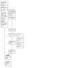
Study flow diagram.
aAltogether, 56 unique studies were excluded in this updated review.
Data extraction and management
Two review authors (JP, TR) independently extracted data regarding study design and methods, participant characteristics, and the primary and secondary outcomes, and recorded the information onto paper data collection forms developed in collaboration with Cochrane Eyes and Vision. Whenever there were discrepancies between review authors, we reached consensus by discussion. When we could not reach a consensus, we consulted a third review author who made the final decision. We contacted study investigators to obtain missing information and to elucidate unclear reporting. When they did not respond within two weeks, we used the information available. One review author (TR) entered data into Review Manager 5 (Review Manager 2020), and a second review author (JP) verified the data entered.
Assessment of risk of bias in included studies
Two review authors (JP, TR) independently assessed each included study for risks of bias as part of the data extraction process. We based our judgments on the tools for assessing risk of bias set in Chapter 8 of the Cochrane Handbook for Systematic Reviews of Interventions (Higgins 2011).
We judged each study with respect to the following risk of bias domains.
Selection bias (sequence generation and allocation concealment before randomization)
Performance bias (masking of participants and personnel)
Detection bias (masking of outcome assessors)
Attrition bias (incomplete outcome data)
Reporting bias (selective outcome reporting)
Other potential sources of bias (e.g. funding source)
We assessed each trial for each risk of bias criterion as being at high, low, or unclear risk of bias (lack of information or uncertainty over the potential for bias).
Measures of treatment effect
Dichotomous outcomes
We analyzed dichotomous outcomes, such as complications and proportion of participants who were drop‐free, using summary risk ratios (RRs) with 95% confidence intervals (CIs).
Continuous outcomes
We estimated the difference between continuous outcomes, such as mean change (or mean) IOP, BCVA, mean number of IOP lowering medications, as the mean difference (MD) with 95% CIs. We planned to analyze IOP fluctuations, visual field changes, quality‐of‐life scores as continuous outcomes, but such data were not available.
Unit of analysis issues
The unit of analysis was the eye that had glaucoma surgery. We recorded whether studies used a parallel‐group design or a paired‐eye design, and whether the study used matched‐analysis when a paired‐eye design was used. When both eyes of all or some participants were allocated to the same intervention group, we recorded the information as available and did not estimate or impute intraperson correlations for individual outcomes.
All studies were parallel‐group designs. Of the eight trials, four included only one eye per participant. Both eyes of some participants were included in another three parallel‐group trials; a mean of 7% of participants across these three trials contributed both eyes to the analysis. One trial was a paired‐eye design in which each participant had one eye in each intervention group. None of the studies that included more than one eye per participant accounted for intraperson correlation.
Dealing with missing data
We contacted study investigators to request missing data or to clarify unclearly reported data or information, including but not limited to information about study methods, effect estimates, and standard deviations of effect estimates. When study investigators did not respond within two weeks or after three attempts to contact them, we used the available information. We did not impute data for this review.
Assessment of heterogeneity
We assessed clinical and methodological heterogeneity among included trials by examining variations in the trial designs and methods, characteristics of the trial participants, variations in interventions, and lengths of follow‐up. We assessed statistical heterogeneity among the reported treatment effect estimates of included trials by examining the overlap of the 95% CIs on estimates from individual trials in forest plots and I² values (Higgins 2003). We considered poor overlap in the 95% CIs and an I² above 50% as indications of substantial statistical heterogeneity.
Assessment of reporting biases
We investigated whether our review was subject to reporting biases. For selective reporting bias, we compared outcomes specified in trial protocols or trial register records with outcomes reported in published full‐text articles. When no trial protocol or trial register record was available, we examined whether outcomes specified in the methods section were reported in the results section of the same published report. We did not use funnel plots to examine signs of asymmetry due to the limited number of studies included in the same meta‐analysis.
Data synthesis
We determined whether data synthesis in meta‐analyses was appropriate based on evidence of heterogeneity. When we considered that there was substantial heterogeneity, we presented results in a narrative summary. In the absence of clinical and methodological heterogeneity across studies, and when the I² statistic was less than 50% (indicating no substantial statistical heterogeneity), we combined study results using a random‐effects meta‐analysis model. Likewise, we applied a random‐effects meta‐analysis model when the I² statistic was greater than 50% but all studies favored the same intervention, or when the I² statistic was greater than 50% but no study showed a clinical difference between groups.
Subgroup analysis and investigation of heterogeneity
We compared a subgroup by the use of device within a single analysis for each outcome where information was available. We did not conduct subgroup analysis for comparisons of outcomes with use of adjuvant antimetabolites (e.g. MMC) because all studies used adjuvant MMC. Also, we were unable to carry out the following planned subgroup analyses as the included studies did not stratify participants based on 1. the status of the lens (i.e. eyes that possessed their natural lens (phakic), eyes without the crystalline lens (aphakic, cataract extraction), or eyes with an intraocular lens implanted that replaced the eye's natural lens (pseudophakic)); 2. ethnicity; 3. baseline IOP; or 4. type of glaucoma.
Sensitivity analysis
We were unable to conduct sensitivity analyses to assess the influence on effect estimates of excluding studies at high risk of reporting bias, as most studies had a low risk of reporting bias. We had also planned to conduct a sensitivity analysis after excluding industry‐funded studies; however, funding information was not always available, so we did not have enough information to conduct such analyses.
Summary of findings and assessment of the certainty of the evidence
Two review authors (JP, TR) independently assessed the certainty of the evidence by outcome using the GRADE system (Guyatt 2011). We reported results in a Table 1.
Our prespecified outcome measures were:
change in IOP;
postoperative mean IOP at one year after the intervention;
mean BCVA in logMAR;
postoperative visual field change at one year after the intervention;
quality of life;
proportion of participants who were drop‐free at one year after the intervention;
frequency of the following complication: proportion of participants with endophthalmitis at end of follow‐up.
Results
Description of studies
Results of the search
For the current update of the review, we amended intervention types in the prespecified inclusion criteria to include only Ex‐PRESS, XEN Gel Stent, and PreserFlo MicroShunt. According to the electronic searches for the previous version of the review as of 22 December 2014, we previously included 39 reports from 33 studies and four ongoing trials. Per the updated inclusion criteria, which excluded antifibrotic materials and devices formerly combined with trabeculectomy but no longer in use assessed in the previous review, we further excluded 28 reports from 28 studies due to ineligible interventions, leaving five previously included studies, and one ongoing trial.
Through an updated search as of 24 August 2021, we retrieved and screened the titles and abstracts of 2801 records after duplicate removal and excluded 2761 of these records. We screened 40 full‐text reports, excluded 34 studies (35 records) with reasons, and classified one study (two records) as awaiting classification. Altogether, we included eight studies (eight reports) and assessed one as ongoing and one as awaiting classification in this version of the review. The ongoing trial is being conducted in Japan and compares Ex‐PRESS with standard trabeculectomy; result are not available yet. We did not identify any additional studies through searching reference lists of included trials.
A flow diagram describing the search and screening process is shown in Figure 1.
Included studies
We included eight trials. All trials were published in either English or Chinese. Details of each trial are presented in the Characteristics of included studies table. We summarized the basic trial characteristics in Table 2.
1. Summary of included studies.
| Device | Study ID | Study design | Country | Participant diagnosis | Interventions | Total number of participants randomized | Total number of eyes randomized | Total number of eyes analyzed | Longest follow‐up period (months) |
| Ex‐PRESS | Dahan 2012 | RCT, paired‐eye design | South Africa | POAG | 1. Trab + MMC 2. Trab + MMC + Ex‐PRESS |
15 | 30 | 30 | 12 |
| De Jong 2005 (abstract) | RCT, parallel‐group design | The Netherlands | OAG | 1. Trab + Ex‐PRESS under a scleral flap 2. Trab + Ex‐PRESS under conjunctiva 3. Trab |
109 | 120 | N/A | 6 | |
| De Jong 2009 | RCT, parallel‐group design | The Netherlands | OAG | 1. Trab 2. Trab + Ex‐PRESS |
78 | 78 | 78 | 60 | |
| El‐Saied 2021 | RCT, parallel‐group design | Egypt | Secondary angle‐closure neovascular glaucoma | 1. Trab 2. Trab + Ex‐PRESS |
20 | 20 | 20 | 12 | |
| Netland 2014 | RCT, parallel‐group design | USA | OAG | 1. Trab + MMC 2. Trab + MMC + Ex‐PRESS |
120 | 120 | 114 | 24 | |
| Wagdy 2021 | RCT, parallel‐group design | Egypt | OAG | 1. Trab + MMC 2. Trab + MMC + Ex‐PRESS |
28 | 28 | 28 | 12 | |
| Wagschal 2015 | RCT, parallel‐group design | Canada | OAG, uncontrolled IOP | 1. Trab + MMC 2. Trab + MMC + Ex‐PRESS |
64 | 64 | 60 | 12 | |
| Subtotal for Ex‐PRESS | 434 | 460 | N/A | Range 6–60 months | |||||
| PreserFlo MicroShunt | Baker 2021 | RCT, parallel‐group design | USA, France, Italy, the Netherlands, Spain, the UK | Mild‐to‐severe POAG | 1. MicroShunt + MMC 2. Trab + MMC |
527 | 527 | 527 | 12 |
| Subtotal for PreserFlo MicroShunt | 527 | 527 | 527 | 12 | |||||
| Total for all included studies | 961 | 987 | N/A | Range 6–60 months | |||||
ACG: angle‐closure glaucoma; MMC: mitomycin C; N/A: not applicable; OAG: open‐angle glaucoma; POAG: primary open‐angle glaucoma; RCT: randomized controlled trial; trab: trabeculectomy.
Types of participants
The eight trials included 989 eyes of 961 participants and had follow‐up periods ranging from six months to five years after surgery. All trials included men and women. Seven trials included participants with OAG; El‐Saied 2021 included participants with NVG. None of the trials stratified participants by type of glaucoma, race, or lens type. They were conducted in North America, Europe, and Africa.
Types of interventions
The eight trials assessed either the Ex‐PRESS with standard trabeculectomy or the PreserFlo MicroShunt. None assessed XEN Gel Stent.
Seven trials assessed trabeculectomy with Ex‐PRESS compared with standard trabeculectomy (Dahan 2012; De Jong 2005; De Jong 2009; El‐Saied 2021; Netland 2014; Wagdy 2021; Wagschal 2015). They enrolled 462 eyes of 395 participants. Six of the seven trials were two‐arm studies that compared standard trabeculectomy versus trabeculectomy and Ex‐PRESS, with MMC applied to both groups. The remaining trial was a three‐arm trial; it compared Ex‐PRESS implanted under a scleral flap with standard trabeculectomy, Ex‐PRESS implanted under the conjunctiva (without creation of a standard trabeculectomy flap), and standard trabeculectomy (De Jong 2005).
One trial (527 eyes of 527 participants) was a two‐arm study that compared PreserFlo MicroShunt with MMC with standard trabeculectomy with MMC (Baker 2021).
Types of outcomes
All trials considered IOP control as their main outcome; however, trials differed in how they reported IOP. One trial reported change of IOP from baseline (El‐Saied 2021); the remaining trials did not report this. All trials reported postoperative IOP at certain time points, and one trial did not report any quantitative data but provided a descriptive summary only (De Jong 2005).
Seven trials reported visual acuity outcomes at different time points (Baker 2021; Dahan 2012; De Jong 2009; El‐Saied 2021; Netland 2014; Wagdy 2021; Wagschal 2015); one trial reported visual field outcome qualitatively (Wagdy 2021); and all studies reported postoperative complications either quantitatively or qualitatively. None of the studies reported IOP fluctuation or quality‐of‐life outcomes.
Funding sources
Seven trials reported the funding sources: industry funded five trials (Baker 2021; Dahan 2012; De Jong 2009; Netland 2014; Wagschal 2015); and two trials reported receiving no funding (El‐Saied 2021; Wagdy 2021). De Jong 2005 did not disclose information about sources of funding.
Excluded studies
According to the updated inclusion criteria, we excluded 56 unique studies and listed the reasons for exclusion in the Characteristics of excluded studies table.
Studies awaiting classification
One study is awaiting classification (Konstantinidis 2021).
Ongoing studies
One study is ongoing (JPRN‐UMIN000008981).
Risk of bias in included studies
Figure 2 shows a summary of the risk of bias assessments. Seven of the eight included trials had a high risk of detection bias. Most trials had either missing or inadequate information in trial reports to assess the risk of selection bias, especially in unclear allocation concealment. All but one trial had a low risk of reporting bias while less than half of included trials received funding from the manufacturer of the device, which was judged as high risk of bias. A description for each domain is summarized below.
2.
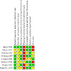
Risk of bias summary: review authors' judgements about each risk of bias item for each included study.
Allocation
Of the eight trials, six specified adequate methods of randomization and were at low risk of bias (Baker 2021; De Jong 2009; El‐Saied 2021; Netland 2014; Wagdy 2021; Wagschal 2015): five out of seven trials for Ex‐PRESS (De Jong 2009; El‐Saied 2021; Netland 2014; Wagdy 2021; Wagschal 2015) and one study for the PreserFlo MicroShunt (Baker 2021). The remaining two trials did not specify methods for random sequence generation, so we judged them at unclear risk of bias (Dahan 2012; De Jong 2005).
Of the eight trials, only Baker 2021, which was a study for PreserFlo MicroShunt, performed proper allocation concealment and was at low risk of bias. The other seven trials did not specify the method for allocation concealment, so they were at unclear risk of bias.
Blinding
Masking (performance bias and detection bias)
Study authors from five trials noted masking of participants: four of seven trials for Ex‐PRESS (Dahan 2012; De Jong 2009; El‐Saied 2021; Wagdy 2021), and one trial for PreserFlo MicroShunt (Baker 2021). The remaining three trials did not report whether participants were masked and were at unclear risk of performance bias (De Jong 2005; Netland 2014; Wagschal 2015). As masking of surgeons is logistically difficult and trabeculectomy is a standardized procedure (all studies described the surgical procedures in detail), we did not consider the lack of masking of surgeons to be an important modifiable source of bias.
In terms of detection bias, only Netland 2014 used a special protocol to minimize bias, so we judged this trial at unclear risk of detection bias. Otherwise, none specified masking of outcome assessors. Due to the easy detection of devices when examining the eye, unmasked outcome assessors could tend to anticipate and thus report favorable changes in IOP among participants with the implant or alternatively, among participants who received the surgery the outcome assessor preferred; therefore, the remaining trials were at high risk of detection bias (Baker 2021; Dahan 2012; De Jong 2005; De Jong 2009; El‐Saied 2021; Wagdy 2021; Wagschal 2015).
Incomplete outcome data
Investigators of five trials reported few or no losses to follow‐up, resulting in our assessment of low risk of attrition bias: four of seven trials of Ex‐PRESS (Dahan 2012; De Jong 2009; Netland 2014; Wagschal 2015), and one study for PreserFlo MicroShunt (Baker 2021). We assessed the remaining three trials at unclear risk of attrition bias as they did not report the number of losses to follow‐up; all were Ex‐PRESS trials (De Jong 2005; El‐Saied 2021; Wagdy 2021).
Selective reporting
We judged seven trials at low risk of reporting bias as they had 1. clinical trial registry records and reported all outcomes listed in the registry (Baker 2021; Dahan 2012; Netland 2014; Wagdy 2021; Wagschal 2015), or 2. reported all outcome measures defined in their methods section of the full‐text reports (De Jong 2005; De Jong 2009). These included six of seven studies for Ex‐PRESS and one for PreserFlo MicroShunt. We judged El‐Saied 2021 to have unclear risk of bias as no protocol or trial registration was publicly available.
Other potential sources of bias
We judged two trials at low risk of other potential sources of bias (El‐Saied 2021; Wagdy 2021). Three trials were at high risk because they received funding from the manufacturer of the device (Baker 2021; Dahan 2012; Netland 2014). The remaining studies were at unclear risk of bias, as funding and methodological details were reported insufficiently to render a judgment of low or high risk of bias (De Jong 2005; De Jong 2009; Wagschal 2015).
Effects of interventions
See: Table 1
Device‐modified trabeculectomy versus trabeculectomy
Seven trials assessed the use of Ex‐PRESS (Dahan 2012; De Jong 2005; De Jong 2009; El‐Saied 2021; Netland 2014; Wagdy 2021; Wagschal 2015), and one trial assessed the use of PreserFlo MicroShunt (Baker 2021). Six of eight trials reported a sample size calculation: Dahan 2012 had a power of 96% to detect a 2.0 mmHg IOP difference between groups; De Jong 2009 had a power of 80% to detect a 32% between‐group difference in IOP; and both Netland 2014 and Wagschal 2015 had power of 80% to detect a 2.0 mmHg IOP difference between groups. Baker 2021 had a power of 90% to detect 15% margin of non‐inferiority, which is a lowering of 2.5 mmHg IOP. Wagdy 2021 performed a post‐hoc power analysis with a post‐hoc power estimation of 0.83. De Jong 2005 and El‐Saied 2021 did not report a power or sample size calculation.
Intraocular pressure
Six trials comparing trabeculectomy plus Ex‐PRESS versus standard trabeculectomy reported postoperative IOP (Dahan 2012; De Jong 2009; Netland 2014; Wagschal 2015; El‐Saied 2021; Wagdy 2021). One trial comparing PreserFlo MicroShunt versus trabeculectomy reported postoperative IOP (Baker 2021).
Dahan 2012 reported IOP data at the last follow‐up time point and presented a figure with IOP reduction over time. The trial encompassed 30 eyes of 15 participants at one year, 20 eyes of 10 participants at two years, and 14 eyes of seven participants at 30 months (last follow‐up). Upon our request, the study investigators shared their original data, so we were able to calculate the mean change in IOP from baseline to one‐year follow‐up and postoperative IOP at various follow‐up time points (months six, 12, and 24). We did not combine trials of Ex‐PRESS and PreserFlo MicroShunt due to substantial statistical and clinical heterogeneity. We instead performed meta‐analyses for this outcome by the device.
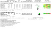
Our primary time frame was at one year, in the subgroup comparing trabeculectomy plus Ex‐PRESS versus trabeculectomy, five trials comprising 213 eyes reported mean IOP (Dahan 2012; De Jong 2009; Wagschal 2015; El‐Saied 2021; Wagdy 2021). The use of Ex‐PRESS may lead to a slightly improved IOP reduction at one year compared to standard trabeculectomy (MD −1.76 mmHg, 95% CI −2.81 to −0.70; I² = 0%; Analysis 1.1; Figure 3). Netland 2014 did not provide quantitative data, but reported that there was no between‐group difference in IOP reduction at one year. We rated the certainty of evidence as low, downgrading for risk of bias and limitations in the design. The trial comparing PreserFlo MicroShunt versus trabeculectomy reported a mean IOP of 446 eyes at one year (Baker 2021). We found that the PreserFlo MicroShunt group had a higher IOP than the trabeculectomy group (MD 3.20 mmHg, 95% CI 2.29 to 4.11). We rated the certainty of evidence as moderate, downgrading one level for risk of bias. There was evidence of a difference in mean IOP at one year between the Ex‐PRESS and PreserFlo MicroShunt groups when tested using the Cochrane test (P < 0.001).
1.1. Analysis.
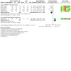
Comparison 1: Device‐modified trabeculectomy (trab) versus trabeculectomy, Outcome 1: Postoperative intraocular pressure (IOP) at 1 year by device type
3.
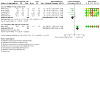
Forest plot of comparison: 2 Trabeculectomy + Ex‐PRESS versus trabeculectomy, outcome: postoperative intraocular pressure at one year.
Only El‐Saied 2021 provided data, so we could calculate the mean change in IOP from baseline to one‐year follow‐up. It is uncertain whether the Ex‐PRESS led to improved IOP reduction at one year compared to standard trabeculectomy (MD 2.00, 95% CI −3.66 to 7.66; P = 0.49; Analysis 1.2; Figure 4). We rated the certainty of evidence as very low, downgrading two levels for risk of bias due to limitations in the design and implementation of available studies and one level for imprecision.
1.2. Analysis.
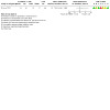
Comparison 1: Device‐modified trabeculectomy (trab) versus trabeculectomy, Outcome 2: Change in IOP from baseline at 1 year
4.
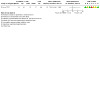
Forest plot of comparison: Trabeculectomy (Trab) + Ex‐PRESS versus trabeculectomy (Trab), outcome: change of intraocular pressure from baseline at one year.
At six months, in the subgroup comparing trabeculectomy plus Ex‐PRESS versus trabeculectomy, five trials comprising 253 eyes reported mean IOP (Dahan 2012; El‐Saied 2021; Netland 2014; Wagdy 2021; Wagschal 2015). It was unclear whether the use of Ex‐PRESS leads to IOP reduction at six months compared to standard trabeculectomy (MD −0.10 mmHg, 95% CI −1.40 to 1.20; I² = 54%; Analysis 1.3). The study comparing PreserFlo MicroShunt versus trabeculectomy suggested that the conventional trabeculectomy led to a further reduction of IOP at six months (MD 3.00 mmHg, 95% CI 1.62 to 4.38; Analysis 1.3).
1.3. Analysis.
Comparison 1: Device‐modified trabeculectomy (trab) versus trabeculectomy, Outcome 3: Postoperative IOP at 6 months by device type
At six months, only El‐Saied 2021 reported the mean change in IOP from baseline. It was unclear whether the use of Ex‐PRESS improved the IOP reduction at six months compared to standard trabeculectomy (MD 0.20, 95% CI −5.46 to 5.86; Analysis 1.5).
1.5. Analysis.
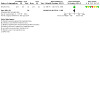
Comparison 1: Device‐modified trabeculectomy (trab) versus trabeculectomy, Outcome 5: Change in IOP from baseline at 6 months
At two years, three trials of Ex‐PRESS comprising 212 eyes reported mean IOP outcome (Dahan 2012; De Jong 2009; Netland 2014). Overall estimate suggested that Ex‐PRESS may slightly improve IOP reduction at two years compared to standard trabeculectomy (MD −1.38 mmHg, 95% CI −2.66 to −0.09; I² = 21%; Analysis 1.4).
1.4. Analysis.
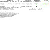
Comparison 1: Device‐modified trabeculectomy (trab) versus trabeculectomy, Outcome 4: Postoperative IOP at 2 years
Postoperative mean best‐corrected visual acuity at one year
Three studies reported logMAR BCVA at one year (Dahan 2012; El‐Saied 2021; Wagschal 2015). It is uncertain whether Ex‐PRESS prevents loss in BCVA compared to standard trabeculectomy (MD −0.04, 95% CI −0.19 to 0.10; I²= 23%; 110 eyes; Analysis 1.6; Figure 5).
1.6. Analysis.
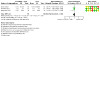
Comparison 1: Device‐modified trabeculectomy (trab) versus trabeculectomy, Outcome 6: Postoperative logMAR best‐corrected visual acuity at 1 year
5.
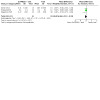
Forest plot of comparison: 1 Trabeculectomy (Trab) + Ex‐PRESS versus trabeculectomy (Trab), outcome: postoperative logMAR best‐corrected visual acuity at one year.
Wagschal 2015 and El‐Saied 2021 reported logMAR BCVA at one year, but Dahan 2012 did not publish quantitative data for this outcome. The authors of Dahan 2012 provided us with original data from which we calculated the postoperative mean logMAR BCVA to be mean 0.41 (standard error [SE] 0.11) for the Ex‐PRESS plus trabeculectomy group and 0.43 (SE 1.33) for the standard trabeculectomy group at one year.
Although De Jong 2009 also assessed visual acuity preoperatively and at each follow‐up visit, quantitative data were not reported. They reported that visual acuity remained equivalent in most participants, with no difference between the groups at one year. Wagdy 2021 did not report quantitative data on visual acuity. They reported one case of visual deterioration in the trabeculectomy group. We rated the certainty of evidence as low, downgrading two levels for high risk of bias due to limitations in the design and implementation of available studies.
Proportion of participants who were drop‐free at one year
Two trials (48 participants) reported the proportion of participants who were drop‐free at one year; both used the Ex‐PRESS (El‐Saied 2021; Wagdy 2021). The effect of Ex‐PRESS on the proportion of participants who were drop‐free at one year compared with standard trabeculectomy was uncertain (RR 2.04, 95% CI 0.42 to 9.82; P = 0.09; I² = 64%; Analysis 1.7; Figure 6). We rated the certainty of evidence as very low, downgrading two levels for high risk of bias and one level for imprecision. The use of the PreserFlo MicroShunt may lead to a lower chance of drop‐free at one year (RR 0.84, 95% CI 0.77 to 0.93; 509 eyes). We rated the certainty of evidence as moderate, downgrading one level for risk of bias. There was no evidence of a difference between the Ex‐PRESS groups and the PreserFlo MicroShunt group when tested using the Cochrane test (P = 0.27). We did not combine trials of Ex‐PRESS and PreserFlo MicroShunt due to substantial statistical and clinical heterogeneity.
1.7. Analysis.
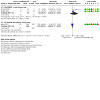
Comparison 1: Device‐modified trabeculectomy (trab) versus trabeculectomy, Outcome 7: Proportion of participants who are drop‐free at 1 year by device type
6.
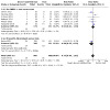
Forest plot of comparison: 1 Trabeculectomy (Trab) + Ex‐PRESS versus trabeculectomy (Trab), outcome: 1.5 Complications.
Mean number of intraocular pressure‐lowering medications at one year
Three trials (170 participants) reported the mean number of IOP‐lowering medications at one year; all used the Ex‐PRESS (Dahan 2012; De Jong 2009; Wagschal 2015). Ex‐PRESS may lead to using a lower number of IOP‐lowering medications at one year compared to standard trabeculectomy (MD −0.34, 95% CI −0.62 to −0.07; I² = 0%; Analysis 1.8). In contrast, the trial of PreserFlo MicroShunt (509 participants) reported a non‐significant effect in the number of IOP‐lowering medications at one year in both groups (MD 0.30, 95% CI 0.11 to 0.49; Analysis 1.8) (Baker 2021).
1.8. Analysis.
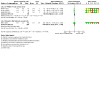
Comparison 1: Device‐modified trabeculectomy (trab) versus trabeculectomy, Outcome 8: Mean number of IOP lowering medications at 1 year by device type
Postoperative mean visual field change at one year
No studies reported postoperative mean visual field change at one year.
Quality of life at one year
No studies reported quality of life at one year.
Complications
Eight trials reported complications in 868 eyes during their respective follow‐up visits (Baker 2021; Dahan 2012; De Jong 2005; De Jong 2009; El‐Saied 2021; Netland 2014; Wagdy 2021; Wagschal 2015). We conducted a meta‐analysis using the proportion of participants with each complication in each group (Analysis 1.9; Analysis 1.10; Analysis 1.11; Analysis 1.12; Analysis 1.13; Analysis 1.14; Figure 6). De Jong 2005 did not report any complications.
1.9. Analysis.
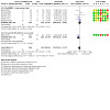
Comparison 1: Device‐modified trabeculectomy (trab) versus trabeculectomy, Outcome 9: Proportion of participants with IOP less than 5 mmHg (hypotony) or shallow anterior chamber by device type
1.10. Analysis.
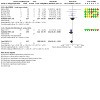
Comparison 1: Device‐modified trabeculectomy (trab) versus trabeculectomy, Outcome 10: Proportion of participants with bleb leakage by device type
1.11. Analysis.
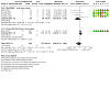
Comparison 1: Device‐modified trabeculectomy (trab) versus trabeculectomy, Outcome 11: Proportion of participants with reoperations for glaucoma by device type
1.12. Analysis.
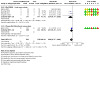
Comparison 1: Device‐modified trabeculectomy (trab) versus trabeculectomy, Outcome 12: Proportion of participants with cataract extraction by device type
1.13. Analysis.
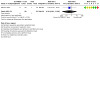
Comparison 1: Device‐modified trabeculectomy (trab) versus trabeculectomy, Outcome 13: Proportion of participants with endophthalmitis
1.14. Analysis.
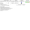
Comparison 1: Device‐modified trabeculectomy (trab) versus trabeculectomy, Outcome 14: Proportion of participants with loss of vision of > 2 lines or loss of light perception
Intraocular pressure less than 5 mmHg (hypotony) or shallow anterior chamber
Seven trials reported either hypotony or shallow anterior chamber, six using the Ex‐PRESS (Dahan 2012; De Jong 2009; El‐Saied 2021; Netland 2014; Wagdy 2021; Wagschal 2015), and one using the PreserFlo MicroShunt (Baker 2021). Overall, we found that participants who received device‐modified trabeculectomy may have a lower risk of hypotony, but the evidence was uncertain (RR 0.73, 95% CI 0.46 to 1.17; I² = 38%; P = 0.14; Analysis 1.9).
In the subgroup of Ex‐PRESS trials comprising 342 eyes, there was no evidence of a difference in the risk of developing hypotony or shallow anterior chamber between the two groups (RR 0.92, 95% CI 0.61 to 1.39; I² = 5%). In contrast, in the subgroup of a PreserFlo MicroShunt trial comprising 526 eyes, there was a lower risk of hypotony or shallow anterior chamber with PreserFlo MicroShunt compared with trabeculectomy (RR 0.44, 95% CI 0.25 to 0.79; Analysis 1.9).
Bleb leakage
Six trials reported bleb leakage, five using the Ex‐PRESS (Dahan 2012; De Jong 2009; El‐Saied 2021; Netland 2014; Wagschal 2015), and one using the PreserFlo MicroShunt (Baker 2021). Overall, participants who received device‐modified trabeculectomy may have a higher risk of bleb leakage than the standard trabeculectomy but the evidence was uncertain (RR 0.63, 95% CI 0.40 to 1.02; P = 0.71; I² = 0%; Analysis 1.9).
In the subgroup of Ex‐PRESS trials comprising 314 eyes, it is unclear whether Ex‐PRESS prevents bleb leakage compared to standard trabeculectomy (RR 0.99, 95% CI 0.45 to 2.16; I² = 0%; Analysis 1.10). In contrast, in the subgroup of a PreserFlo MicroShunt trial comprising 526 eyes, participants who received PreserFlo MicroShunt had a lower risk of bleb leakage compared with participants with trabeculectomy (RR 0.51, 95% CI 0.28 to 0.90; P = 0.02; Analysis 1.10).
Reoperations for glaucoma
Five trials reported reoperations for glaucoma, four using the Ex‐PRESS (Dahan 2012; De Jong 2009; El‐Saied 2021; Wagschal 2015), and one using the PreserFlo MicroShunt (Baker 2021). Overall, there was no difference in the risk of reoperation for glaucoma between device‐modified trabeculectomy and trabeculectomy (RR 0.69, 95% CI 0.24 to 1.98; P = 0.20; I² = 33%). In the subgroup of Ex‐PRESS trials comprising 194 eyes, there was no difference in risk of reoperation for glaucoma between groups (RR 0.34, 95% CI 0.09 to 1.26; I² = 0%; Analysis 1.11). In the subgroup of a PreserFlo MicroShunt trial comprising 526 eyes, there was no difference in risk of reoperations for glaucoma between groups (RR 1.30, 95% CI 0.77 to 2.22; Analysis 1.11).
Device migration or exposure
Two trials of Ex‐PRESS comprising 144 eyes reported this complication (De Jong 2009; Wagschal 2015). Obviously, device migration or exposure occurred only in the device‐modified trabeculectomy group. De Jong 2005 found that 1/40 participants (2.5%) reported device migration or exposure and Wagschal 2015 reported that 2/33 participants (6%) reported device migration or exposure. We did not perform a meta‐analysis on this outcome as device migration or exposure cannot occur with standard trabeculectomy.
Cataract surgery
Five trials reported subsequent requirement for cataract surgery after glaucoma surgery, four using the Ex‐PRESS (Dahan 2012; De Jong 2009; Netland 2014; Wagschal 2015), and one trial using the PreserFlo MicroShunt (Baker 2021). Overall, device‐modified trabeculectomy was associated with less frequent subsequent cataract surgery (RR 0.46, 95% CI 0.27 to 0.80; I² = 0%). Subgroup analysis in the Ex‐PRESS group comprising 294 eyes showed that Ex‐PRESS may lead to a lower risk of subsequent cataract surgery than standard trabeculectomy (RR 0.34, 95% CI 0.14 to 0.80; I² = 0%; only 3 studies included in meta‐analysis as 1 study had no events in either group). In the subgroup of a PreserFlo MicroShunt trial comprising 526 eyes, it was unclear whether the use of PreserFlo MicroShunt reduced subsequent cataract surgery (RR 0.57, 95% CI 0.28 to 1.17; P = 0.13; Analysis 1.12).
Endophthalmitis
Only Netland 2014 reported endophthalmitis. The trial found that only one participant who underwent a standard trabeculectomy developed endophthalmitis, and there was no difference on risk of endophthalmitis between the Ex‐PRESS plus trabeculectomy group and standard trabeculectomy (RR 0.34, 95% CI 0.01 to 8.29; Analysis 1.13). We rated the certainty of evidence for this complication as very low, downgrading two levels for high risk of bias, and one level for imprecision.
Loss of vision of more than two lines or loss of light perception
Only Baker 2021 reported the proportion of participants with loss of vision of more than two lines, or loss of light perception. PreserFlo MicroShunt appears to be associated with less vision loss, but the evidence was uncertain (RR 0.57, 95% CI 0.30 to 1.07; P = 0.08; Analysis 1.14). Dahan 2012 reported no cases with loss of vision of more than two lines in either group.
Discussion
Summary of main results
The addition of an Ex‐PRESS to trabeculectomy may result in lower IOP than standard trabeculectomy, based on data from five trials comprising 213 eyes (Dahan 2012; De Jong 2009; El‐Saied 2021; Wagdy 2021; Wagschal 2015). PreserFlo MicroShunt may not lower IOP as well as standard trabeculectomy. Data from three studies comprising 55 eyes found no difference in CVA between device‐modified and standard trabeculectomy groups at one year (Dahan 2012; El‐Saied 2021; Wagschal 2015). There was no difference in the number of IOP‐lowering medications at one year between groups. PreserFlo MicroShunt may prevent postoperative hypotony and bleb leakage compared with trabeculectomy. Other complications such as reoperations for glaucoma, device migration, endophthalmitis, or loss of vision were similar between the two groups. Device‐modified trabeculectomy appears to have a lower risk of subsequent cataract surgery.
Overall completeness and applicability of evidence
For Ex‐PRESS, of the seven included trials, five were powered to detect between‐group differences (Dahan 2012; De Jong 2009; Netland 2014; Wagschal 2015; Wagdy 2021). These trials were conducted in China, South Africa, the Netherlands, the USA, Canada, and Egypt. They included a mix of white, African American, Asian, and Indian participants. In four trials, the mean age was around 65 years (Dahan 2012; De Jong 2009; Netland 2014; Wagschal 2015), whereas Wagdy 2021 included participants aged between 42 and 55 years. Since six trials included participants with OAG, the Ex‐PRESS results are most applicable to people with OAG (Dahan 2012; De Jong 2005; De Jong 2009; Netland 2014; Wagdy 2021; Wagschal 2015). Wagdy 2021 included participants with failed trabeculectomy previously.
For the PreserFlo MicroShunt, one study comparing PreserFlo MicroShunt with trabeculectomy was conducted in the US and Europe (Baker 2021). This study included a mix of white, Black/African American, and Asian participants, with a mean age of around 65 years. There was a higher proportion of Black/African American participants in the PreserFlo MicroShunt group compared with the trabeculectomy group. This study included participants with POAG and excluded participants with secondary OAG or ACG. Thus, the effectiveness and safety in people with other types of glaucoma remain uncertain for both devices.
We found no eligible trials assessing the XEN Gel Stent.
Quality of the evidence
Overall, the agreement in absolute IOP measurements at one year across the included studies was high within each device. The widths of the 95% CI for MD in IOP measurement between device‐modified and standard trabeculectomy were small, ranging from −2.81 mmHg to −0.7 mmHg for the Ex‐PRESS and from 2.29 mmHg to 4.11 mmHg for the PreserFlo MicroShunt. Only one small study of Ex‐PRESS reported mean change in IOP, finding it varied, with a large 95% CI ranging from −3.66 mmHg to 7.66 mmHg.
Most of the trials were at high risk of detection bias for lack of masking outcome assessors. Most trials had either missing or inadequate information in trial reports to assess the risk of selection bias, especially in unclear allocation concealment. Furthermore, some Ex‐PRESS and PreserFlo MicroShunt studies had potential conflicts of interest due to receiving funding support from the device manufacturer, which may suggest high likelihood of bias in the study design and implementation of available studies. Overall, we graded the certainty of the evidence as low or very low for most outcomes due to potential high risks of detection bias and imprecision.
Potential biases in the review process
We conducted comprehensive electronic searches for studies with no imposed date or language restrictions to minimize potential biases in the study selection process. We followed standard Cochrane Review methodology.
Agreements and disagreements with other studies or reviews
Ex‐PRESS plus trabeculectomy versus trabeculectomy
Our meta‐analyses of five trials (at one year) and three trials (at two years) found that the use of Ex‐PRESS plus trabeculectomy may lead to greater IOP reduction compared with standard trabeculectomy, while it was uncertain whether the risk of complications, such as hypotony, bleb leakage, operations, device migration, and endophthalmitis differed between the groups. The proportion of participants requiring subsequent cataract extraction was lower in the device‐modified trabeculectomy group than in the trabeculectomy group.
One retrospective comparative series by Maris 2007 of 100 eyes, Good 2011 of 70 eyes, Moisseiev 2015 of 200 eyes, and Bustros 2017 of 56 eyes found no difference between Ex‐PRESS plus trabeculectomy and trabeculectomy in lowering IOP. One retrospective review of 153 eyes showed a lower risk of postoperative hypotony with Ex‐PRESS plus trabeculectomy compared with trabeculectomy, but no difference in lowering IOP (Marzette 2011).
One systematic review concluded that Ex‐PRESS has the same effectiveness in IOP reduction compared with standard trabeculectomy, with a lower frequency of hypotony and hyphema compared with standard trabeculectomy (Wang 2013a). However, these pooled results were from a mix of randomized controlled trials, prospective non‐randomized controlled trials, and retrospective studies, which limits the reliability of their inference.
One meta‐analysis found no reduction in IOP between Ex‐PRESS plus trabeculectomy and standard trabeculectomy, and a lower frequency of hyphema with Ex‐PRESS plus trabeculectomy (Chen 2014). The other complications, such as hypotony, shallow or flat anterior chamber, choroidal effusion, and encapsulated bleb were no different between groups. However, this review was flawed in that it mixed the different follow‐up periods from different studies for IOP control (e.g. six months and one year) in one meta‐analysis. Also, one included study was a subset of another (both were references from Wagschal 2015), and its meta‐analyses of complications included both studies, thereby double‐counting the data.
One meta‐analysis reported that Ex‐PRESS implantation achieved better outcomes in terms of long‐term IOP control, complete success rate, and lower numbers of IOP lowering medications (Zhang 2022). However, this review included trials with short follow‐up periods and mixed the different follow‐up periods for IOP. Also, this review compared Ex‐PRESS, trabeculectomy, and Ahmed glaucoma valve implant together, including more participants with secondary glaucoma compared with studies included in our review.
Some of the reviews, including the one presented here, reported that Ex‐PRESS plus trabeculectomy may lead to greater IOP reduction compared with standard trabeculectomy, whereas some other reviews reported there was no difference in reduction of IOP between Ex‐PRESS and trabeculectomy. Some reviews reported that Ex‐PRESS showed a lower rate of complications, such as hypotony and hyphema. Only our review reported that the risk of subsequent cataract extraction was lower in Ex‐PRESS plus trabeculectomy group than the standard trabeculectomy group.
PreserFlo MicroShunt versus trabeculectomy
Baker 2021 is the only study in our meta‐analysis that compared PreserFlo MicroShunt versus trabeculectomy. This study found that the PreserFlo MicroShunt was inferior in IOP‐lowering effect compared with conventional trabeculectomy, but produced smaller proportions of participants with hypotony and bleb leakage compared with the trabeculectomy group. One non‐randomized study of 52 eyes that were treated with PreserFlo MicroShunt or trabeculectomy found no differences in the reduction of IOP (Pillunat 2021). The incidence of early (within four weeks) hypotony was higher in the PreserFlo MicroShunt group compared with the trabeculectomy group, but the incidences of hypotony requiring anterior chamber formation, hypotony leading to choroidal effusion, hypotony maculopathy, or prolonged hypotony were not different between groups. We found no previous meta‐analysis of PreserFlo MicroShunt versus standard trabeculectomy.
Authors' conclusions
Implications for practice.
Our findings suggest that the use of Ex‐PRESS plus trabeculectomy may lead to slightly greater intraocular pressure (IOP) reduction at one‐year follow‐up than standard trabeculectomy. The PreserFlo MicroShunt was inferior to trabeculectomy with respect to mean IOP at one‐year follow‐up but it may be effective in preventing postoperative hypotony. Overall complication rates were not different between the two groups, but device‐modified trabeculectomy is associated with less frequent need for cataract extraction after trabeculectomy. Conclusions for each type of device are limited due to methodological concerns for bias and poor reporting of outcomes. Currently, these devices increase costs for insurance companies and patients compared with those incurred for a standard trabeculectomy. Whether the greater IOP reduction or improved safety that can be achieved with these devices is sufficient to outweigh these additional costs will need to be determined on a case‐by‐case basis. As it has been reported that a 1 mmHg reduction in IOP can be associated with a 10% decrease in the risk of glaucomatous progression, the additional IOP reduction that may be obtained at one‐year follow‐up may be valuable in selected populations (Heijl 2002). As these devices are also intended to reduce surgical risk and simplify postoperative management, their benefits and harms need to be considered for each individual patient.
Implications for research.
Because the certainty in evidence of this review is low, better‐quality trials with higher‐certainty evidence are warranted to determine the comparative effectiveness of all devices included in this review. These studies are limited and the applicability of the evidence to other populations or settings remains unclear. Therefore, more research is needed to generate evidence for or against the use of devices such as Ex‐PRESS, PreserFlo MicroShunt, and XEN Gel Stent.
In the absence of definitive evidence, we need more trials of better quality for most comparisons and outcomes. These should account for losses to follow‐up at each follow‐up time point measured and for the correlation of outcomes between two eyes when applicable. They also need to consider the appropriate use of adjunctive agents, such as mitomycin C, in both groups to ensure comparability. It would be helpful for future trials to specify the types of glaucoma, and also to consider stratifying participants by type of glaucoma, race, and perhaps lens status. Data reporting needs to be improved by reporting differences between groups to allow more robust inferences when applicable. Future trials should also report the elements of trial quality identified above and ensure consistency between protocols and published studies.
What's new
| Date | Event | Description |
|---|---|---|
| 13 March 2023 | New search has been performed | Updated search on studies comparing three devices only: Ex‐PRESS shunts, XEN GelStent, and PreserFlo MicroShunt. |
| 13 March 2023 | New citation required and conclusions have changed | Inclusion criteria for the update revised, not affecting search strategies. In the current updates of this review, we included the Ex‐PRESS shunt, XEN GelStent, and PreserFlo MicroShunt, which are the major devices available to patients in the current US or EU market. We excluded some devices assessed in the previous review (i.e. silicone tube implant, SOLX Gold Shunt, Ologen, amniotic membrane, expanded polytetrafluoroethylene (ePTFE), and Gelfilms) as they are no longer in current use combined with trabeculectomy or they are adjuvant materials rather than devices. |
History
Protocol first published: Issue 4, 2013 Review first published: Issue 12, 2015
Acknowledgements
We would like to thank Gianni Virgili (Queen's University Belfast), Miriam Kolko (University of Copenhagen), and Barbara Hawkins (Johns Hopkins University) for their comments on earlier drafts of this update.
Editorial and peer‐reviewer contributions
CEV@US supported the authors in the development of this update. The following people conducted the editorial process for this update.
Sign‐off Editors (final editorial decision): Dr Tianjing Li (University of Colorado Anschutz Medical Campus) and Dr Gianni Virgilli (Queen's University Belfast)
Managing Editor and Assistant Managing Editors (selected peer reviewers, collated peer‐reviewer comments): Anupa Shah (Queen's University Belfast); Louis Leslie (University of Colorado Anschutz Medical Campus), and Genie Han (Johns Hopkins University)
Methodologist (provided methodological and editorial guidance to authors, edited the article): Sueko Ng and Alison Su‐Hsun Liu (University of Colorado Anschutz Medical Campus)
Information Specialist: Lori Rosman (CEV)
Copy Editor: Anne Lawson (Central Production Service, Cochrane)
Peer reviewers: Renee Bovelle (Howard University), Anthony King (Nottingham University)
Appendices
Appendix 1. CENTRAL search strategy
#1 MeSH descriptor: [Trabeculectomy] explode all trees #2 MeSH descriptor: [Glaucoma] explode all trees and with qualifiers: [Surgery ‐ SU] #3 MeSH descriptor: [Trabecular Meshwork] explode all trees and with qualifiers: [Surgery ‐ SU] #4 MeSH descriptor: [Filtering Surgery] explode all trees #5 Trabeculectom* or Trabeculoplast* or Trabeculotom* or Goniotom* or Microtrabeculectom* #6 (Glaucoma* near/5 (surg* or filter* or filtrate*)) #7 #1 or #2 or #3 or #4 or #5 or #6 #8 MeSH descriptor: [Glaucoma Drainage Implants] explode all trees #9 (modif* near/5 (Trabeculectom* or Trabeculoplast* or Trabeculotom* or Goniotom* or Microtrabeculectom*)) #10 MeSH descriptor: [Polytetrafluoroethylene] explode all trees #11 (Polytef or Politef or "E PTFE" or EPTFE or PTFE or TFE or FEP or SOLX or polytetrafluoroethylen* or polytetrafluorethylen* or polytetrafluoroethen* or Fluoroflex or Fluoroplast or Ftoroplast or Halon or Polyfene or Tetron or Tarflen or "GORE TEX" or Goretex or gortex or Teflon or Fluon or Ex‐press or ologen or Baerveldt or Krupin or Ahmed or Molteno or ExPress or collagen matrix or collagen‐GAG or collagen‐glycosaminoglycan copolymer matrix) #12 Device* or implant* or shunt* or valve* or tube* #13 #8 or #9 or #10 or #11 or #12 #14 MeSH descriptor: [Fluorouracil] explode all trees #15 5FU or 5‐FU or Fluorouracil* or Fluoruracil* or 5‐HU or Adrucil or Carac or Efudix or Fluoro Uracile or Fluoro‐Uracile or Efudex or Fluoroplex or Flurodex or Fluracedyl or Haemato‐fu or Neofluor or Onkofluor or Ribofluor or 5‐Fluorouracil #16 MeSH descriptor: [Mitomycin] explode all trees #17 Mitomycin* or NSC‐26980 or NSC 26980 or NSC26980 or Mutamycin or Ametycine or Mitocin‐C or MitocinC or mytomycin* or mitomicin* or mytomicin* or MMC #18 MeSH descriptor: [Mitomycins] explode all trees #19 #18 from 1966 to 1991 #20 MeSH descriptor: [Antimetabolites] explode all trees #21 MeSH descriptor: [Antimetabolites, Antineoplastic] explode all trees #22 MeSH descriptor: [Nucleic Acid Synthesis Inhibitors] explode all trees #23 Antimetabolite* or anti‐metabolite* #24 Antifibrotic* or anti‐fibrotic* #25 #14 or #15 or #16 or #17 or #19 or #20 or #21 or #22 or #23 or #24 #26 #7 and (#13 or #25)
Appendix 2. MEDLINE (OvidSP) search strategy
1. Randomized Controlled Trial.pt. 2. Controlled Clinical Trial.pt. 3. (randomized or randomised).ab,ti. 4. placebo.ab,ti. 5. drug therapy.fs. 6. randomly.ab,ti. 7. trial.ab,ti. 8. groups.ab,ti. 9. 1 or 2 or 3 or 4 or 5 or 6 or 7 or 8 10. exp animals/ not humans.sh. 11. 9 not 10 12. exp Trabeculectomy/ 13. exp Glaucoma/su [Surgery] 14. exp Trabecular Meshwork/su [Surgery] 15. (Trabeculectom* or Trabeculoplast* or Trabeculotom* or Goniotom* or Microtrabeculectomy).tw. 16. (Glaucoma$ adj5 (surg$ or filter$ or filtrat$)).tw. 17. exp filtering surgery/ 18. 12 or 13 or 14 or 15 or 16 or 17 19. exp Glaucoma Drainage Implants/ 20. (modif* adj5 (Trabeculectom* or Trabeculoplast* or Trabeculotom* or Goniotom* or Microtrabeculectomy)).tw. 21. exp Polytetrafluoroethylene/ 22. (Polytef or Politef or "E PTFE" or EPTFE or PTFE or TFE or FEP or SOLX or polytetrafluoroethylen* or polytetrafluorethylen* or polytetrafluoroethen* or Fluoroflex or Fluoroplast or Ftoroplast or Halon or Polyfene or Tetron or Tarflen or "GORE TEX" or Goretex or gortex or Teflon or Fluon or Ex‐press or ologen or Baerveldt or Krupin or Ahmed or Molteno or ExPress or collagen matrix or collagen‐GAG or collagen‐glycosaminoglycan copolymer matrix).tw. 23. (Device* or implant* or shunt* or valve* or tube*).tw. 24. 19 or 20 or 21 or 22 or 23 25. exp Fluorouracil/ 26. (5FU or 5‐FU or Fluorouracil* or Fluoruracil* or 5‐HU or Adrucil or Carac or Efudix or Fluoro Uracile or Fluoro‐Uracile or Efudex or Fluoroplex or Flurodex or Fluracedyl or Haemato‐fu or Neofluor or Onkofluor or Ribofluor or 5‐Fluorouracil).tw. 27. exp Mitomycin/ 28. (Mitomycin* or NSC‐26980 or NSC 26980 or NSC26980 or Mutamycin or Ametycine or Mitocin‐C or MitocinC or mytomycin* or mitomicin* or mytomicin* or MMC).tw. 29. exp Mitomycins/ 30. limit 29 to yr="1966 ‐ 1991" 31. antimetabolites/ 32. Antimetabolites, Antineoplastic/ 33. Nucleic Acid Synthesis Inhibitors/ 34. (Antimetabolite* or anti‐metabolite*).tw. 35. (Antifibrotic* or anti‐fibrotic*).tw. 36. 25 or 26 or 27 or 28 or 30 or 31 or 32 or 33 or 34 or 35 37. 11 and 18 and (24 or 36) The search filter for trials at the beginning of the MEDLINE strategy is from the published paper by Glanville et al (Glanville 2006).
Appendix 3. Embase search strategy
1. 'randomized controlled trial'/exp 2. 'randomization'/exp 3. 'double blind procedure'/exp 4. 'single blind procedure'/exp 5. random*:ab,ti 6. 1 OR 2 OR 3 OR 4 OR 5 7. 'animal'/exp OR 'animal experiment'/exp 8. 'human'/exp 9. 7 AND 8 10. 7 NOT 9 11. 6 NOT 10 12. 'clinical trial'/exp 13. (clin* NEAR/3 trial*):ab,ti 14. ((singl* OR doubl* OR trebl* OR tripl*) NEAR/3 (blind* OR mask*)):ab,ti 15. 'placebo'/exp 16. placebo*:ab,ti 17. random*:ab,ti 18. 'experimental design'/exp 19. 'crossover procedure'/exp 20. 'control group'/exp 21. 'latin square design'/exp 22. 12 OR 13 OR 14 OR 15 OR 16 OR 17 OR 18 OR 19 OR 20 OR 21 23. 22 NOT 10 24. 23 NOT 11 25. 'comparative study'/exp 26. 'evaluation'/exp 27. 'prospective study'/exp 28. control*:ab,ti OR prospectiv*:ab,ti OR volunteer*:ab,ti 29. 25 OR 26 OR 27 OR 28 30. 29 NOT 10 31. 30 NOT (11 OR 23) 32. 11 OR 24 OR 31 33. 'trabeculectomy'/exp 34. 'trabeculoplasty'/exp 35. 'trabeculotomy'/exp 36. trabeculectom*:ab,ti OR trabeculoplast*:ab,ti OR trabeculotom*:ab,ti OR goniotom*:ab,ti OR microtrabeculectom*:ab,ti 37. 'glaucoma surgery'/de 38. 'trabecular meshwork'/exp 39. (glaucoma* NEAR/5 (surg* OR filter* OR filtrate*)):ab,ti 40. 'filtering operation'/de 41. 33 OR 34 OR 35 OR 36 OR 37 OR 38 OR 39 OR 40 42. 'glaucoma drainage implant'/exp 43. (modif* NEAR/5 (trabeculectom* OR trabeculoplast* OR trabeculotom* OR goniotom* OR microtrabeculectom*)):ab,ti 44. 'politef'/exp 45. (Polytef or Politef or 'E PTFE' or EPTFE or PTFE or TFE or FEP or SOLX or polytetrafluoroethylen* or polytetrafluorethylen* or polytetrafluoroethen* or Fluoroflex or Fluoroplast or Ftoroplast or Halon or Polyfene or Tetron or Tarflen or 'GORE TEX' or Goretex or gortex or Teflon or Fluon or Ex‐press or ologen or Baerveldt or Krupin or Ahmed or Molteno or ExPress or 'collagen matrix' or 'collagen‐GAG' or 'collagen‐glycosaminoglycan copolymer matrix'):ab,ti 46. device*:ab,ti OR implant*:ab,ti OR shunt*:ab,ti OR valve*:ab,ti OR tube*:ab,ti 47. 42 OR 43 OR 44 OR 45 48. 'fluorouracil'/exp 49. 5fu:ab,ti OR '5 fu':ab,ti OR fluorouracil*:ab,ti OR fluoruracil*:ab,ti OR '5 hu':ab,ti OR adrucil:ab,ti OR carac:ab,ti OR efudix:ab,ti OR fluoro:ab,ti AND uracile:ab,ti OR 'fluoro uracile':ab,ti OR efudex:ab,ti OR fluoroplex:ab,ti OR flurodex:ab,ti OR fluracedyl:ab,ti OR 'haemato fu':ab,ti OR neofluor:ab,ti OR onkofluor:ab,ti OR ribofluor:ab,ti OR '5 fluorouracil':ab,ti OR '5 fluoro 2':ab,ti OR '4 pyrimidinedione':ab,ti OR '5 fu':ab,ti OR accusite:ab,ti OR 'actino hermal':ab,ti OR effluderm:ab,ti OR efurix:ab,ti OR f6627:ab,ti OR fivoflu:ab,ti OR fluoroblastin:ab,ti OR fluouracil:ab,ti OR fluoxan:ab,ti OR fluracil:ab,ti OR fluracilium:ab,ti OR fluril:ab,ti OR 'fluro uracil':ab,ti OR fluroblastin:ab,ti OR ifacil:ab,ti OR 'nsc 18913':ab,ti OR 'nsc 19893':ab,ti OR 'nsc18913':ab,ti OR nsc19893:ab,ti OR 'oncofu':ab,ti OR 'ro 2‐9757':ab,ti OR 'ro 2 9757':ab,ti OR 'ro2‐9757':ab,ti OR 'ro2 9757':ab,ti OR uflahex:ab,ti OR utoral:ab,ti OR verrumal:ab,ti OR '51 21 8':ab,ti 50. 'mitomycin'/exp 51. mitomycin*:ab,ti OR 'nsc 26980':ab,ti OR nsc:ab,ti AND 26980:ab,ti OR nsc26980:ab,ti OR mutamycin:ab,ti OR ametycine:ab,ti OR 'mitocin c':ab,ti OR mitocinc:ab,ti OR mytomycin*:ab,ti OR mitomicin*:ab,ti OR mytomicin*:ab,ti OR mmc:ab,ti OR datisan:ab,ti OR metomit:ab,ti OR mitocyna:ab,ti OR mitosol:ab,ti OR mixandex:ab,ti OR mytocine:ab,ti OR mytozytrex:ab,ti OR vetio:ab,ti OR '1404 00 8':ab,ti 52. 'antimetabolite'/de 53. 'antineoplastic antimetabolite'/de 54. 'nucleic acid synthesis inhibitor'/de 55. antimetabolite*:ab,ti OR (anti NEAR/1 metabolite*):ab,ti 56. antifibrotic*:ab,ti OR (anti NEAR/1 fibrotic*):ab,ti 57. 48 OR 49 OR 50 OR 51 OR 52 OR 53 OR 54 OR 55 OR 56 58. 32 AND 41 AND (47 OR 57)
Appendix 4. PubMed search strategy
#1 ((randomized controlled trial[pt]) OR (controlled clinical trial[pt]) OR (randomised[tiab] OR randomized[tiab]) OR (placebo[tiab]) OR (drug therapy[sh]) OR (randomly[tiab]) OR (trial[tiab]) OR (groups[tiab])) NOT (animals[mh] NOT humans[mh]) #2 (Trabeculectom*[tiab] OR Trabeculoplast*[tiab] OR Trabeculotom*[tiab] OR Goniotom*[tiab] OR Microtrabeculectomy[tiab]) NOT MEDLINE[sb] #3 (Glaucoma*[tiab] AND (surge*[tiab] OR surgi*[tiab] OR filter*[tiab] OR filtrate*[tiab])) NOT MEDLINE[sb] #4 #2 OR #3 #5 (modif*[tiab] AND (Trabeculectom*[tiab] OR Trabeculoplast*[tiab] OR Trabeculotom*[tiab] OR Goniotom*[tiab] OR Microtrabeculectomy[tiab])) NOT MEDLINE[sb] #6 (Polytef[tiab] OR Politef[tiab] OR "E PTFE"[tiab] OR EPTFE[tiab] OR PTFE[tiab] OR TFE[tiab] OR FEP[tiab] OR SOLX[tiab] OR polytetrafluoroethylen*[tiab] OR polytetrafluorethylen*[tiab] OR polytetrafluoroethen*[tiab] OR Fluoroflex[tiab] OR Fluoroplast[tiab] OR Ftoroplast[tiab] OR Halon[tiab] OR Polyfene[tiab] OR Tetron[tiab] OR Tarflen[tiab] OR "GORE TEX"[tiab] OR Goretex[tiab] OR gortex[tiab] OR Teflon[tiab] OR Fluon[tiab] OR Ex‐press[tiab] OR ologen[tiab] OR Baerveldt[tiab] OR Krupin[tiab] OR Ahmed[tiab] OR Molteno[tiab] OR ExPress[tiab] OR collagen matrix[tiab] OR collagen‐GAG[tiab] OR collagen‐glycosaminoglycan copolymer matrix[tiab]) NOT MEDLINE[sb] #7 (Device*[tiab] OR implant*[tiab] OR shunt*[tiab] OR valve*[tiab] OR tube[tiab] OR tubes[tiab]) NOT MEDLINE[sb] #8 (5FU[tiab] OR 5‐FU[tiab] OR Fluorouracil*[tiab] OR Fluoruracil*[tiab] OR 5‐HU[tiab] OR Adrucil[tiab] OR Carac[tiab] OR Efudix[tiab] OR Fluoro Uracile[tiab] OR Fluoro‐Uracile[tiab] OR Efudex[tiab] OR Fluoroplex[tiab] OR Flurodex[tiab] OR Fluracedyl[tiab] OR Haemato‐fu[tiab] OR Neofluor[tiab] OR Onkofluor[tiab] OR Ribofluor[tiab] OR 5‐Fluorouracil[tiab]) NOT MEDLINE[sb] #9 (Mitomycin*[tiab] OR NSC‐26980[tiab] OR NSC 26980[tiab] OR NSC26980[tiab] OR Mutamycin[tiab] OR Ametycine[tiab] OR Mitocin‐C[tiab] OR MitocinC[tiab] OR mytomycin*[tiab] OR mitomicin*[tiab] OR mytomicin*[tiab] OR MMC[tiab]) NOT MEDLINE[sb] #10 (Antimetabolite*[tiab] OR anti‐metabolite*[tiab]) NOT MEDLINE[sb] #11 (Antifibrotic*[tiab] OR anti‐fibrotic*[tiab]) NOT MEDLINE[sb] #12 #5 OR #6 OR #7 OR #8 OR #9 OR #10 OR #11 #13 #1 AND #4 AND #12
Appendix 5. LILACS Controlled Trials search strategy
(Trabeculectom$ or Trabeculoplast$ or Trabeculotom$ or Goniotom$ or Microtrabeculectom$ or "trabecular meshwork" or "filtering surgery" or glaucoma$) AND (Polytef or Politef or "E PTFE"or EPTFE or PTFE or TFE or FEP or SOLX or polytetrafluoroethylen$ or polytetrafluorethylen$ or polytetrafluoroethen$ or Fluoroflex or Fluoroplast or Ftoroplast or Halon or Polyfene or Tetron or Tarflen or "GORE TEX" or Goretex or gortex or Teflon or Fluon or Ex‐press or ologen or Baerveldt or Krupin or Ahmed or Molteno or ExPress or "collagen matrix" or "collagen‐GAG" or "collagen‐glycosaminoglycan copolymer matrix" or Device$ or implant$ or shunt$ or valve$ or tube$ or (modif$ and Trabeculectom$ or Trabeculoplast$ or Trabeculotom$ or Goniotom$ or Microtrabeculectom$) or Fluorouracil$ or 5FU or 5‐FU or Fluoruracil$ or 5‐HU or Adrucil or Carac or Efudix or Fluoro Uracile or Fluoro‐Uracile or Efudex or Fluoroplex or Flurodex or Fluracedyl or Haemato‐fu or Neofluor or Onkofluor or Ribofluor or 5‐Fluorouracil or Mitomycin$ or NSC‐26980 or NSC 26980 or NSC26980 or Mutamycin or Ametycine or Mitocin‐C or MitocinC or mytomycin$ or mitomicin$ or mytomicin$ or MMC or Antimetabolite$ or anti‐metabolite$ or Antifibrotic$ or anti‐fibrotic$)
Appendix 6. metaRegister of Controlled Trials search strategy
(Trabeculectomy OR (glaucoma surgery)) AND (device OR implant OR implants OR shunt OR valve OR tube OR 5FU OR 5‐FU OR Fluorouracil OR Fluoruracil OR Fluoro Uracile OR 5‐Fluorouracil OR Mitomycin OR MMC OR Antimetabolite OR Antimetabolites OR Antifibrotic)
Appendix 7. ClinicalTrials.gov search strategy
(search terms) Trabeculectomy OR Trabeculoplasty OR Trabeculotomy OR Goniotomy OR Microtrabeculectomy OR glaucoma
(intervention) Device OR implant OR implants OR shunt OR valve OR tube OR Fluorouracil OR 5‐ Fluorouracil OR 5‐FU OR Fluoruracil OR Mitomycin OR mytomycin OR mitomicin OR mytomicin OR MMC OR Antimetabolite OR Antifibrotic
Appendix 8. ICTRP search strategy
(condition) Trabeculectomy OR Trabeculoplasty OR Trabeculotomy OR Goniotomy OR Microtrabeculectomy OR Goniotomy OR Microtrabeculectomy OR glaucoma (intervention) Device OR implant OR implants OR shunt OR valve OR tube OR Fluorouracil OR 5‐ Fluorouracil OR 5‐FU OR Fluoruracil OR Mitomycin OR mytomycin OR mitomicin OR mytomicin OR MMC OR Antimetabolite OR Antifibrotic
Data and analyses
Comparison 1. Device‐modified trabeculectomy (trab) versus trabeculectomy.
| Outcome or subgroup title | No. of studies | No. of participants | Statistical method | Effect size |
|---|---|---|---|---|
| 1.1 Postoperative intraocular pressure (IOP) at 1 year by device type | 6 | Mean Difference (IV, Random, 95% CI) | Subtotals only | |
| 1.1.1 Ex‐PRESS + trab versus trab | 5 | 213 | Mean Difference (IV, Random, 95% CI) | ‐1.76 [‐2.81, ‐0.70] |
| 1.1.2 PreserFlo MicroShunt versus trab | 1 | 446 | Mean Difference (IV, Random, 95% CI) | 3.20 [2.29, 4.11] |
| 1.2 Change in IOP from baseline at 1 year | 1 | Mean Difference (IV, Random, 95% CI) | Totals not selected | |
| 1.3 Postoperative IOP at 6 months by device type | 6 | Mean Difference (IV, Random, 95% CI) | Subtotals only | |
| 1.3.1 Ex‐PRESS + trab versus trab | 5 | 253 | Mean Difference (IV, Random, 95% CI) | ‐0.10 [‐1.40, 1.20] |
| 1.3.2 PreserFlo MicroShunt versus trab | 1 | 446 | Mean Difference (IV, Random, 95% CI) | 3.00 [1.62, 4.38] |
| 1.4 Postoperative IOP at 2 years | 3 | 212 | Mean Difference (IV, Random, 95% CI) | ‐1.38 [‐2.66, ‐0.09] |
| 1.5 Change in IOP from baseline at 6 months | 1 | 20 | Mean Difference (IV, Random, 95% CI) | 0.20 [‐5.46, 5.86] |
| 1.6 Postoperative logMAR best‐corrected visual acuity at 1 year | 3 | 110 | Mean Difference (IV, Random, 95% CI) | ‐0.04 [‐0.19, 0.10] |
| 1.7 Proportion of participants who are drop‐free at 1 year by device type | 3 | Risk Ratio (M‐H, Random, 95% CI) | Subtotals only | |
| 1.7.1 Ex‐PRESS + trab versus trab | 2 | 48 | Risk Ratio (M‐H, Random, 95% CI) | 2.04 [0.42, 9.82] |
| 1.7.2 PreserFlo MicroShunt versus trab | 1 | 509 | Risk Ratio (M‐H, Random, 95% CI) | 0.84 [0.77, 0.93] |
| 1.8 Mean number of IOP lowering medications at 1 year by device type | 4 | Mean Difference (IV, Random, 95% CI) | Subtotals only | |
| 1.8.1 Ex‐PRESS + trab versus trab | 3 | 170 | Mean Difference (IV, Random, 95% CI) | ‐0.34 [‐0.62, ‐0.07] |
| 1.8.2 PreserFlo MicroShunt versus trab | 1 | 509 | Mean Difference (IV, Random, 95% CI) | 0.30 [0.11, 0.49] |
| 1.9 Proportion of participants with IOP less than 5 mmHg (hypotony) or shallow anterior chamber by device type | 7 | 868 | Risk Ratio (M‐H, Random, 95% CI) | 0.73 [0.46, 1.17] |
| 1.9.1 Ex‐PRESS + trab versus trab | 6 | 342 | Risk Ratio (M‐H, Random, 95% CI) | 0.92 [0.61, 1.39] |
| 1.9.2 PreserFlo MicroShunt versus trab | 1 | 526 | Risk Ratio (M‐H, Random, 95% CI) | 0.44 [0.25, 0.79] |
| 1.10 Proportion of participants with bleb leakage by device type | 6 | 840 | Risk Ratio (M‐H, Random, 95% CI) | 0.64 [0.40, 1.02] |
| 1.10.1 Ex‐PRESS + trab versus trab | 5 | 314 | Risk Ratio (M‐H, Random, 95% CI) | 0.99 [0.45, 2.16] |
| 1.10.2 PreserFlo MicroShunt versus trab | 1 | 526 | Risk Ratio (M‐H, Random, 95% CI) | 0.51 [0.28, 0.90] |
| 1.11 Proportion of participants with reoperations for glaucoma by device type | 5 | 720 | Risk Ratio (M‐H, Random, 95% CI) | 0.69 [0.24, 1.98] |
| 1.11.1 Ex‐PRESS + trab versus trab | 4 | 194 | Risk Ratio (M‐H, Random, 95% CI) | 0.34 [0.09, 1.26] |
| 1.11.2 PreserFlo MicroShunt versus trab | 1 | 526 | Risk Ratio (M‐H, Random, 95% CI) | 1.30 [0.77, 2.22] |
| 1.12 Proportion of participants with cataract extraction by device type | 5 | 820 | Risk Ratio (M‐H, Random, 95% CI) | 0.46 [0.27, 0.80] |
| 1.12.1 Ex‐PRESS + trab versus trab | 4 | 294 | Risk Ratio (M‐H, Random, 95% CI) | 0.34 [0.14, 0.80] |
| 1.12.2 PreserFlo MicroShunt versus trab | 1 | 526 | Risk Ratio (M‐H, Random, 95% CI) | 0.57 [0.28, 1.17] |
| 1.13 Proportion of participants with endophthalmitis | 1 | 120 | Risk Ratio (M‐H, Random, 95% CI) | 0.34 [0.01, 8.29] |
| 1.14 Proportion of participants with loss of vision of > 2 lines or loss of light perception | 1 | 526 | Risk Ratio (M‐H, Random, 95% CI) | 0.57 [0.30, 1.07] |
Characteristics of studies
Characteristics of included studies [ordered by study ID]
Baker 2021.
| Study characteristics | ||
| Methods |
Study design: multicenter, parallel‐group, randomized controlled trial Country: the US, France, Italy, the Netherlands, Spain, the UK Number randomized: 527 eyes of 527 participants total; 395 for PreserFlo MicroShunt + MMC and 132 for trabeculectomy + MMC Exclusions after randomization: none reported Losses to follow‐up: 21 for total; 14 for PreserFlo MicroShunt + MMC and 7 for trabeculectomy + MMC Unit of analysis: eye Number analyzed: 527 eyes of 527 participants total; 395 for PreserFlo MicroShunt + MMC and 132 for trabeculectomy + MMC How were missing data handled? imputed Power calculation: (quote) "sample size calculations were based on a Z‐test with normal distribution approximation and assuming an annual dropout rate of 6%." |
|
| Participants |
Mean age: none reported for overall; 66.4 (SD 9.3) years for PreserFlo MicroShunt + MMC group; 67.8 (SD 9.3) years for trabeculectomy + MMC group Gender 181/395 (45.8%) men and 214/395 (54.2%) women in PreserFlo MicroShunt + MMC group; 59/132 (44.7%) men and 73/132 (55.3%) women in trabeculectomy + MMC group Inclusion criteria: aged 40–85 years with mild‐to‐severe POAG inadequately controlled on maximum tolerated medical therapy, with IOP > 15 mmHg and < 40 mmHg and a visual field mean deviation of ≤ −3.00 dB Exclusion criteria: secondary OAG such as post‐trauma, pseudoexfoliative, or pigment dispersion (pigmentary glaucoma); ACG; aphakia; vision level of no light perception; previous incisional ophthalmic surgery involving the conjunctiva; prior clear corneal cataract, angle, or trabecular meshwork surgery conducted within the past 6 months; ocular steroid use in the planned study eye or systemic steroid use any time within 3 months of the procedure; BCVA < 20/80 in the non‐study eye; and laser surgery within 90 days of enrollment Equivalence of baseline characteristics: yes, apart from a higher proportion of Black/African American people (18.0% in the PreserFlo MicroShunt group vs 8.3% in the trabeculectomy group; P < 0.01). |
|
| Interventions |
Intervention 1: PreserFlo MicroShunt + MMC Intervention 2: trabeculectomy + MMC Length of follow‐up Planned: 1 years Actual: 1 years |
|
| Outcomes |
Primary outcomes, as defined: ≥ 20% reduction in mean diurnal IOP from baseline at 1 year follow‐up visit without increasing the number of glaucoma medications. Secondary outcomes, as defined: mean diurnal IOP change from baseline at 1 year; requirement for postoperative intervention by 1 year; number of glaucoma medications per participant at each follow‐up visit; incidence of adverse events; presence of cataract in phakic eyes; time for postoperative BCVA to return to baseline; change in endothelial cell density Intervals at which outcomes assessed: 1 day; 1 and 4 weeks; 3, 6, 12, 18, and 24 months |
|
| Notes |
Publication type: published article Funding sources: InnFocus Inc, a Santen Pharmaceutical Co Ltd Company and Santen Inc (quote: "the sponsor participated in the design and conduct of the study, data collection, and management. This analysis was also sponsored by Santen Inc., which participated in the data analysis, interpretation of the data, and preparation, review, and approval of the manuscript"). Disclosures of interest: "N.D.B.: Consultant/Advisor: Molteno Ophthalmic, Santen; Investigator: Santen. H.S.B.: Consultant/Advisor: Santen; Lecture fees Aerie, Bausch & Lomb. M.R.M.: Consultant/Advisor: Aerie, Alcon, Allergan, Qura, Santen; Lecture fees: Aerie, Alcon, Allergan, Bausch & Lomb, Iridex, MedEdicus, Novartis; Grant support: Aerie, Alcon, Allergan, Bausch & Lomb, Glaukos, InnFocus, Iridex, Santen; Equity/Owner: Qura. M.C.S.: Grant support Allergan, Glaukos, Ivantis, Santen. S.D.V.: Consultant/Advisor: Aerie, Alcon, Allergan, Bausch & Lomb, Carl Zeiss Meditec, Glaukos, Iridex, iSTAR Medical, Ivantis, New World Medical, Sight Sciences, Volk Optical; Grant support: Aerie, Alcon, Allergan, Bausch & Lomb, Carl Zeiss Meditec, Glaukos, Ivantis, RxSight, Santen, Sight Sciences; Equity/Owner: Alphaeon, Ivantis, O3 Optix; Patents/Royalty: Iridex, Volk Optical. A.K.K.: Consultant/Advisor: Gore Inc, Ivantis, Reliance/Haag‐Streit; Lecture fees: Aerie, Ivantis; Grant support: Glaukos, InnFocus, Santen. B.E.F.: Consultant/Advisor: Alcon, Bausch & Lomb, Eyenovia, Glaukos, InnFocus, iSTAR Medical, Ivantis, New World Medical, l, Sight Sciences; Grant support: Eyenovia, Glaukos, InnFocus, iSTAR Medical, Ivantis, Santen, Sight Sciences. D.S.G.: Consultant/Advisor: Allergan, MicroOptx, New World Medical, Reichert Instruments, Santen; Lecture fees: Aerie, Allergan, Bausch & Lomb, New World Medical, Reichert Instruments; Grant support: Allergan. N.G.S.: Funding: NIHR Biomedical Research Centre, Moorfields Eye Hospital NHS Foundation Trust, and UCL Institute of Ophthalmology. The sponsor or funding organization had no role in the design or conduct of this research. The views expressed are those of the authors and not necessarily those of the NHS, NIHR, or UK Department of Health. J.F.P.: Consultant/Advisor: Aerie, Allergan, Cornea Gen, Glaukos, New World Medical, Santen; Grant support: Allergan." Trial registry: NCT01881425 Study period: December 2015 to November 2017 Subgroup analyses: (quote) "Prespecified subgroup analyses in patients with baseline mean diurnal IOP<18, 18–20, and >21 mmHg were conducted." Publication language: English |
|
| Risk of bias | ||
| Bias | Authors' judgement | Support for judgement |
| Random sequence generation (selection bias) | Low risk | Study authors clearly reported that randomization was performed in a 3:1 ratio, which was stratified by investigational site and within site by lens status. The baseline characteristics in both groups were similar, suggesting effective randomization. |
| Allocation concealment (selection bias) | Low risk | Allocation concealment was performed using envelopes containing the randomization assignments. |
| Masking of participants and personnel (performance bias) | Low risk | Participants were masked to the assignment, and it is unlikely that they were aware of their group assignment after the operation given the similar possible complications. |
| Masking of outcome assessment (detection bias) | High risk | Outcome assessors were not masked to the participant's group assignment. The study authors did not explicitly specify the method used to measure IOP. However, as some methods of IOP measurement, e.g. using Goldmann applanation, could be subjective, it is likely that the unmasking of outcome assessors would affect the outcome. |
| Incomplete outcome data (attrition bias) All outcomes | Low risk | Only 3–5% of each group were lost to follow‐up. This missingness was unlikely related to worsening participants' health status due to device or procedure. |
| Selective reporting (reporting bias) | Low risk | The trial was analyzed according to the prespecified plan in terms of outcome measurements (e.g. definition, scales, and time points) within each outcome. |
| Other bias | High risk | This study was sponsored by the InnFocus company and many of the authors had industrial support. |
Dahan 2012.
| Study characteristics | ||
| Methods |
Study design: paired‐eye randomized controlled trial Country: South Africa Number randomized: 30 eyes of 15 participants total; each participant had 1 eye in each intervention group Exclusions after randomization: none reported Losses to follow‐up: 0 up to 1 year after surgery; 1 participant died at 13 months and 2 were subsequently lost to follow‐up Unit of analysis: eye Number analyzed: 30 eyes of 15 participants total; each participant had 1 eye in each intervention group How were missing data handled? no missing data at 1 year Power calculation: a power of 96% to detect a 2 mmHg IOP difference between groups |
|
| Participants |
Mean age: 65 years; not reported by intervention group Gender: 10/15 (67%) men and 5/15 (33%) women; not reported by intervention group Inclusion criteria: aged ≥ 18 years and presented with medically uncontrolled POAG requiring bilateral incisional surgery for IOP reduction. Patients with prior cataract operation or failed filtration surgery in either eye were eligible if surgery took place ≥ 3 months prior to enrolment Exclusion criteria: any form of glaucoma other than POAG; history of active uveitis; any ocular abnormality that would preclude accurate IOP assessment Equivalence of baseline characteristics: yes |
|
| Interventions |
Intervention 1: trabeculectomy + MMC + Ex‐PRESS X200 Intervention 2: trabeculectomy + MMC Length of follow‐up Planned: ≥ 1 year Actual: all participants followed ≥ 1 year; the longest follow‐up visit for a participant was 30 months |
|
| Outcomes | Primary and secondary outcomes not distinguished Outcomes, as reported: IOP, visual acuity, number of medicines for IOP control, complications Intervals at which outcomes assessed: 1 and 7 days; 1, 3, 6, 9, 12, 18, 24, and 30 months after surgery |
|
| Notes |
Publication type: published article Funding sources: (quote) "the study was supported by a financial grant from Alcon Laboratories." Disclosures of interest: "E Dahan is a paid consultant in Alcon Laboratories. GJ Ben Simon and A Lafuma has no financial or proprietary interest in any of the drugs or materials mentioned in this study. A Lafuma is employed by CEMKAEVAL, a company that provides services in statistical analyses and epidemiology." Trial registry: NCT00698438 Study period: not reported Subgroup analyses: none reported Publication language: English Study authors contacted and outcome data shared (IOP reduction, number of medications, and mean IOP at 1 and 7 days; and 1, 3, 6, 12, 18, 24, and 30 months after surgery) |
|
| Risk of bias | ||
| Bias | Authors' judgement | Support for judgement |
| Random sequence generation (selection bias) | Unclear risk | Quote: "Randomisation of contralateral operations was achieved by opening an envelope in which the procedure (trabeculectomy or Ex‐PRESS implantation) that would be applied to the first eye was stated, thereby determining the procedure in the other eye." Comment: the method of randomization was not described and thus its adequacy could not be judged. |
| Allocation concealment (selection bias) | Unclear risk | Allocation concealment was not reported. |
| Masking of participants and personnel (performance bias) | Low risk | Because trabeculectomy is a surgical procedure with informed consent, masking of the participants and personnel becomes impossible. However, given that (quote) "after sub‐tenonian local anaesthesia, surgery was performed by one experienced surgeon (ED), for consistency, using a standardized technique for both procedures," the risk of performance bias was comparably low for a surgical procedure. |
| Masking of outcome assessment (detection bias) | High risk | The study mentioned: (quote) "It was not possible to mask the surgical technique as trabeculectomy is easily differentiated from Ex‐PRESS implantation during postoperative follow‐ups. However, this limitation is overcome by the fact that all patients were followed up concurrently by their referring ophthalmologists from the first month postoperatively till completion of the study." Comment: although it was not possible to mask outcome assessors, devices could be easily seen during exam of the eye. |
| Incomplete outcome data (attrition bias) All outcomes | Low risk | Quote: "All 15 patients were followed‐up for 12 months after surgery. One patient died 13 months after surgery and two patients were subsequently lost to follow‐up. All data available for these patients (i.e., up to 1 year) are included in the analyses." |
| Selective reporting (reporting bias) | Low risk | The study was registered at www.ClinicalTrials.gov. All defined outcomes in www.ClinicalTrials.gov were reported in full text. Complete and qualified success was reported and defined using IOP. |
| Other bias | High risk | Received industry monetary support from device manufacturer; (quotes) "ED is a paid consultant to Alcon Laboratories." "The study was supported by a financial grant from Alcon Laboratories." To consider intraperson correlation between eyes, the analysis used Wilcoxon matched‐pairs t‐test to compare preoperative and final IOP values. No other sources of bias identified. |
De Jong 2005.
| Study characteristics | ||
| Methods |
Study design: parallel‐group randomized controlled trial (11 participants both eyes included) Country: the Netherlands Number randomized: 120 eyes of 109 participants; not reported by intervention group Exclusions after randomization: not reported Losses to follow‐up: not reported Unit of analysis: eye Number analyzed: not reported How were missing data handled? not reported Power calculation: not reported |
|
| Participants |
Mean age: overall not reported 61.8 years for trabeculectomy + Ex‐PRESS R50 implanted under a scleral flap group; 61.8 years for trabeculectomy + Ex‐PRESS R50 implanted under the conjunctiva group; 68.7 years for trabeculectomy group Gender: not reported Inclusion criteria: OAG; medical treatment failure, indicated for glaucoma surgery Exclusion criteria: not reported Equivalence of baseline characteristics: yes |
|
| Interventions |
Intervention 1: trabeculectomy + Ex‐PRESS R50 implanted under a scleral flap Intervention 2: trabeculectomy + Ex‐PRESS R50 implanted under the conjunctiva Intervention 3: trabeculectomy Length of follow‐up Planned: not reported Actual: 6 months |
|
| Outcomes | Primary and secondary outcomes not distinguished Outcomes, as reported: success rate (defined as % IOP reduction and medication reduction), IOP, and use of IOP‐lowering medications Intervals at which outcomes assessed: 6 months after surgery |
|
| Notes |
Publication type: published abstract Funding sources: not reported Disclosures of interest: not reported Trial registry: not registered Study period: not reported Subgroup analyses: none reported Publication language: English Attempted to contact author, but unable to find contact information in abstract. |
|
| Risk of bias | ||
| Bias | Authors' judgement | Support for judgement |
| Random sequence generation (selection bias) | Unclear risk | Not reported. |
| Allocation concealment (selection bias) | Unclear risk | Not reported. |
| Masking of participants and personnel (performance bias) | Unclear risk | Not reported. |
| Masking of outcome assessment (detection bias) | High risk | It is not possible to mask outcome assessors, as devices can be easily seen during eye exam. |
| Incomplete outcome data (attrition bias) All outcomes | Unclear risk | Not reported. |
| Selective reporting (reporting bias) | Low risk | Protocol was not available. All defined outcomes were reported. |
| Other bias | Unclear risk | Did not report source of support or conflict of interest. Insufficient information from the abstract. |
De Jong 2009.
| Study characteristics | ||
| Methods |
Study design: parallel‐group randomized controlled trial (2 participants both eyes included) Number randomized: 80 eyes of 78 participants total; 40 eyes in each group Exclusions after randomization: none reported Losses to follow‐up: 5 eyes total at 1 year; 3 eyes in trabeculectomy + MMC + Ex‐PRESS group; 2 eyes in trabeculectomy + MMC group Unit of analysis: eye Number analyzed: 75 eyes total; 37 eyes in trabeculectomy + MMC + Ex‐PRESS group; 38 eyes in trabeculectomy + MMC group How were missing data handled? 5 eyes with missing data excluded from analysis Power calculation: a power of 80% to detect 32% between‐group difference in IOP |
|
| Participants |
Country: the Netherlands Mean age: 66 years; 62.3 years for trabeculectomy + MMC + Ex‐PRESS group 68.9 years for trabeculectomy + MMC group Gender: 46/80 (58%) men and 34/80 (42%) women 19/40 (48%) men and 21/40 (52%) women in trabeculectomy + MMC + Ex‐PRESS group; 27/40 (68%) men and 13/40 (32%) women in trabeculectomy + MMC group Inclusion criteria: aged > 18 years with a diagnosis of OAG that could not be controlled with maximal‐tolerated medical therapy Exclusion criteria: any other ocular disease or previous ocular surgery other than cataract extraction Equivalence of baseline characteristics: not reported |
|
| Interventions |
Intervention 1: trabeculectomy + Ex‐PRESS Intervention 2: trabeculectomy Length of follow‐up Planned: 5 years Actual: mean 262 weeks for Ex‐PRESS group and 266 weeks for trabeculectomy group |
|
| Outcomes |
Primary outcome, as defined: complete success (final IOP > 4 mmHg and ≤ 18 mmHg without antiglaucoma medication) and overall success (final IOP > 4 mmHg and ≤ 18 mmHg with or without medications) Secondary outcomes, as defined: IOP, postoperative medication use, surgical failure (IOP > 18 mmHg or the requirement for further glaucoma surgery), stringent target (final IOP > 4 mmHg and ≤ 15 mmHg), complications, and visual acuity Intervals at which outcomes assessed: 1 day; 1 week; 1, 3, and 6 months; and 1, 2, 3, 4, and 5 years after surgery Length of follow‐up Planned: not reported Actual: 60‐month follow‐up |
|
| Notes |
Publication type: published article Funding sources: Alcon Management SA, Geneva, Switzerland Disclosures of interest: (quote) "L. de J. has no proprietary interest in any of the products mentioned here." Trial registry: not registered Study period: October 2003 to November 2004 Subgroup analyses: none reported Publication language: English Authors contacted to retrieve number of participants lost to follow‐up at 2–5 years, but no response received |
|
| Risk of bias | ||
| Bias | Authors' judgement | Support for judgement |
| Random sequence generation (selection bias) | Low risk | Quotes: "The participants were assigned randomly to receive either Ex‐PRESS implantation under a scleral flap (Group A), or trabeculectomy (Group B) in the study eye, according to a computer‐generated randomization list." "Randomization was determined before surgery according to a block randomization sequence prepared by SAS (version 9.1; SAS Institute Inc., Cary, NC, USA)." |
| Allocation concealment (selection bias) | Unclear risk | Allocation concealment was not reported. |
| Masking of participants and personnel (performance bias) | Low risk | Because trabeculectomy is a surgical procedure with informed consent, masking of the participants and personnel becomes impossible. However, given that a strict and standardized surgical protocol was followed and differences in the surgical protocol of the 2 groups were minimized, the risk of performance bias is comparably low for a surgical procedure. |
| Masking of outcome assessment (detection bias) | High risk | Quote: "Secondly, the evaluator was not blinded to the procedure used in each case; however, it is difficult to carry out truly blinded evaluation as the type of surgery used is usually visible to the assessor." Comment: but how they controlled the risk was not specified. |
| Incomplete outcome data (attrition bias) All outcomes | Low risk | There are 2 articles related to this study, as reported in 2009, the number analyzed was 40 eyes per treatment group (no loss to follow‐up); however, as reported in 2011, the number analyzed was 38 eyes per treatment group. |
| Selective reporting (reporting bias) | Low risk | Protocol was not available. All defined outcomes were reported. |
| Other bias | Unclear risk | Total industry support but no other source of potential bias identified. Quote: "There were no significant differences between the two groups except for age; the Ex‐PRESS group (Group A) included significantly younger patients compared with the trabeculectomy group (Group B)." Comment: age‐adjusted values are reported in de Jong 2011 and do not significantly change the results. |
El‐Saied 2021.
| Study characteristics | ||
| Methods |
Study design: parallel‐group randomized controlled trial (2 participants both eyes included) Country: Egypt Number randomized: 20 eyes of 20 participants total; 10 eyes in each group Exclusions after randomization: not reported Losses to follow‐up: not reported Unit of analysis: eye Number analyzed: 20 eyes total; 10 eyes in trabeculectomy + Ex‐PRESS mini shunt + MMC group; 10 eyes in trabeculectomy + MMC group How were missing data handled? not reported Power calculation: the sample size was calculated using MedCalc 10.2.0.0, by referring to success rates of glaucoma surgeries for NVG from literature. The power and alpha were not reported. |
|
| Participants |
Mean age: 55.7(SD 5.6) years 55.4 (SD 5.8) years for trabeculectomy + Ex‐PRESS mini shunt group 56 (SD 5.3) years for trabeculectomy group Gender 5/10 (50%) men and 5/10 (50%) women in trabeculectomy + Ex‐PRESS mini shunt group; 4/10 (40%) men and 6/10 (60%) women in trabeculectomy group Inclusion criteria: IOP > 21 mmHg on maximum tolerated topical antiglaucoma medication (medication score 3 for all the eyes); had secondary angle‐closure NVG, as confirmed by gonioscopy. Exclusion criteria: not reported Equivalence of baseline characteristics: yes |
|
| Interventions |
Intervention 1: trabeculectomy + Ex‐PRESS mini shunt Intervention 2: trabeculectomy Length of follow‐up Planned: 1 year Actual: 1 year |
|
| Outcomes |
Primary outcomes, as defined: IOP, BCVA, central foveal thickness, intraoperative bleeding, postoperative complications, and secondary intervention: needling or diode‐cyclo photocoagulation Secondary outcomes, as defined: postoperative hypotony, surgical success rate Intervals at which outcomes assessed: 1 day; 1 week; 1, 3, and 6 months; 1 year |
|
| Notes |
Publication type: published article Funding sources: none Disclosures of interest: authors declared that they had no conflict of interest Trial registry: not reported Study period: October 2016 to November 2017 Subgroup analyses: none reported Publication language: English |
|
| Risk of bias | ||
| Bias | Authors' judgement | Support for judgement |
| Random sequence generation (selection bias) | Low risk | Study authors clearly reported that randomization was performed using computer‐generated numbers. Baseline characteristics in both groups were similar, suggesting effective randomization. |
| Allocation concealment (selection bias) | Unclear risk | Study authors did not mention how the allocation concealment was performed. |
| Masking of participants and personnel (performance bias) | Low risk | There was no description about masking study participants and personnel. However, as the IOP was an objective measurement for participants and personnel, it was unlikely that the unmasking of participants and personnel would affect the outcome. |
| Masking of outcome assessment (detection bias) | High risk | Outcome assessors were not masked to the participant's group assignment. As the IOP measurement using Goldmann applanation could be subjective, there were some possibilities that the unmasking of outcome assessors would affect the outcome. |
| Incomplete outcome data (attrition bias) All outcomes | Unclear risk | Study authors did not explicitly report the number of losses to follow‐up. |
| Selective reporting (reporting bias) | Unclear risk | No statistical analysis plan was available, so it was unclear if the reported approach to analyzing this outcome was prespecified or influenced by the results. |
| Other bias | Low risk | Study authors had no financial or proprietary interest in this paper. |
Netland 2014.
| Study characteristics | ||
| Methods |
Study design: parallel‐group randomized controlled trial Country: USA Number randomized: 120 eyes of 120 participants total; 59 participants in trabeculectomy + MMC + Ex‐PRESS group; 61 participants in trabeculectomy + MMC group Exclusions after randomization: 1 participant randomized to receive treatment but was withdrawn prior to surgery because of thin sclera Losses to follow‐up: 6 participants total; 2 participants in trabeculectomy + MMC + Ex‐PRESS group; 4 participants in the trabeculectomy + MMC group Unit of analysis: individual (1 eye per participant) Number analyzed: 114 participants total; 57 participants in trabeculectomy + MMC + Ex‐PRESS group; 57 participants in trabeculectomy + MMC group How were missing data handled? 6 participants excluded from analysis Power calculation: a power of 80% to detect a 2 mmHg IOP difference between groups with a sample size of 60 participants in each group |
|
| Participants |
Mean age: 69 years 69.4 years for trabeculectomy + MMC + Ex‐PRESS group 67.8 years for trabeculectomy + MMC group Gender 32/59 (54%) men and 27/59 (46%) women in trabeculectomy + MMC + Ex‐PRESS group 33/61 (54%) men and 28/61 (46%) women in trabeculectomy + MMC group Inclusion criteria: aged > 18 years; diagnosed with OAG (including POAG, PEXG, and pigmentary glaucoma); previously treated with ocular hypotensive medications; candidate for glaucoma surgery with intraoperative MMC; IOP ≥ 18 mmHg Exclusion criteria: ACG, normal tension glaucoma, or NVG; previous incisional glaucoma surgery, penetrating keratoplasty, extracapsular cataract extraction; visually significant cataract planned for extraction at time of filtering surgery or within 12 months thereafter; any significant ocular disease or history in the operated eye other than glaucoma and cataract; ocular pathology that could interfere with accurate IOP measurements; vitreous present in the anterior chamber for which vitrectomy is anticipated; participation in any other concurrent ophthalmic clinical trial Equivalence of baseline characteristics: yes |
|
| Interventions |
Intervention 1: trabeculectomy + MMC + Ex‐PRESS (Alco Laboratories, Fort Worth, Texas, USA) Intervention 2: trabeculectomy + MMC Length of follow‐up Planned: 2 years Actual: 2 years |
|
| Outcomes |
Primary outcomes, as defined: IOP, medication reduction, and surgical success (5 mmHg ≤ IOP ≤ 18 mmHg) Secondary outcomes, as defined: visual acuity, complications, and IOP at 2 weeks' follow‐up Intervals at which outcomes assessed: 1 and 7 days; 1, 3, 6, 12, 18, and 24 months |
|
| Notes |
Publication type: published article Funding sources: (quote) "research support for this investigator‐initiated trial was obtained from Optonol Ltd. (Neve Ilan, Israel) and Alcon Laboratories, Inc. (Fort Worth, TX)." Disclosures of interest: several co‐authors received research support, consulting fees, and speaker honoraria from industries, but no company wrote or influenced the writing of the manuscript Trial registry: NCT00444080 Study period: not reported Subgroup analyses: none reported Publication language: English Authors contacted for 1‐year IOP and visual acuity data, but no response received. |
|
| Risk of bias | ||
| Bias | Authors' judgement | Support for judgement |
| Random sequence generation (selection bias) | Low risk | Quote: "Randomization was performed separately for each study site. Each subject was assigned a 3‐digit identifying number, and all subjects were randomized using a computer‐based random‐number generator to undergo treatment with EX‐PRESS glaucoma filtration implant under scleral flap or trabeculectomy." |
| Allocation concealment (selection bias) | Unclear risk | Not reported. |
| Masking of participants and personnel (performance bias) | Unclear risk | No‐one was masked in this study. We are uncertain whether this has introduced bias. |
| Masking of outcome assessment (detection bias) | Unclear risk | The outcome assessors were not masked. However, the authors mentioned that (quote) "we did provide standardized methods for measurement of IOP and documentation of other clinical findings, which may reduce, to some degree, the potential for bias." |
| Incomplete outcome data (attrition bias) All outcomes | Low risk | The study had 6/120 participants lost to follow‐up and with approximately even numbers of participants lost in the 2 groups. |
| Selective reporting (reporting bias) | Low risk | The study was registered at www.ClinicalTrials.gov. All defined outcomes in www.ClinicalTrials.gov were reported in full text. |
| Other bias | High risk | Received funding from manufacturer of device. No other sources of bias identified. |
Wagdy 2021.
| Study characteristics | ||
| Methods |
Study design: parallel‐group randomized controlled trial Country: Egypt Number randomized: 28 eyes of 28 participants total; 14 participants in trabeculectomy + MMC + Ex‐PRESS implant group; 14 participants in trabeculectomy + MMC group Exclusions after randomization: none reported Losses to follow‐up: none reported Unit of analysis: individual (1 eye per participant) Number analyzed: 28 participants total; 14 participants in each group How were missing data handled? none reported Power calculation: (quote) "The effect size for this study was 2.28, considered to be large using Cohen’s (1988) criteria, with an alpha=0.05 and sample size=28 distributed as 14 for group I and 14 for group II." |
|
| Participants |
Mean age: overall 42–55 years; not reported by intervention group Gender: 11/28 (39.3%) men and 17/28 (60.7%) women overall; not reported by intervention group Inclusion criteria: OAG, despite maximally tolerated medication and previous subscleral trabeculectomy with a fibrotic bleb for > 4 months Exclusion criteria: other types of glaucoma; complained of any diseases that interfered with wound healing such as diabetes and other vascular or autoimmune disorders; subscleral trabeculectomy failure other than fibrotic bleb; followed < 4 months after the first surgery or < 12 months after the second surgery Equivalence of baseline characteristics: yes; baseline age and IOP were comparable |
|
| Interventions |
Intervention 1: trabeculectomy + MMC + Ex‐PRESS implant group Intervention 2: trabeculectomy + MMC group Length of follow‐up Planned: 1 year Actual: 1 year |
|
| Outcomes |
Primary outcome, as defined: reduction of IOP throughout 1‐year follow‐up Secondary outcomes, as defined: complete success (defined as an IOP < 21 mmHg without treatment), postoperative changes in visual acuity, postoperative changes in visual field, bleb‐related complications throughout 1‐year follow‐up Intervals at which outcomes assessed: 1 day; 1 and 6 months; and 1 year after surgery |
|
| Notes |
Publication type: published article Funding sources: none reported Disclosures of interest: (quote) "the authors reported no conflict of interest." Trial registry: NCT04417920 Study period: July 2017 to May 2019 Subgroup analyses: none reported Publication language: English |
|
| Risk of bias | ||
| Bias | Authors' judgement | Support for judgement |
| Random sequence generation (selection bias) | Low risk | Randomization performed using computer‐generated random number table. |
| Allocation concealment (selection bias) | Unclear risk | No comment about concealment of allocation. |
| Masking of participants and personnel (performance bias) | Low risk | No description about masking study participants and personnel. However, as the IOP was an objective measurement for participants and personnel, it was unlikely that the unmasking of participants and personnel would affect the outcome. |
| Masking of outcome assessment (detection bias) | High risk | Outcome assessors were not masked to the participant's group assignment. As the IOP measurement using Goldmann applanation could be subjective, it was likely that the unmasking of outcome assessors would affect the outcome. |
| Incomplete outcome data (attrition bias) All outcomes | Unclear risk | Study authors did not report the number of losses to follow‐up. |
| Selective reporting (reporting bias) | Low risk | Trial was analyzed according to the prespecified plan in terms of outcome measurements (e.g. definition, scales, and time points) within each outcome. |
| Other bias | Low risk | Study did not receive any additional funding. Study authors reported no conflict of interest. |
Wagschal 2015.
| Study characteristics | ||
| Methods |
Study design: parallel‐group randomized controlled trial Country: Canada Number randomized: 64 eyes of 64 participants total 33 participants in trabeculectomy + MMC + Ex‐PRESS P50 group 31 participants in trabeculectomy + MMC group Exclusions after randomization: none reported Losses to follow‐up: 4 participants total at 1 year 3 participants in trabeculectomy + MMC + Ex‐PRESS P50 group 1 participant in trabeculectomy + MMC group Unit of analysis: individual (1 eye per participant) Number analyzed: 60 participants total; 30 participants in each group How were missing data handled? 4 participants excluded from analysis Power calculation: a power of 80% to detect a 2 mmHg IOP difference with a sample size of 52 eyes |
|
| Participants |
Mean age: overall not reported 61.9 years for trabeculectomy + MMC + Ex‐PRESS P50 group 65.9 years for trabeculectomy + MMC group Gender: 41/64 (64%) men and 23/64 (36%) women overall; not reported by intervention group Inclusion criteria: aged 18–85 years with OAG and uncontrolled IOP on maximum tolerated medication and trabeculectomy as the planned surgical procedure Exclusion criteria: previous ocular incisional surgery (except for clear cornea cataract surgery or 1 trabeculectomy), history of uveitis, unwilling or unable to give consent, unwilling to accept randomization, or unable to return for scheduled protocol visits Equivalence of baseline characteristics: yes; baseline IOP and visual acuity were comparable |
|
| Interventions |
Intervention 1: trabeculectomy + MMC + Ex‐PRESS P50 Intervention 2: trabeculectomy + MMC Length of follow‐up Planned: 1 year Actual: 1 year |
|
| Outcomes |
Primary outcome, as defined: IOP, complete success ("IOP between 5 and 18 mmHg and a 20% reduction from baseline without medication and qualified success was defined as above with hypotensive medications"), failure ("reoperation for glaucoma or loss of light perception") Secondary outcomes, as defined: visual acuity, surgery time, glaucoma medication usage, IOP, bleb morphology, Seidel test, additional procedures, complications, and potential risk factors for vision loss Intervals at which outcomes assessed: 1 day; 1 and 2 weeks; and 1, 2, 3, and 6 months after surgery |
|
| Notes |
Publication type: published article Funding sources: (quote) "some Ex‐PRESS shunts were provided at no cost by Imed and Alcon Canada" Disclosures of interest: (quote) "Y.M.B. has received speaking honoraria from Alcon Canada. The remaining authors declare no conflict of interest." Trial registry: NCT01263561 Study period: May 2009 to July 2011 Subgroup analyses: subgroup of 43 participants randomly chosen for cost‐effectiveness analysis Publication language: English Authors contacted for randomization method, reasons for lost to follow‐up, etc. |
|
| Risk of bias | ||
| Bias | Authors' judgement | Support for judgement |
| Random sequence generation (selection bias) | Low risk | As this was not provided in the published report, we contacted the study investigator, and received the following response: (quote) "randomization was done by drawing a piece of paper with procedure name from a bag." |
| Allocation concealment (selection bias) | Unclear risk | Not reported. |
| Masking of participants and personnel (performance bias) | Unclear risk | By contacting the study investigators, we found participants were not masked, but whether this would introduce bias was uncertain as current evidence did not show 1 procedure significantly better than the other. |
| Masking of outcome assessment (detection bias) | High risk | By contacting the study investigators, we found the outcome assessors were not masked. Although it is not possible to mask outcome assessors, devices can be easily seen during exam of the eye. |
| Incomplete outcome data (attrition bias) All outcomes | Low risk | Study had 4/64 participants lost to follow‐up, and little difference in numbers in the 2 groups (lost 3 versus 1). Also, exclusions were due to death (3 participants) and 1 participant not adhering to the assigned procedure. |
| Selective reporting (reporting bias) | Low risk | Study registered in www.ClinicalTrials.gov. All defined outcomes in www.ClinicalTrials.gov were reported in full text. |
| Other bias | Unclear risk | Partial industry support and other source(s) of potential bias. |
ACG: angle‐closure glaucoma; BCVA: best‐corrected visual acuity; dB: decibel; IOP: intraocular pressure; MMC: mitomycin C; NVG: neovascular glaucoma; OAG: open‐angle glaucoma; PEXG: pseudoexfoliation glaucoma; POAG: primary open‐angle glaucoma; SD: standard deviation.
Characteristics of excluded studies [ordered by study ID]
| Study | Reason for exclusion |
|---|---|
| Birt 1998 | Not the intervention of interest |
| Bruno 2008 | Not the intervention of interest |
| Cai 2012 | Not the intervention of interest |
| ChiCTR‐IPC‐17011900 | Not the intervention of interest |
| Cho 2013 | Not the intervention of interest |
| Cillino 2008 | Not the intervention of interest |
| Cillino 2011 | Not the intervention of interest |
| CTRI/2011/06/001836 | Not the intervention of interest |
| CTRI/2016/12/007595 | Not a randomized controlled trial |
| Dhalla 2016 | Combined phacoemulsification |
| Donoso 1998 | Not the intervention of interest |
| El 2019 | Not the intervention of interest |
| Eldaly 2017 | Not the intervention of interest |
| Eliezer 2006 | Not the intervention of interest |
| EUCTR2013‐000395‐15‐GB | Not the comparator of interest |
| Fang 2019 | Not the intervention of interest |
| Huang 2007 | Not the intervention of interest |
| IRCT201212284166N | Not the intervention of interest |
| IRCT2015020620979N | Not the intervention of interest |
| Ji 2013 | Not the intervention of interest |
| JPRN‐UMIN000008391 | Early termination |
| JPRN‐UMIN000011069 | Not a randomized controlled trial |
| Khairy 2015 | Not the intervention of interest |
| Li 2010 | Not the intervention of interest |
| Lia 2016 | Not the intervention of interest |
| Liu 2009 | Not the intervention of interest |
| Maheshwari 2012 | Not the intervention of interest |
| Marey 2013 | Not the intervention of interest |
| Mitra 2012 | Not the intervention of interest |
| Mohamed 2018 | Pediatric population |
| NCT00449098 | Not the intervention of interest |
| NCT00472810 | Not the intervention of interest |
| NCT00524758 | Not the intervention of interest |
| NCT00538590 | Not the intervention of interest |
| NCT01440751 | Not the intervention of interest |
| NCT01753492 | Not a randomized controlled trial |
| NCT01912638 | Not the intervention of interest |
| NCT02121171 | Pediatric population |
| NCT03541551 | Not the population of interest |
| Papaconstantinou 2010 | Not the intervention of interest |
| Ren 2009 | Not the intervention of interest |
| Rosentreter 2010 | Not the intervention of interest |
| Rosentreter 2014 | Not the intervention of interest |
| Sarker 2019 | Not the intervention of interest |
| Sen 2018 | Not the intervention of interest |
| Senthil 2013 | Not the intervention of interest |
| Sheha 2008 | Not the intervention of interest |
| Stavrakas 2012 | Not the intervention of interest |
| Wang 2008 | Not the intervention of interest |
| Wang 2009 | Not the intervention of interest |
| Yadava 2017 | Not the intervention of interest |
| Yan 2004 | Not the intervention of interest |
| Yang 2004 | Not the intervention of interest |
| Yu 2017 | Not the intervention of interest |
| Zhang 2009 | Not the intervention of interest |
| Zheng 2005 | Not the intervention of interest |
IOP: intraocular pressure; MMC: mitomycin C.
Characteristics of studies awaiting classification [ordered by study ID]
Konstantinidis 2021.
| Methods |
Study design: prospective comparative study (unclear randomization), single center Country: Greece Number analyzed: 30 eyes of 30 participants total; 19 for in trabeculectomy + Ex‐PRESS mini shunt group and 11 for trabeculectomy group Losses to follow‐up: 0 eyes of 0 participants total; 0 for in trabeculectomy + Ex‐PRESS mini shunt group and 0 for trabeculectomy group Unit of analysis: eye How were missing data handled? not reported Power calculation: "The power of all statistical tests used was greater than 0.8, suggesting that the size of our sample was sufficient (G*Power 3.1.9.2, University of Dusseldorf, Dusseldorf, Germany)" |
| Participants |
Age Range 16–81 (mean 62.4) years for trabeculectomy + Ex‐PRESS mini shunt group; Range 60–78 (mean 67.2) years for trabeculectomy group Gender 10/19 (53%) men and 9/19 (47%) women in trabeculectomy + Ex‐PRESS mini shunt group; 6/11 (55%) men and 5/11 (54%) women in trabeculectomy group Inclusion criteria: not reported Exclusion criteria: previous ocular trauma, ocular surgery other than phacoemulsification, previous disease of the ocular surface, and congenital glaucoma Equivalence of baseline characteristics: yes |
| Interventions |
Intervention 1: trabeculectomy + Ex‐PRESS mini shunt Intervention 2: trabeculectomy Length of follow‐up Planned: 1, 6, and 12 months Actual: 1, 6, and 12 months |
| Outcomes |
Primary outcomes, as defined: corneal resistance factor and corneal hysteresis measured using the Ocular Response Analyzer Secondary outcomes, as defined: IOP measured with the Goldmann Applanation Tonometry Intervals at which outcomes assessed: 1, 6, and 12 months |
| Notes |
Publication type: published article Funding sources: none Disclosures of interest: authors declared that they had no conflicts of interest Trial registry: not reported Study period: July 2013 to May 2016 Subgroup analyses: none reported Publication language: English |
IOP: intraocular pressure.
Characteristics of ongoing studies [ordered by study ID]
JPRN‐UMIN000008981.
| Study name | Prospective comparative study of the Ex‐PRESS mini glaucoma shunt with standard trabeculectomy |
| Methods |
Study design: interventional, randomized, parallel, open label clinical trial Number randomized: 200 participants (number of eyes not reported) total; not reported by intervention group Unit of analysis: not reported Power calculation: not reported |
| Participants |
Country: Japan Inclusion criteria: aged ≥ 20 years with open‐angle glaucoma Exclusion criteria: angle‐closure glaucoma; uveitis |
| Interventions |
Intervention 1: trabeculectomy + Ex‐PRESS mini shunt implantation Intervention 2: trabeculectomy Length of follow‐up: planned: not reported |
| Outcomes |
Primary outcome, as defined in study reports: IOP reduction Secondary outcomes, as defined in study reports: not reported Adverse events reported: not reported Intervals at which outcomes assessed: not reported |
| Starting date | 1 October 2012 |
| Contact information | Hideki Mochizuki: mochizuki‐h@hiroshima‐u.ac.jp 1‐2‐3 Kasumi Minamiku Hiroshima, Japan Hiroshima University Department of Ophthalmology |
| Notes |
Funding source: self‐funding Last updated: 3 June 2014 This study is completed but no results published. |
IOP: intraocular pressure.
Differences between protocol and review
We made the following change from our review protocol (Wang 2013b).
We performed all meta‐analyses using a random‐effects model regardless of the number of pooled studies.
For complications reported for each study, the time of measurement may be different. We combined them in the analyses, based on the assumption that most complications occur within a short time after the surgery. These data should be interpreted with caution.
Contributions of authors
Conceiving the review: JP, TR, XW, JE, AKB
Designing the review: JP, TR, XW, JE, AKB
Co‐ordinating the review: TR
Screening search results: JP, TR, XW, JE
Organizing retrieval of papers: TR
Screening retrieved papers against inclusion criteria: JP, TR
Appraising quality of papers: JP, TR
Extracting data from papers: JP, TR
Writing to authors of papers for additional information: TR
Providing additional data about papers: TR
Obtaining and screening data on unpublished studies: TR
Data management for the review: TR
Entering data into Review Manager: TR
Analysis of data: TR
Interpretation of data: JP, TR, AKB
Writing the review: JP, TR, AKB
Critical revision: JP, TR, XW, JE, AKB
Performing previous work that was the foundation of current study: XW, RK (Rabeea Khan), AC (Anne Coleman)
Sources of support
Internal sources
-
None, Other
No internal source of support.
External sources
-
National Eye Institute, National Institutes of Health, USA
Cochrane Eyes and Vision US Project, supported by grant UG1EY020522 (Principal Investigator: Tianjing Li, MD, MHS, PhD).
-
Public Health Agency, UK
The HSC Research and Development (R&D) Division of the Public Health Agency funds the Cochrane Eyes and Vision editorial base at Queen's University Belfast.
-
Queen's University Belfast, UK
The work of Gianni Virgili, Co‐ordinating Editor for Cochrane Eyes and Vision, is funded by the Centre for Public Health, Queen’s University of Belfast, Northern Ireland.
Declarations of interest
JP: none.
TR: none.
XW: none.
JE: none.
AB reports a consulting relationship with W.L. Gore & Associates, Inc. developing a novel glaucoma drainage implant. This device is currently in preclinical testing and is not intended for use with trabeculectomy.
New search for studies and content updated (conclusions changed)
References
References to studies included in this review
Baker 2021 {published data only}
- Baker ND, Barnebey HS, Moster MR, Stiles MC, Vold SD, Khatana AK, et al, INN005 Study Group. Ab-externo microshunt versus trabeculectomy in primary open-angle glaucoma: one-year results from a 2-year randomized, multicenter study. Ophthalmology 2021;128(12):1710-21. [DOI: 10.1016/j.ophtha.2021.05.023] [DOI] [PubMed] [Google Scholar]
Dahan 2012 {published data only}
- Dahan E, Ben Simon GJ, Lafuma A. Comparison of trabeculectomy and Ex-PRESS implantation in fellow eyes of the same patient: a prospective, randomised study. Eye 2012;26(5):703-10. [DOI] [PMC free article] [PubMed] [Google Scholar]
De Jong 2005 {published data only}
- De Jong LA. Ex-PRESS® tiva in patients with open angle glaucoma. A prospective comparison randomized 3-arms study. Investigative Ophthalmology and Visual Science 2005;46:ARVO E-abstract 68.
De Jong 2009 {published data only}
- De Jong L, Lafuma A, Aguade AS, Berdeaux G. Five-year extension of a clinical trial comparing the Ex-PRESS glaucoma filtration device and trabeculectomy in primary open-angle glaucoma. Clinical Ophthalmology 2011;5:527-33. [DOI] [PMC free article] [PubMed] [Google Scholar]
- De Jong L, Lafuma A, Aguade AS, Clement O, Berdeaux G. Cost-effectiveness of the Ex-PRESS glaucoma filtration device in the Netherlands. Value in Health 2011;14(7):A250. [Google Scholar]
- De Jong LA. Ex-PRESS minishunt under scleral flap compared to standard trabeculectomy. Investigative Ophthalmology and Visual Science 2006;47:ARVO E-abstract 3544.
- De Jong LA. The Ex-PRESS glaucoma shunt versus trabeculectomy in open-angle glaucoma: a prospective randomized study. Advances in Therapy 2009;26(3):336-45. [DOI] [PubMed] [Google Scholar]
El‐Saied 2021 {published data only}
- El-Saied HM, Abdelhakim M. Various modalities for management of secondary angle closure neovascular glaucoma in diabetic eyes: 1-year comparative study. International Ophthalmology 2021;41(4):1179-90. [DOI: 10.1007/s10792-020-01673-1] [DOI] [PubMed] [Google Scholar]
Netland 2014 {published data only}
- Netland PA, Sarkisian SR Jr, Moster MR, Ahmed II, Condon G, Salim S, et al. Randomized, prospective, comparative trial of Ex-PRESS glaucoma filtration device versus trabeculectomy (XVT study). American Journal of Ophthalmology 2014;157(2):433-40. [DOI] [PubMed] [Google Scholar]
Wagdy 2021 {published data only}
- Wagdy F, Mokbel TH, Elsorogy H, Alnagdy A, Elfattah DA, Elhesy AA. An Ex-Press implant versus trabeculectomy in a fibrotic bleb with late failure after previous trabeculectomy. International Journal of Ophthalmology 2021;14(3):383-7. [DOI: 10.18240/ijo.2021.03.08] [DOI] [PMC free article] [PubMed] [Google Scholar]
Wagschal 2015 {published data only}
- Beltran-Agullo L, Trope GE, Jin Y, Wagschal LD, Jinapriya D, Buys YM. Comparison of visual recovery following Ex-PRESS versus trabeculectomy: results of a prospective randomized controlled trial. Journal of Glaucoma 2015;24(3):181-6. [DOI] [PubMed] [Google Scholar]
- Patel H, Wagschal LD, Jinapriya D, Trope G, Buys Y. Prospective randomized study comparing Ex-PRESS miniature glaucoma device to trabeculectomy. Clinical and Experimental Ophthalmology 2011;39:25. [Google Scholar]
- Patel HY, Wagschal LD, Trope GE, Buyes YM. Economic analysis of the Ex-PRESS miniature glaucoma device versus trabeculectomy. Journal of Glaucoma 2014;23(6):385-90. [DOI] [PubMed] [Google Scholar]
- Wagschal LD, Trope GE, Jinapriya D, Jin YP, Buys YM. Prospective randomized study comparing Ex-PRESS to trabeculectomy: 1-year results. Journal of Glaucoma 2015;24(8):624-9. [DOI] [PubMed] [Google Scholar]
References to studies excluded from this review
Birt 1998 {unpublished data only}
- Birt CM. Gelfilm® as a wound healing modulator in filtration surgery: a pilot study. Investigative Ophthalmology and Visual Science 1998;39:ARVO E-abstract 3202. [Google Scholar]
Bruno 2008 {unpublished data only}
- Bruno CA, Radenbaugh PA, Trzcinka A, Kim DS, John DA, Lutz D, et al. Effect of amniotic membrane on trabeculectomy outcome in a prospective, randomized pilot study of patients at high risk for filtration failure. In: American Glaucoma Society. 2008.
Cai 2012 {published data only}
- Cai JS, Li FZ, Liao YJ, Gu ZH. Modified trabeculectomy combined with amniotic membrane implantation for treatment of refractory glaucoma [改良双瓣型小梁切除联合羊膜移植治疗难治性青光眼]. International Eye Science 2012;12(5):928-30. [Google Scholar]
ChiCTR‐IPC‐17011900 {published data only}
- ChiCTR-IPC-17011900. Effect of subconjunctival anti-VEGF conbercept as an adjuvant to filtration surgery for open angle glaucoma patients. trialsearch.who.int/?TrialID=ChiCTR-IPC-17011900 (first received 6 July 2017).
Cho 2013 {published data only}
- Cho YL, Huang P, Zhang C. Randomized-controlled trial of anti-scarring effectiveness on filtrating surgery combined with amniotic membrane [羊膜在小梁切除术中抗瘢痕作用的随机对照研究]. Zhonghua Shiyan Yanke Zazhi [Chinese Journal of Experimental Ophthalmology] 2013;31(3):265-9. [Google Scholar]
Cillino 2008 {published data only}
- Cillino S, Zeppa L, Pace F, Casuccio A, Morreale D, Bocchetta F, et al. E-PTFE (Gore-Tex) implant with or without low-dosage mitomycin-C as an adjuvant in penetrating glaucoma surgery: 2 year randomized clinical trial. Acta Ophthalmologica 2008;86(3):314-21. [DOI] [PubMed] [Google Scholar]
Cillino 2011 {published data only}
- Cillino S, Casuccio A, Di Pace F, Cagini C, Ferraro LL, Cillino G. Biodegradable collagen matrix implant versus mitomycin-C in trabeculectomy: five-year follow-up. BMC Ophthalmology 2016;16:24. [DOI: 10.1186/s12886-016-0198-0] [DOI] [PMC free article] [PubMed] [Google Scholar]
- Cillino S, Pace FD, Cillino G, Casuccio A. Biodegradable collagen matrix implant vs mitomycin-C as an adjuvant in trabeculectomy: a 24-months, randomized clinical trial. Eye 2011;25(12):1598-606. [DOI] [PMC free article] [PubMed] [Google Scholar]
CTRI/2011/06/001836 {published data only}
- CTRI/2011/06/001836. A study to see how two different agents, mitomycin C (commonly used) and bevacizumab (newer agent)influence the outcome of surgery for medically uncontrolled glaucoma. trialsearch.who.int/?TrialID=CTRI/2011/06/001836 (first received 28 June 2011).
CTRI/2016/12/007595 {published data only}
- CTRI/2016/12/007595. Clinical trial to study the effect of using a drug to reduce scarring called mitomycin C alone compared to a combination of mitomycin C and a biodegradable implant (Ologen) in surgery for glaucoma. trialsearch.who.int/?TrialID=CTRI/2016/12/007595 (first received 19 December 2016).
Dhalla 2016 {published data only}
- Dhalla K, Cousens S, Bowman R, Wood M, Murdoch I. Is beta radiation better than 5 fluorouracil as an adjunct for trabeculectomy surgery when combined with cataract surgery? A randomised controlled trial. PLOS One 2016;11(9):e0161674. [DOI: 10.1371/journal.pone.0161674] [DOI] [PMC free article] [PubMed] [Google Scholar]
Donoso 1998 {published data only}
- Donoso R, Armas D, Charlín R, Eggers A, Schweikart A, Varela H, et al. Prospective randomized study of single intraoperative exposure to 5-FU in primary trabeculectomy in open angle glaucoma. Archivos Chilenos Oftalmologia 1998;55(2):17-25. [Google Scholar]
El 2019 {published data only}
- El MH, Mandour S, Mostafa M, Elmorsy O. Augmented subscleral trabeculectomy with beta radiation and mitomycin C in Egyptian glaucoma patients. Journal of Glaucoma 2019;28(7):637-42. [DOI: 10.1097/IJG.0000000000001255] [DOI] [PubMed] [Google Scholar]
Eldaly 2017 {published data only}
- Eldaly ZH, Maasoud AA, Saad MS, Mohamed AA. Comparison between Ologen implant and different concentrations of mitomycin C as an adjuvant to trabeculectomy surgery. Oman Journal of Ophthalmology 2017;10(3):184-92. [DOI: 10.4103/ojo.OJO_199_2016] [DOI] [PMC free article] [PubMed] [Google Scholar]
Eliezer 2006 {published data only}
- Eliezer RN, Kasahara N, Caixeta-Umbelino C, Pinheiro RK, Mandia C Jr, Malta RF. Use of amniotic membrane in trabeculectomy for the treatment of glaucoma: a pilot study. Arquivos Brasileiros de Oftalmologia 2006;69(3):309-12. [DOI] [PubMed] [Google Scholar]
EUCTR2013‐000395‐15‐GB {published data only}
- EUCTR2013-000395-15-GB. Role of avastin and 5-fluorouracil in trabeculectomy surgery [The use of bevacizumab as a modulator of wound healing following trabeculectomy surgery: a single centre randomised controlled phase III pilot study. Role of avastin and 5-fluorouracil in trabeculectomy surgery (RAFTS)]. www.clinicaltrialsregister.eu/ctr-search/search?query=EUCTR2013-000395-15-GB (first received 10 March 2016).
Fang 2019 {published data only}
- Fang C, Ma N, Qian L. Ex-PRESS glaucoma shunt implantation and trabeculectomy combined with mitomycin C in the treatment of uveitic glaucoma. Minerva Medica 2019;110(3):267-9. [DOI: 10.23736/S0026-4806.18.05911-6] [DOI] [PubMed] [Google Scholar]
Huang 2007 {published data only}
- Huang H. Trabeculectomy with use of amnion transplantation in glaucoma [羊膜移植小梁切除术的应用]. Chinese Journal of Prime Medicine and Pharmacology 2007;14(5):729-30. [Google Scholar]
IRCT201212284166N {published data only}
- IRCT201212284166N. Trabeculectomy with collagen matrix in comparison with mitomycin C in patient with open angle glaucoma. trialsearch.who.int/?TrialID=IRCT201212284166N5 (first received 17 June 2013).
IRCT2015020620979N {published data only}
- IRCT2015020620979N. Trabeculectomy with mytomycin c versus trabeculectomy with Ologen: clinical results in glaucoma. trialsearch.who.int/?TrialID=IRCT2015020620979N1 (first received 16 March 2016).
Ji 2013 {published data only}
- Ji QS, Qi B, Liu L, Lao W, Yang ZH, Wang GF, et al. Comparison of trabeculectomy and trabeculectomy with amniotic membrane transplantation in the same patient with bilateral glaucoma. International Journal of Ophthalmology 2013;6(4):448-51. [DOI] [PMC free article] [PubMed] [Google Scholar]
JPRN‐UMIN000008391 {unpublished data only}
- JPRN-UMIN000008391. Comparison between Ex-PRESS shunt surgery and trabeculectomy for refractory glaucoma. apps.who.int/trialsearch/Trial2.aspx?TrialID=JPRN-UMIN000008391 (first received 12 July 2012).
JPRN‐UMIN000011069 {published data only}
- JPRN-UMIN000011069. Prospective study for glaucoma surgery outcome. trialsearch.who.int/?TrialID=JPRN-UMIN000011069 (first received 1 August 2013).
Khairy 2015 {published data only}
- Khairy HA, Elsawy MF. Trabeculectomy with mitomycin-C versus trabeculectomy with amniotic membrane transplant: a medium-term randomized, controlled trial. Journal of Glaucoma 2015;24(7):556-9. [DOI] [PubMed] [Google Scholar]
Li 2010 {published data only}
- Li L, Wang XM, Cui GD, Jiang H, Liu CY. Clinical study of bio-amnion implantation used in combined trabeculectomy for refractory glaucoma [生物羊膜在难治性青光眼复合式小梁切除术中应用的临床观察]. International Journal of Ophthalmology 2010;10(10):1897-9. [Google Scholar]
Lia 2016 {published data only}
- Lia H, Wu JG. Clinical effect of modified glaucoma surgery in patients with primary angle closure glaucoma. International Eye Science 2016;16(9):1683-6. [DOI: 10.3980/j.issn.1672-5123.2016.9.21] [DOI] [Google Scholar]
Liu 2009 {published data only}
- Liu H, Nie Q, Chen X, Zhu Y, Li X, Lu Y, et al. Clinical research of amniotic membrane transplantation in complex trabeculectomy for the treatment of refractory glaucoma [羊膜移植在复合式小梁切除术治疗难治性青光眼的临床研究]. Journal of China Medical University 2009;38(8):615-7. [Google Scholar]
Maheshwari 2012 {published data only}
- Maheshwari D, Gupta A, Ramakrishnan R. Comparative study of MMC augmented trabeculectomy vs an Ologen implant in open angle glaucoma. Glaucoma-II Free Papers 2012:363-5.
Marey 2013 {published data only}
- Marey HM, Mandour SS, Ellakwa AF. Subscleral trabeculectomy with mitomycin-C versus Ologen for treatment of glaucoma. Journal of Ocular Pharmacology and Therapeutics 2013;29(3):330-4. [DOI] [PubMed] [Google Scholar]
Mitra 2012 {published data only}
- Mitra A, Krishnan R, Kadar MA, Chaudhury D. To compare the outcome, complications and management of complications of trabeculectomy with Ologen implant versus trabeculectomy with MMC. Glaucoma-II Free Papers 2012:330-5.
Mohamed 2018 {published data only}
- Mohamed TH, Salman AG, Elshinawy RF. Trabeculectomy with Ologen implant versus mitomycin C in congenital glaucoma secondary to Sturge Weber Syndrome. International Journal of Ophthalmology 2018;11(2):251-5. [DOI: 10.18240/ijo.2018.02.12] [DOI] [PMC free article] [PubMed] [Google Scholar]
NCT00449098 {unpublished data only}
- NCT00449098. Ologen (OculusGen)-glaucoma MMC control trial in India. clinicaltrials.gov/ct2/show/NCT00449098 (first received 19 March 2007).
NCT00472810 {unpublished data only}
- NCT00472810. Ologen (OculusGen)-glaucoma MMC control in Pakistan. clinicaltrials.gov/ct2/show/NCT00472810 (first received 14 May 2007).
NCT00524758 {unpublished data only}
- NCT00524758. Oculusgen (Ologen) glaucoma MMC control in Estonia. clinicaltrials.gov/ct2/show/NCT00524758 (first received 5 September 2007).
NCT00538590 {published data only}
- NCT00538590. Ologen collagen matrix safety and effective comparison with mitomycin-C (MMC) in glaucoma surgery. clinicaltrials.gov/ct2/show/NCT00538590 (first received 2 October 2007).
NCT01440751 {unpublished data only}
- NCT01440751. Comparative study of the safety and effectiveness of Ologen collagen matrix versus mitomycin-C in glaucoma filtering surgery. clinicaltrials.gov/ct2/show/NCT01440751 (first received 27 September 2011).
NCT01753492 {published data only}
- NCT01753492. Trabeculectomy with Ologen. clinicaltrials.gov/ct2/show/NCT01753492 (first received 20 December 2012).
NCT01912638 {unpublished data only}
- NCT01912638. Trabeculectomy with Ologen collagen implant vs trabeculectomy with provisc in steroid induced glaucoma. clinicaltrials.gov/ct2/show/study/NCT01912638 (first received 31 July 2013).
NCT02121171 {published data only}
- NCT02121171. Combined Trab+Trab versus combined Trab+Trab with subconjunctival implantation of Ologen for primary congenital glaucoma. clinicaltrials.gov/ct2/show/study/NCT02121171 (first received 23 April 2014).
NCT03541551 {published data only}
- NCT03541551. Ologen® collagen matrix in patients With Primary Congenital Glaucoma Undergoing Trabeculectomy. clinicaltrials.gov/ct2/show/NCT03541551 (first received 30 May 2018).
Papaconstantinou 2010 {published data only}
- Papaconstantinou D, Georgalas I, Karmiris E, Diagourtas A, Koutsandrea C, Ladas I, et al. Trabeculectomy with OloGen versus trabeculectomy for the treatment of glaucoma: a pilot study. Acta Ophthalmologica 2010;88(1):80-5. [DOI] [PubMed] [Google Scholar]
Ren 2009 {published data only}
- Ren Y. Clinical study of bio-amnion implantation combined with trabeculectomy in angle-closure glaucoma [小梁切除联合生物羊膜植入术治疗闭角型青光眼的临床观察]. Journal of Clinical Ophthalmology [Lin Chuang Yan Ke Za Zhi] 2009;17(3):220-2. [Google Scholar]
Rosentreter 2010 {published data only}
- Rosentreter A, Schild AM, Jordan JF, Krieglstein GK, Dietlein TS. A prospective randomised trial of trabeculectomy using mitomycin C vs an Ologen implant in open angle glaucoma. Eye 2010;24(9):1449-57. [DOI] [PubMed] [Google Scholar]
Rosentreter 2014 {published data only}
- Rosentreter A, Gaki S, Cursiefen C, Dietlein TS. Trabeculectomy using mitomycin C versus an atelocollagen implant: clinical results of a randomized trial and histopathologic findings. Ophthalmologica 2014;231(3):133-40. [DOI] [PubMed] [Google Scholar]
Sarker 2019 {published data only}
- Sarker BK, Abdullahi SM, Hassan Z, Kabir J, Badmus S, Alam S, et al. Outcome of trabeculectomy with Ologen versus mitomycin C: a comparative prospective study in Bangladesh. European Journal of Ophthalmology 2019;29(2):183-8. [DOI: 10.1177/1120672118771841] [DOI] [PubMed] [Google Scholar]
Sen 2018 {published data only}
- Sen M, Midha N, Sidhu T, Angmo D, Sihota R, Dada T. Prospective randomized trial comparing mitomycin C combined with Ologen implant versus mitomycin C alone as adjuvants in trabeculectomy. Ophthalmology. Glaucoma 2018;1(2):88-98. [DOI: 10.1016/j.ogla.2018.07.003] [DOI] [PubMed] [Google Scholar]
Senthil 2013 {published data only}
- Senthil S, Rao HL, Babu JG, Mandal AK, Garudadri CS. Comparison of outcomes of trabeculectomy with mitomycin C vs. Ologen implant in primary glaucoma. Indian Journal of Ophthalmology 2013;61(7):338-42. [DOI] [PMC free article] [PubMed] [Google Scholar]
Sheha 2008 {published data only}
- Sheha H, Kheirkhah A, Taha H. Amniotic membrane transplantation in trabeculectomy with mitomycin C for refractory glaucoma. Journal of Glaucoma 2008;17(4):303-7. [DOI] [PubMed] [Google Scholar]
Stavrakas 2012 {published data only}
- Stavrakas P, Georgopoulos G, Milia M, Papaconstantinou D, Bafa M, Stavrakas E, et al. The use of amniotic membrane in trabeculectomy for the treatment of primary open-angle glaucoma: a prospective study. Clinical Ophthalmology 2012;6:205-12. [DOI] [PMC free article] [PubMed] [Google Scholar]
Wang 2008 {unpublished data only}
- Wang M. Clinical Trial of Trabeculectomy with Finished-Product Amniotic Membrane Transplantation [成品生物羊膜在青光眼小梁切除术中应用的临床研究]. [Unpublished doctoral thesis] 2008.
Wang 2009 {published data only}
- Wang Z, Shen J, Zhang S. Trabeculectomy with mitomycin C and amniotic membrane transplantation and adjustable suture for refractory glaucoma [小梁切除术联合MMC及生物羊膜移植可调整缝线治疗难治性青光眼的临床观察]. Journal of Otolaryngolophthalogy and Ophthalmology Shandong University 2009;23(6):67-70. [Google Scholar]
Yadava 2017 {published data only}
- Yadava U, Jaisingh K, Dangda S, Thacker P, Singh K, Goel Y. Simultaneous use of amniotic membrane and mitomycin C in trabeculectomy for primary glaucoma. Indian Journal of Ophthalmology 2017;65(11):1151-5. [DOI: ] [DOI] [PMC free article] [PubMed] [Google Scholar]
Yan 2004 {published data only}
- Yan 2004. Trabeculectomy with use of amnion and releasable sutures for sclera flap in glaucoma [可调整缝线联合羊膜移植在小梁切除术中的应用]. Medical Journal of Wuhan University 2004;25(6):728-30, 738. [Google Scholar]
Yang 2004 {published data only}
- Yang J, Sun L, Liu S, Peng H, Wu S. A clinical applicative study of trabeculectomy with amniotic membrane transplanted [小梁切除术联合羊膜植入术的临床应用研究]. Chinese Journal of Ophthalmology and Otorhinolaryngology 2004;4(4):228-9. [Google Scholar]
Yu 2017 {published data only}
- Yu L, Qin H. Collagen matrix compared with mitomycin C for treatment of primary open-angle glaucoma with trabeculectomy performed. International Eye Science 2017;17(9):1712-5. [DOI: ] [Google Scholar]
Zhang 2009 {published data only}
- Zhang Y, Cui W. Clinical efficacy of trabeculectomy combined with sub-flap implantation with amniotic membrane preserved in glycerin for angle closure glaucoma [小梁切除联合双瓣下甘油保存羊膜植入术治疗闭角型青光眼的临床观察]. Inner Mongolia Medical Journal 2009;41(3):473-6. [Google Scholar]
Zheng 2005 {published data only}
- Zheng K, Huang Z, Zou H, Li H, Huang Y, Xie M. The comparison study of glaucoma trabeculectomy applying amniotic membrane or mitomycin C [青光眼小梁切除术中应用羊膜与丝裂霉素C的对比研究]. Yan Ke Xue Bao [Eye science] 2005;21(2):84-7, 91. [PubMed] [Google Scholar]
References to studies awaiting assessment
Konstantinidis 2021 {published data only}
- Konstantinidis A, Panagiotopoulou EK, Panos GD, Sideroudi H, Mehmet A, Labiris G. The effect of antiglaucoma procedures (trabeculectomy vs. Ex-PRESS glaucoma drainage implant) on the corneal biomechanical properties. Journal of Clinical Medicine 2021;10(4):802. [DOI: 10.3390/jcm10040802] [DOI] [PMC free article] [PubMed] [Google Scholar]
- NCT04648943. The effect of trabeculectomy & Ex-PRESS glaucoma drainage implant on the corneal biomechanical properties. clinicaltrials.gov/ct2/show/study/NCT04648943 (first received 2 December 2020). [CLINICALTRIAL.GOV: NCT04648943]
References to ongoing studies
JPRN‐UMIN000008981 {unpublished data only}
- JPRN-UMIN000008981. Prospective comparative study of the Ex-PRESS mini glaucoma shunt with standard trabeculectomy. trialsearch.who.int/?TrialID=JPRN-UMIN000008981 (first received 25 September 2012).
Additional references
Al‐Haddad 2015
- Al-Haddad C, Abdulaal M, Al-Moujahed A, Ervin AM. Fornix-based versus limbal-based conjunctival trabeculectomy flaps for glaucoma. Cochrane Database of Systematic Reviews 2015, Issue 11. Art. No: CD009380. [DOI: 10.1002/14651858.CD009380.pub2] [DOI] [PMC free article] [PubMed] [Google Scholar]
Batlle 2021
- Batlle JF, Corona A, Albuquerque R. Long-term results of the PRESERFLO microShunt in patients with primary open-angle glaucoma from a single-center nonrandomized study. Journal of Glaucoma 2021;30:281-6. [PMID: ] [DOI] [PMC free article] [PubMed] [Google Scholar]
Boland 2008
- Boland MV, Zhang L, Broman AT, Jampel HD, Quigley HA. Comparison of optic nerve head topography and visual field in eyes with open-angle and angle-closure glaucoma. Ophthalmology 2008;115(2):239-45. [DOI] [PubMed] [Google Scholar]
Bustros 2017
- Bustros YD, Fechtner R, Khouri AS. Outcomes of Ex-PRESS and trabeculectomy in a glaucoma population of African origin: one year results. Journal of Current Glaucoma Practice 2017;11(2):42-7. [PMID: ] [DOI] [PMC free article] [PubMed] [Google Scholar]
Cabourne 2015
- Cabourne E, Clarke JC, Schlottmann PG, Evans JR. Mitomycin C versus 5-fluorouracil for wound healing in glaucoma surgery. Cochrane Database of Systematic Reviews 2015, Issue 11. Art. No: CD006259. [DOI: 10.1002/14651858.CD006259.pub2] [DOI] [PMC free article] [PubMed] [Google Scholar]
Cairns 1968
- Cairns JE. Trabeculectomy. Preliminary report of a new method. American Journal of Ophthalmology 1968;66(4):673-9. [PubMed] [Google Scholar]
Chan 2015
- Chan JE, Netland PA. EX-PRESS glaucoma filtration device: efficacy, safety, and predictability. Medical Devices (Auckland N.Z.) 2015;8:381-8. [DOI] [PMC free article] [PubMed] [Google Scholar]
Chen 2014
- Chen G, Li W, Jiang F, Mao S, Tong Y. Ex-PRESS implantation versus trabeculectomy in open-angle glaucoma: a meta-analysis of randomized controlled clinical trials. PLOS One 2014;9(1):e86045. [DOI] [PMC free article] [PubMed] [Google Scholar]
Coleman 2012
- Coleman AL. Advances in glaucoma treatment and management: surgery. Investigative Ophthalmology and Visual Science 2012;58(5):2491-4. [DOI] [PubMed] [Google Scholar]
Douglas 1975
- Douglas GR, Drance SM, Schulzer M. The visual field and nerve head in angle-closure glaucoma. A comparison of the effects of acute and chronic angle closure. Archives of Ophthalmology 1975;93(6):409-11. [DOI] [PubMed] [Google Scholar]
Fea 2020
- Fea AM, Durr GM, Marolo P, Malinverni L, Economou MA, Ahmed I. XEN ® gel stent: a comprehensive review on its use as a treatment option for refractory glaucoma. Clinical Ophthalmology (Auckland N.Z.) 2020;14:1805-32. [DOI] [PMC free article] [PubMed] [Google Scholar]
Foster 2002
- Foster PJ, Buhrmann R, Quigley HA, Johnson GJ. The definition and classification of glaucoma in prevalence surveys. British Journal of Ophthalmology 2002;86(2):238-42. [DOI] [PMC free article] [PubMed] [Google Scholar]
Francis 2011
- Francis BA, Singh K, Lin SC, Hodapp E, Jampel HD, Samples JR, et al. Novel glaucoma procedures: a report by the American Academy of Ophthalmology. Ophthalmology 2011;118(7):1466-80. [DOI] [PubMed] [Google Scholar]
Gallego‐Pinazo 2009
- Gallego-Pinazo R, Lopez-Sanchez E, Marin-Montiel J. Postoperative outcomes after combined glaucoma surgery. Comparison of Ex-PRESS miniature implant with standard trabeculectomy. Archivos de la Sociedad Espanola de Oftalmologia 2009;84(6):293-8. [DOI] [PubMed] [Google Scholar]
GBD 2021
- GBD 2019 Blindness and Vision Impairment Collaborators, Vision Loss Expert Group of the Global Burden of Disease Study. Causes of blindness and vision impairment in 2020 and trends over 30 years, and prevalence of avoidable blindness in relation to VISION 2020: the Right to Sight: an analysis for the Global Burden of Disease Study. Lancet Global Health 2021;9(2):E144-60. [Google Scholar]
Ghate 2015
- Ghate D, Wang X. Surgical interventions for primary congenital glaucoma. Cochrane Database of Systematic Reviews 2015, Issue 1. Art. No: CD008213. [DOI: 10.1002/14651858.CD008213.pub2] [DOI] [PMC free article] [PubMed] [Google Scholar]
Glanville 2006
- Glanville JM, Lefebvre C, Miles JN, Camosso-Stefinovic J. How to identify randomized controlled trials in MEDLINE: ten years on. Journal of the Medical Library Association 2006;94(2):130-6. [PMC free article] [PubMed] [Google Scholar]
Good 2011
- Good TJ, Kahook MY. Assessment of bleb morphologic features and postoperative outcomes after Ex-PRESS drainage device implantation versus trabeculectomy. American Journal of Ophthalmology 2011;151(3):507-13. [DOI] [PubMed] [Google Scholar]
Green 2014
- Green E, Wilkins M, Bunce C, Wormald R. 5-Fluorouracil for glaucoma surgery. Cochrane Database of Systematic Reviews 2014, Issue 2. Art. No: CD001132. [DOI: 10.1002/14651858.CD001132.pub2] [DOI] [PMC free article] [PubMed] [Google Scholar]
Guyatt 2011
- Guyatt G, Oxman AD, Akl EA, Kunz R, Vist G, Brozek J, et al. GRADE guidelines: 1. Introduction – GRADE evidence profiles and summary of findings tables. Journal of Clinical Epidemiology 2011;64(4):383-94. [DOI] [PubMed] [Google Scholar]
Heijl 2002
- Heijl A, Leske MC, Bengtsson B, Hyman L, Begtsson B, Hussein M, et al. Reduction of intraocular pressure and glaucoma progression: results from the Early Manifest Glaucoma Trial. Archives of Ophthalmology 2002;120(10):1268-79. [DOI] [PubMed] [Google Scholar]
Higgins 2003
- Higgins JP, Thompson SG, Deeks JJ, Altman DG. Measuring inconsistency in meta-analyses. BMJ 2003;327(7414):557-60. [DOI] [PMC free article] [PubMed] [Google Scholar]
Higgins 2011
- Higgins JP, Altman DG, Sterne JA, editor(s). Chapter 8: Assessing risk of bias in included studies. In: Higgins JP, Green S, editor(s). Cochrane Handbook for Systematic Reviews of Interventions Version 5.1.0 (updated March 2011). The Cochrane Collaboration, 2011. Available from training.cochrane.org/handbook/archive/v5.1/.
Maris 2007
- Maris PJ Jr, Ishida K, Netland PA. Comparison of trabeculectomy with Ex-PRESS miniature glaucoma device implanted under scleral flap. Journal of Glaucoma 2007;16(1):14-9. [DOI] [PubMed] [Google Scholar]
Marzette 2011
- Marzette L, Herndon LW. A comparison of the Ex-PRESS™ mini glaucoma shunt with standard trabeculectomy in the surgical treatment of glaucoma. Ophthalmic Surgery, Lasers and Imaging 2011;42(6):453-9. [DOI] [PubMed] [Google Scholar]
Moisseiev 2015
- Moisseiev E, Zunz E, Tzur R, Kurtz S, Shemesh G. Standard trabeculectomy and Ex-PRESS miniature glaucoma shunt: a comparative study and literature review. Journal of Glaucoma 2015;24(6):410-6. [DOI] [PMC free article] [PubMed] [Google Scholar]
Pan 2011
- Pan Y, Varma P. Natural history of glaucoma. Indian Journal of Ophthalmology 2011;59(Suppl 1):S19-23. [DOI] [PMC free article] [PubMed] [Google Scholar]
Pillunat 2021
- Pillunat KR, Herber R, Haase MA, Jamke M, Jasper CS, Pillunat LE. PRESERFLO™ MicroShunt versus trabeculectomy: first results on efficacy and safety. Acta Ophthalmologica 2021;100(3):e779-90. [DOI] [PubMed] [Google Scholar]
Pinchuk 2008
- Pinchuk L, Wilson GJ, Barry JJ, Schoephoerster RT, Parel JM, Kennedy JP. Medical applications of poly(styrene-block-isobutylene-block-styrene) ("SIBS"). Biomaterials 2008;29:448-60. [DOI] [PubMed] [Google Scholar]
Pinchuk 2017
- Pinchuk L, Riss I, Batlle JF, Kato YP, Martin JB, Arrieta E, et al. The development of a micro-shunt made from poly(styrene-block-isobutylene-block-styrene) to treat glaucoma. Journal of Biomedical Materials Research. Part B, Applied Biomaterials 2017;105:211-21. [DOI] [PMC free article] [PubMed] [Google Scholar]
Quigley 2006
- Quigley HA, Broman AT. The number of people with glaucoma worldwide in 2010 and 2020. British Journal of Ophthalmology 2006;90(3):262-7. [DOI] [PMC free article] [PubMed] [Google Scholar]
Quigley 2011
- Quigley HA. Glaucoma. Lancet 2011;377(9774):1367-77. [DOI] [PubMed] [Google Scholar]
Review Manager 2020 [Computer program]
- Review Manager 5 (RevMan 5). Version 5.4. Copenhagen: The Nordic Cochrane Centre, The Cochrane Collaboration, 2020.
Small 1986
- Small RG. Ophthalmology Notes. Philadelphia (PA): Lippincott Williams & Wilkins, 1986. [Google Scholar]
Tham 2014
- Tham YC, Li X, Wong TY, Quigley HA, Aung T, Cheng CY. Global prevalence of glaucoma and projections of glaucoma burden through 2040: a systematic review and meta-analysis. Ophthalmology 2014;121(11):2081-90. [DOI] [PubMed] [Google Scholar]
Turkoski 2012
- Turkoski BB. Glaucoma and glaucoma medications. Orthopaedic Nursing 2012;31(1):37-41. [DOI] [PubMed] [Google Scholar]
Wang 2013a
- Wang W, Zhou M, Huang W, Zhang X. Ex-PRESS implantation versus trabeculectomy in uncontrolled glaucoma: a meta-analysis. PLOS One 2013;8(5):e63591. [DOI] [PMC free article] [PubMed] [Google Scholar]
Watson 1972
- Watson PG. Surgery of the glaucomas. British Journal of Ophthalmology 1972;56(3):299-306. [DOI] [PMC free article] [PubMed] [Google Scholar]
Watson 1981
- Watson PG. Trabeculectomy. Developments in Ophthalmology 1981;1:61-70. [PubMed] [Google Scholar]
Wilkins 2005
- Wilkins M, Indar A, Wormald R. Intraoperative mitomycin C for glaucoma surgery. Cochrane Database of Systematic Reviews 2005, Issue 4. Art. No: CD002897. [DOI: 10.1002/14651858.CD002897.pub2] [DOI] [PMC free article] [PubMed] [Google Scholar]
Zhang 2022
- Zhang X, Wang B, Liu R, Chen Y, Leng X, Wu Y, et al. The effectiveness of AGV, Ex-PRESS, or trabeculectomy in the treatment of primary and secondary glaucoma: a systematic review and network meta-analysis. Annals of Palliative Medicine 2022;11(1):321-31. [DOI] [PubMed] [Google Scholar]
References to other published versions of this review
Wang 2013b
- Wang X, Wang R, Coleman A. Device modified trabeculectomy for glaucoma. Cochrane Database of Systematic Reviews 2013, Issue 4. Art. No: CD010472. [DOI: 10.1002/14651858.CD010472] [DOI] [PMC free article] [PubMed] [Google Scholar]
Wang 2015
- Wang X, Khan R, Coleman A. Device-modified trabeculectomy for glaucoma. Cochrane Database of Systematic Reviews 2015, Issue 12. Art. No: CD010472. [DOI: 10.1002/14651858.CD010472.pub2] [DOI] [PMC free article] [PubMed] [Google Scholar]


