Abstract
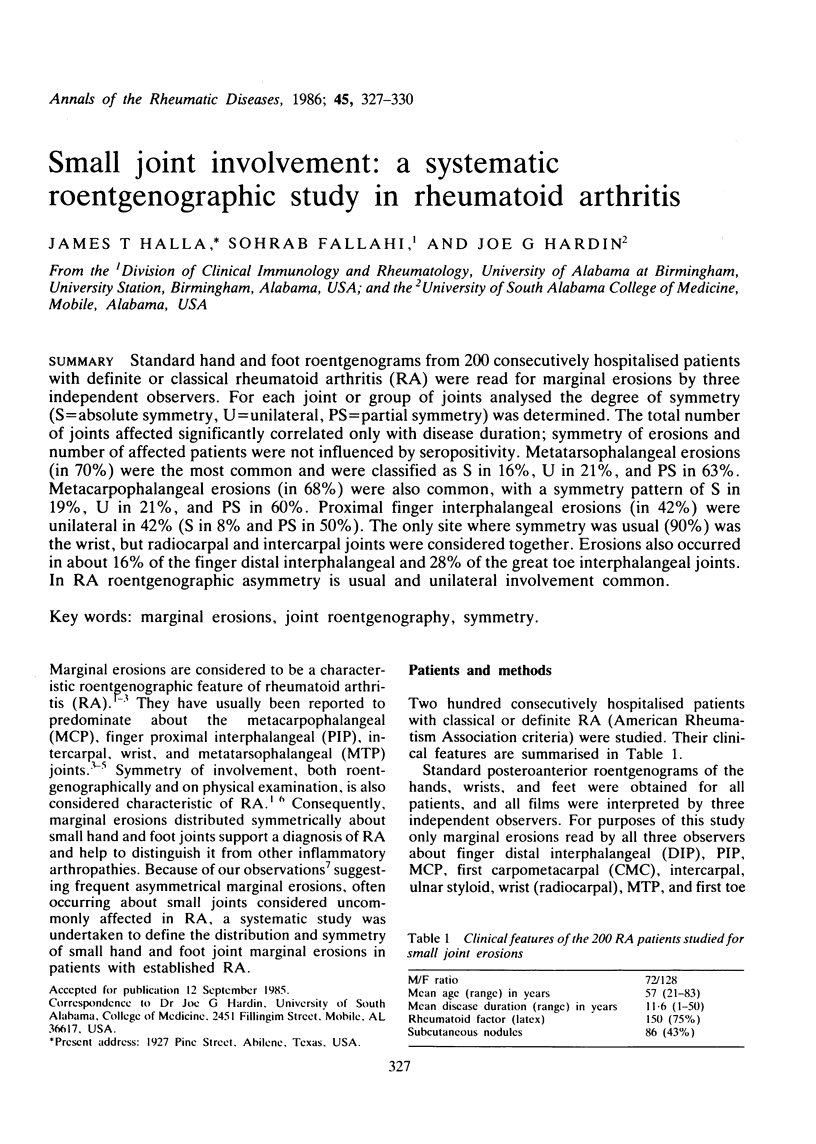
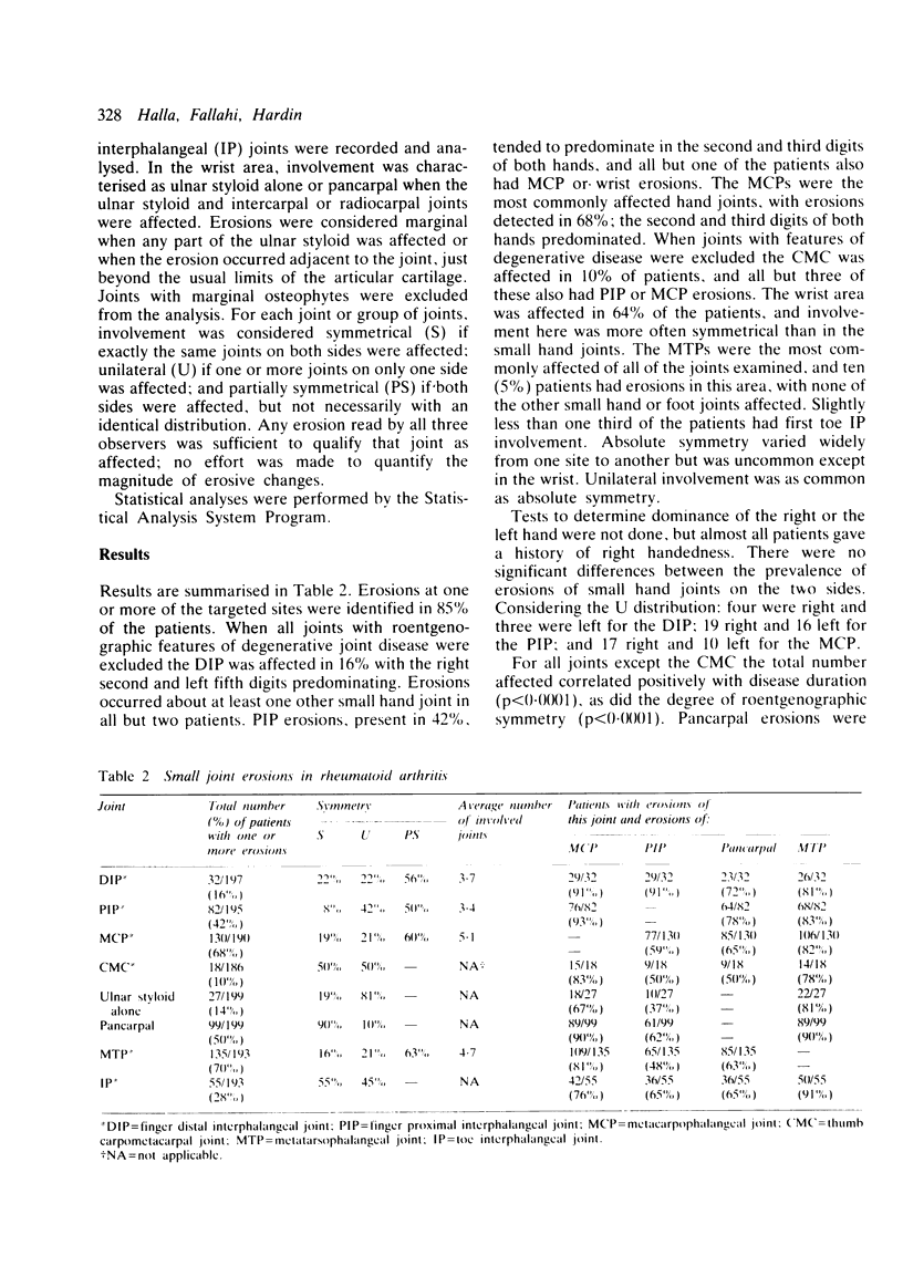
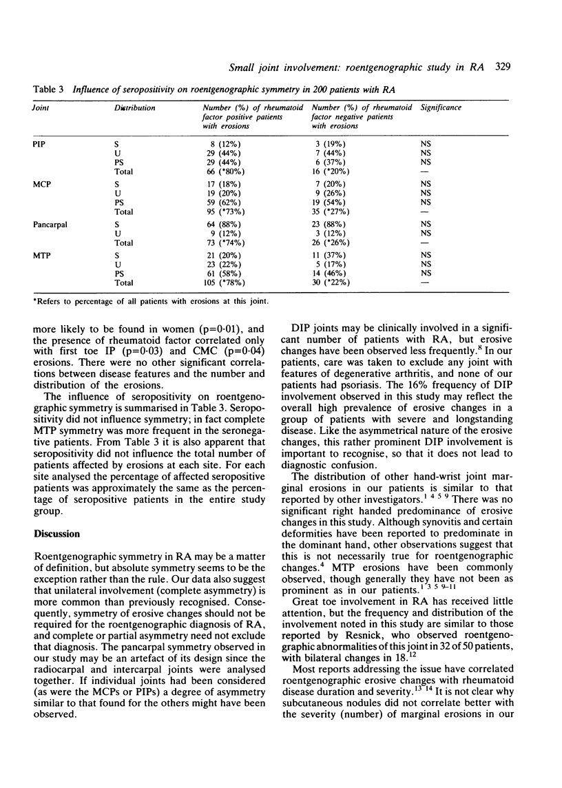
Standard hand and foot roentgenograms from 200 consecutively hospitalised patients with definite or classical rheumatoid arthritis (RA) were read for marginal erosions by three independent observers. For each joint or group of joints analysed the degree of symmetry (S = absolute symmetry, U = unilateral, PS = partial symmetry) was determined. The total number of joints affected significantly correlated only with disease duration; symmetry of erosions and number of affected patients were not influenced by seropositivity. Metatarsophalangeal erosions (in 70%) were the most common and were classified as S in 16%, U in 21%, and PS in 63%. Metacarpophalangeal erosions (in 68%) were also common, with a symmetry pattern of S in 19%, U in 21%, and PS in 60%. Proximal finger interphalangeal erosions (in 42%) were unilateral in 42% (S in 8% and PS in 50%). The only site where symmetry was usual (90%) was the wrist, but radiocarpal and intercarpal joints were considered together. Erosions also occurred in about 16% of the finger distal interphalangeal and 28% of the great toe interphalangeal joints. In RA roentgenographic asymmetry is usual and unilateral involvement common.
Full text
PDF


Selected References
These references are in PubMed. This may not be the complete list of references from this article.
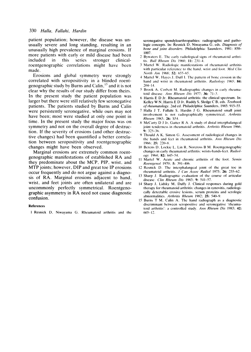
- BERENS D. L., LOCKIE L. M., LIN R. K., NORCROSS B. M. ROENTGEN CHANGES IN EARLY RHEUMATOID ARTHRITIS. WRISTS--HANDS--FEET. Radiology. 1964 Apr;82:645–654. doi: 10.1148/82.4.645. [DOI] [PubMed] [Google Scholar]
- Burns T. M., Calin A. The hand radiograph as a diagnostic discriminant between seropositive and seronegative 'rheumatoid arthritis': a controlled study. Ann Rheum Dis. 1983 Dec;42(6):605–612. doi: 10.1136/ard.42.6.605. [DOI] [PMC free article] [PubMed] [Google Scholar]
- Martel W. Radiologic manifestations of rheumatoid arthritis with particular reference to the hand, wrist and foot. Med Clin North Am. 1968 May;52(3):655–665. [PubMed] [Google Scholar]
- Resnick D. The interphalangeal joint of the great toe in rheumatoid arthritis. J Can Assoc Radiol. 1975 Dec;26(4):255–262. [PubMed] [Google Scholar]
- Sharp J. T., Lidsky M. D., Duffy J. Clinical responses during gold therapy for rheumatoid arthritis. Changes in synovitis, radiologically detectable erosive lesions, serum proteins, and serologic abnormalities. Arthritis Rheum. 1982 May;25(5):540–549. doi: 10.1002/art.1780250508. [DOI] [PubMed] [Google Scholar]
- Sharp J. T. Radiographic evaluation of the course of articular disease. Clin Rheum Dis. 1983 Dec;9(3):541–557. [PubMed] [Google Scholar]


