Abstract
Spontaneous rupture of the extensor pollicis longus is a rare injury. Distal radius fractures and rheumatisms are the principal causes, but there is an increase in cases related to some professional or sports activities. We report a case of a semi-professional bodybuilder who presented with a full loss of interphalangeal thumb extension and retropulsion following trivial trauma of the left wrist. Ultrasound confirmed the diagnosis of spontaneous rupture of extensor pollicis longus, and MRI revealed a very rare and aggressive anatomical variant of Lister's tubercle. The patient underwent a transfer of the extensor indicis propius, which is the most popular technique used for hyperextension and retropulsion restitution. Aggressive anatomical forms of Lister's tubercle can explain the frequent occurrence of spontaneous rupture of extensor pollicis longus in patients who practice sports or professions with a repetitive movement of the wrist.
Keywords: bodybuilder, lister tubercle, extensor policis longus, tendon reconstruction, sport injury
Introduction
Subcutaneous rupture of the extensor pollicis longus is a very rare injury [1]. Most often, it is a complication of a distal radius fracture, rheumatoid arthritis, and corticosteroid use, but currently, more cases are being reported in patients with a particular profession or practicing certain sports [2]. Surgical treatment is the gold standard, based either on tendon graft or tendon transfer.
Case presentation
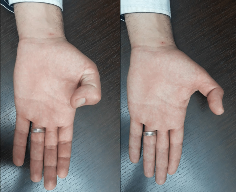
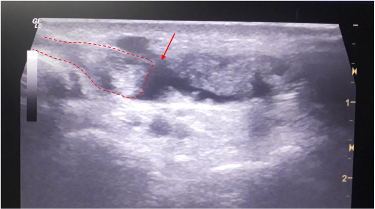
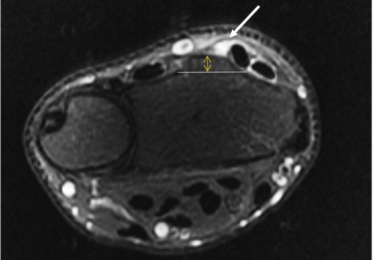
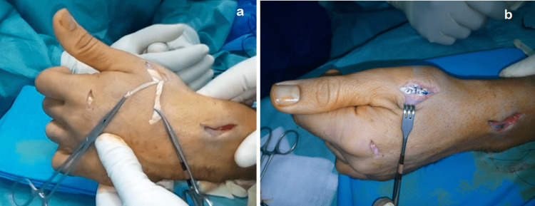
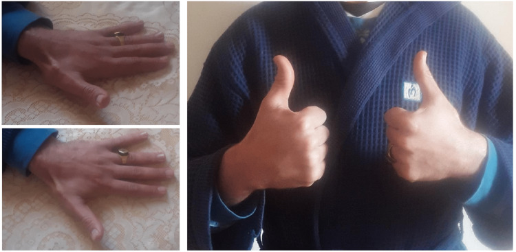
We report the case of a 27-year-old male, a semi-professional bodybuilder with no prior medical history. The patient experienced pain in the left wrist while trying to open a door, followed by an immediate loss of active hyper-extension in the thumb. The patient sought medical attention two weeks later and, upon examination, we found a mallet thumb with a loss of active interphalangeal hyper-extension and retropulsion (Figure 1). Standard radiography did not show any abnormalities. Ultrasound revealed a loss of continuity in the extensor pollicis longus at the wrist level (Figure 2), confirmed by MRI, in association with a 3B form of the Lister's tubercle anatomical variants classification as suggested by Chan et al. (Figure 3), which is the rarest and most aggressive form. The patient underwent transfer of the extensor indicis tendon (Figure 4), resulting in complete recovery of the interphalangeal hyper-extension of the thumb. The three-year evolution is very satisfactory (Figure 5).
Figure 1. Preoperative image showing mallet thumb and active retropulsion loss.
Figure 2. Ultrasound image showing discontinuous extensor pollicis longus in longitudinal scans (red dots) with hematoma (arrow).
Figure 3. Axial fat-saturation (FAT-SAT) image of the wrist level demonstrating a complete rupture of the extensor pollicis longus (arrow) with no radial peak of Lister's tubercle.
Figure 4. Peroperative images showing (a) the tensioning step before suturing the transferred extensor indicis proprius, (b) the pulvertaft suture of the extensor indicis proprius on the extensor pollicis longus.
Figure 5. Satisfactory recovery of extension and retropulsion of the thumb (results after three years).
Discussion
Subcutaneous ruptures of the extensor pollicis longus are very rare injuries [1]. Two theories can explain the mechanism of injury. The mechanical theory incriminates friction and tendon path around Lister's tubercle. Then the vascular theory explains that the risk of ischemia at this level is related to the increase in pressure in this inextensible osteofibrous tunnel, exacerbated by the poor vascular irrigation of the tendon in view of Lister's tubercle, making it even more vulnerable [2-4]. The most common etiologies are distal radius fractures, rheumatological disorders, corticosteroid use, and some sports activities (skiers, kick-boxers…) and professions involving repetitive forced wrist movements (cooking, cow milking…) [5-8]. Typically, the patient presents with a loss of active extension of the interphalangeal joint of the thumb for several days or weeks, following sometimes a history of pain at the radial edge of the wrist. The clinical examination finds a thumb in the swan's neck or, more rarely, in mallet deformity, a loss of active interphalangeal joint extension, with the impossibility of the thumb's retropulsion [9]. Ultrasound and MRI can locate the lesion and measure the gap between the tendon edges when possible. Chan et al. proposed a morphological classification in their study of the anatomical variations of Lister's tubercle. By studying 360 wrist MRIs, they have described three anatomical types of Lister's tubercle with two subtypes each. Type 1a is the most common and least aggressive, and 3b is the rarest and the most aggressive [10]. The management of spontaneous ruptures of extensor pollicis longus is always surgical. Two techniques are mostly used. The first is the longus palmaris tendon graft. This technique has the disadvantage of requiring that the graft passes inside the third compartment with a pulvertaft suture further away, to recover the retropulsion and avoid lateral sweeping of the new tendon in the absence of a pulley. The second and most used technique is the extensor indicis proprius transfer. This technique provides excellent results, and it offers the advantage of performing a single suture of a vascularized tendon, providing a course and strength similar to those of the native extensor pollicis longus [11,12].
Conclusions
The spontaneous rupture of the extensor pollicis longus is a rare injury. Some sports and professional activities requiring repetitive movement of the wrist are increasingly reported in the literature as an etiology of this injury. Lister's tubercle is probably the principal cause. Extensor indicis proprius tendon transfer is a simple technique that leads to excellent results.
The content published in Cureus is the result of clinical experience and/or research by independent individuals or organizations. Cureus is not responsible for the scientific accuracy or reliability of data or conclusions published herein. All content published within Cureus is intended only for educational, research and reference purposes. Additionally, articles published within Cureus should not be deemed a suitable substitute for the advice of a qualified health care professional. Do not disregard or avoid professional medical advice due to content published within Cureus.
The authors have declared that no competing interests exist.
Human Ethics
Consent was obtained or waived by all participants in this study
References
- 1.Spontaneous rupture of extensor pollicis longus tendon: clinical and occupational implications, treatment approaches and prognostic outcome in non-rheumatoid arthritis patients: a retrospective study. Al-Omari AA, Ar Altamimi A, ALuran E, Saleh AA, Alyafawee QM, Audat MZ, Bashaireh K. Open Access Rheumatol. 2020;12:47–54. doi: 10.2147/OARRR.S253583. [DOI] [PMC free article] [PubMed] [Google Scholar]
- 2.Rupture of the extensor pollicis longus tendon after fracture of the lower end of the radius: a clinical and microangiographic study. Engkvist O, Lundborg G. Hand. 1979;11:76–86. doi: 10.1016/s0072-968x(79)80015-7. [DOI] [PubMed] [Google Scholar]
- 3.Rupture of the extensor pollicis longus tendon: a study of aetiological factors. Björkman A, Jörgsholm P. Scand J Plast Reconstr Surg Hand Surg. 2004;38:32–35. doi: 10.1080/02844310310013046. [DOI] [PubMed] [Google Scholar]
- 4.Spontaneous rupture of the extensor pollicis longus tendon. Kim CH. Arch Plast Surg. 2012;39:680–682. doi: 10.5999/aps.2012.39.6.680. [DOI] [PMC free article] [PubMed] [Google Scholar]
- 5.Skiing-induced rupture of the extensor pollicis longus tendon: a report of three cases. Uemura T, Kazuki K, Hashimoto Y, Takaoka K. Clin J Sport Med. 2008;18:292–294. doi: 10.1097/JSM.0b013e31816a1c83. [DOI] [PubMed] [Google Scholar]
- 6.Spontaneous rupture of extensor pollicis longus tendon in a kick boxer. Lloyd TW, Tyler MP, Roberts AH. Br J Sports Med. 1998;32:178–179. doi: 10.1136/bjsm.32.2.178. [DOI] [PMC free article] [PubMed] [Google Scholar]
- 7.Spontaneous rupture of the extensor pollicis longus tendon in a tailor. Choi JC, Kim WS, Na HY, Lee YS, Song WS, Kim DH, Park TH. Clin Orthop Surg. 2011;3:167–169. doi: 10.4055/cios.2011.3.2.167. [DOI] [PMC free article] [PubMed] [Google Scholar]
- 8.Spontaneous rupture of the extensor pollicis longus tendon due to unusual etiology. Taş S, Balta S, Benlier E. https://doi.org/10.5152/balkanmedj.2013.9027. Balkan Med J. 2014;31:105–106. doi: 10.5152/balkanmedj.2013.9027. [DOI] [PMC free article] [PubMed] [Google Scholar]
- 9.Spontaneous rupture of the extensor pollicis longus in a break-dancer. Shifflett GD, Ek ET, Weiland AJ. https://www.ncbi.nlm.nih.gov/pmc/articles/PMC3899809/ Eplasty. 2014;14:0. [PMC free article] [PubMed] [Google Scholar]
- 10.Anatomical variants of Lister’s tubercle: a new morphological classification based on magnetic resonance imaging. Chan WY, Chong LR. https://doi.org/10.3348/kjr.2017.18.6.957. Korean J Radiol. 2017;18:957–963. doi: 10.3348/kjr.2017.18.6.957. [DOI] [PMC free article] [PubMed] [Google Scholar]
- 11.Palmaris longus tendon grafting for extensor pollicis longus tendon rupture bScrew tip after 20 years. Pal JN, Bera AK, Roy AN, Bari W. https://www.ncbi.nlm.nih.gov/pmc/articles/PMC5245929/ J Orthop Case Rep. 2016;6:25–27. doi: 10.13107/jocr.2250-0685.486. [DOI] [PMC free article] [PubMed] [Google Scholar]
- 12.Spontaneous rupture of the extensor pollicis longus tendon in a lacrosse player. Aruma JF, Herickhoff P, Taylor K, Seidenberg P. Phys Sportsmed. 2022;50:553–556. doi: 10.1080/00913847.2022.2093619. [DOI] [PubMed] [Google Scholar]


