Abstract
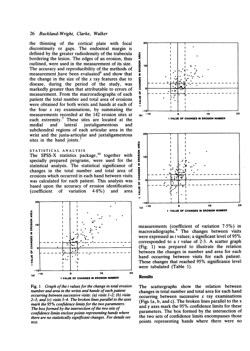
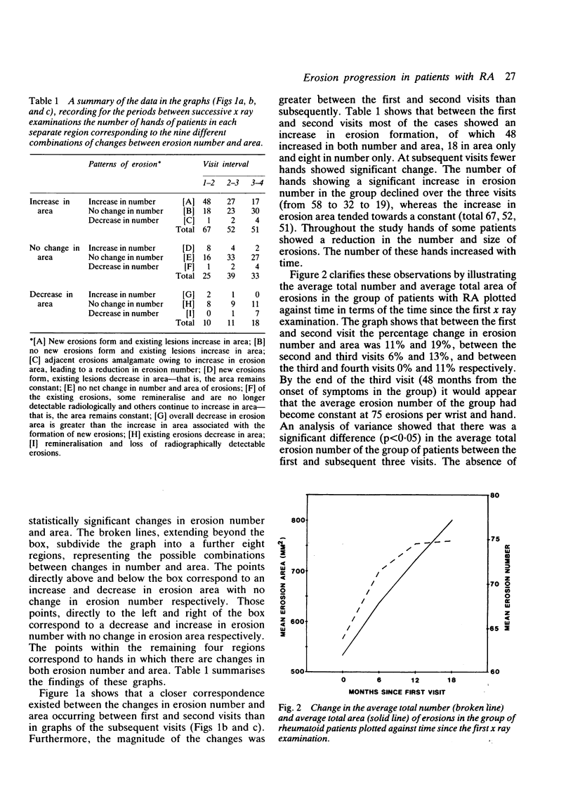
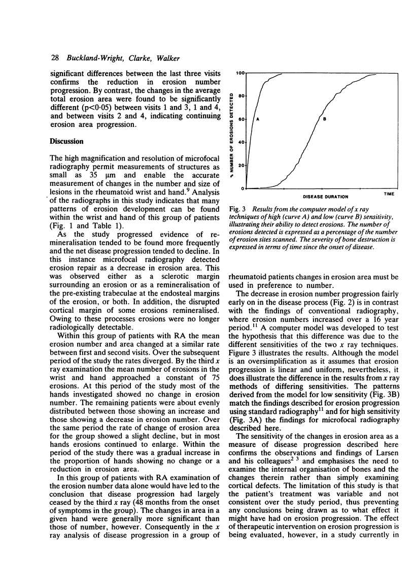
Microfocal radiography has been used to evaluate the relation between erosion number and erosion area in the hands and wrists of 51 patients with early to moderately advanced rheumatoid arthritis. The hands of these patients showed different patterns of erosion progression, in terms of the relation between changes in number and area, and included those showing a decrease in one or both of the erosion parameters. The mean number of erosions in the group increased between the first and second visits. By the third visit (a mean of 48 months from the onset of symptoms) the mean number of erosions in the wrist and hand of the group had approached a constant value of 75 erosions. Over the same period the mean erosion area of the group continued to increase. Measurement of changes in erosion area is a more sensitive indicator of erosion progression than erosion number, both within the group and in individual patients.
Full text
PDF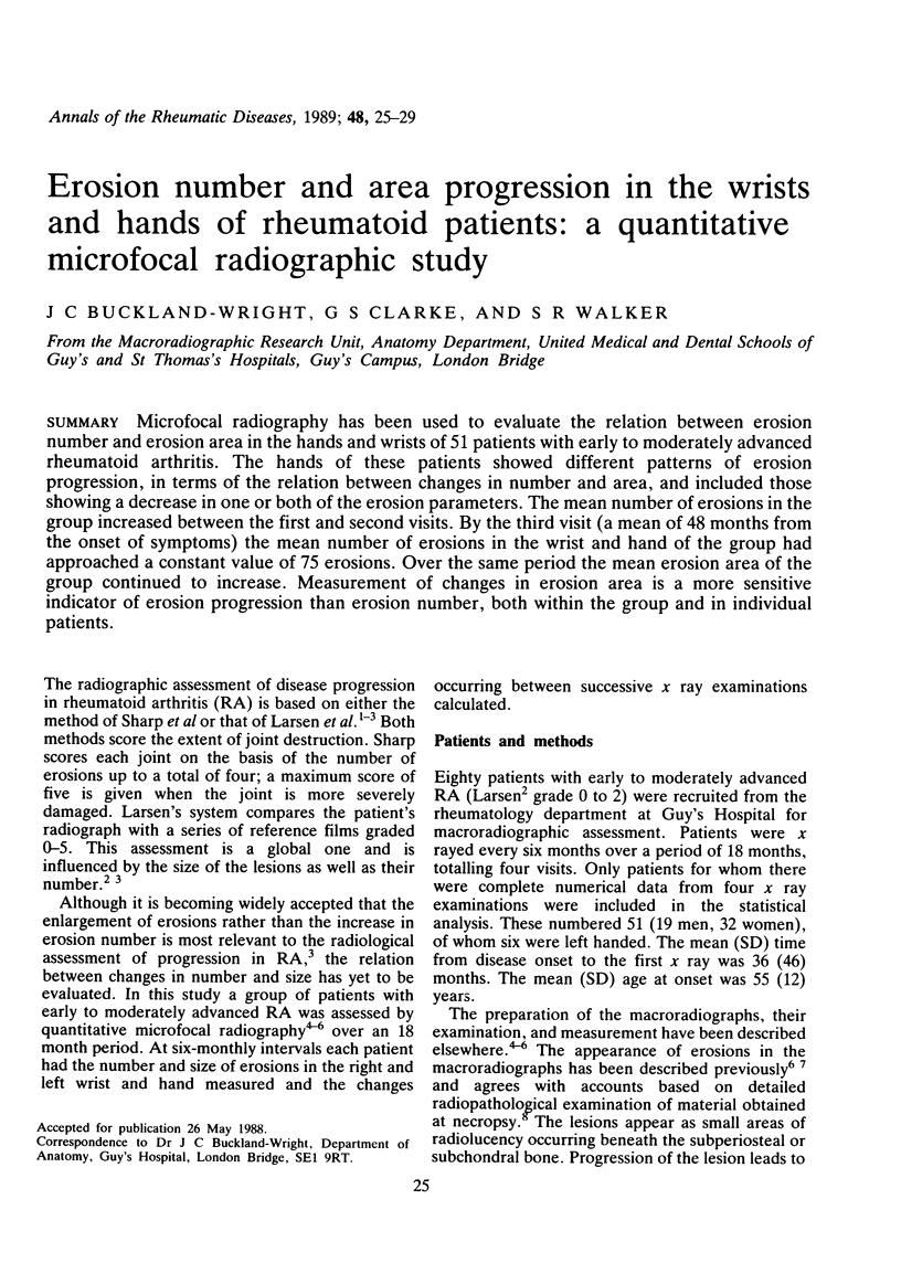
Selected References
These references are in PubMed. This may not be the complete list of references from this article.
- Buckland-Wright J. C. Advances in the radiological assessment of rheumatoid arthritis. Br J Rheumatol. 1983 Aug;22(3 Suppl):34–43. doi: 10.1093/rheumatology/xxii.suppl_1.34. [DOI] [PubMed] [Google Scholar]
- Buckland-Wright J. C., Carmichael I., Walker S. R. Quantitative microfocal radiography accurately detects joint changes in rheumatoid arthritis. Ann Rheum Dis. 1986 May;45(5):379–383. doi: 10.1136/ard.45.5.379. [DOI] [PMC free article] [PubMed] [Google Scholar]
- Buckland-Wright J. C. Microfocal radiographic examination of erosions in the wrist and hand of patients with rheumatoid arthritis. Ann Rheum Dis. 1984 Apr;43(2):160–171. doi: 10.1136/ard.43.2.160. [DOI] [PMC free article] [PubMed] [Google Scholar]
- Buckland-Wright J. C., Walker S. R. Incidence and size of erosions in the wrist and hand of rheumatoid patients: a quantitative microfocal radiographic study. Ann Rheum Dis. 1987 Jun;46(6):463–467. doi: 10.1136/ard.46.6.463. [DOI] [PMC free article] [PubMed] [Google Scholar]
- Buckland-Wright J. C. X-ray assessment of activity in rheumatoid disease. Br J Rheumatol. 1983 Feb;22(1):3–10. doi: 10.1093/rheumatology/22.1.3. [DOI] [PubMed] [Google Scholar]
- Larsen A., Dale K., Eek M. Radiographic evaluation of rheumatoid arthritis and related conditions by standard reference films. Acta Radiol Diagn (Stockh) 1977 Jul;18(4):481–491. doi: 10.1177/028418517701800415. [DOI] [PubMed] [Google Scholar]
- Sharp J. T., Lidsky M. D., Collins L. C., Moreland J. Methods of scoring the progression of radiologic changes in rheumatoid arthritis. Correlation of radiologic, clinical and laboratory abnormalities. Arthritis Rheum. 1971 Nov-Dec;14(6):706–720. doi: 10.1002/art.1780140605. [DOI] [PubMed] [Google Scholar]
- Sharp J. T. Radiographic evaluation of the course of articular disease. Clin Rheum Dis. 1983 Dec;9(3):541–557. [PubMed] [Google Scholar]


