Abstract
The condition of having a healthy, functional proteome is known as protein homeostasis, or proteostasis. Establishing and maintaining proteostasis is the province of the proteostasis network, approximately 2,700 components that regulate protein synthesis, folding, localization, and degradation. The proteostasis network is a fundamental entity in biology that is essential for cellular health and has direct relevance to many diseases of protein conformation. However, it is not well defined or annotated, which hinders its functional characterization in health and disease. In this series of manuscripts, we aim to operationally define the human proteostasis network by providing a comprehensive, annotated list of its components. We provided in a previous manuscript a list of chaperones and folding enzymes as well as the components that make up the machineries for protein synthesis, protein trafficking into and out of organelles, and organelle-specific degradation pathways. Here, we provide a curated list of 838 unique high-confidence components of the autophagy-lysosome pathway, one of the two major protein degradation systems in human cells.
Introduction
Proteostasis is the maintenance of the proteome in a healthy, functional state.1 Proteostasis is enabled by the proteostasis network, a collection of cellular components responsible for managing the synthesis, folding, trafficking, and degradation of proteins.1–5 Although the term “proteostasis network” was introduced over a decade ago,1 the network has remained poorly defined as there is no comprehensive accounting of its components. This problem is especially acute considering the important role of proteostasis in aging-related neurodegenerative diseases.6–11 To address this problem, we have been creating a detailed enumeration of the proteostasis network. The first installment of this list was reported recently and included the machinery of protein synthesis, chaperones, folding enzymes, the systems for trafficking proteins into and out of organelles, and organelle-specific degradation pathways.12 Here we report the next installment, a comprehensive list of the components of the autophagy-lysosome pathway (ALP). The ALP is one of two major systems for the degradation of proteins (the other being the ubiquitin-proteasome system, or UPS, which is the subject of an upcoming manuscript). In the ALP, the substrates to be degraded are surrounded by a double-membrane vesicle called the autophagosome, which is transported to and fuses with the lysosome for degradation.13–19 Within the proteostasis network, the ALP is of particular interest because mutations in many of its components have been found to increase the risk of diseases of failed proteostasis, like Alzheimer’s disease, frontotemporal dementia, Parkinson’s disease, and amyotrophic lateral sclerosis, among others.11,20
Results
Taxonomic system for annotating proteostasis network components, and criteria for inclusion
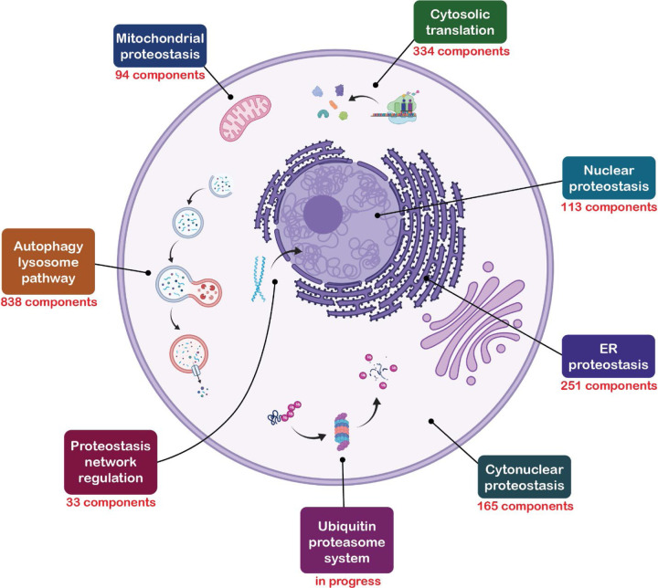
We previously introduced a taxonomic annotation system for the proteostasis network.12 This system was constructed to convey at a glance a component’s role in proteostasis. It consists of five levels: Branch, Class, Group, Type, and Subtype. The Branch annotation is the broadest and refers to a component’s localization or membership in an overarching pathway. There are eight Branches of the proteostasis network (PN; Figure 1), six of which we described previously (cytonuclear proteostasis, ER proteostasis, mitochondrial proteostasis, nuclear proteostasis, cytosolic translation, and proteostasis network regulation).12 The ALP is the seventh Branch of the proteostasis network, and the UPS is the eighth. The Class, Group, Type and Subtype annotations give increasingly specific descriptions of a component’s role in proteostasis. We endeavored to use as few descriptors as possible for each component. Thus, many components lack Type and Subtype annotations. Also, some components have multiple roles in proteostasis and therefore have multiple sets of annotations.
Figure 1.
The proteostasis network. The branches of the proteostasis network are called out in boxes. The number of unique components is shown for the ALP and the six branches described in our previous manuscript, from which this figure is adapted.12 Parts of this figure were created with BioRender.com.
We previously described two criteria that we employ to decide whether components should be included in the proteostasis network.12 The first is the “entity-based” criterion, in which entities (proteins, protein complexes, non-coding RNA, etc.) are included because there is experimental evidence for their having a role in proteostasis. The second is the “domain-based” criterion, in which components are included because they have at least one structural domain that is characteristic of protein families known to be involved in proteostasis. We primarily used the entity-based criterion applied to existing lists of autophagy components (for example, autophagy-related Gene Ontology annotations,21,22 the KEGG pathway “Autophagy - Animal”,23 and previous compilations of ALP components,24–26 especially the recent list by Bordi and co-workers27) to populate our initial list of ALP components. This initial list was then expanded through a comprehensive literature search as described in the Methods section. The domain-based criterion was also used in a few cases. For example, MAP1LC3B2 was included because it is a homolog of the yeast protein ATG8.28,29 ATG8 is central to the yeast ALP30,31 and six of its seven human homologs have also been shown to function in the ALP, but to our knowledge MAP1LC3B2 has not.28
While the scope of the PN naturally encompasses the fate of proteins, our enumeration extends slightly beyond the bounds of the protein world. For example, our description of selective autophagy includes all forms of it, including those by which biomolecules other than proteins are degraded, such as lipophagy.32 We also included all the catabolic enzymes of the lysosome in the ALP, not just those that catalyze the cleavage of peptide bonds, as well as those components involved in maintaining the acidic lysosomal environment. However, we did not include components that are generally involved in lysosomal or endosomal trafficking. We also did not include components associated only with non-canonical autophagy or with secretory autophagy.33–35 These pathways may be included in later versions of the ALP when their significance and mechanisms are better understood.
Application of the taxonomic scheme to the ALP
Our taxonomy of the ALP is underpinned by the temporal progression of autophagy, much of which was first elucidated by Ohsumi and co-workers.36 Autophagy is governed by signaling pathways that regulate flux through the ALP. These inputs—including, for example, nutrient, hormone, energy, and stress signals—are mostly communicated through mTORC1 (mammalian target of rapamycin complex 1) and AMPK (AMP-activated protein kinase), which are regulators of numerous key pathways in addition to autophagy, like cell metabolism and energy homeostasis.37–42 These kinases modulate autophagy through modification of the ULK complex (unc-51 like autophagy activating kinase), with mTORC1 and AMPK exerting inhibitory and activating effects, respectively, through phosphorylation at specific sites (for example, Ser757 of ULK1 by mTORC1 and Ser317/Ser777 of ULK1 by AMPK).38,41 When the ULK complex is activated it is recruited to the site where the autophagophore (as the nascent autophagosome is known) formation is initiated; sometimes, this is at a structure in the ER membrane known as the omegasome.43 There, the ULK complex activates the PI3KC3 complex 1 (class III phosphatidylinositol 3-kinase)44 which phosphorylates phosphatidylinositol (PI) at the 3 position to yield phosphatidylinositol 3-phosphate (PI(3)P).14,45–47 PI(3)P recruits the ATG2-WIPI complex (ATG = autophagy related; WIPI = WD repeat domain, phosphoinositide interacting), which rapidly transports lipids to the growing autophagophore.14,48,49 The ATG2-WIPI complex also recruits the ATG5-ATG12-ATG16 complex,50 which catalyzes the conjugation of phosphatidylethanolamine to human ATG8 orthologs like the MAP1LC3 or GABARAP proteins on the autophagophore membrane (MAP1LC3 = microtubule associated protein light chain 3; GABARAP = GABA type A receptor-associated protein).51–53 These lipidated proteins are critical components of the autophagosome,28,54–57 especially for substrate recognition.58–61
The autophagophore grows until it surrounds its substrate, whether that is a portion of cytosol, as in bulk macroautophagy, or other substrates such as protein aggregates or entire organelles, as in selective autophagy. The substrates of selective autophagy are bound by receptors that recognize substrates by various mechanisms (often involving ubiquitin59), and recognize autophagosomes via their displayed ATG8 homologs.58,60,61 The autophagophore is sealed with the assistance of ESCRT complexes (endosomal complexes required for transport), yielding an autophagosome.14,62 Autophagosomes are transported to through the cell19,63 to fuse with lysosomes, creating autolysosomes,15 in which the cargo from the autophagosome is degraded by lysosomal enzymes.64 Lysosomes are then regenerated from the autolysosomes by the process of autophagic lysosome reformation.65
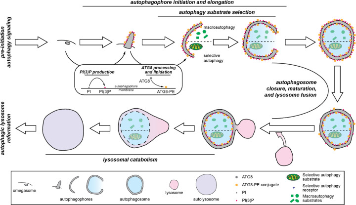
The Classes in the ALP Branch of the proteostasis network largely reflect the stages described above in the autophagy process and are illustrated in the schematic overview of the ALP in Figure 2. These Classes are “pre-initiation autophagy signaling”; “autophagophore initiation and elongation”; “autophagy substrate selection”; “autophagosome closure, maturation, and lysosome fusion”; “lysosomal catabolism”; and “autophagic lysosome reformation”. The remaining classes are “autophagy gene expression”, which comprises the transcription factors and transcriptional and translational regulators that control the expression of autophagy genes; “chaperone directed autophagy”, which includes components involved in chaperone-mediated autophagy66 and chaperone assisted selective autophagy67; and “specific function in autophagy unknown”, a small Class that contains components that clearly affect autophagy but through mechanisms that are not yet known. The Groups, Types, and Subtypes further specify a component’s function within the processes denoted by their Class.
Figure 2.
A schematic overview of the ALP. The temporal stages that correspond to Classes in the ALP are highlighted. Pre-initiation autophagy signaling: Flux through the ALP is regulated by signaling pathways that are upstream of autophagy initiation. Autophagophore initiation and elongation: Once autophagy is initiated, the autophagophore double membrane begins to grow. Initiation sometimes occurs at structures in the ER membrane known as omegasomes, but there are likely many mechanisms by which autophagophores can be nucleated. The growing autophagophore is decorated with PI(3)P (phosphatidylinositol 3-phosphate) and ATG8-PE (ATG8-phosphatidylethanolamine conjugate). Autophagy substrate selection: As the autophagophore grows, it encloses whatever components of the cytosol are in its vicinity (in macroautophagy) or components that are recruited via interactions between ATG8 and autophagy receptors (in selective autophagy). Autophagosome closure, maturation, and lysosome fusion: When it is large enough, the autophagophore membrane closes to form the autophagosome. As the autophagosome is transported in the cell, its membrane composition is modified in the process of autophagosome maturation, which readies it for fusion with the lysosome to form the autolysosome. Lysosomal catabolism: After lysosomal fusion, the autophagosome’s interior membrane and the cargo it contains are digested by lysosomal enzymes. Autophagic lysosome reformation: After lysosomal catabolism is complete, the components of the autolysosome are recycled to form new lysosomes.
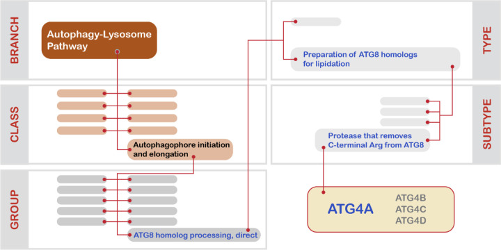
The application of our taxonomic scheme to an ALP component is illustrated in Figure 3 for ATG4A, a cysteine protease that trims the C-terminal Arg from ATG8 homologs in preparation for their conjugation to phosphatidylethanolamine.68,69 The following annotations were assigned to ATG4A: Branch = “autophagy-lysosome pathway”; Class = “autophagophore initiation and elongation”; Group = “ATG8 homolog processing, direct”; Type = “preparation of ATG8 homologs for lipidation”; Subtype = “peptidase that removes C-terminal Arg from ATG8”. It is important to note that these annotations are not unique, as they are shared with ATG4A’s paralogs in the human genome: ATG4B, ATG4C, and ATG4D.69 Also, like many other ALP components, ATG4A has two distinct sets of annotations because in addition to its role in preparing ATG8 homologs for lipidation, it is also responsible for delipidating ATG8 homologs and removing them from the autophagosome membrane as the autophagosome matures.68,70 The annotation for this role of ATG4A in the ALP is: Branch = “autophagy-lysosome pathway”; Class = “autophagosome closure, maturation, and lysosome fusion”; Group = “regulation of autophagosome membrane composition”; Type = “ATG8 homolog de-lipidation”; Subtype = “releases ATG8 homologs from maturing autophagosome”.
Figure 3.
Illustration of the five-level taxonomic scheme applied to proteostasis network components herein using the cysteine protease ATG4A. Blank boxes correspond to categories within each taxonomic level to which ATG4A does not belong. ATG4A has two annotations (see text), but here we illustrate only ATG4A’s annotation in the “autophagophore initiation and elongation” Class.
The autophagy-lysosome pathway consists of at least 838 unique protein-coding genes
The list of components of the ALP is presented in Supplemental Table 1 (see the Methods section for details on how the list was constructed). There are 985 entries in our list of ALP components, representing 838 unique components. Of these, 714 have a single entry, 108 have two, 11 have three, 4 have four, and 1 (SQSTM1, or sequestosome 1) has six. All the entries have been assigned to one of the nine Classes of the ALP Branch, and all but three have one of the 76 Group annotations. The three exceptions are those components in the “specific function in autophagy unknown” Class. Most of the entries have one of the 138 Type annotations (899 out of 985), but fewer than half of the entries have one of the 78 Subtype annotations (365 out of 985).
To illustrate how the finer-grained annotations were assigned in the ALP, we show how the components of the Class “autophagosome closure, maturation, and lysosome fusion” are distributed within this Class’s various Groups, Types, and Subtypes in Table 1.
Table 1.
Groups, Types and Subtypes within the “autophagosome closure, maturation, and lysosome fusion” Class.
| Group | Type | Subtype | Members |
|---|---|---|---|
| Sealing of autophagophore membrane | ESCRT-I complex component | – | 10 |
| ESCRT-III complex component | – | 11 | |
| ESCRT-III complex activity modulator | – | 4 | |
| Localization of the ESCRT-III complex | – | 3 | |
| Specific function in sealing of autophagophore membrane unknown | ATG8 homolog | 1 | |
| Specific function in sealing of autophagophore membrane unknown | – | 1 | |
| Localization of the autophagosome | Movement of autophagosomes along microtubules | ATG8 homolog | 1 |
| – | 7 | ||
| Movement of autophagosomes along actin | – | 4 | |
| Retrograde transport along axons | – | 2 | |
| Recruitment of autophagosome to vicinity of ER | – | 2 | |
| Regulation of autophagosome membrane composition | ATG8 homolog de-lipidation | Releases ATG8 homologs from maturing autophagosome | 4 |
| PIKFYVE complex component | – | 3 | |
| – | – | 3 | |
| Class 3 PI3K complex 2, direct | Class 3 PI3K complex 2 component | – | 7 |
| Modulator of class 3 PI3K complex 2 activity | – | 6 | |
| Autophagosome-endosome docking | Lysosome-endosome SNARE complex | – | 3 |
| Autophagosome-lysosome docking | Tethering factor recruitment | – | 10 |
| Lysosome-autophagosome SNARE complex | – | 6 | |
| Lysosome-autophagosome SNARE complex regulator | – | 7 | |
| BORC complex component | – | 8 | |
| HOPS complex component | – | 6 | |
| HOPS-BORC interaction mediator | – | 4 | |
| Recruitment of HOPS complex to autophagosome | ATG8 homolog | 6 | |
| ATG8 homolog phosphorylation | 2 | ||
| Bridges ATG8 and HOPS complex | 1 | ||
| Bridges STX17, RUBCNL and HOPS complex | 1 | ||
| Activator of PLECKHM1 | 2 | ||
| ATG8-LAMP1 interaction modulator | – | 1 | |
| ATG8-LAMP2 interaction modulator | – | 1 | |
| Lysosome-autophagosome interaction modulator | – | 13 | |
| Localization of the lysosome | – | – | 1 |
| Ca2+ efflux | – | – | 2 |
| Specific function in autophagosome maturation and lysosome fusion unknown | – | – | 12 |
Discussion
The ALP is much larger than any of the proteostasis network branches described in our preceding manuscript, consisting of over 800 components. The size of the ALP can be attributed both to the complexity of the physical processes involved—membrane nucleation and growth, substrate selection, vesicle trafficking and fusion—and to the extensive regulation of these processes. This regulation happens at all points in the ALP, but mTORC1,37,40 which regulates autophagy initiation, and PI3KC3 complex 1,71,72 which regulates the PI(3)P content of the autophagophore, are especially dense nodes for regulatory activity. This level of evolutionary investment into the control systems of the ALP shows how critical it is to tune the level of catabolism to the state of the cell. The significance of the ALP to cellular health is also manifested in the outsized representation of ALP mutations in many aging-related neurodegenerative diseases, as noted above.11,20 We expect that the list of ALP components described herein will enable a deeper understanding of the ways that failures of the ALP contribute to the pathogenesis of such diseases by enabling those studying them to see the full array of connections between, say, the results obtained from patient-derived multiomic datasets and the ALP. In this way, new targets could be discovered for diseases with known links to the ALP. Moreover, our comprehensive accounting of ALP components could reveal new roles for the ALP in diseases that are not currently associated with it, enabling the exploration of new therapeutic strategies.
Finally, we note that we view the list of annotations presented herein as a first version. We will regularly update our list of ALP components as new information becomes available. Suggestions for components that should be included or removed, or annotations that should be added, deleted, or changed can be sent to pnannotation@gmail.com.
Methods
Our “entity-based” and “domain-based” criteria for inclusion of components in the proteostasis network were described in the previous manuscript in this series.12 To generate the initial lists of ALP components we relied on the existing autophagy-related Gene Ontology annotations21,22 the KEGG pathway “Autophagy - Animal”,23 and previous compilations of ALP components24–27 supplemented with recent reviews of either autophagy in general or specific aspects of autophagy.11,13–20,28,37,58–61,63–66,73–76 The initial list consisted of ~730 components. We then explored the literature for genes with associations with autophagy that were either newly discovered or not recognized in the sources referenced above. We used the Gene database from NCBI77 to find papers related to each gene in the genome that had been published as of June 2021. The titles, abstracts, and MeSH terms for these papers were downloaded and scanned for the word fragment “-autopha-” to identify papers that could conceivably report a role for the gene of interest in autophagy. We selected ~420 genes for further evaluation that had four or more related papers in which the fragment “-autopha-” occurred and were not already in our preliminary list. The relevant abstracts (and the manuscripts themselves, as necessary) for each of these genes were read by members of the Consortium to determine if they should be considered as candidates for inclusion in the ALP. Candidate genes were proposed to the Consortium’s PN annotation subgroup and inclusion and exclusion decisions were made collectively by the subgroup. Approximately 110 components were added to the preliminary list through this process. Each ALP component entry in Supplemental Table 1 has a note justifying its inclusion in the ALP and explaining the annotation, as well as links to supporting literature, in the last few columns of the table.
Supplementary Material
Acknowledgements
We gratefully acknowledge funding from the National Institutes of Health (National Institute on Aging P01AG054407 to R. I. M., D. F., S. F., J. E. G., E. T. P., J. W. K., and J. F.; P01AG017617 and R01AG062376 to R. A. N.; K99AG068544 to T. S.; and K99AG080109 to E. P. T.)
References
- 1.Balch W.E., Morimoto R.I., Dillin A., and Kelly J.W. (2008). Adapting proteostasis for disease intervention. Science 319, 916–919. 10.1126/science.1141448. [DOI] [PubMed] [Google Scholar]
- 2.Jayaraj G.G., Hipp M.S., and Hartl F.U. (2020). Functional Modules of the Proteostasis Network. Cold Spring Harb Perspect Biol 12. 10.1101/cshperspect.a033951. [DOI] [PMC free article] [PubMed] [Google Scholar]
- 3.Powers E.T., and Gierasch L.M. (2021). The Proteome Folding Problem and Cellular Proteostasis. J Mol Biol 433, 167197. 10.1016/j.jmb.2021.167197. [DOI] [PMC free article] [PubMed] [Google Scholar]
- 4.Powers E.T., Morimoto R.I., Dillin A., Kelly J.W., and Balch W.E. (2009). Biological and chemical approaches to diseases of proteostasis deficiency. Annu Rev Biochem 78, 959–991. 10.1146/annurev.biochem.052308.114844. [DOI] [PubMed] [Google Scholar]
- 5.Sala A.J., Bott L.C., and Morimoto R.I. (2017). Shaping proteostasis at the cellular, tissue, and organismal level. J Cell Biol 216, 1231–1241. 10.1083/jcb.201612111. [DOI] [PMC free article] [PubMed] [Google Scholar]
- 6.Hipp M.S., Kasturi P., and Hartl F.U. (2019). The proteostasis network and its decline in ageing. Nat Rev Mol Cell Biol 20, 421–435. 10.1038/s41580-019-0101-y. [DOI] [PubMed] [Google Scholar]
- 7.Kaushik S., and Cuervo A.M. (2015). Proteostasis and aging. Nat Med 21, 1406–1415. 10.1038/nm.4001. [DOI] [PubMed] [Google Scholar]
- 8.Labbadia J., and Morimoto R.I. (2015). The biology of proteostasis in aging and disease. Annu Rev Biochem 84, 435–464. 10.1146/annurev-biochem-060614-033955. [DOI] [PMC free article] [PubMed] [Google Scholar]
- 9.Lopez-Otin C., Blasco M.A., Partridge L., Serrano M., and Kroemer G. (2013). The hallmarks of aging. Cell 153, 1194–1217. 10.1016/j.cell.2013.05.039. [DOI] [PMC free article] [PubMed] [Google Scholar]
- 10.Sonninen T.M., Goldsteins G., Laham-Karam N., Koistinaho J., and Lehtonen S. (2020). Proteostasis Disturbances and Inflammation in Neurodegenerative Diseases. Cells 9. 10.3390/cells9102183. [DOI] [PMC free article] [PubMed] [Google Scholar]
- 11.Finkbeiner S. (2020). The Autophagy Lysosomal Pathway and Neurodegeneration. Cold Spring Harb Perspect Biol 12. 10.1101/cshperspect.a033993. [DOI] [PMC free article] [PubMed] [Google Scholar]
- 12.Consortium P. (2022). A Comprehensive Enumeration of the Human Proteostasis Network. 1. Components of Translation, Protein Folding, and Organelle-Specific Systems. bioRxiv, 2022.2008.2030.505920. 10.1101/2022.08.30.505920. [DOI]
- 13.Bento C.F., Renna M., Ghislat G., Puri C., Ashkenazi A., Vicinanza M., Menzies F.M., and Rubinsztein D.C. (2016). Mammalian Autophagy: How Does It Work? Annu Rev Biochem 85, 685–713. 10.1146/annurev-biochem-060815-014556. [DOI] [PubMed] [Google Scholar]
- 14.Chang C., Jensen L.E., and Hurley J.H. (2021). Autophagosome biogenesis comes out of the black box. Nat Cell Biol 23, 450–456. 10.1038/s41556-021-00669-y. [DOI] [PMC free article] [PubMed] [Google Scholar]
- 15.Lorincz P., and Juhasz G. (2020). Autophagosome-Lysosome Fusion. J Mol Biol 432, 2462–2482. 10.1016/j.jmb.2019.10.028. [DOI] [PubMed] [Google Scholar]
- 16.Mizushima N., and Komatsu M. (2011). Autophagy: renovation of cells and tissues. Cell 147, 728–741. 10.1016/j.cell.2011.10.026. [DOI] [PubMed] [Google Scholar]
- 17.Reggiori F., and Ungermann C. (2017). Autophagosome Maturation and Fusion. J Mol Biol 429, 486–496. 10.1016/j.jmb.2017.01.002. [DOI] [PubMed] [Google Scholar]
- 18.Yu L., Chen Y., and Tooze S.A. (2018). Autophagy pathway: Cellular and molecular mechanisms. Autophagy 14, 207–215. 10.1080/15548627.2017.1378838. [DOI] [PMC free article] [PubMed] [Google Scholar]
- 19.Zhao Y.G., and Zhang H. (2019). Autophagosome maturation: An epic journey from the ER to lysosomes. J Cell Biol 218, 757–770. 10.1083/jcb.201810099. [DOI] [PMC free article] [PubMed] [Google Scholar]
- 20.Choi A.M., Ryter S.W., and Levine B. (2013). Autophagy in human health and disease. N Engl J Med 368, 651–662. 10.1056/NEJMra1205406. [DOI] [PubMed] [Google Scholar]
- 21.Denny P., Feuermann M., Hill D.P., Lovering R.C., Plun-Favreau H., and Roncaglia P. (2018). Exploring autophagy with Gene Ontology. Autophagy 14, 419–436. 10.1080/15548627.2017.1415189. [DOI] [PMC free article] [PubMed] [Google Scholar]
- 22.Consortium G.O. (2021). The Gene Ontology resource: enriching a GOld mine. Nucleic Acids Res 49, D325–D334. 10.1093/nar/gkaa1113. [DOI] [PMC free article] [PubMed] [Google Scholar]
- 23.Kanehisa M., Sato Y., Kawashima M., Furumichi M., and Tanabe M. (2016). KEGG as a reference resource for gene and protein annotation. Nucleic Acids Res 44, D457–462. 10.1093/nar/gkv1070. [DOI] [PMC free article] [PubMed] [Google Scholar]
- 24.Di Fruscio G., Schulz A., De Cegli R., Savarese M., Mutarelli M., Parenti G., Banfi S., Braulke T., Nigro V., and Ballabio A. (2015). Lysoplex: An efficient toolkit to detect DNA sequence variations in the autophagy-lysosomal pathway. Autophagy 11, 928–938. 10.1080/15548627.2015.1043077. [DOI] [PMC free article] [PubMed] [Google Scholar]
- 25.Wang N.N., Dong J., Zhang L., Ouyang D., Cheng Y., Chen A.F., Lu A.P., and Cao D.S. (2018). HAMdb: a database of human autophagy modulators with specific pathway and disease information. J Cheminform 10, 34. 10.1186/s13321-018-0289-4. [DOI] [PMC free article] [PubMed] [Google Scholar]
- 26.Deng W., Ma L., Zhang Y., Zhou J., Wang Y., Liu Z., and Xue Y. (2018). THANATOS: an integrative data resource of proteins and post-translational modifications in the regulation of autophagy. Autophagy 14, 296–310. 10.1080/15548627.2017.1402990. [DOI] [PMC free article] [PubMed] [Google Scholar]
- 27.Bordi M., De Cegli R., Testa B., Nixon R.A., Ballabio A., and Cecconi F. (2021). A gene toolbox for monitoring autophagy transcription. Cell Death Dis 12, 1044. 10.1038/s41419-021-04121-9. [DOI] [PMC free article] [PubMed] [Google Scholar]
- 28.Schaaf M.B., Keulers T.G., Vooijs M.A., and Rouschop K.M. (2016). LC3/GABARAP family proteins: autophagy-(un)related functions. FASEB J 30, 3961–3978. 10.1096/fj.201600698R. [DOI] [PubMed] [Google Scholar]
- 29.Shpilka T., Weidberg H., Pietrokovski S., and Elazar Z. (2011). Atg8: an autophagy-related ubiquitin-like protein family. Genome Biol 12, 226. 10.1186/gb-2011-12-7-226. [DOI] [PMC free article] [PubMed] [Google Scholar]
- 30.Kirisako T., Baba M., Ishihara N., Miyazawa K., Ohsumi M., Yoshimori T., Noda T., and Ohsumi Y. (1999). Formation process of autophagosome is traced with Apg8/Aut7p in yeast. J Cell Biol 147, 435–446. 10.1083/jcb.147.2.435. [DOI] [PMC free article] [PubMed] [Google Scholar]
- 31.Nakatogawa H., Ichimura Y., and Ohsumi Y. (2007). Atg8, a ubiquitin-like protein required for autophagosome formation, mediates membrane tethering and hemifusion. Cell 130, 165–178. 10.1016/j.cell.2007.05.021. [DOI] [PubMed] [Google Scholar]
- 32.Zechner R., Madeo F., and Kratky D. (2017). Cytosolic lipolysis and lipophagy: two sides of the same coin. Nat Rev Mol Cell Biol 18, 671–684. 10.1038/nrm.2017.76. [DOI] [PubMed] [Google Scholar]
- 33.Hooper K.M., Jacquin E., Li T., Goodwin J.M., Brumell J.H., Durgan J., and Florey O. (2022). V-ATPase is a universal regulator of LC3-associated phagocytosis and non-canonical autophagy. J Cell Biol 221. 10.1083/jcb.202105112. [DOI] [PMC free article] [PubMed] [Google Scholar]
- 34.Codogno P., Mehrpour M., and Proikas-Cezanne T. (2011). Canonical and non-canonical autophagy: variations on a common theme of self-eating? Nat Rev Mol Cell Biol 13, 7–12. 10.1038/nrm3249. [DOI] [PubMed] [Google Scholar]
- 35.Ponpuak M., Mandell M.A., Kimura T., Chauhan S., Cleyrat C., and Deretic V. (2015). Secretory autophagy. Curr Opin Cell Biol 35, 106–116. 10.1016/j.ceb.2015.04.016. [DOI] [PMC free article] [PubMed] [Google Scholar]
- 36.Ohsumi Y. (2014). Historical landmarks of autophagy research. Cell Res 24, 9–23. 10.1038/cr.2013.169. [DOI] [PMC free article] [PubMed] [Google Scholar]
- 37.Dunlop E.A., and Tee A.R. (2014). mTOR and autophagy: a dynamic relationship governed by nutrients and energy. Semin Cell Dev Biol 36, 121–129. 10.1016/j.semcdb.2014.08.006. [DOI] [PubMed] [Google Scholar]
- 38.Egan D.F., Shackelford D.B., Mihaylova M.M., Gelino S., Kohnz R.A., Mair W., Vasquez D.S., Joshi A., Gwinn D.M., Taylor R., et al. (2011). Phosphorylation of ULK1 (hATG1) by AMP-activated protein kinase connects energy sensing to mitophagy. Science 331, 456–461. 10.1126/science.1196371. [DOI] [PMC free article] [PubMed] [Google Scholar]
- 39.Galluzzi L., Pietrocola F., Levine B., and Kroemer G. (2014). Metabolic control of autophagy. Cell 159, 1263–1276. 10.1016/j.cell.2014.11.006. [DOI] [PMC free article] [PubMed] [Google Scholar]
- 40.Jung C.H., Ro S.H., Cao J., Otto N.M., and Kim D.H. (2010). mTOR regulation of autophagy. FEBS Lett 584, 1287–1295. 10.1016/j.febslet.2010.01.017. [DOI] [PMC free article] [PubMed] [Google Scholar]
- 41.Kim J., Kundu M., Viollet B., and Guan K.L. (2011). AMPK and mTOR regulate autophagy through direct phosphorylation of Ulk1. Nat Cell Biol 13, 132–141. 10.1038/ncb2152. [DOI] [PMC free article] [PubMed] [Google Scholar]
- 42.Tamargo-Gomez I., and Marino G. (2018). AMPK: Regulation of Metabolic Dynamics in the Context of Autophagy. Int J Mol Sci 19. 10.3390/ijms19123812. [DOI] [PMC free article] [PubMed] [Google Scholar]
- 43.Ktistakis N.T., and Tooze S.A. (2016). Digesting the Expanding Mechanisms of Autophagy. Trends Cell Biol 26, 624–635. 10.1016/j.tcb.2016.03.006. [DOI] [PubMed] [Google Scholar]
- 44.Russell R.C., Tian Y., Yuan H., Park H.W., Chang Y.Y., Kim J., Kim H., Neufeld T.P., Dillin A., and Guan K.L. (2013). ULK1 induces autophagy by phosphorylating Beclin-1 and activating VPS34 lipid kinase. Nat Cell Biol 15, 741–750. 10.1038/ncb2757. [DOI] [PMC free article] [PubMed] [Google Scholar]
- 45.Backer J.M. (2008). The regulation and function of Class III PI3Ks: novel roles for Vps34. Biochem J 410, 1–17. 10.1042/BJ20071427. [DOI] [PubMed] [Google Scholar]
- 46.Nascimbeni A.C., Codogno P., and Morel E. (2017). Phosphatidylinositol-3-phosphate in the regulation of autophagy membrane dynamics. FEBS J 284, 1267–1278. 10.1111/febs.13987. [DOI] [PubMed] [Google Scholar]
- 47.Petiot A., Ogier-Denis E., Blommaart E.F., Meijer A.J., and Codogno P. (2000). Distinct classes of phosphatidylinositol 3’-kinases are involved in signaling pathways that control macroautophagy in HT-29 cells. J Biol Chem 275, 992–998. 10.1074/jbc.275.2.992. [DOI] [PubMed] [Google Scholar]
- 48.Valverde D.P., Yu S., Boggavarapu V., Kumar N., Lees J.A., Walz T., Reinisch K.M., and Melia T.J. (2019). ATG2 transports lipids to promote autophagosome biogenesis. J Cell Biol 218, 1787–1798. 10.1083/jcb.201811139. [DOI] [PMC free article] [PubMed] [Google Scholar]
- 49.Maeda S., Otomo C., and Otomo T. (2019). The autophagic membrane tether ATG2A transfers lipids between membranes. Elife 8. 10.7554/eLife.45777. [DOI] [PMC free article] [PubMed] [Google Scholar]
- 50.Dooley H.C., Razi M., Polson H.E., Girardin S.E., Wilson M.I., and Tooze S.A. (2014). WIPI2 links LC3 conjugation with PI3P, autophagosome formation, and pathogen clearance by recruiting Atg12-5-16L1. Mol Cell 55, 238–252. 10.1016/j.molcel.2014.05.021. [DOI] [PMC free article] [PubMed] [Google Scholar]
- 51.Hanada T., Noda N.N., Satomi Y., Ichimura Y., Fujioka Y., Takao T., Inagaki F., and Ohsumi Y. (2007). The Atg12-Atg5 conjugate has a novel E3-like activity for protein lipidation in autophagy. J Biol Chem 282, 37298–37302. 10.1074/jbc.C700195200. [DOI] [PubMed] [Google Scholar]
- 52.Mizushima N., Yamamoto A., Hatano M., Kobayashi Y., Kabeya Y., Suzuki K., Tokuhisa T., Ohsumi Y., and Yoshimori T. (2001). Dissection of autophagosome formation using Apg5-deficient mouse embryonic stem cells. J Cell Biol 152, 657–668. 10.1083/jcb.152.4.657. [DOI] [PMC free article] [PubMed] [Google Scholar]
- 53.Mizushima N., Sugita H., Yoshimori T., and Ohsumi Y. (1998). A new protein conjugation system in human. The counterpart of the yeast Apg12p conjugation system essential for autophagy. J Biol Chem 273, 33889–33892. 10.1074/jbc.273.51.33889. [DOI] [PubMed] [Google Scholar]
- 54.Kabeya Y., Mizushima N., Ueno T., Yamamoto A., Kirisako T., Noda T., Kominami E., Ohsumi Y., and Yoshimori T. (2000). LC3, a mammalian homologue of yeast Apg8p, is localized in autophagosome membranes after processing. EMBO J 19, 5720–5728. 10.1093/emboj/19.21.5720. [DOI] [PMC free article] [PubMed] [Google Scholar]
- 55.Weidberg H., Shvets E., Shpilka T., Shimron F., Shinder V., and Elazar Z. (2010). LC3 and GATE-16/GABARAP subfamilies are both essential yet act differently in autophagosome biogenesis. EMBO J 29, 1792–1802. 10.1038/emboj.2010.74. [DOI] [PMC free article] [PubMed] [Google Scholar]
- 56.Xie Z., Nair U., and Klionsky D.J. (2008). Atg8 controls phagophore expansion during autophagosome formation. Mol Biol Cell 19, 3290–3298. 10.1091/mbc.e07-12-1292. [DOI] [PMC free article] [PubMed] [Google Scholar]
- 57.Kabeya Y., Mizushima N., Yamamoto A., Oshitani-Okamoto S., Ohsumi Y., and Yoshimori T. (2004). LC3, GABARAP and GATE16 localize to autophagosomal membrane depending on form-II formation. J Cell Sci 117, 2805–2812. 10.1242/jcs.01131. [DOI] [PubMed] [Google Scholar]
- 58.Johansen T., and Lamark T. (2020). Selective Autophagy: ATG8 Family Proteins, LIR Motifs and Cargo Receptors. J Mol Biol 432, 80–103. 10.1016/j.jmb.2019.07.016. [DOI] [PubMed] [Google Scholar]
- 59.Khaminets A., Behl C., and Dikic I. (2016). Ubiquitin-Dependent And Independent Signals In Selective Autophagy. Trends Cell Biol 26, 6–16. 10.1016/j.tcb.2015.08.010. [DOI] [PubMed] [Google Scholar]
- 60.Stolz A., Ernst A., and Dikic I. (2014). Cargo recognition and trafficking in selective autophagy. Nat Cell Biol 16, 495–501. 10.1038/ncb2979. [DOI] [PubMed] [Google Scholar]
- 61.Kirkin V., and Rogov V.V. (2019). A Diversity of Selective Autophagy Receptors Determines the Specificity of the Autophagy Pathway. Mol Cell 76, 268–285. 10.1016/j.molcel.2019.09.005. [DOI] [PubMed] [Google Scholar]
- 62.Vietri M., Radulovic M., and Stenmark H. (2020). The many functions of ESCRTs. Nat Rev Mol Cell Biol 21, 25–42. 10.1038/s41580-019-0177-4. [DOI] [PubMed] [Google Scholar]
- 63.Soreng K., Neufeld T.P., and Simonsen A. (2018). Membrane Trafficking in Autophagy. Int Rev Cell Mol Biol 336, 1–92. 10.1016/bs.ircmb.2017.07.001. [DOI] [PubMed] [Google Scholar]
- 64.Yim W.W., and Mizushima N. (2020). Lysosome biology in autophagy. Cell Discov 6, 6. 10.1038/s41421-020-0141-7. [DOI] [PMC free article] [PubMed] [Google Scholar]
- 65.Nanayakkara R., Gurung R., Rodgers S.J., Eramo M.J., Ramm G., Mitchell C.A., and McGrath M.J. (2022). Autophagic lysosome reformation in health and disease. Autophagy, 1–18. 10.1080/15548627.2022.2128019. [DOI] [PMC free article] [PubMed]
- 66.Kaushik S., and Cuervo A.M. (2018). The coming of age of chaperone-mediated autophagy. Nat Rev Mol Cell Biol 19, 365–381. 10.1038/s41580-018-0001-6. [DOI] [PMC free article] [PubMed] [Google Scholar]
- 67.Tedesco B., Vendredy L., Timmerman V., and Poletti A. (2023). The chaperone-assisted selective autophagy complex dynamics and dysfunctions. Autophagy, 1–23. 10.1080/15548627.2022.2160564. [DOI] [PMC free article] [PubMed]
- 68.Kirisako T., Ichimura Y., Okada H., Kabeya Y., Mizushima N., Yoshimori T., Ohsumi M., Takao T., Noda T., and Ohsumi Y. (2000). The reversible modification regulates the membrane-binding state of Apg8/Aut7 essential for autophagy and the cytoplasm to vacuole targeting pathway. J Cell Biol 151, 263–276. 10.1083/jcb.151.2.263. [DOI] [PMC free article] [PubMed] [Google Scholar]
- 69.Agrotis A., Pengo N., Burden J.J., and Ketteler R. (2019). Redundancy of human ATG4 protease isoforms in autophagy and LC3/GABARAP processing revealed in cells. Autophagy 15, 976–997. 10.1080/15548627.2019.1569925. [DOI] [PMC free article] [PubMed] [Google Scholar]
- 70.Kauffman K.J., Yu S., Jin J., Mugo B., Nguyen N., O’Brien A., Nag S., Lystad A.H., and Melia T.J. (2018). Delipidation of mammalian Atg8-family proteins by each of the four ATG4 proteases. Autophagy 14, 992–1010. 10.1080/15548627.2018.1437341. [DOI] [PMC free article] [PubMed] [Google Scholar]
- 71.Backer J.M. (2016). The intricate regulation and complex functions of the Class III phosphoinositide 3-kinase Vps34. Biochem J 473, 2251–2271. 10.1042/BCJ20160170. [DOI] [PubMed] [Google Scholar]
- 72.Kang R., Zeh H.J., Lotze M.T., and Tang D. (2011). The Beclin 1 network regulates autophagy and apoptosis. Cell Death Differ 18, 571–580. 10.1038/cdd.2010.191. [DOI] [PMC free article] [PubMed] [Google Scholar]
- 73.Jiang W., Chen X., Ji C., Zhang W., Song J., Li J., and Wang J. (2021). Key Regulators of Autophagosome Closure. Cells 10. 10.3390/cells10112814. [DOI] [PMC free article] [PubMed] [Google Scholar]
- 74.Behrends C., Sowa M.E., Gygi S.P., and Harper J.W. (2010). Network organization of the human autophagy system. Nature 466, 68–76. 10.1038/nature09204. [DOI] [PMC free article] [PubMed] [Google Scholar]
- 75.Remec Pavlin M., and Hurley J.H. (2020). The ESCRTs - converging on mechanism. J Cell Sci 133. 10.1242/jcs.240333. [DOI] [PMC free article] [PubMed] [Google Scholar]
- 76.Fullgrabe J., Klionsky D.J., and Joseph B. (2014). The return of the nucleus: transcriptional and epigenetic control of autophagy. Nat Rev Mol Cell Biol 15, 65–74. 10.1038/nrm3716. [DOI] [PubMed] [Google Scholar]
- 77.Brown G.R., Hem V., Katz K.S., Ovetsky M., Wallin C., Ermolaeva O., Tolstoy I., Tatusova T., Pruitt K.D., Maglott D.R., and Murphy T.D. (2015). Gene: a gene-centered information resource at NCBI. Nucleic Acids Res 43, D36–42. 10.1093/nar/gku1055. [DOI] [PMC free article] [PubMed] [Google Scholar]
Associated Data
This section collects any data citations, data availability statements, or supplementary materials included in this article.


