Abstract
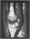
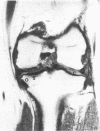
To date, MRI has primarily been used to study anatomical changes, and at a resolution that makes detailed analysis of focal change difficult. This is primarily because cost limits the development and use of tailor made research systems. The detailed analysis of soft tissue, cartilage, and bone marrow images should provide a fruitful non-invasive method to study OA. However, the development of MRI methods to study movement, diffusion and perfusion, and the spatial localisation of spectroscopic information, promises a revolution in the study of the living joint in man.
Full text
PDF
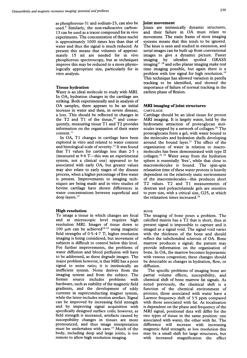
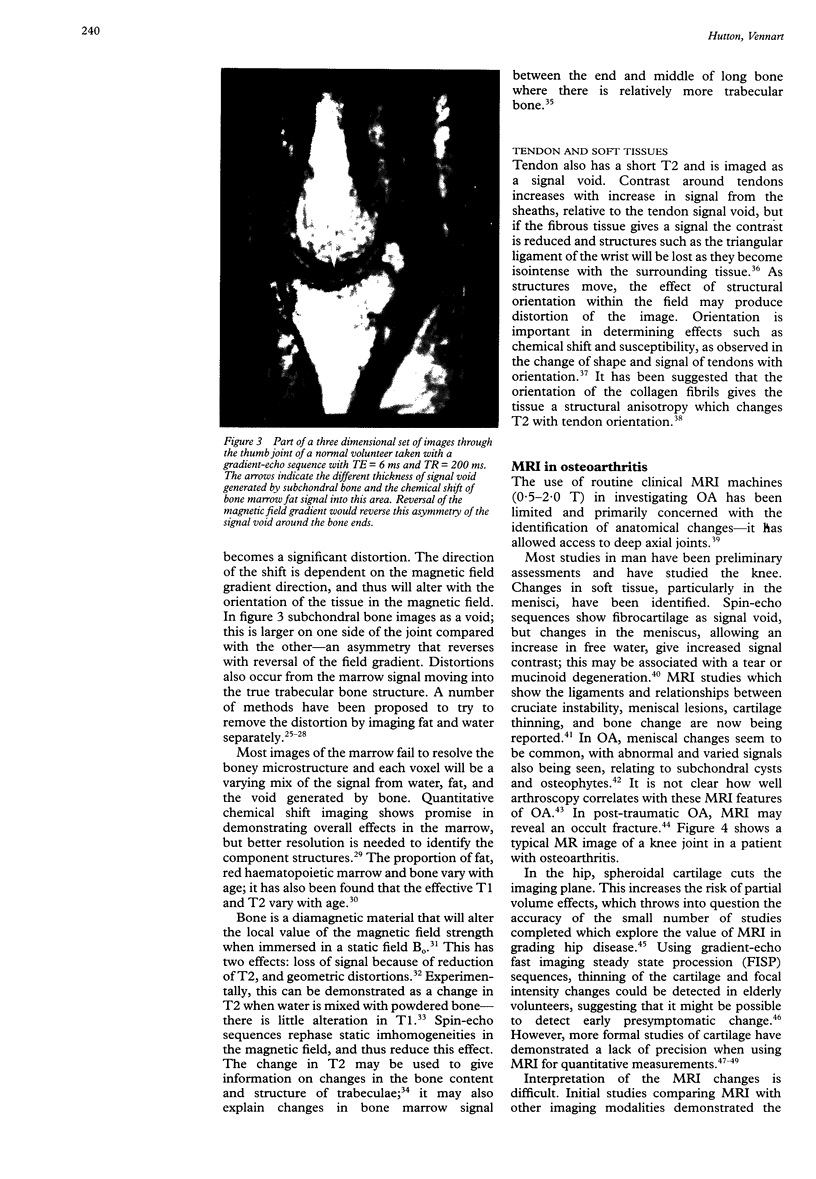
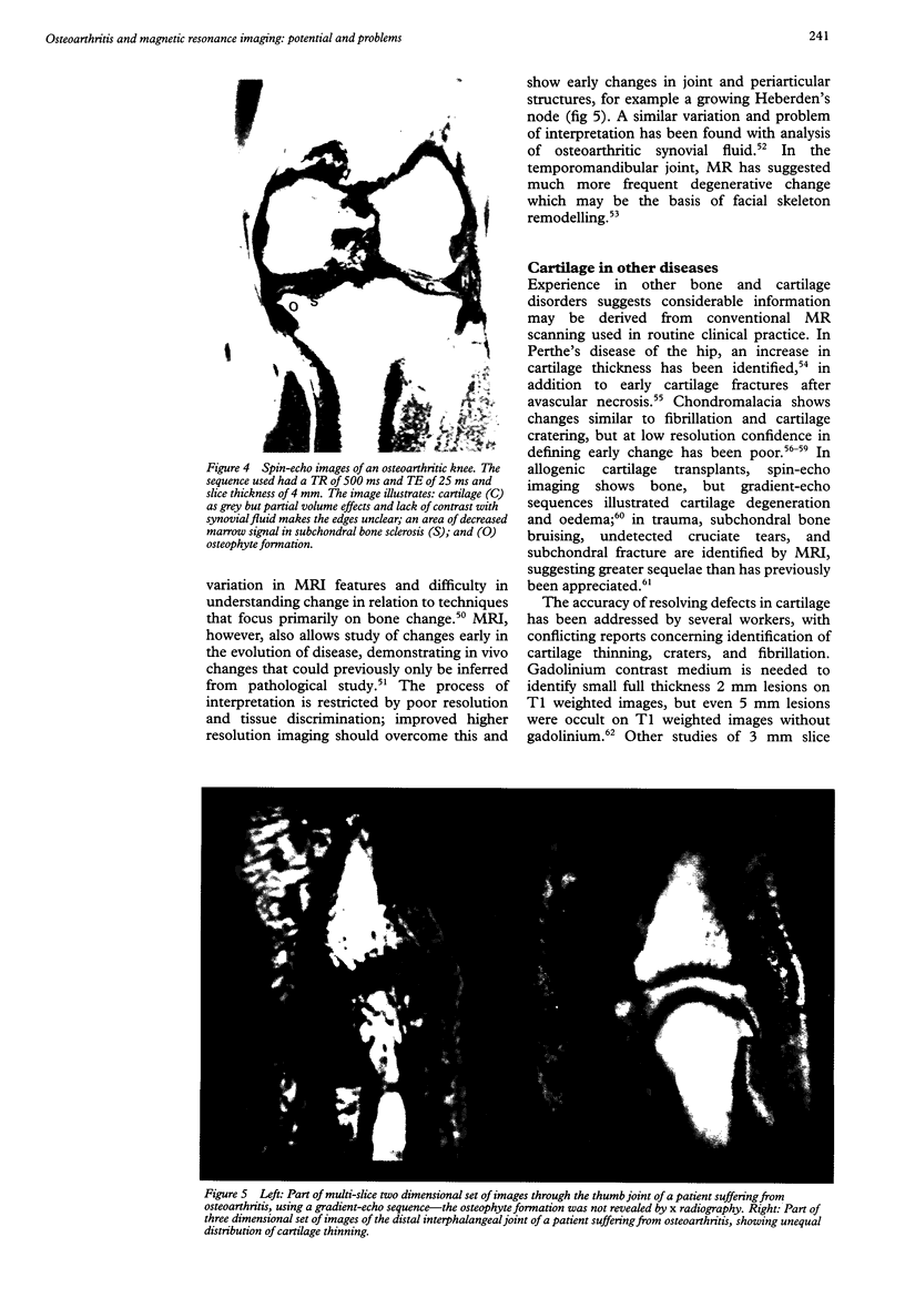
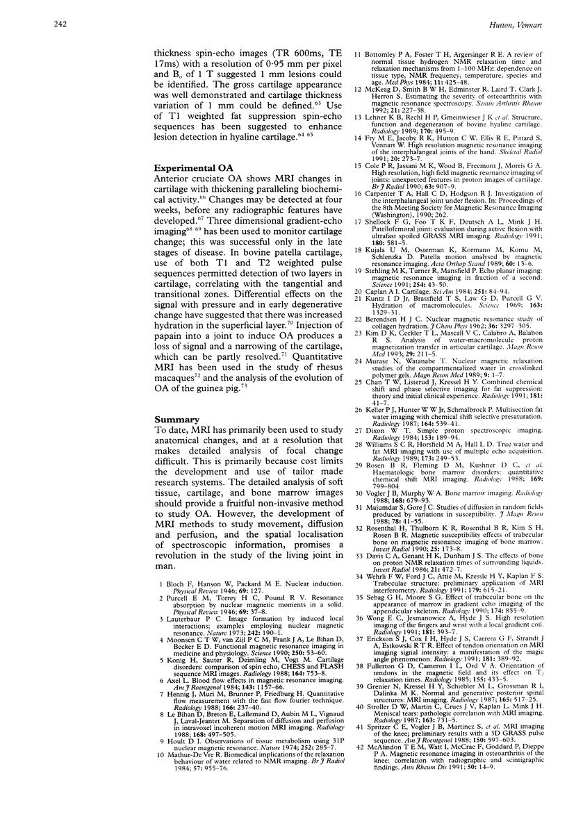
Images in this article
Selected References
These references are in PubMed. This may not be the complete list of references from this article.
- Adam G., Nolte-Ernsting C., Prescher A., Bühne M., Bruchmüller K., Küpper W., Günther R. W. Experimental hyaline cartilage lesions: two-dimensional spin-echo versus three-dimensional gradient-echo MR imaging. J Magn Reson Imaging. 1991 Nov-Dec;1(6):665–672. doi: 10.1002/jmri.1880010608. [DOI] [PubMed] [Google Scholar]
- Adams M. E., Li D. K., McConkey J. P., Davidson R. G., Day B., Duncan C. P., Tron V. Evaluation of cartilage lesions by magnetic resonance imaging at 0.15 T: comparison with anatomy and concordance with arthroscopy. J Rheumatol. 1991 Oct;18(10):1573–1580. [PubMed] [Google Scholar]
- Axel L. Blood flow effects in magnetic resonance imaging. AJR Am J Roentgenol. 1984 Dec;143(6):1157–1166. doi: 10.2214/ajr.143.6.1157. [DOI] [PubMed] [Google Scholar]
- Bongartz G., Bock E., Horbach T., Requardt H. Degenerative cartilage lesions of the hip--magnetic resonance evaluation. Magn Reson Imaging. 1989 Mar-Apr;7(2):179–186. doi: 10.1016/0730-725x(89)90702-9. [DOI] [PubMed] [Google Scholar]
- Braunstein E. M., Brandt K. D., Albrecht M. MRI demonstration of hypertrophic articular cartilage repair in osteoarthritis. Skeletal Radiol. 1990;19(5):335–339. doi: 10.1007/BF00193086. [DOI] [PubMed] [Google Scholar]
- Chan T. W., Listerud J., Kressel H. Y. Combined chemical-shift and phase-selective imaging for fat suppression: theory and initial clinical experience. Radiology. 1991 Oct;181(1):41–47. doi: 10.1148/radiology.181.1.1887054. [DOI] [PubMed] [Google Scholar]
- Chan W. P., Lang P., Stevens M. P., Sack K., Majumdar S., Stoller D. W., Basch C., Genant H. K. Osteoarthritis of the knee: comparison of radiography, CT, and MR imaging to assess extent and severity. AJR Am J Roentgenol. 1991 Oct;157(4):799–806. doi: 10.2214/ajr.157.4.1892040. [DOI] [PubMed] [Google Scholar]
- Chandnani V. P., Ho C., Chu P., Trudell D., Resnick D. Knee hyaline cartilage evaluated with MR imaging: a cadaveric study involving multiple imaging sequences and intraarticular injection of gadolinium and saline solution. Radiology. 1991 Feb;178(2):557–561. doi: 10.1148/radiology.178.2.1987624. [DOI] [PubMed] [Google Scholar]
- Cole P. R., Jasani M. K., Wood B., Freemont A. J., Morris G. A. High resolution, high field magnetic resonance imaging of joints: unexpected features in proton images of cartilage. Br J Radiol. 1990 Nov;63(755):907–909. doi: 10.1259/0007-1285-63-755-907. [DOI] [PubMed] [Google Scholar]
- Davis C. A., Genant H. K., Dunham J. S. The effects of bone on proton NMR relaxation times of surrounding liquids. Invest Radiol. 1986 Jun;21(6):472–477. doi: 10.1097/00004424-198606000-00005. [DOI] [PubMed] [Google Scholar]
- Dixon W. T. Simple proton spectroscopic imaging. Radiology. 1984 Oct;153(1):189–194. doi: 10.1148/radiology.153.1.6089263. [DOI] [PubMed] [Google Scholar]
- Erickson S. J., Cox I. H., Hyde J. S., Carrera G. F., Strandt J. A., Estkowski L. D. Effect of tendon orientation on MR imaging signal intensity: a manifestation of the "magic angle" phenomenon. Radiology. 1991 Nov;181(2):389–392. doi: 10.1148/radiology.181.2.1924777. [DOI] [PubMed] [Google Scholar]
- Fry M. E., Jacoby R. K., Hutton C. W., Ellis R. E., Phil M., Pittard S., Vennart W. High-resolution magnetic resonance imaging of the interphalangeal joints of the hand. Skeletal Radiol. 1991;20(4):273–277. doi: 10.1007/BF02341664. [DOI] [PubMed] [Google Scholar]
- Fullerton G. D., Cameron I. L., Ord V. A. Orientation of tendons in the magnetic field and its effect on T2 relaxation times. Radiology. 1985 May;155(2):433–435. doi: 10.1148/radiology.155.2.3983395. [DOI] [PubMed] [Google Scholar]
- Gahunia H. K., Lemaire C., Cross A. R., Babyn P., Kessler M. J., Pritzker K. P. Osteoarthritis in rhesus macaques: assessment of cartilage matrix quality by quantitative magnetic resonance imaging. Agents Actions Suppl. 1993;39:255–259. doi: 10.1007/978-3-0348-7442-7_31. [DOI] [PubMed] [Google Scholar]
- Grenier N., Kressel H. Y., Schiebler M. L., Grossman R. I., Dalinka M. K. Normal and degenerative posterior spinal structures: MR imaging. Radiology. 1987 Nov;165(2):517–525. doi: 10.1148/radiology.165.2.3659376. [DOI] [PubMed] [Google Scholar]
- Gylys-Morin V. M., Hajek P. C., Sartoris D. J., Resnick D. Articular cartilage defects: detectability in cadaver knees with MR. AJR Am J Roentgenol. 1987 Jun;148(6):1153–1157. doi: 10.2214/ajr.148.6.1153. [DOI] [PubMed] [Google Scholar]
- Hayes C. W., Sawyer R. W., Conway W. F. Patellar cartilage lesions: in vitro detection and staging with MR imaging and pathologic correlation. Radiology. 1990 Aug;176(2):479–483. doi: 10.1148/radiology.176.2.2367664. [DOI] [PubMed] [Google Scholar]
- Hennig J., Müri M., Brunner P., Friedburg H. Quantitative flow measurement with the fast Fourier flow technique. Radiology. 1988 Jan;166(1 Pt 1):237–240. doi: 10.1148/radiology.166.1.3336686. [DOI] [PubMed] [Google Scholar]
- Ho C., Cervilla V., Kjellin I., Haghigi P., Amiel D., Trudell D., Resnick D. Magnetic resonance imaging in assessing cartilage changes in experimental osteoarthrosis of the knee. Invest Radiol. 1992 Jan;27(1):84–90. doi: 10.1097/00004424-199201000-00017. [DOI] [PubMed] [Google Scholar]
- Hodler J., Trudell D., Pathria M. N., Resnick D. Width of the articular cartilage of the hip: quantification by using fat-suppression spin-echo MR imaging in cadavers. AJR Am J Roentgenol. 1992 Aug;159(2):351–355. doi: 10.2214/ajr.159.2.1632354. [DOI] [PubMed] [Google Scholar]
- Hoult D. I., Busby S. J., Gadian D. G., Radda G. K., Richards R. E., Seeley P. J. Observation of tissue metabolites using 31P nuclear magnetic resonance. Nature. 1974 Nov 22;252(5481):285–287. doi: 10.1038/252285a0. [DOI] [PubMed] [Google Scholar]
- Karvonen R. L., Negendank W. G., Fraser S. M., Mayes M. D., An T., Fernandez-Madrid F. Articular cartilage defects of the knee: correlation between magnetic resonance imaging and gross pathology. Ann Rheum Dis. 1990 Sep;49(9):672–675. doi: 10.1136/ard.49.9.672. [DOI] [PMC free article] [PubMed] [Google Scholar]
- Keller P. J., Hunter W. W., Jr, Schmalbrock P. Multisection fat-water imaging with chemical shift selective presaturation. Radiology. 1987 Aug;164(2):539–541. doi: 10.1148/radiology.164.2.3602398. [DOI] [PubMed] [Google Scholar]
- Kim D. K., Ceckler T. L., Hascall V. C., Calabro A., Balaban R. S. Analysis of water-macromolecule proton magnetization transfer in articular cartilage. Magn Reson Med. 1993 Feb;29(2):211–215. doi: 10.1002/mrm.1910290209. [DOI] [PubMed] [Google Scholar]
- Kujala U. M., Osterman K., Kormano M., Komu M., Schlenzka D. Patellar motion analyzed by magnetic resonance imaging. Acta Orthop Scand. 1989 Feb;60(1):13–16. doi: 10.3109/17453678909150081. [DOI] [PubMed] [Google Scholar]
- Kuntz I. D., Jr, Brassfield T. S., Law G. D., Purcell G. V. Hydration of macromolecules. Science. 1969 Mar 21;163(3873):1329–1331. doi: 10.1126/science.163.3873.1329. [DOI] [PubMed] [Google Scholar]
- König H., Sauter R., Deimling M., Vogt M. Cartilage disorders: comparison of spin-echo, CHESS, and FLASH sequence MR images. Radiology. 1987 Sep;164(3):753–758. doi: 10.1148/radiology.164.3.3615875. [DOI] [PubMed] [Google Scholar]
- König H., Skalej M., Höntzsch D., Aicher K. Kernspintomographie von Knochen-Knorpel-Transplantaten im Kniegelenk: Transplantat-Morphologie und Versuch einer quantitativen Beurteilung der Knorpelveränderungen. Rofo. 1988 Feb;148(2):176–182. doi: 10.1055/s-2008-1048172. [DOI] [PubMed] [Google Scholar]
- Le Bihan D., Breton E., Lallemand D., Aubin M. L., Vignaud J., Laval-Jeantet M. Separation of diffusion and perfusion in intravoxel incoherent motion MR imaging. Radiology. 1988 Aug;168(2):497–505. doi: 10.1148/radiology.168.2.3393671. [DOI] [PubMed] [Google Scholar]
- Lehner K. B., Rechl H. P., Gmeinwieser J. K., Heuck A. F., Lukas H. P., Kohl H. P. Structure, function, and degeneration of bovine hyaline cartilage: assessment with MR imaging in vitro. Radiology. 1989 Feb;170(2):495–499. doi: 10.1148/radiology.170.2.2911674. [DOI] [PubMed] [Google Scholar]
- Li K. C., Higgs J., Aisen A. M., Buckwalter K. A., Martel W., McCune W. J. MRI in osteoarthritis of the hip: gradations of severity. Magn Reson Imaging. 1988 May-Jun;6(3):229–236. doi: 10.1016/0730-725x(88)90396-7. [DOI] [PubMed] [Google Scholar]
- Mathur-De Vré R. Biomedical implications of the relaxation behaviour of water related to NMR imaging. Br J Radiol. 1984 Nov;57(683):955–976. doi: 10.1259/0007-1285-57-683-955. [DOI] [PubMed] [Google Scholar]
- McCauley T. R., Kier R., Lynch K. J., Jokl P. Chondromalacia patellae: diagnosis with MR imaging. AJR Am J Roentgenol. 1992 Jan;158(1):101–105. doi: 10.2214/ajr.158.1.1727333. [DOI] [PubMed] [Google Scholar]
- McKeag D., Smith B. W., Edminster R., Laird T., Clark J., Herron S. Estimating the severity of osteoarthritis with magnetic resonance spectroscopy. Semin Arthritis Rheum. 1992 Feb;21(4):227–238. doi: 10.1016/0049-0172(92)90053-g. [DOI] [PubMed] [Google Scholar]
- Mink J. H., Deutsch A. L. Occult cartilage and bone injuries of the knee: detection, classification, and assessment with MR imaging. Radiology. 1989 Mar;170(3 Pt 1):823–829. doi: 10.1148/radiology.170.3.2916038. [DOI] [PubMed] [Google Scholar]
- Moonen C. T., van Zijl P. C., Frank J. A., Le Bihan D., Becker E. D. Functional magnetic resonance imaging in medicine and physiology. Science. 1990 Oct 5;250(4977):53–61. doi: 10.1126/science.2218514. [DOI] [PubMed] [Google Scholar]
- Murase N., Watanabe T. Nuclear magnetic relaxation studies of the compartmentalized water in crosslinked polymer gels. Magn Reson Med. 1989 Jan;9(1):1–7. doi: 10.1002/mrm.1910090102. [DOI] [PubMed] [Google Scholar]
- Recht M. P., Kramer J., Marcelis S., Pathria M. N., Trudell D., Haghighi P., Sartoris D. J., Resnick D. Abnormalities of articular cartilage in the knee: analysis of available MR techniques. Radiology. 1993 May;187(2):473–478. doi: 10.1148/radiology.187.2.8475293. [DOI] [PubMed] [Google Scholar]
- Rosen B. R., Fleming D. M., Kushner D. C., Zaner K. S., Buxton R. B., Bennet W. P., Wismer G. L., Brady T. J. Hematologic bone marrow disorders: quantitative chemical shift MR imaging. Radiology. 1988 Dec;169(3):799–804. doi: 10.1148/radiology.169.3.3187003. [DOI] [PubMed] [Google Scholar]
- Rosenthal H., Thulborn K. R., Rosenthal D. I., Kim S. H., Rosen B. R. Magnetic susceptibility effects of trabecular bone on magnetic resonance imaging of bone marrow. Invest Radiol. 1990 Feb;25(2):173–178. doi: 10.1097/00004424-199002000-00013. [DOI] [PubMed] [Google Scholar]
- Rush B. H., Bramson R. T., Ogden J. A. Legg-Calvé-Perthes disease: detection of cartilaginous and synovial change with MR imaging. Radiology. 1988 May;167(2):473–476. doi: 10.1148/radiology.167.2.3357958. [DOI] [PubMed] [Google Scholar]
- Sabiston C. P., Adams M. E., Li D. K. Magnetic resonance imaging of osteoarthritis: correlation with gross pathology using an experimental model. J Orthop Res. 1987;5(2):164–172. doi: 10.1002/jor.1100050203. [DOI] [PubMed] [Google Scholar]
- Schellhas K. P., Piper M. A., Omlie M. R. Facial skeleton remodeling due to temporomandibular joint degeneration: an imaging study of 100 patients. AJNR Am J Neuroradiol. 1990 May;11(3):541–551. [PMC free article] [PubMed] [Google Scholar]
- Sebag G. H., Moore S. G. Effect of trabecular bone on the appearance of marrow in gradient-echo imaging of the appendicular skeleton. Radiology. 1990 Mar;174(3 Pt 1):855–859. doi: 10.1148/radiology.174.3.2305069. [DOI] [PubMed] [Google Scholar]
- Shellock F. G., Foo T. K., Deutsch A. L., Mink J. H. Patellofemoral joint: evaluation during active flexion with ultrafast spoiled GRASS MR imaging. Radiology. 1991 Aug;180(2):581–585. doi: 10.1148/radiology.180.2.2068335. [DOI] [PubMed] [Google Scholar]
- Shuman W. P., Castagno A. A., Baron R. L., Richardson M. L. MR imaging of avascular necrosis of the femoral head: value of small-field-of-view sagittal surface-coil images. AJR Am J Roentgenol. 1988 May;150(5):1073–1078. doi: 10.2214/ajr.150.5.1073. [DOI] [PubMed] [Google Scholar]
- Spritzer C. E., Vogler J. B., Martinez S., Garrett W. E., Jr, Johnson G. A., McNamara M. J., Lohnes J., Herfkens R. J. MR imaging of the knee: preliminary results with a 3DFT GRASS pulse sequence. AJR Am J Roentgenol. 1988 Mar;150(3):597–603. doi: 10.2214/ajr.150.3.597. [DOI] [PubMed] [Google Scholar]
- Stehling M. K., Turner R., Mansfield P. Echo-planar imaging: magnetic resonance imaging in a fraction of a second. Science. 1991 Oct 4;254(5028):43–50. doi: 10.1126/science.1925560. [DOI] [PubMed] [Google Scholar]
- Stoller D. W., Martin C., Crues J. V., 3rd, Kaplan L., Mink J. H. Meniscal tears: pathologic correlation with MR imaging. Radiology. 1987 Jun;163(3):731–735. doi: 10.1148/radiology.163.3.3575724. [DOI] [PubMed] [Google Scholar]
- Vellet A. D. The evolution of post-traumatic osteoarthritis: its relationship to occult post-traumatic subcortical fractures identified on MRI. Bildgebung. 1992 Sep;59(3):121–122. [PubMed] [Google Scholar]
- Watson P. J., Hall L. D., Carpenter T. A., Tyler J. A. A magnetic resonance imaging study of joint degeneration in the guinea pig knee. Agents Actions Suppl. 1993;39:261–265. doi: 10.1007/978-3-0348-7442-7_32. [DOI] [PubMed] [Google Scholar]
- Wehrli F. W., Ford J. C., Attie M., Kressel H. Y., Kaplan F. S. Trabecular structure: preliminary application of MR interferometry. Radiology. 1991 Jun;179(3):615–621. doi: 10.1148/radiology.179.3.2027962. [DOI] [PubMed] [Google Scholar]
- Williams S. C., Horsfield M. A., Hall L. D. True water and fat MR imaging with use of multiple-echo acquisition. Radiology. 1989 Oct;173(1):249–253. doi: 10.1148/radiology.173.1.2781016. [DOI] [PubMed] [Google Scholar]
- Wojtys E., Wilson M., Buckwalter K., Braunstein E., Martel W. Magnetic resonance imaging of knee hyaline cartilage and intraarticular pathology. Am J Sports Med. 1987 Sep-Oct;15(5):455–463. doi: 10.1177/036354658701500505. [DOI] [PubMed] [Google Scholar]
- Wong E. C., Jesmanowicz A., Hyde J. S. High-resolution, short echo time MR imaging of the fingers and wrist with a local gradient coil. Radiology. 1991 Nov;181(2):393–397. doi: 10.1148/radiology.181.2.1924778. [DOI] [PubMed] [Google Scholar]
- Yulish B. S., Montanez J., Goodfellow D. B., Bryan P. J., Mulopulos G. P., Modic M. T. Chondromalacia patellae: assessment with MR imaging. Radiology. 1987 Sep;164(3):763–766. doi: 10.1148/radiology.164.3.3615877. [DOI] [PubMed] [Google Scholar]


