Abstract
Background
Neovascular glaucoma (NVG) is a potentially blinding, secondary glaucoma. It is caused by the formation of abnormal new blood vessels, which prevent normal drainage of aqueous from the anterior segment of the eye. Anti‐vascular endothelial growth factor (anti‐VEGF) medications are specific inhibitors of the primary mediators of neovascularization. Studies have reported the effectiveness of anti‐VEGF medications for the control of intraocular pressure (IOP) in NVG.
Objectives
To assess the effectiveness of intraocular anti‐VEGF medications, alone or with one or more types of conventional therapy, compared with no anti‐VEGF medications for the treatment of NVG.
Search methods
We searched CENTRAL (which contains the Cochrane Eyes and Vision Trials Register); MEDLINE; Embase; PubMed; and LILACS to 19 October 2021; metaRegister of Controlled Trials to 19 October 2021; and two additional trial registers to 19 October 2021. We did not use any date or language restrictions in the electronic search for trials.
Selection criteria
We included randomized controlled trials (RCTs) of people treated with anti‐VEGF medications for NVG.
Data collection and analysis
Two review authors independently assessed the search results for trials, extracted data, and assessed risk of bias, and the certainty of the evidence. We resolved discrepancies through discussion.
Main results
We included five RCTs (356 eyes of 353 participants). Each trial was conducted in a different country: two in China, and one each in Brazil, Egypt, and Japan. All five RCTs included both men and women; the mean age of participants was 55 years or older. Two RCTs compared intravitreal bevacizumab combined with Ahmed valve implantation and panretinal photocoagulation (PRP) with Ahmed valve implantation and PRP alone. One RCT randomized participants to receive an injection of either intravitreal aflibercept or placebo at the first visit, followed by non‐randomized treatment according to clinical findings after one week. The remaining two RCTs randomized participants to PRP with and without ranibizumab, one of which had insufficient details for further analysis. We assessed the RCTs to have an unclear risk of bias for most domains due to insufficient information to permit judgment.
Four RCTs examined achieving control of IOP, three of which reported our time points of interest. Only one RCT reported our critical time point at one month; it found that the anti‐VEGF group had a 1.3‐fold higher chance of achieving control of IOP at one month (RR 1.32, 95% 1.10 to 1.59; 93 participants) than the non‐anti‐VEGF group (low certainty of evidence). For other time points, one RCT found a three‐fold greater achievement in control of IOP in the anti‐VEGF group when compared with the non‐anti‐VEGF group at one year (RR 3.00; 95% CI:1.35 to 6.68; 40 participants). However, another RCT found an inconclusive result at the time period ranging from 1.5 years to three years (RR 1.08; 95% CI: 0.67 to 1.75; 40 participants).
All five RCTs examined mean IOP, but at different time points. Very‐low‐certainty evidence showed that anti‐VEGFs were effective in reducing mean IOP by 6.37 mmHg (95% CI: ‐10.09 to ‐2.65; 3 RCTs; 173 participants) at four to six weeks when compared with no anti‐VEGFs. Anti‐VEGFs may reduce mean IOP at three months (MD ‐4.25; 95% CI ‐12.05 to 3.54; 2 studies; 75 participants), six months (MD ‐5.93; 95% CI ‐18.13 to 6.26; 2 studies; 75 participants), one year (MD ‐5.36; 95% CI ‐18.50 to 7.77; 2 studies; 75 participants), and more than one year (MD ‐7.05; 95% CI ‐16.61 to 2.51; 2 studies; 75 participants) when compared with no anti‐VEGFs, but such effects remain uncertain.
Two RCTs reported the proportion of participants who achieved an improvement in visual acuity with specified time points. Participants receiving anti‐VEGFs had a 2.6 times (95% CI 1.60 to 4.08; 1 study; 93 participants) higher chance of improving visual acuity when compared with those not receiving anti‐VEGFs at one month (very low certainty of evidence). Likewise, another RCT found a similar result at 18 months (RR 4.00, 95% CI 1.33 to 12.05; 1 study; 40 participants).
Two RCTs reported the outcome, complete regression of new iris vessels, at our time points of interest. Low‐certainty evidence showed that anti‐VEGFs had a nearly three times higher chance of complete regression of new iris vessels when compared with no anti‐VEGFs (RR 2.63, 95% CI 1.65 to 4.18; 1 study; 93 participants). A similar finding was observed at more than one year in another RCT (RR 3.20, 95% CI 1.45 to 7.05; 1 study; 40 participants).
Regarding adverse events, there was no evidence that the risks of hypotony and tractional retinal detachment were different between the two groups (RR 0.67; 95% CI: 0.12 to 3.57 and RR 0.33; 95% CI: 0.01 to 7.72, respectively; 1 study; 40 participants). No RCTs reported incidents of endophthalmitis, vitreous hemorrhage, no light perception, and serious adverse events. Evidence for the adverse events of anti‐VEGFs was low due to limitations in the study design due to insufficient information to permit judgments and imprecision of results due to the small sample size.
No trial reported the proportion of participants with relief of pain and resolution of redness at any time point.
Authors' conclusions
Anti‐VEGFs as an adjunct to conventional treatment could help reduce IOP in NVG in the short term (four to six weeks), but there is no evidence that this is likely in the longer term. Currently available evidence regarding the short‐ and long‐term effectiveness and safety of anti‐VEGFs in achieving control of IOP, visual acuity, and complete regression of new iris vessels in NVG is insufficient. More research is needed to investigate the effect of these medications compared with, or in addition to, conventional surgical or medical treatment in achieving these outcomes in NVG.
Keywords: Female; Humans; Male; Middle Aged; Bevacizumab; Bevacizumab/therapeutic use; Glaucoma, Neovascular; Glaucoma, Neovascular/drug therapy; Ranibizumab; Ranibizumab/therapeutic use; Vascular Endothelial Growth Factor A; Vascular Endothelial Growth Factor A/antagonists & inhibitors
Plain language summary
Anti‐vascular endothelial growth factor for neovascular glaucoma
What was the aim of this review? To compare treatment with and without anti‐vascular endothelial growth factor (anti‐VEGF) medications for people with neovascular glaucoma (NVG).
Key message It is uncertain whether treatment with anti‐VEGF medications is more beneficial than treatment without anti‐VEGF medications for people with NVG. More research is needed to investigate the effect of anti‐VEGF medications compared with, or in addition to, conventional treatment.
What did we study in this review? VEGF is a protein produced by cells in your body, and produces new blood vessels when needed. When cells produce too much VEGF, abnormal blood vessels can grow in the eye. NVG is a type of glaucoma where the angle between the iris (colored part of the eye) and the cornea (transparent front part of the eye) is closed by new blood vessels growing in the eye, hence, the name 'neovascular'. New blood vessels can cause scarring and narrowing, which can eventually lead to complete closure of the angle. This results in increased eye pressure since the fluid in the eye cannot drain properly. In NVG, the eye is often red and painful, and the vision is abnormal. High pressure in the eye can lead to blindness.
Anti‐VEGF medication is a type of medicine that blocks VEGF, therefore, slowing the growth of blood vessels. It is administered by injection into the eye. It can be used at an early stage, when conventional treatment may not be possible. Most studies report short‐term (generally four to six weeks) benefits of anti‐VEGF medication, but long‐term benefits are not clear.
What were the main results of this review? We included five studies enrolling a total of 356 eyes of 353 participants with NVG.
Three studies reported different results for achieving control of IOP at various time points of interest. One study showed that anti‐VEGF medications were more effective at one month. In the longer term, one study reported the superiority of anti‐VEGF medications while another showed inconclusive results. Therefore, available evidence is insufficient to recommend the routine use of anti‐VEGF medication in individuals with NVG.
How up‐to‐date is the review? We searched for studies that were published up to 19 October 2021.
Summary of findings
Summary of findings 1. Anti‐VEGF versus no anti‐VEGF for neovascular glaucoma.
| Anti‐VEGF medication compared with no anti‐VEGF medication for neovascular glaucoma | ||||||
| Patient or population: people with neovascular glaucoma Setting: ophthalmology hospital or clinic Intervention: intravitreal anti‐VEGF medication injection Comparison: no anti‐VEGF medication | ||||||
| Outcomes | Anticipated absolute effects* (95% CI) | Relative effect (95% CI) | № of participants (studies) | Certainty of the evidence (GRADE) | Comments | |
| Risk with no anti‐VEGF | Risk with anti‐VEGF | |||||
| Proportion of participants with IOP ≤ 21 mmHg, with or without ocular hypotensive medications, 4 to 6 weeks follow‐up |
723 per 1000 | 954 per 1000 (795 to 1150) | RR 1.32 (95% CI 1.10 to 1.59) | 93 (1) | ⊕⊕⊝⊝ Lowa,b | None |
| Mean IOP, 4 to 6 weeks follow‐up | The mean IOP in the no anti‐VEGF group was 24.2 mm Hg, ranged from 23.6 to 24.8 mm Hg |
The mean IOP in the anti‐VEGF group was 17.8 mm Hg, ranged from 14.1 to 21.6 mm Hg |
MD ‐6.37 (95% CI ‐10.09 to ‐2.65) | 173 (3) | ⊕⊝⊝⊝ Very lowa,b,c | None |
| Proportion of participants with improvement in visual acuity of 2 ETDRS lines or 0.2 logMAR units, 4 to 6 weeks follow‐up | 298 per 1000 | 760 per 1000 (477 to 1216) | RR 2.55 (95% CI 1.60 to 4.08) | 93 (1) | ⊕⊝⊝⊝ Very lowa,b,d | The trial did not clearly specify a definition of the improvement in visual acuity |
| Proportion of participants with complete regression of new iris vessels, 4 to 6 weeks follow‐up |
298 per 1000 | 784 per 1000 (492 to 1246) | RR 2.63 (95% CI 1.65 to 4.18) | 93 (1) | ⊕⊕⊝⊝ Lowa,b | None |
| Proportion of participants with relief of pain and resolution of redness, 1 year follow‐up |
Included studies did not report data for this outcome |
|||||
| Proportion of participants with adverse events at various follow‐up times |
Hypotony (IOP ≤ 6 mmHg) | |||||
| 15 per 100 |
10 per 100 (2 to 54) | RR 0.67 (95% CI 0.12 to 3.57) | 40 (1) | ⊕⊕⊝⊝ Lowa,b | The follow‐up period was 18 months. | |
| Tractional retinal detachment | ||||||
| 5 per 100 | 2 per 100 (0 to 39) | RR 0.33 (95% CI 0.01 to 7.72) | 40 (1) | ⊕⊕⊝⊝ Lowa,b | The follow‐up period was 24 months. | |
| Serious adverse events (e.g. systemic thrombosis, stroke, and coronary thrombosis) | ||||||
| Included studies did not report data for this outcome | Inatani 2021 reported an occurrence of serious ocular treatment‐emergent adverse events in both groups during the non‐randomized period, where participants could receive both sham injection and aflibercept injection if the re‐treatment criteria were met. | |||||
| *The risk in the intervention group (and its 95% CI) is based on the assumed risk in the comparison group and the relative effect of the intervention (and its 95% CI). Anti‐VEGF: anti‐vascular endothelial growth factor; CI: confidence interval; IOP: intraocular pressure; MD: mean difference; RR: risk ratio. | ||||||
| GRADE Working Group grades of evidence High certainty: We are very confident that the true effect lies close to that of the estimate of the effect Moderate certainty: We are moderately confident in the effect estimate: The true effect is likely to be close to the estimate of the effect, but there is a possibility that it is substantially different Low certainty: Our confidence in the effect estimate is limited: The true effect may be substantially different from the estimate of the effect Very low certainty: We have very little confidence in the effect estimate: The true effect is likely to be substantially different from the estimate of effect | ||||||
aDowngraded (‐1) due to limitations in the design bDowngraded (‐1) due to imprecision of results
cDowngraded (‐1) due to inconsistency dDowngraded (‐1) due to indirectness of evidence
Background
Description of the condition
Neovascular glaucoma (NVG) is a secondary glaucoma in which new vessels, and subsequently fibrous tissue, form in the anterior chamber angle of the eye. This leads to blockage of the angle, which inhibits aqueous drainage, causing elevated intraocular pressure (IOP). This condition was described as early as 1871 (Pagenstecher 1871; Tsai 2008). Historically, it has also been referred to as rubeotic glaucoma, hemorrhagic glaucoma, thrombotic glaucoma, congestive glaucoma, and diabetic hemorrhagic glaucoma.
Clinical conditions causing retinal ischemia, such as proliferative diabetic retinopathy (PDR), central retinal vein occlusion (CRVO), and ocular ischemic syndrome, are associated with NVG. The condition can be unilateral or bilateral, depending on the underlying cause for the NVG. Diabetic retinopathy is usually bilateral; CRVO is usually unilateral. Retinal ischemia results in the release of angiogenic factors, such as vascular endothelial growth factor (VEGF). The angiogenic factors diffuse into the aqueous and anterior segment, and trigger neovascularization of the iris and anterior chamber angle. This process leads to fibrous tissue proliferation, and subsequent synechial angle closure (closure of the angle because the iris is adhering to the cornea). Increased levels of VEGF have been measured in the aqueous of people with NVG (Aiello 1994; Sone 1996; Tripathi 1998). Elevated IOP is a direct result of secondary angle closure glaucoma.
NVG is a potentially devastating glaucoma. Delayed diagnosis or poor management can result in complete loss of vision, with intractable pain. It is imperative to diagnose it early, and treat it immediately and aggressively. In managing NVG, it is essential to treat both the elevated IOP and the underlying cause of the disease.
General principles for treating people with NVG include identifying the underlying etiology, controlling or eliminating retinal ischemia and reducing the IOP. Panretinal photocoagulation (PRP) ablates the ischemic retina by shrinking and eliminating the abnormal blood vessels. When most of the angle is closed due to synechiae, consequent to the angle neovascularization, medical and/or surgical treatment is necessary to control IOP. Surgical procedures for treating NVG are: trabeculectomy, implantation of aqueous drainage devices (Minckler 2006; Yalvac 2007), Nd‐Yag cyclophotocoagulation (Delgado 2003), vitrectomy with PRP and trabeculectomy (Kiuchi 2006), and cyclocryotherapy (Kovacic 2004). They may be done in conjunction with anti‐metabolites, such as 5‐fluorouracil or mitomycin C, which modify wound healing and reduce scarring (Wilkins 2005; Wormald 2001).
Description of the intervention
Currently, anti‐VEGF medications are used for various conditions in which hypoxia‐induced VEGF release and subsequent neovascularization lead to ocular damage. Initially used in ophthalmology for the treatment of choroidal neovascularization in age‐related macular degeneration (Solomon 2019), the application of anti‐VEGF medications has expanded rapidly to include treatment for other conditions, such as NVG, diabetic macular edema, and retinopathy of prematurity (Andreoli 2007). Some of the anti‐VEGF medications most frequently used in the eye are bevacizumab, ranibizumab, pegaptanib sodium, and aflibercept (VEGF Trap‐eye).
How the intervention might work
In treating NVG, it is critical to address the underlying pathology – angiogenic factors released by the ischemic retina. The issue of retinal ischemia can be addressed by PRP, which ablates the ischemic retina and reduces further production of angiogenic medications. However, in many people, the view of the fundus is poor, due to corneal edema or vitreous hemorrhage and, therefore, precludes PRP. Hence, interventions aimed at directly blocking angiogenic factors could help reduce the formation of new vessels, and possibly reverse the neovascularization (Andreoli 2007; Arcieri 2015; Tripathi 1998). Intraocular injection of bevacizumab has been shown to reduce the levels of VEGF in the aqueous (Grover 2009).
In eyes in which PRP can be done, variable times for regression of new vessels have been reported, and the newly formed vessels may not regress until four to six weeks after treatment. In one study, Doft and Blankenship reported regression of new vessels in 20% of participants at three days, 50% at two weeks, 72% at three weeks, and 62% at six months (Doft 1984). In another study, Blankenship reported regression in 97% of participants at one month (Blankenship 1988). Comparison of studies is difficult, due to variation in the laser treatments, variation in the response to laser between type 1 and 2 diabetics, and the variation in the definition of substantial regression in different studies.
On the other hand, anti‐VEGF medications have been shown to cause regression of new vessels in the anterior chamber angle and a drop in IOP within a few days (Avery 2006; Iliev 2006). Intravitreal (Iliev 2006; Yazdani 2007), and less commonly, intracameral (Grover 2009) anti‐VEGF medications have been used in the management of NVG to control angiogenesis in the angle and iris. However, the effects of anti‐VEGF medications for treating NVG are temporary, generally lasting four to six weeks (Wakabayashi 2008). Thus, many studies have combined the use of anti‐VEGF medications with traditional treatments, such as PRP (Ehlers 2008; Ha 2017), with or without other surgery (Arcieri 2015; Gupta 2009; Kang 2014; Mahdy 2013; Noor 2017; Olmos 2016; Wakabayashi 2008; Wittstrom 2012; Yazdani 2009).
Why it is important to do this review
Various case reports, prospective and retrospective case series, and a few randomized controlled trials (RCTs) have shown good short‐term benefit of anti‐VEGF use in NVG, when combined with conventional treatment that included PRP and IOP‐lowering procedures, such as trabeculectomy, insertion of aqueous drainage devices, cyclocryotherapy and Nd Yag cyclophotocoagulation. These studies reported better regression of iris new vessels and reduced postoperative incidence of hyphema. However, the sustained long‐term benefit of better IOP control and improved visual outcomes is not clear; while a few studies showed better outcomes, most studies showed no difference with the use of anti‐VEGF medications. Variation in participant allocation, number and doses of anti‐VEGF injections, and conventional treatment used in the studies makes comparison difficult.
On the basis of studies that showed that ischemic CRVO tends to eventually subside to a state of quiescence (Hayreh 2003), Gandhi 2008 suggested that anti‐VEGF medications alone can treat NVG secondary to CRVO effectively. In two participants with CRVO who had persisting neovascularization and high IOP in spite of PRP, Yazdani 2007 reported regression of new vessels and control of IOP following intravitreal bevacizumab. Maintenance of IOP control was reported for as long as six months following a second dose of intravitreal bevacizumab in both of these participants, one at eight weeks, and the other at six weeks. So the question arises about the effectiveness of anti‐VEGF in managing NVG in both the short and long term.
A previous version of this review (Simha 2020) did not find any evidence to draw conclusions regarding the long‐term benefits of the use of intraocular anti‐VEGF for NVG treatment and suggested the need for further RCTs.
Objectives
To assess the effectiveness of intraocular anti‐vascular endothelial growth factor (VEGF) medications, alone or with one or more types of conventional therapy, compared with no anti‐VEGF medications for the treatment of neovascular glaucoma.
Methods
Criteria for considering studies for this review
Types of studies
We included RCTs only.
Types of participants
We included studies of people with NVG. We included all age groups and ocular comorbidities.
Types of interventions
Intervention group
People with NVG who received intraocular anti‐VEGF medications, with one or more types of conventional therapy, which included laser PRP, trabeculectomy, insertion of aqueous drainage devices, cyclophotocoagulation, cryotherapy, and medical therapy.
In the subgroup of people with NVG due to CRVO, the intervention group could receive intraocular anti‐VEGF injection alone, without additional conventional therapy.
Control group
People who underwent the same conventional therapy as the intervention group, but without intraocular anti‐VEGF medications.
In the subgroup of people with NVG due to CRVO, the control group could receive placebo injections, or no treatment, including no conventional therapy.
We did not include dosing studies, in which one dose of anti‐VEGF medication was compared to another dose, unless the study also had a control arm.
Types of outcome measures
Primary outcomes
The critical outcome of this review was the proportion of participants who achieved control of IOP, measured at four to six weeks after treatment. Control of IOP was defined as IOP ≤ 21 mmHg, with or without ocular hypotensive medications.
Secondary outcomes
We examined each important outcome described below at four to six weeks, three months, six months, one year, and thereafter as available throughout follow‐up.
IOP
Proportion of participants with IOP ≤ 21 mmHg, with or without ocular hypotensive medications or other treatment at three months, six months, one year, and thereafter as available throughout follow‐up
Mean IOP, with or without ocular hypotensive medications
Visual acuity
Proportion of participants with improvement in visual acuity
Regression of new vessels
Proportion of participants with complete regression of new iris vessels
Relief of symptoms
Proportion of participants with relief of pain and resolution of redness
Adverse events
Infection: proportion of participants with intraocular infection or inflammation (endophthalmitis) within six weeks of the intervention
Low IOP (hypotony): proportion of participants with IOP ≤ 6 mmHg
Vitreous hemorrhage: proportion of participants with development of vitreous hemorrhage
Tractional retinal detachment: proportion of participants who experienced tractional retinal detachment
No light perception: proportion of participants with no light perception
Other serious adverse events, including systemic thrombosis, stroke and coronary thrombosis, up to one‐year follow‐up
Search methods for identification of studies
Electronic searches
The Cochrane Eyes and Vision Information Specialist updated searches in the following electronic databases for RCTs and controlled clinical trials. There were no restrictions on language or year of publication. The electronic databases were last searched on 21 Oct 2021. The last search of metaRegister of Controlled Trials was on 13 August 2013.
Cochrane Central Register of Controlled Trials (CENTRAL; 2021, Issue 10), which contains the Cochrane Eyes and Vision Trials Register, in the Cochrane Library (searched 21 Oct 2021; Appendix 1);
MEDLINE Ovid (1946 to 21 Oct 2021; Appendix 2);
Embase.com (1947 to 21 Oct 2021; Appendix 3);
PubMed (1948 to 21 Oct 2021; Appendix 4);
Latin American and Caribbean Health Sciences Literature Database (LILACS; 1982 to 21 Oct 2021; Appendix 5);
metaRegister of Controlled Trials (mRCT; www.controlled-trials.com; searched 13 August 2013; Appendix 6), the service of which was discontinued in 2014.
US National Institutes of Health Ongoing Trials Register ClinicalTrials.gov (www.clinicaltrials.gov; searched 21 Oct 2021; Appendix 7);
World Health Organization (WHO) International Clinical Trials Registry Platform (ICTRP; www.who.int/ictrp; searched 21 Oct 2021; Appendix 8).
Searching other resources
We handsearched the reference lists of eligible studies to identify other potentially relevant trials. We contacted investigators of ongoing studies for information about ongoing studies.
Data collection and analysis
Selection of studies
Two review authors independently screened the titles and abstracts of all the reports of studies identified by the electronic searches, and handsearching, using Covidence (Covidence). Each review author classified the studies as: (1) definitely include (Yes), (2) possibly include (Maybe), and (3) definitely exclude (No). Each review author obtained and independently assessed the full‐text report(s) of each study classified by either review author as (1) or (2), and reclassified them as: (a) include, (b) awaiting classification, or (c) exclude. For reports from studies classified as (b), we attempted to contact study investigators for clarification. The two review authors compared their individual classifications and discussed discrepancies. When they could not reach consensus after discussion, a third review author reclassified the studies. We documented all studies classified as (c) exclude, and took note of any studies that are currently ongoing. We retrieved and reviewed all pertinent references from each potentially relevant study, in order to provide the most complete published information about study design, methods, and findings.
Data extraction and management
Two review authors independently extracted data from included studies, using Covidence. We resolved all discrepancies through discussion. One review author entered data into Review Manager 5, and a second review author verified the data entries (Review Manager 2014).
Categories of information extracted for each study included: methods (study design, number of participants, and setting), intervention details, outcomes (definitions and time points), and results for each outcome (sample size, missing data, summary data for each intervention).
Assessment of risk of bias in included studies
Two review authors independently assessed the risk of bias as recommended in Chapter 8 of the Cochrane Handbook for Systematic Reviews of Interventions (Higgins 2011a). We provided judgment for each domain as low risk of bias, high risk of bias, or unclear risk of bias, which indicated either lack of information or uncertainty over the potential for bias. Specific criteria for assessing risk of bias focused on adequate sequence generation; allocation concealment; masking (blinding) of study participants, personnel, and outcome assessors; adequate handling of incomplete outcome data; absence of selective outcome reporting; and absence of other potential sources of bias. We attempted to contact the principal investigators if information was insufficient to judge risk of bias.
Measures of treatment effect
Data analysis followed guidelines set forth in Chapter 9 of the Cochrane Handbook for Systematic Reviews of Interventions (Deeks 2011).
We had planned to present dichotomous data as risk ratios (RRs) with 95% confidence intervals (CIs) for the following outcomes:
The proportion of participants with control of IOP (defined as IOP ≤ 21 mmHg, with or without ocular hypotensive medications);
The proportion of participants with improvement in visual acuity of 2 ETDRS lines or 0.2 logMAR units;
The proportion of participants with complete regression of new iris vessels;
The proportion of participants with relief of pain and resolution of redness;
The proportion of participants with an adverse event.
In the absence of dichotomous data, we reported continuous IOP values as means with standard deviations, when data were available.
Unit of analysis issues
The unit of analysis was the affected eye of an individual participant. We documented studies that included participants with bilateral NVG, and used data based on the individual when possible (e.g. average of both eyes or one eye selected per participant). When data were not available based on the individual, or appropriate methods were not used to account for paired data due to the correlation between eyes, we extracted the data as reported, and performed a sensitivity analysis if we planned to include the data in a meta‐analysis.
Dealing with missing data
We consulted the guidelines in Chapter 16 of the Cochrane Handbook for Systematic Reviews of Interventions to inform the analysis of studies with missing data (Higgins 2011b). Where data were missing due to loss of follow‐up, or there was a mismatch between reported time endpoints and our endpoints of interest, we conducted a primary analysis based on the data as reported. Where essential data needed for statistical analysis were incomplete or missing, we attempted to contact the principal investigators for details. Whenever possible, outcome data were derived from the study reports, and we described any assumptions made when extracting data. We did not impute data for the purposes of this review.
Assessment of heterogeneity
We assessed heterogeneity by examining study characteristics, and forest plots of the results. We used the I² value to assess the impact of statistical heterogeneity, interpreting an I² value of 50% or more as substantial.
Assessment of reporting biases
We did not examine small study effects using funnel plots, as we did not perform a meta‐analysis. We assessed incomplete outcome reporting at the trial level as part of the Risk of bias assessment.
Data synthesis
We conducted a meta‐analysis for the mean IOP outcome, in spite of the high I2 value, as there was no significant clinical or methodological heterogeneity across RCTs. For the other outcomes, we did not conduct a meta‐analysis but reported results qualitatively and in tabular form only, due to substantial heterogeneity amongst trials.
Subgroup analysis and investigation of heterogeneity
We planned to perform subgroup analysis on the primary outcome by numbers of injections administered to participants and route of administration (intracamerular versus intravitreal) to identify the best therapeutic protocol to be adopted, in terms of the number of injections and route of administration.
Sensitivity analysis
We did not perform sensitivity analysis to investigate the influence of studies with quasi‐random allocation methods, or those without masking of participants, providers, or outcome assessors, on the overall estimates of effect.
Summary of findings and assessment of the certainty of the evidence
We prepared a Summary of findings table with the following outcomes of interest at one‐year follow‐up: (1) the proportion of participants who achieved control of IOP defined as IOP ≤ 21 mmHg, with or without ocular hypotensive medications, (2) mean IOP, (3) the proportion of participants with improvement in visual acuity of 2 ETDRS lines or 0.2 logMAR units, (4) the proportion of participants with complete regression of new iris vessels, (5) the proportion of participants with relief of pain and resolution of redness, and (6) the proportion of participants with adverse events. As a post hoc decision, we also included mean IOP at one year (see Differences between protocol and review). We assessed the certainty of evidence for each quantitative outcome by using the GRADE classification system (GRADEpro GDT). We graded the certainty of evidence as very low, low, moderate, or high, based on these five criteria: risk of bias, imprecision, inconsistency, indirectness, and publication bias.
Results
Description of studies
Results of the search
According to the electronic searches of the previous version of the review as of 22 March 2019, we had already included six reports of four studies (Arcieri 2015; Jiang 2015; Mahdy 2013; NCT02396316), and one ongoing trial (NCT02914626), which was further excluded in the updated review due to being a not completed or confirmed study.
For this version of the review, we updated the electronic searches on 19 October 2021, and identified 395 unique records (Figure 1). Of these, we excluded 366 records after screening the titles and abstracts, and assessed 29 full‐text reports for eligibility. Of 29 total records, we excluded a further 26 records; we included three records of two unique RCTs (Deng 2018; Inatani 2021), one of which (Inatani 2021) was the same study as the previously included one (NCT02396316). Ultimately, we included five trials (Arcieri 2015; Deng 2018; Inatani 2021; Jiang 2015; Mahdy 2013) for the updated version of this review.
1.
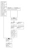
PRISMA flow diagram
Included studies
Types of studies
We included five RCTs that met the inclusion criteria, and summarized the details for each (Arcieri 2015; Deng 2018; Inatani 2021; Jiang 2015; Mahdy 2013) in the 'Characteristics of included studies' table. All RCTs were of parallel‐group design, except for Inatani 2021, which applied parallel‐group design only during the first week; after week one, the study applied a non‐randomized design where participants in the sham group were allowed to receive anti‐VEGF injections if meeting re‐treatment criteria. Of all five RCTs, the maximum planned or stated length of follow‐up varied from one month (Deng 2018), to 13 weeks (Inatani 2021), to 18 months (Arcieri 2015), and 24 months (Mahdy 2013). Two RCTs, both multicentered studies, were registered in a clinical trials registry (Arcieri 2015 and Inatani 2021). Results for Arcieri 2015, Deng 2018, Jiang 2015, and Mahdy 2013 came from journal publications. All were published in English, except Deng 2018 and Jiang 2015, which were published in Chinese. Deng 2018 and Mahdy 2013 declared no conflict of interest, and did not report information about a funding source; Jiang 2015 did not report the source of funding nor conflict of interest; Arcieri 2015 was an unfunded study; and Inatani 2021 was sponsored by Bayer and Regeneron Pharmaceuticals.
Types of participants
All together, the five RCTs enrolled 356 eyes of 353 adult participants with uncontrolled NVG from China (Deng 2018 and Jiang 2015), Brazil (Arcieri 2015), Egypt (Mahdy 2013), and Japan (Inatani 2021). All five RCTs included both men and women; the mean age of participants was 55 years or older. In Mahdy 2013 and Arcieri 2015, the numbers of participants who had CRVO or PDR as the underlying cause for NVG at baseline were comparable between the intervention and control groups. In Inatani 2021, while the number of participants who had PDR and ocular ischemic syndrome as the underlying cause for NVG at baseline was comparable between groups, approximately twice as many participants in the intervention group had CRVO or other conditions at baseline. Data on the underlying cause for NVG were unavailable in the remaining two RCTs.
Arcieri 2015 required that all participants underwent PRP at least two weeks before enrolment; Inatani 2021 required an administration of a combination of three topical IOP‐lowering drugs during a run‐in phase before the first treatment; Mahdy 2013 also recruited participants undergoing PRP, but did not specify the exact timing. In Arcieri 2015, mean preoperative IOP was 40.10 mmHg (standard deviation [SD] 13.33) in the anti‐VEGF group, and 38.35 mmHg (SD 10.34) in the control group; in Mahdy 2013, it was 38.4 mmHg (SD 4.7) in the anti‐VEGF group, and 38.5 mmHg (SD 7.5) in the control group. Similarly, Inatani 2021 reported mean preoperative IOP of 33 mmHg (SD 10) in the anti‐VEGF group and 37 mmHg (SD 9) in the control group. Data on mean baseline IOP were unavailable in the remaining two RCTs.
Types of interventions
The anti‐VEGF medications the RCTs examined included intravitreal ranibizumab (Deng 2018; Jiang 2015), bevacizumab (Arcieri 2015; Mahdy 2013), and aflibercept (Inatani 2021). The adjunct treatments were PRP (as required: Deng 2018; Inatani 2021; Jiang 2015) and PRP combined with an Ahmed glaucoma valve implant (Arcieri 2015; Mahdy 2013). Inatani 2021 used sham injections in the control group and intravitreal anti‐VEGF injections were allowed in the control group if re‐treatment criteria were met after week one. At week one, 81.5% of patients in the sham arm met re‐treatment criteria and received a single dose of anti‐VEGF injection. In all RCTs, participants were treated with anti‐glaucoma medications, as required, to improve control of their IOP.
Types of outcomes
Critical outcome
Proportion of participants who achieved control of IOP
Of five RCTs, four reported achieving control of IOP using different definition at various time points (Arcieri 2015; Deng 2018; Inatani 2021; Mahdy 2013). Arcieri 2015 defined achieving control of IOP as (1) achieving a postoperative IOP between 6 mmHg and 21 mmHg, with or without anti‐glaucoma medications, and (2) IOP reduction of at least 30% from baseline, at 1 day, 1 week, 2 weeks, and 1, 3, 6, 12, 18, and 24 months. Deng 2018 defined such an outcome as (1) for markedly effective: IOP < 16 mmHg without further damage to the visual field or need for postoperative pharmacologic treatment for NVG; (2) effective: IOP 16 to 21 mmHg without further damage to the visual field and required postoperative local pharmacologic treatment at one month. Mahdy 2013 defined achieving control of IOP as achieving an unmedicated IOP ≤ 21 mmHg but ≥ 10 mmHg, without the need for additional glaucoma surgery or visually devastating complications at 3, 5, 7, 10, and 15 days, and at 1, 3, 6, 9, 12 and 18 months. Inatani 2021 reported the proportion of participants who achieved IOP ≤ 21 mmHg at weeks 1, 2, 5, 9, and 13.
Important outcomes
Mean intraocular pressure
All five RCTs reported mean IOP. Arcieri 2015 reported mean intraocular pressure at baseline, 1, 7, 15 days, and 1, 3, 6, 9, 12, 18, and 24 months. Deng 2018 reported mean intraocular pressure at postoperative 1 month. Mahdy 2013 reported such an outcome at 1, 3, 6 months, 1 year, and 18 months. Jiang 2015 reported the mean IOP immediately following treatment; however, the interpretation of the results is uncertain, because it is unclear whether this analysis accounted for the potential unit of analysis issues in this study. Inatani 2021 examined the change in IOP from baseline to 1 week as the critical outcome, and reported the change in IOP from baseline to weeks 2, 5, 9, and 13 as the important outcomes.
Proportion of participants with improvement in visual acuity
Of all included RCTs, only two RCTs reported the improvement in visual acuity at different time points. Deng 2018 and Mahdy 2013 reported the proportion of visual acuity improvement at one month and 18 months, respectively, without clearly specifying the definition of visual acuity improvement. Arcieri 2015 reported differences in postoperative visual acuity without specifying the measurement time. Likewise, Jiang 2015 reported the visual acuity outcome; however, the interpretation of such results is uncertain because it is unclear whether this analysis accounted for the potential unit of analysis issues and which unit of measurement was used for this visual acuity outcome.
Proportion of participants with complete regression of new iris vessels
Of five RCTs, three (Arcieri 2015; Deng 2018, Mahdy 2013) reported the proportion of participants with complete regression of new iris vessels at different time points. Mahdy 2013 reported the complete regression of new vessels outcome in the anti‐VEGF medications arm at one week, but did not provide results for the control group. Deng 2018 and Arcieri 2015 reported such an outcome at one month, and from 1.5 to 3 years, respectively.
Proportion of participants with relief of symptoms
No RCTs reported on the proportion of participants with relief of pain and resolution of redness at any time points.
Adverse events
We specified six adverse events to compare such effects of anti‐VEGFs in the protocol: proportion of participants with intraocular infection or inflammation (endophthalmitis), hypotony (IOP ≤ 6 mmHg), development of vitreous hemorrhage, tractional retinal detachment, no light perception, and other serious adverse events, including systemic thrombosis, stroke, and coronary thrombosis. Only Mahdy 2013 reported the proportion of participants with hypotony. Only Arcieri 2015 documented occurrences of tractional retinal detachment. Inatani 2021 was the only RCT that reported serious adverse events occurring during the non‐randomized period.
Excluded studies
We excluded 27 studies after full‐text review (see reasons in the 'Characteristics of excluded studies' table), including 26 studies identified from the updated searches and one study from the Characteristics of ongoing studies section of the previous review due to its inactive status since 2016 (NCT02914626).
In summary, of all 27 excluded studies, 14 studies were not RCTs, four studies did not include participants with NVG, three studies did not evaluate interventions eligible for this review, four studies did not evaluate comparator eligible for this review, and two studies were registered in a clinical trial register and listed as 'unknown/incomplete' for more than eight years. If these two incomplete studies are completed, or their status is updated, we will reassess them for eligibility in future versions of this review (EUCTR2007‐000585‐21‐IE and NCT02914626).
Risk of bias in included studies
Figure 2 presents our assessment of the risk of bias in the included RCTs.
2.
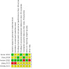
Risk of bias summary: review authors' judgments about each risk of bias item for each included study
Allocation
Most included RCTs had either low or unclear risk of bias for sequence generation and allocation concealment domains, except for Jiang 2015, which was judged to have a high risk of bias for both domains. Arcieri 2015 described using a computer‐generated randomization table to generate the randomization sequence but did not describe how this sequence was concealed. Deng 2018 and Mahdy 2013 provided no information about generating the random sequence or concealment of allocation. Inatani 2021 used stratified randomization with a 1:1 ratio and performed allocation concealment using an interactive voice response system. Jiang 2015 assigned participants to interventions based on a medical record number, which was not truly random. Accordingly, we assessed Arcieri 2015 as having low risk of bias for sequence generation and unclear risk of bias for allocation concealment; we assessed Deng 2018 and Mahdy 2013 as having unclear risk of bias for both sequence generation and allocation concealment; we assessed Inatani 2021 as having low risk of bias for both sequence generation and allocation concealment; we assessed Jiang 2015 as having high risk of bias for both sequence generation and allocation concealment.
Blinding
We assessed Deng 2018 and Inatani 2021 as having low risk of bias for masking of participants and personnel and assessed the remaining three studies (Arcieri 2015; Jiang 2015; Mahdy 2013) as having unclear risk of bias for masking of participants and personnel because they did not provide sufficient information. In terms of masking of outcome assessment, Arcieri 2015 and Inatani 2021 commented that the IOP assessors did not know which group participants were assigned to; thus, we assessed these two RCTs as having low risk of bias for masking of outcome assessors for the primary outcome. We assessed all the remaining four RCTs (Arcieri 2015; Deng 2018; Jiang 2015; Mahdy 2013) as having unclear risk of bias for masking of outcome assessment because they did not provide sufficient information.
Incomplete outcome data
We assessed Deng 2018 as having low risk of bias for incomplete outcome data as the authors reported data for all participants included in the study. However, we assessed Inatani 2021 as having high risk of bias for incomplete outcome data as 22% of the participants did not complete the RCT mainly due to progressive diseases, and such losses of follow‐up due to progressive diseases were unbalanced between groups. Since the RCT handled the missing data using last‐observation‐carried‐forward, it could bias the results. For the remaining RCTs (Arcieri 2015; Jiang 2015; Mahdy 2013), we assessed them as having unclear risk of bias for incomplete outcome data because we did not have sufficient information to permit judgment. In Arcieri 2015, the data for five participants (25%) in each arm were not included at the one‐year follow‐up, but the reasons for exclusion were not reported.
Selective reporting
We assessed Arcieri 2015 and Inatani 2021 as having low risk of bias for selective reporting of outcomes because the full‐text reports included all outcomes specified on clinical trial registries. We judged the remaining three RCTs (Deng 2018; Jiang 2015; Mahdy 2013) as having unclear risk of bias for this domain for because the protocols or trial registrations were not available.
Other potential sources of bias
We assessed Inatani 2021 as having high risk of bias for other potential sources of bias for three reasons: 1) IOP was assessed using applanation tonometry or Tono‐Pen, but no further details were reported about the number of patients assessed with each of them. It is known that these two tonometers provide measurements that are not interchangeable; 2) the funding was sponsored by a pharmaceutical company, which was involved in study design, data collection, data analysis and interpretation, review preparation, and manuscript submission; and 3) participants who were randomized to sham injection could receive aflibercept injections after one week. We assessed the remaining four RCTs as having unclear risk of bias for other potential sources of bias: Arcieri 2015 did not report conflicts of interest; sources of funding were unclear in Deng 2018, Jiang 2015, and Mahdy 2013.
Effects of interventions
See: Table 1
Critical outcome
Proportion of participants who achieved control of IOP
Four RCTs reported the proportion of participants achieving IOP ≤ 21 mmHg with or without anti‐glaucoma medications (i.e. success) at one month or beyond (Arcieri 2015; Deng 2018; Inatani 2021; Mahdy 2013). We did not conduct meta‐analyses, because of small numbers of included RCTs for each time point.
Two RCTs (Deng 2018 and Inatani 2021) reported our critical time point, which was at four to six weeks. However, Inatani 2021 applied a non‐randomized design during that time period. Deng 2018 found that the anti‐VEGF group had a 1.3‐fold higher chance of achieving control of IOP at one month than the non‐anti‐VEGF group (RR 1.32, 95% 1.10 to 1.59; 93 participants; Analysis 1.1). We graded certainty of the evidence as low due to limitations in the study design due to insufficient information to permit judgment (‐1) and imprecision of results due to the small sample size (‐1) (Table 1).
1.1. Analysis.
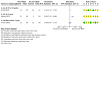
Comparison 1: Anti‐VEGF medications vs no anti‐VEGF medications, Outcome 1: Proportion of participants who achieved control of IOP
The two remaining RCTs (Arcieri 2015 and Mahdy 2013) reported time points at one year or beyond. Arcieri 2015 found that anti‐VEGFs may increase the chance of achieving control of IOP by one‐fold as compared with no anti‐VEGFs (RR 1.08; 95% CI: 0.67 to 1.75; 40 participants; range 1.5 years to 3 years; Analysis 1.1). However, Mahdy 2013 reported that the anti‐VEGF group had a three times higher chance of achieving control of IOP at one year (RR 3.00; 95% CI: 1.35 to 6.68; 40 participants; Analysis 1.1).
Important outcomes
Mean intraocular pressure
All five RCTs reported mean IOP, ranging from one week to 18 months (Table 2; Table 3), three (Arcieri 2015; Deng 2018; Mahdy 2013) of which reported mean IOP at four to six weeks (Analysis 1.2). Based on data from 173 participants, anti‐VEGFs decreased the mean IOP by 6.37 mmHg (95% CI: ‐10.09 to ‐2.65; P = 0.0008) as compared with no anti‐VEGFs (I2 = 95%, P < 0.00001), suggesting that anti‐VEGFs may reduce mean IOP at four to six weeks when compared with no anti‐VEGFs. We graded the certainty of the evidence for this outcome as very low due to unclear risk of bias (‐1), imprecision of results due to small sample size (‐1), and inconsistency due to high statistical heterogeneity (‐1) (Table 1).
1. Arcieri 2015 – IOP at baseline and follow‐up.
| Time point | IVB + PRP + AGV IOP (mean ± SD) | PRP + AGV (control) IOP (mean ± SD) | P value |
| Baseline | 40.10 ± 13.33 (N = 20) | 38.35 ± 10.34 (N = 20) | 0.6454 |
| 1 day | 10.68 ± 5.74 (N = 20) | 10.85 ± 6.74 (N = 20) | 0.9348 |
| 7 days | 10.35 ± 4.76 (N = 20) | 11.45 ± 5.77 (N = 20) | 0.5148 |
| 15 days | 14.00 ± 6.13 (N = 20) | 16.50 ± 7.34 (N = 20) | 0.2498 |
| 1 month | 17.45 ± 4.65 (N = 20 ) | 19.05 ± 6.16 (N = 20) | 0.3597 |
| 3 months | 18.30 ± 6.55 (N = 18 ) | 18.33 ± 5.44 (N = 17) | 0.9866 |
| 6 months | 16.78 ± 7.47 (N = 16) | 16.33 ± 4.35 (N = 17) | 0.3827 |
| 9 months | 18.31 ± 8.93 (N = 16) | 16.17 ± 4.60 (N = 16) | 0.8898 |
| 12 months | 17.40 ± 9.99 (N = 15) | 16.00 ± 3.98 (N = 15) | 0.4598 |
| 18 months | 14.57 ± 1.72 (N = 15) | 18.37 ± 1.06 (N = 14) | 0.0002 |
| 24 months | 14.43 ± 0.53 (N = 14) | 16.67 ± 4.40 (N = 12) | 0.0526 |
AGV: Ahmed glaucoma valve IOP: intraocular pressure (mmHg) IVB: intravitreal bevacizumab N: number of eyes PRP: pan retinal photocoagulation SD: standard deviation
2. Mahdy 2012 – IOP at baseline and follow‐up.
| Time point | Avastin + PRP + AGV (N = 20 eyes) IOP (mean ± SD) | PRP + AGV (control) (N = 20 eyes) IOP (mean ± SD) |
| Preoperative | 38.4 ± 4.7 | 38.5 ± 7.5 |
| 1 week postoperative | 10.0 ± 3.1 | 13.5 ± 4.1 |
| 1 month postoperative | 13 ± 2.2 | 19.5 ± 2.4 |
| 3 months postoperative | 14 ± 1.9 | 22 ± 1.6 |
| 6 months postoperative | 16 ± 2.0 | 28 ± 3.1 |
| 12 months postoperative | 16 ± 7.0 | 28 ± 8.4 |
| 18 months postoperative | 16 ± 4.2 | 28 ± 6.5 |
AGV: Ahmed glaucoma valve IOP: intraocular pressure (mmHg) N: number of eyes PRP: pan retinal photocoagulation SD: standard deviation
1.2. Analysis.
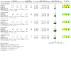
Comparison 1: Anti‐VEGF medications vs no anti‐VEGF medications, Outcome 2: Mean intraocular pressure
Two (Arcieri 2015; Mahdy 2013) of the five RCTs reported mean IOP at three months, six months, one year, and more than one year (Analysis 1.2). Based on the combined estimate from 75 participants, anti‐VEGFs may decrease mean IOP by 4.25 mmHg (95% CI ‐12.05 to 3.54, P = 0.28; I2 = 93%, P for I2 = 0.0002) at three months, by 5.93 mmHg (95% CI ‐18.13 to 6.26, P = 0.34; I2 = 97%, P for I2 < 0.00001) at six months, by 5.36 mmHg (95% CI ‐18.50 to 7.77, P = 0.42; I2 = 92%, P for I2 = 0.0003) at one year, and by 7.05 mmHg (95% CI ‐16.61 to 2.51, P = 0.15; I2 = 95%, P for I2 < 0.00001) at more than one year when compared with no anti‐VEGF. However, these results remain uncertain.
Proportion of participants with improvement in visual acuity
Two RCTs reported the proportion of participants who achieved improvement in visual acuity at one month, or at 18 months (Deng 2018; Mahdy 2013).
Deng 2018 was the only RCT that reported this outcome at one month. Participants receiving anti‐VEGFs had 2.6 times higher chance of improving visual acuity when compared with those not receiving anti‐VEGFs (RR 2.55, 95% CI 1.60 to 4.08; 93 participants; Analysis 1.3); however, the study did not clearly specify a definition of the improvement in visual acuity. We graded the certainty of the evidence for this outcome measured at four to six weeks as very low due to imprecision (‐1), indirectness (‐1) of results, as well as limitations in the design due to insufficient information to permit judgment (‐1) (Table 1).
1.3. Analysis.
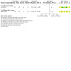
Comparison 1: Anti‐VEGF medications vs no anti‐VEGF medications, Outcome 3: Proportion of participants with improvement in visual acuity
Mahdy 2013 reported this outcome at 18 months. They found that the anti‐VEGF group had a four‐fold higher chance of improving visual acuity than the non‐anti‐VEGF group (RR 4.00, 95% CI 1.33 to 12.05; 40 participants; Analysis 1.3). Arcieri 2015 reported no statistically significant difference in postoperative visual acuity (P > 0.1270), but did not specify the measurement time point. Jiang 2015 reported that visual acuity was higher in the anti‐VEGF group compared to the non‐anti‐VEGF group, but the results were uncertain, due to the limitations of study design.
Proportion of participants with complete regression of new iris vessels
All five RCTs noted that a larger proportion of participants in the anti‐VEGF medications arm had more regression of iris new vessels at various time points. Three out of five (Arcieri 2015; Deng 2018, Mahdy 2013) reported on the proportion of participants with complete regression of new iris vessels at 1 week, 1 month, and from 1.5 to 3 years, respectively.
Deng 2018 was the only RCT that reported this outcome at one month. Anti‐VEGFs had a 2.6 times higher chance of complete regression of new iris vessels when compared with no anti‐VEGFs (RR 2.63, 95% CI 1.65 to 4.18; 93 participants; Analysis 1.4). We graded the certainty of the evidence for this outcome measured at four to six weeks as low due to imprecision from a small sample size (‐1) and limitations in the design due to insufficient information to permit judgment (‐1) (Table 1).
1.4. Analysis.
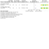
Comparison 1: Anti‐VEGF medications vs no anti‐VEGF medications, Outcome 4: Proportion of participants with complete regression of new iris vessels
Arcieri 2015 reported the proportion of participants with complete regression of new iris vessels at more than one year, and found that the anti‐VEGF group had 3.2 times higher chance of complete regression of iris new vessels when compared with the non‐anti‐VEGF group (RR 3.20, 95% CI 1.45 to 7.05; 40 participants; Analysis 1.4)
Proportion of participants with relief of symptoms
No RCTs reported on the proportion of participants with relief of pain and resolution of redness at any time points.
Adverse events
Proportion of participants with intraocular infection or inflammation (endophthalmitis)
No RCTs reported on the proportion of participants with intraocular infection or inflammation (endophthalmitis) at any time points.
Proportion of participants with hypotony (IOP ≤ 6 mmHg)
Only Mahdy 2013 reported incidents of hypotony. The estimated RR was 0.67 (95% CI: 0.12 to 3.57; 40 participants; Analysis 1.5), suggesting no evidence of differences in hypotony incidence comparing anti‐VEGFs with no anti‐VEGFs. We graded the certainty of the evidence as low due to limitations in the design due to insufficient information to permit judgment (‐1) and imprecision (‐1) of results from a small sample size (Table 1).
1.5. Analysis.
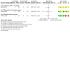
Comparison 1: Anti‐VEGF medications vs no anti‐VEGF medications, Outcome 5: Complications
Proportion of participants with development of vitreous hemorrhage.
No RCTs reported on the proportion of participants with vitreous hemorrhage at any time points.
Proportion of participants with tractional retinal detachment
Only Arcieri 2015 reported the incident of tractional retinal detachment. The estimated RR was 0.33 (95% CI: 0.01 to 7.72; 40 participants; Analysis 1.5), suggesting no evidence of differences in tractional retinal detachment incidence comparing anti‐VEGFs with no anti‐VEGFs. We graded the certainty of the evidence as low due to limitations in the design due to insufficient information to permit judgment (‐1) and imprecision (‐1) of results from a small sample size (Table 1).
Proportion of participants with no light perception
No RCTs reported on the proportion of participants with no light perception at any time points.
Proportion of participants with serious adverse events (e.g., systemic thrombosis, stroke, and coronary thrombosis)
No RCTs reported incidents of serious adverse events. Inatani 2021 reported that severe ocular treatment‐emergent adverse events occurred in both groups during the non‐randomized period, where participants could receive both sham injection and aflibercept injection if the re‐treatment criteria were met. During the non‐randomized period, three severe ocular treatment‐emergent adverse events were reported. For the anti‐VEGF medications arm, one participant (3.7%) developed retinal artery occlusion and one (3.7%) developed retinal vein occlusion. For the sham/anti‐VEGF arm, one (3.7%) developed diabetic retinopathy. In addition, one participant (3.7%) in the sham/anti‐VEGF arm developed nonfatal myocardial infarction during this non‐randomized study period.
Discussion
Summary of main results
We included five eligible RCTs (Arcieri 2015; Deng 2018; Inatani 2021; Jiang 2015; Mahdy 2013) in this updated review. The five RCTs, taken together, randomized 356 eyes of 353 adult participants to treatment with either anti‐VEGF medications or to treatment without anti‐VEGF medications. When compared with no anti‐VEGFs, anti‐VEGFs may be effective in achieving control of IOP, improving visual acuity, and achieving complete regression of new iris vessels, with very low to low certainty of evidence. Likewise, low certainty of evidence suggested that anti‐VEGFs may be effective in reducing mean IOP in the short term (four to six weeks) while the long‐term effects remain uncertain. Anti‐VEGFs had a comparable risk of hypotony and tractional retinal detachment to no anti‐VEGFs; however, the certainty of evidence for both adverse outcomes was low due to insufficient information to permit judgment.
Overall completeness and applicability of evidence
Of the five RCTs included, three were available through journal publications and trial registries. Four included RCTs were conducted in different geographic locations, including Brazil, China, Egypt, and Japan. The applicability to other populations including Caucasians is uncertain. All included RCTs recruited middle‐aged male and female participants, which were well representative of the typical NVG patient population (Rodrigues 2016). Regarding the interventions, three main anti‐VEGF medications were used across RCTs, including ranibizumab, bevacizumab, and aflibercept, which covered all available traditional options on the market (Andrés‐Guerrero 2017). The adjunct treatment was either PRP alone, or with a glaucoma drainage device using the Ahmed glaucoma valve implantation, the first treatment option for refractory glaucoma (Rodrigues 2016). All RCTs allocated no pharmacologic treatment to the control group, except for one RCT that allowed the control group to receive anti‐VEGFs if re‐treatment criteria were met after the first week. For the review's critical outcome, achieving control of IOP, each RCT reported such an outcome at different time points and using different definitions, which did not permit pooling via meta‐analyses. Likewise, similar issues occurred for the important outcomes of improvement in visual acuity and complete regression of new iris vessels. For the most commonly reported review outcome, mean IOP, most RCTs did not specify the method of IOP measurement. Such variations in population's ethnicities, pharmacologic treatment, adjunct treatment, as well as insufficient information on outcome measurements across included RCTs are possibly potential sources of between‐study heterogeneity. Therefore, these factors should be considered when interpreting the evidence.
Quality of the evidence
We graded the certainty of the evidence as low to very low for all outcomes reported by the included studies, mostly because of small sample size and unclear risk of bias domains due to insufficient information to permit judgment. No relevant and sufficient data were available for meta‐analysis for all but one outcome (mean IOP control) which we specified as a critical outcome for this review.
Potential biases in the review process
We followed standard Cochrane methods outlined in the Cochrane Handbook for Systematic Reviews of Interventions to minimize potential for introducing bias in the review process (Higgins 2011a). We worked with an information specialist to design a comprehensive search strategy and we searched multiple electronic databases, including clinical trial registries. We did not limit our search by date or by language. The review team constituted content experts and methodologists; two review authors completed tasks, such as screening references for inclusion and assessing studies, in duplicate, in order to minimize errors and bias.
Agreements and disagreements with other studies or reviews
This review showed better IOP reduction and regression of iris neovascularization in the short term with the use of anti‐VEGF medications in NVG, which were consistent with other non‐randomized studies (Grover 2009; Gupta 2009; Inatani 2020). Trials included in this review reported varying results in controlling IOP with the use of anti‐VEGF medications in the long term. Published meta‐analyses showed inconsistent findings, possibly due to methodological limitations of these reviews (Dong 2018; Hwang 2015; Hwang 2021; Zhou 2016). Dong 2018 conducted incomprehensive literature searches while Hwang 2021 focused only on bevacizumab as the adjuvant pharmacologic treatment, included not only RCTs but also observational studies, and used inappropriate statistical methods for analyzing their data.
Authors' conclusions
Implications for practice.
Anti‐VEGFs as an adjunct to conventional treatment could help reduce IOP in NVG in the short term (four to six weeks). However, such an effect remains uncertain in the longer term. We did not find sufficient evidence from which to draw definite conclusions regarding the benefits of the use of anti‐VEGF medications alone, or as an adjunct to existing modalities for the treatment of NVG. Likewise, evidence is inadequate to assess the differences in adverse events with or without the use of anti‐VEGF medications.
Implications for research.
Future trials should target a larger sample size and adopt standardized conventional therapy and treatment groups.
To increase the translatability of the results into clinical practice, a therapeutic algorithm should be clearly defined, thus involving the time of the injection in NVG management (i.e. how many days before surgery), the site of the injection, additional therapy, and the number of injections during the follow‐up.
Moreover, randomization in future trials should be stratified by underlying etiology for NVG or proliferative diabetic retinopathy, and the extent of peripheral anterior synechiae or angle closure, because both factors may modify the effectiveness of treatment, and imbalance in either could confound the results.
Lastly, using a core outcome set (mean IOP and regression of new iris vessels) measured at standardized time of follow‐up would allow data to be combined in a meta‐analysis.
What's new
| Date | Event | Description |
|---|---|---|
| 9 June 2023 | Amended | Typos in the Summary of Finding table corrected. |
History
Protocol first published: Issue 3, 2009 Review first published: Issue 10, 2013
| Date | Event | Description |
|---|---|---|
| 3 April 2023 | New citation required but conclusions have not changed | Updated searches included 2 new trials; conclusion not changed. |
| 3 April 2023 | New search has been performed | Updated searches, with eligibility criteria and analysis plan (subgroup analysis) modified |
| 29 February 2020 | New citation required and conclusions have changed | Issue 2, 2020: 4 new studies added: Arcieri 2015; Jiang 2015; Mahdy 2013; NCT02396316 |
| 29 February 2020 | New search has been performed | Issue 2, 2020: Searches updated 22 March 2019 |
Acknowledgements
This review was produced with the assistance of protocol development and review completion workshops conducted by the South Asian Cochrane Network, based at the Professor B.V. Moses and ICMR Advanced Center for Research Training in Evidence Informed Healthcare, Christian Medical College, Vellore, India. We would like to acknowledge Dr. Prathap Tharyan, the director of the Center for his never ending encouragement and help. The South Asian Cochrane Network is funded by the ICMR.
We thank Dr. Soumik Kalita for guidance through the process of training and working with Cochrane software. We thank Iris Gordon, Information Specialist for Cochrane Eyes and Vision (CEV), for designing and undertaking the electronic search strategies. We thank Dr. Barbara Hawkins and CEV methodologists for comments on the protocol and full review.
The authors are grateful to the following peer reviewers for their time and comments: Amanda Bicket (Johns Hopkins Medicine), Bill Vaughan (National Committee to Preserve Social Security and Medicare), and one peer reviewer who wished to remain anonymous.
The 2022 review update was managed by CEV@US and was signed off for publication by Tianjing Li and Richard Wormald.
Appendices
Appendix 1. CENTRAL search strategy
#1 MeSH descriptor: [Glaucoma, Neovascular] explode all trees #2 (glaucoma* or angle* or iris or anterior) near/4 (neovascular*) #3 (haemorrhagic or hemorrhagic or thrombotic or congestive or rubeotic or secondary) near/4 (glaucoma*) #4 NVG or NVI #5 {or #1‐#4} #6 MeSH descriptor: [Angiogenesis Inhibitors] explode all trees #7 (Angiogenesis or Neovascularization or Angiogenic or Angiogenetic) near/2 (Inhibitor* or Antagonist*) #8 (Angiostatic or "Anti Angiogenetic" or "Anti Angiogenic" or Antiangiogenic or "Anti Angiogenesis" or Antiangiogenesis) near/1 (Agent* or drug* or effect*) #9 MeSH descriptor: [Angiogenesis Inducing Agents] explode all trees #10 (Angiogenesis or Neovascularization or Angiogenic or Angiogenetic) near/2 (agent* or Stimulator* or Inducer* or factor* or effect*) #11 MeSH descriptor: [Endothelial Growth Factors] explode all trees #12 MeSH descriptor: [Vascular Endothelial Growth Factors] explode all trees #13 VEGF or Vasculotropin or Vascular Permeability Factor* #14 macugen* or pegaptanib* or "eye 001" or eye001 or "NX 1838" or nx1838 or "222716‐86‐1" #15 MeSH descriptor: [Ranibizumab] explode all trees #16 lucentis* or lucentris or rhufab* or ranibizumab* or "347396‐82‐1" #17 MeSH descriptor: [Bevacizumab] explode all trees #18 bevacizumab* or avastin* or altuzan or "nsc 704865" or nsc704865 or "216974‐75‐3" #19 aflibercept* or Eylea or Zaltrap or "AVE 0005" or "AVE 005" or "845771‐78‐0" or "862111‐32‐8" #20 antiVEGF #21 (endothelial near/2 growth near/2 factor*) #22 {or #6‐#21} #23 #5 and #22
Appendix 2. MEDLINE Ovid search strategy
1. Glaucoma, Neovascular/ 2. ((glaucoma* or angle* or iris or anterior) adj4 neovascular*).tw. 3. ((haemorrhagic or hemorrhagic or thrombotic or congestive or rubeotic or secondary) adj4 glaucoma*).tw. 4. (NVG or NVI).tw. 5. or/1‐4 6. exp angiogenesis inhibitors/ 7. ((Angiogenesis or Neovascularization or Angiogenic or Angiogenetic) adj2 (Inhibitor* or Antagonist*)).tw. 8. ((Angiostatic or "Anti Angiogenetic" or "Anti Angiogenic" or Antiangiogenic or "Anti Angiogenesis" or Antiangiogenesis) adj1 (Agent* or drug* or effect*)).tw. 9. exp angiogenesis inducing agents/ 10. ((Angiogenesis or Neovascularization or Angiogenic or Angiogenetic) adj2 (agent* or Stimulator* or Inducer* or factor* or effect*)).tw. 11. exp endothelial growth factors/ 12. exp vascular endothelial growth factors/ 13. (VEGF or Vasculotropin or Vascular Permeability Factor*).tw. 14. (macugen* or pegaptanib* or "eye 001" or eye001 or "NX 1838" or nx1838 or "222716‐86‐1").tw. 15. exp Ranibizumab/ 16. (lucentis* or lucentris or rhufab* or ranibizumab* or "347396‐82‐1").tw. 17. exp Bevacizumab/ 18. (bevacizumab* or avastin* or altuzan or "nsc 704865" or nsc704865 or "216974‐75‐3").tw. 19. (aflibercept* or Eylea or Zaltrap or "AVE 0005" or "AVE 005" or "845771‐78‐0" or "862111‐32‐8").tw. 20. antiVEGF.tw. 21. (endothelial adj2 growth adj2 factor*).tw. 22. or/6‐21 23. 5 and 22
Appendix 3. Embase.com search strategy
1. 'neovascular glaucoma'/exp 2. ((glaucoma* OR angle* OR iris OR anterior) NEAR/4 neovascular*):ab,ti 3. ((haemorrhagic OR hemorrhagic OR thrombotic OR congestive OR rubeotic OR secondary) NEAR/4 glaucoma*):ab,ti 4. (NVG OR NVI):ab,ti 5. #1 OR #2 OR #3 OR #4 6. 'angiogenesis inhibitor'/exp 7. ((Angiogenesis OR Neovascularization OR Angiogenic OR Angiogenetic) near/2 (Inhibitor* OR Antagonist*)):ab,ti 8. ((Angiostatic OR "Anti Angiogenetic" OR "Anti Angiogenic" OR Antiangiogenic OR "Anti Angiogenesis" OR Antiangiogenesis) near/1 (Agent* OR drug* OR effect*)):ab,ti 9. 'angiogenesis'/exp 10. 'angiogenic factor'/exp 11. ((Angiogenesis OR Neovascularization OR Angiogenic OR Angiogenetic) near/2 (agent* OR Stimulator* OR Inducer* OR factor* OR effect*)):ab,ti 12. 'endothelial cell growth factor'/exp 13. 'vasculotropin'/exp 14. (VEGF OR Vasculotropin OR "Vascular Permeability Factor*"):ab,ti 15. (macugen* OR pegaptanib* OR "eye 001" OR eye001 OR "NX 1838" OR nx1838 OR "222716‐86‐1"):ab,ti,tn 16. (lucentis* OR lucentris OR rhufab* OR ranibizumab* OR "347396‐82‐1"):ab,ti,tn 17. (bevacizumab* OR avastin* OR altuzan OR "nsc 704865" OR nsc704865 OR "216974‐75‐3"):ab,ti,tn 18. (aflibercept* OR Eylea OR Zaltrap OR "AVE 0005" OR "AVE 005" OR "845771‐78‐0" OR "862111‐32‐8"):ab,ti,tn 19. antiVEGF:ab,ti 20. (endothelial near/2 growth near/2 factor*):ab,ti,tn 21. #6 OR #7 OR #8 OR #9 OR #10 OR #11 OR #12 OR #13 OR #14 OR #15 OR #16 OR #17 OR #18 OR #19 OR #20 22. #5 AND #21
Appendix 4. PubMed search strategy
1. ((glaucoma* [tw] OR angle* [tw] OR iris [tw] OR anterior [tw]) AND neovascular* [tw]) 2. ((haemorrhagic [tw] OR hemorrhagic [tw] OR thrombotic [tw] OR congestive [tw] OR rubeotic [tw] OR secondary [tw]) AND glaucoma* [tw]) 3. NVG [tw] OR NVI [tw] 4. #1 OR #2 OR #3 5. ((Angiostatic[tw] OR "Anti Angiogenetic"[tw] OR "Anti Angiogenic"[tw] OR Antiangiogenic[tw] OR "Anti Angiogenesis"[tw] OR Antiangiogenesis[tw]) AND (Agent*[tw] OR drug*[tw] OR effect*[tw])) 6. ((Angiogenesis[tw] OR Neovascularization[tw] OR Angiogenic[tw] OR Angiogenetic[tw]) AND (agent*[tw] OR Stimulator*[tw] OR Inducer*[tw] OR factor*[tw] OR effect*[tw])) 7. (VEGF[tw] OR Vasculotropin[tw] OR Vascular Permeability Factor*[tw]) 8. macugen*[tw] OR pegaptanib*[tw] OR "eye 001"[tw] OR eye001[tw] OR "NX 1838"[tw] OR nx1838[tw] OR "222716‐86‐1"[tw] 9. lucentis*[tw] OR lucentris[tw] OR rhufab*[tw] OR ranibizumab*[tw] OR "347396‐82‐1"[tw] 10. bevacizumab*[tw] OR avastin*[tw] OR altuzan[tw] OR "nsc 704865"[tw] OR nsc704865[tw] OR "216974‐75‐3"[tw] 11. aflibercept*[tw] OR Eylea[tw] OR Zaltrap[tw] OR "AVE 0005"[tw] OR "AVE 005"[tw] OR "845771‐78‐0"[tw] OR "862111‐32‐8"[tw] 12. antiVEGF[tw] 13. (endothelial[tw] AND growth[tw] AND factor*[tw]) 14. #5 OR #6 OR #7 OR #8 OR #9 OR #10 OR #11 OR #12 OR #13 15. #4 AND #14 16. Medline[sb] 17. #15 NOT #16
Appendix 5. LILACS search strategy
(MH:C11.525.381.348$ OR ((glaucoma* OR angle* OR iris OR anterior) AND neovascular*) OR ((haemorrhagic OR hemorrhagic OR thrombotic OR congestive OR rubeotic OR secondary) AND glaucoma*) OR NVG OR NVI) AND (MH:D27.505.696.377.077.099$ OR MH:D27.505.696.377.450.100$ OR MH:D27.505.954.248.025$ OR ((Angiogenesis OR Neovascularization OR Angiogenic OR Angiogenetic) AND (Inhibitor$ OR Antagonist$)) OR ((Angiostatic OR "Anti Angiogenetic" OR "Anti Angiogenic" OR Antiangiogenic OR "Anti Angiogenesis" OR Antiangiogenesis) AND (Agent$ OR drug$ OR effect$)) OR MH:D27.505.696.377.077.077$ OR ((Angiogenesis OR Neovascularization OR Angiogenic OR Angiogenetic) AND (agent$ OR Stimulator$ OR Inducer$ OR factor$ OR effect$)) OR MH:D12.644.276.390$ OR MH:D12.776.467.390$ OR MH:D23.529.390$ OR MH:D12.644.276.100.800$ OR MH:D12.776.467.100.800$ OR MH:D23.529.100.800$ OR VEGF OR Vasculotropin OR (Vascular Permeability Factor$) OR Macugen$ OR pegaptanib$ OR "eye 001" OR eye001 OR "NX 1838" OR nx1838 OR "222716‐86‐1" OR MH:D12.776.124.486.485.114.224.060.868$ OR MH:D12.776.124.790.651.114.224.060.868$ OR MH:D12.776.377.715.548.114.224.200.868$ OR lucentis$ OR lucentris OR rhufab$ OR ranibizumab$ OR "347396‐82‐1" OR MH:D12.776.124.486.485.114.224.060.375$ OR Bevacizumab$ OR avastin$ OR altuzan OR "nsc 704865" OR nsc704865 OR "216974‐75‐3" OR aflibercept$ OR Eylea OR Zaltrap OR "AVE 0005" OR "AVE 005" OR "845771‐78‐0" OR "862111‐32‐8" OR antiVEGF OR (endothelial AND growth AND factor$))
Appendix 6. metaRegister of Controlled Trials search strategy
neovascular glaucoma
Appendix 7. ClinicalTrials.gov search strategy
"secondary glaucoma" OR (neovascular AND (glaucoma OR angle OR iris OR anterior))
Appendix 8. ICTRP search strategy
glaucoma AND VEGF OR glaucoma AND Vasculotropin OR glaucoma AND Vascular Permeability Factor OR glaucoma AND macugen OR glaucoma AND pegaptanib OR glaucoma AND eye 001 OR glaucoma AND eye001 OR glaucoma AND NX 1838 OR glaucoma AND nx1838 OR glaucoma AND lucentis OR glaucoma AND lucentris OR glaucoma AND rhufab OR glaucoma AND ranibizumab OR glaucoma AND bevacizumab OR glaucoma AND avastin OR glaucoma AND altuzan OR glaucoma AND nsc704865 OR glaucoma AND aflibercept OR glaucoma AND Eylea OR glaucoma AND Zaltrap OR glaucoma AND antiVEGF OR glaucoma AND endothelial growth factor
Data and analyses
Comparison 1. Anti‐VEGF medications vs no anti‐VEGF medications.
| Outcome or subgroup title | No. of studies | No. of participants | Statistical method | Effect size |
|---|---|---|---|---|
| 1.1 Proportion of participants who achieved control of IOP | 3 | Risk Ratio (M‐H, Random, 95% CI) | Totals not selected | |
| 1.1.1 At 4 to 6 weeks | 1 | Risk Ratio (M‐H, Random, 95% CI) | Totals not selected | |
| 1.1.2 At 1 year | 1 | Risk Ratio (M‐H, Random, 95% CI) | Totals not selected | |
| 1.1.3 More than 1 year | 1 | Risk Ratio (M‐H, Random, 95% CI) | Totals not selected | |
| 1.2 Mean intraocular pressure | 3 | Mean Difference (IV, Random, 95% CI) | Subtotals only | |
| 1.2.1 At 4 to 6 weeks | 3 | 173 | Mean Difference (IV, Random, 95% CI) | ‐6.37 [‐10.09, ‐2.65] |
| 1.2.2 At 3 months | 2 | 75 | Mean Difference (IV, Random, 95% CI) | ‐4.25 [‐12.05, 3.54] |
| 1.2.3 At 6 months | 2 | 73 | Mean Difference (IV, Random, 95% CI) | ‐5.93 [‐18.13, 6.26] |
| 1.2.4 At 1 year | 2 | 70 | Mean Difference (IV, Random, 95% CI) | ‐5.36 [‐18.50, 7.77] |
| 1.2.5 More than 1 year | 2 | 66 | Mean Difference (IV, Random, 95% CI) | ‐7.05 [‐16.61, 2.51] |
| 1.3 Proportion of participants with improvement in visual acuity | 2 | Risk Ratio (M‐H, Random, 95% CI) | Totals not selected | |
| 1.3.1 At 4 to 6 weeks | 1 | Risk Ratio (M‐H, Random, 95% CI) | Totals not selected | |
| 1.3.2 More than 1 year | 1 | Risk Ratio (M‐H, Random, 95% CI) | Totals not selected | |
| 1.4 Proportion of participants with complete regression of new iris vessels | 2 | Risk Ratio (M‐H, Random, 95% CI) | Totals not selected | |
| 1.4.1 At 4 to 6 weeks | 1 | Risk Ratio (M‐H, Random, 95% CI) | Totals not selected | |
| 1.4.2 More than 1 year | 1 | Risk Ratio (M‐H, Random, 95% CI) | Totals not selected | |
| 1.5 Complications | 3 | Risk Ratio (M‐H, Random, 95% CI) | Totals not selected | |
| 1.5.1 Hypotony (IOP ≤ 6 mmHg) | 1 | Risk Ratio (M‐H, Random, 95% CI) | Totals not selected | |
| 1.5.2 Tractional retinal detachment | 1 | Risk Ratio (M‐H, Random, 95% CI) | Totals not selected | |
| 1.5.3 Serious adverse events | 1 | Risk Ratio (M‐H, Random, 95% CI) | Totals not selected |
Characteristics of studies
Characteristics of included studies [ordered by study ID]
Arcieri 2015.
| Study characteristics | ||
| Methods |
Study design: parallel‐group RCT Setting: multicenter trial in Brazil Number randomized: 40 participants Unit of analysis: participant (one study eye per individual) Maximum planned (or stated) length of follow‐up: 24 months Number not included in final analysis: 14 participants |
|
| Participants |
Number of men: 13 in the intervention group and 11 in the comparator group Number of women: 7 in the intervention group and 9 in the comparator group Mean age: 59 years in the intervention group and 62 years in the comparator group Mean IOP at baseline: 40 mmHg in the intervention group and 38 mmHg in the comparator group Inclusion criteria: older than 18 years with uncontrolled NVG, defined as an eye with IOP above 22 mm Hg using maximum tolerated glaucoma medication; PRP at least 2 weeks before enrolment Exclusion criteria: no light perception; NVG secondary to intraocular tumors or uveitis; unwilling or unable to return for follow‐up; pregnancy; learning difficulties, mental illness or dementia; previous cyclodestructive procedure, scleral buckle procedure, or silicone oil surgery |
|
| Interventions |
Intervention (N = 20): 0.05 mL intravitreal bevacizumab (concentration of 25 mg/mL) with Ahmed glaucoma valve implant Comparator (N = 20): 0.05 mL of sterile saline salt solution (placebo) with Ahmed glaucoma valve implant All participants underwent PRP at least 2 weeks prior to enrolment. |
|
| Outcomes |
From prospective clinical trial registration Primary: IOP control, measured six months after randomization with Goldman applanation tonometer Secondary: safety of intravitreal bevacizumab up to six months after randomization |
|
| Notes |
Trial registration: ACTRN12607000577415 Study dates: not reported |
|
| Risk of bias | ||
| Bias | Authors' judgement | Support for judgement |
| Random sequence generation (selection bias) | Low risk | "Eligible patients with NVG were randomised to the following groups using a computer‐generated randomization table". |
| Allocation concealment (selection bias) | Unclear risk | Insufficient information – method of sequence allocation not clearly mentioned to permit judgment of low risk or high risk |
| Blinding of participants and personnel (performance bias) All outcomes | Unclear risk | Unclear risk of bias, insufficient information to permit judgment of low risk or high risk |
| Blinding of outcome assessment (detection bias) All outcomes | Low risk | Low risk of bias as outcome assessor did not know the group to which the participant was assigned: "Ophthalmologists responsible for the patients’ follow‐up were masked to the use of IVB". |
| Incomplete outcome data (attrition bias) All outcomes | Unclear risk | Unclear risk of bias as data from 10 participants, 5 (25%) from each arm were unavailable at the 1‐year follow‐up |
| Selective reporting (reporting bias) | Low risk | No selective reporting identified; outcomes described in trial registration record were reported in full‐text publication. |
| Other bias | Unclear risk | Unclear risk of bias – insufficient information to permit judgment of low risk or high risk of bias; this was an unfunded study. |
Deng 2018.
| Study characteristics | ||
| Methods |
Study design: parallel‐group RCT Setting: Single‐site, university‐affiliated eye center, in China Number randomized: 93 eyes (90 participants) Unit of analysis: eye Maximum planned (or stated) length of follow‐up: 1 month Number not included in final analysis: 0 participants |
|
| Participants |
Number of men: 23 in the intervention group and 27 in the comparator group Number of women: 23 in the intervention group and 20 in the comparator group Mean age: 58.5 years in the intervention group and 57.2 years in the comparator group Mean IOP at baseline: 46.8 in total Inclusion criteria: (1) aged 18‐75 years; (2) in line with the diagnostic criteria of NVG; (3) conventional intraocular pressure lowering drugs are ineffective and require further treatment; (4) before treatment, the patients and their families signed an informed consent for surgery consent. Exclusion criteria: (1) withdrew from the treatment halfway; (2) combined with severe uncontrolled hypertension; (3) combined with congestive heart failure; (4) combined with abnormal liver and kidney function; (5) combined with other eye infections and hyperplasia/sexual diseases; (6) combined with malignant tumors; (7) breastfeeding or pregnant women; (8) received eye surgery |
|
| Interventions |
Intervention (N = 46): Received intravitreal injection of ranibizumab at first. After seven days, treated with 532 nm argon‐green laser for retinal laser photocoagulation (the method was the same as that of the control group) [ranibizumab + PRP]. Comparator (N = 47): Treated with 532 nm argon‐green laser for retinal laser photocoagulation. The treatment was performed 3 to 4 times, with an interval of 5 to 7 days, and pranoprofen eye drops were applied to the eyes for 7 days after the operation, 4 times a day [PRP]. The anti‐VEGF injection was performed one week before PRP. |
|
| Outcomes |
From study method Primary: The clinical efficacy at 1 month is based on the criteria for judging clinical efficacy: ‐ markedly effective: intraocular pressure < 16 mmHg, no further damage to the optic disc visual field, no need for postoperative drug treatment for NVG; ‐ effective: intraocular pressure 16‐21 mmHg, no further damage to the optic disc visual field, and postoperative local drug treatment was required; ‐ ineffective: the above indicators did not change significantly. Effective rate = (marked + effective)/total number of eyes + 100%. Secondary: ‐ The proportion of disappearance of iris neovascularization at 1 month ‐ The proportion of visual acuity improvement at 1 month ‐ Retinal venous circulation time (s) at 1 month ‐ Mean intraocular pressure at 1 month ‐ Mean RNFL thickness at 1 month ‐ Mean visual field defect at 1 month ‐ Mean level of VEGF in the aqueous humor at 1 month ‐ Mean level of PDGF‐C in the aqueous humor at 1 month |
|
| Notes |
Trial registration: none reported Study dates: November 2016 to November 2017 |
|
| Risk of bias | ||
| Bias | Authors' judgement | Support for judgement |
| Random sequence generation (selection bias) | Unclear risk | It is unclear how the randomization was performed. Authors reported that patients were divided by chance but no other details were reported. |
| Allocation concealment (selection bias) | Unclear risk | The authors did not mention how the allocation concealment was performed. |
| Blinding of participants and personnel (performance bias) All outcomes | Low risk | The authors did not mention whether or not participants were masked. However, the risk of bias may be low, as all outcomes were objective parameters. |
| Blinding of outcome assessment (detection bias) All outcomes | Unclear risk | Unclear risk of bias, insufficient information to permit judgment of low risk or high risk |
| Incomplete outcome data (attrition bias) All outcomes | Low risk | Authors reported data of all participants included in the study. |
| Selective reporting (reporting bias) | Unclear risk | The protocol was not available. So, it was unclear whether or not the trial was analyzed according to the prespecified plan. |
| Other bias | Unclear risk | The funding source was not specified. So, it was unclear if the sponsor had influenced the study results. |
Inatani 2021.
| Study characteristics | ||
| Methods |
Study design: parallel‐group RCT Setting: multicenter trial in Japan Number randomized: 54 participants Unit of analysis: participant (one study eye per individual) Maximum planned (or stated) length of follow‐up: 13 weeks Number not included in final analysis: 0 participants |
|
| Participants |
Number of men: 22 in the intervention group and 23 in the comparator group Number of women: 5 in the intervention group and 4 in the comparator group Mean age: 68.1 years in the intervention group and 66.2 years in the comparator group Mean IOP at baseline: 33 mmHg in the intervention group and 37 mmHg in the comparator group Inclusion criteria: "Japanese patients aged >= 20 years were eligible for inclusion if they had a diagnosis of NVG with neovascularization in the anterior segment (iris and anterior chamber angle) and IOP > 25 mmHg due to neovascularization in the study eye." Exclusion criteria: "Angle closure from conditions other than NVG, use of topical ophthalmic atropine sulfatehydrate in the study eye ≤ 30 days before day 1, and use of systemic IOP‐lowering drugs 24 h before the pre‐injection IOP evaluation on day 1." |
|
| Interventions |
Intervention (N = 27): Background therapy plus intravitreal aflibercept 2 mg (IVT‐AFL) at day 1 Comparator (N = 27): Background therapy plus sham injection at day 1 "Additional treatment was administered only if re‐treatment criteria (IOP > 21 mmHg, incomplete regression of iris neovascularization, and IVT‐AFL was deemed necessary) were met. A combination of 3 topical IOP‐lowering drugs was administered during a run‐in phase before the first treatment and was kept unchanged until the pre‐injection IOP evaluation at week 1. PRP was performed on and after day 1 as needed." |
|
| Outcomes |
From study method Primary: Mean change in IOP from baseline to week 1 Secondary: Mean change in IOP from baseline to weeks 2, 5, 9, and 13; proportion of patients with a change >= 1 NVI grade from baseline to week 1, 2, 5, 9, and 13; proportion of patients with a change of >= 1 NVA grade from baseline to weeks 1, 2, 5, 9, and 13; proportion of patients who achieved IOP <= 21 mmHg at weeks 1, 2, 5, 9, and 13; safety outcomes |
|
| Notes |
Trial registration: NCT02396316 Study dates: 2 April 2015 to 6 September 2016 |
|
| Risk of bias | ||
| Bias | Authors' judgement | Support for judgement |
| Random sequence generation (selection bias) | Low risk | The authors clearly reported that randomization was performed in a 1:1 ratio, which was stratified by baseline in the stage of NVG (open‐ or closed‐angle). The baseline characteristics in both groups were similar in terms of sex, age, stage of NVG, and mean IOP at baseline, suggesting effective randomization. |
| Allocation concealment (selection bias) | Low risk | Randomization and participation allocation were double‐masked throughout the study. Allocation concealment was performed using an interactive voice response system and interactive web response system. |
| Blinding of participants and personnel (performance bias) All outcomes | Low risk | The author mentioned that the trial was double‐masked without clearly specifying whether or not participants were masked. However, the risk of bias may be low, as all outcomes were objective parameters. |
| Blinding of outcome assessment (detection bias) All outcomes | Low risk | According to the trial registry, the author mentioned that outcome assessors were masked |
| Incomplete outcome data (attrition bias) All outcomes | High risk | 22% of participants did not complete the study mainly due to progressive diseases. The losses of follow‐up due to progressive diseases were unbalanced between groups. The missing data were handled using last‐observation‐carried‐forward, which was not an appropriate method to handle missing data. |
| Selective reporting (reporting bias) | Low risk | The trial was analyzed according to the prespecified plan in terms of outcome measurements (e.g. definition, scales, and time points) within each outcome. |
| Other bias | High risk | 1) IOP was assessed using applanation tonometry or Tono‐Pen, but no further details were reported about the number of patients assessed with each of them. It is known that these two tonometers provide measurements that are not interchangeable. 2) The funding was sponsored by a pharmaceutical company, which was involved in study design, data collection, data analysis and interpretation, review preparation, and manuscript submission. |
Jiang 2015.
| Study characteristics | ||
| Methods |
Study design: parallel‐group RCT Setting: single center trial in China Number randomized: 129 participants Unit of analysis: eyes (both eyes analyzed separately) Maximum planned (or stated) length of follow‐up: unclear Number not included in final analysis: unclear |
|
| Participants |
Number of men: 33 in the intervention group and 35 in the comparator group Number of women: 29 in the intervention group and 32 in the comparator group Mean age: 60.3 years in the intervention group and 60.24 years in the comparator group Mean IOP at baseline: not reported Inclusion criteria: not reported Exclusion criteria: history of eye trauma, recent Received other eye surgery and retinal photocoagulation therapy |
|
| Interventions | Intervention (N = 62): “retinal laser photocoagulation combined with intravitreal ranibizumab treatment” Comparator (N = 67): “retinal laser photocoagulation” |
|
| Outcomes | From the abstract “After the treatment, the degeneration of iris neovascularization, visual acuity, intraocular pressure, ocular fundus, and the adverse reactions were evaluated.” | |
| Notes |
Trial registration: not reported Study dates: 2012 to 2014 |
|
| Risk of bias | ||
| Bias | Authors' judgement | Support for judgement |
| Random sequence generation (selection bias) | High risk | The sequence generation was not truly random (investigators assigned participants to treatment based on medical record number or a similar identifier). |
| Allocation concealment (selection bias) | High risk | Investigators enrolling participants could possibly foresee assignments, introducing bias. |
| Blinding of participants and personnel (performance bias) All outcomes | Unclear risk | Unclear risk of bias – insufficient information to permit judgment of low risk or high risk of bias |
| Blinding of outcome assessment (detection bias) All outcomes | Unclear risk | Unclear risk of bias – insufficient information to permit judgment of low risk or high risk of bias |
| Incomplete outcome data (attrition bias) All outcomes | Unclear risk | Unclear risk of bias – insufficient information to permit judgment of low risk or high risk of bias |
| Selective reporting (reporting bias) | Unclear risk | Unclear risk of bias – insufficient information to permit judgment of low risk or high risk of bias |
| Other bias | Unclear risk | Unclear risk of bias – insufficient information to permit judgment of low risk or high risk of bias; study was not registered in a clinical trials registry and funding sources were not clearly reported. |
Mahdy 2013.
| Study characteristics | ||
| Methods |
Study design: parallel‐group RCT Setting: single center trial in Egypt Number randomized: 40 participants Unit of analysis: participant (one study eye per individual) Maximum planned (or stated) length of follow‐up: 18 months Number not included in final analysis: all participants included at 18 months |
|
| Participants |
Number of men: 12 in the intervention group and 11 in the comparator group Number of women: 8 in the intervention group and 9 in the comparator group Mean age: 55 years in the intervention group and 56 years in the comparator group Mean IOP at baseline: 38 mmHg in the intervention group and 39 mmHg in the comparator group Inclusion criteria: uncontrolled NVG using maximum tolerated glaucoma medication, with evident iris neovascularization and active retinal pathology; no previous PRP Exclusion criteria: no light perception; unwilling or unable to provide written informed consent; uncontrolled hypertension, renal disease, or a history of thromboembolic events, including myocardial infarction, cerebral insult |
|
| Interventions |
Intervention (N = 20): 0.05 mL intravitreal bevacizumab (1.25 mg) and PRP; Ahmed glaucoma valve implant two weeks after injection Comparator (N = 20): PRP with Ahmed glaucoma valve implant |
|
| Outcomes |
From study methods "At each visit, complete ophthalmic evaluation included best corrected visual acuity, corneal appearance, iris neovascularization, anterior chamber depth, IOP measurements, bleb appearance, and fundus examination". |
|
| Notes |
Trial registration: not reported Study dates: not reported |
|
| Risk of bias | ||
| Bias | Authors' judgement | Support for judgement |
| Random sequence generation (selection bias) | Unclear risk | Unclear risk of bias – insufficient information to permit judgment of low risk or high risk of bias |
| Allocation concealment (selection bias) | Unclear risk | Unclear risk of bias – insufficient information to permit judgment of low risk or high risk of bias |
| Blinding of participants and personnel (performance bias) All outcomes | Unclear risk | Unclear risk of bias – insufficient information to permit judgment of low risk or high risk of bias |
| Blinding of outcome assessment (detection bias) All outcomes | Unclear risk | Unclear risk of bias – insufficient information to permit judgment of low risk or high risk of bias |
| Incomplete outcome data (attrition bias) All outcomes | Unclear risk | Unclear risk of bias – insufficient information to permit judgment of low risk or high risk of bias |
| Selective reporting (reporting bias) | Unclear risk | Unclear risk of bias – insufficient information to permit judgment of low risk or high risk of bias |
| Other bias | Unclear risk | Unclear risk of bias ‐ insufficient information to permit judgment of low risk or high risk of bias; study was not registered in a clinical trials registry; it declared "no conflict of interest"; there was no information about source of funding. |
IOP: intraocular pressure IVB: intravitreal bevacizumab IVT‐AFL: intravitreal aflibercept mmHg: millimeters of mercury NVA: neovascularization of the angle NVG: neovascular glaucoma NVI: neovascularization of the iris PDGF‐C: platelet‐derived growth factor C PRP: panretinal photocoagulation RCT: randomized controlled trial RNFL: retinal nerve fiber layer VEGF: vascular endothelial growth factor
Characteristics of excluded studies [ordered by study ID]
| Study | Reason for exclusion |
|---|---|
| Bai 2021 | Wrong study design |
| ChiCTR‐IPR‐15006695 | Wrong intervention |
| ChiCTR‐OPN‐16008147 | Wrong study design |
| Dorrans 2020 | Wrong study design |
| Elwehidy 2019 | Wrong intervention |
| EUCTR2007‐000585‐21‐IE | Study not completed or confirmed |
| EUCTR2008‐005464‐14‐DE | Wrong study design |
| Gou 2020 | Wrong study design |
| IRCT2016042527595N1 | Wrong patient population |
| Jiang 2019 | Wrong study design |
| JPRN‐UMIN000000895 | Wrong study design |
| JPRN‐UMIN000001136 | Wrong study design |
| JPRN‐UMIN000003854 | Wrong study design |
| JPRN‐UMIN000013974 | Wrong study design |
| Liu 2020 | Wrong comparator |
| Muhsen 2019 | Wrong patient population |
| NCT02914626 | Study not completed or confirmed |
| NCT04970251 | Wrong study design |
| RBR‐9wkw73j | Wrong patient population |
| Song 2019 | Wrong study design |
| Sun 2019 | Wrong study design |
| TCTR20160826002 | Wrong intervention |
| Wu 2020 | Wrong comparator |
| Yang 2020 | Wrong comparator |
| Yuryevich 2019 | Wrong study design |
| Zarei 2021 | Wrong patient population |
| Zhao 2020 | Wrong comparator |
anti‐VEGF: anti‐vascular endothelial growth factor IOP: intraocular pressure IVB: intravitreal bevacizumab mg: milligram MMC: mitomycin C NVG: neovascular glaucoma PRP: panretinal photocoagulation RCT: randomized controlled trial
Differences between protocol and review
1. Based on peer review comments on the first review manuscript (Simha 2013), we added the following adverse events after publication of the protocol (Simha 2009):
Vitreous hemorrhage: proportion of participants with development of vitreous hemorrhage at six weeks and one year
Tractional retinal detachment: proportion of participants who experienced tractional retinal detachment at six weeks and one year
No light perception: proportion of participants with no light perception at six weeks and one year
We planned to assess IOP outcomes as dichotomous data; however, the included studies reported IOP only as continuous data. Thus, we reported IOP outcomes as continuous data, as reported by the included studies. We added methods for reporting a Summary of findings table and grading the certainty of evidence based on the current Cochrane methodological expectations (Higgins 2022).
2. We conducted a meta‐analysis only for the mean IOP outcome, in spite of the high I2 value, as there was not any significant clinical or methodological heterogeneity across RCTs. For the other outcomes, we did not conduct a meta‐analysis but reported results qualitatively and in tabular form due to substantial heterogeneity among trials.
Contributions of authors
Conceiving the review: TR, GR, MM Designing the review: TR, GR, MM Co‐ordinating the review: TR Data collection for the review ‐ Designing electronic search strategies: Iris Gordon, Cochrane Eyes and Vision Group ‐ Undertaking manual searches: TR ‐ Screening search results: TR, GR, MM ‐ Organizing retrieval of papers: TR ‐ Screening retrieved papers against inclusion criteria: TR, GR, MM ‐ Appraising quality of papers: TR, GR, MM ‐ Extracting data from papers: TR, GR, MM ‐ Writing to authors of papers for additional information: TR ‐ Providing additional data about papers: TR ‐ Obtaining and screening data on unpublished studies: TR Data management for the review ‐ Entering data into RevMan 5: TR ‐ Analysis of data: TR Interpretation of data ‐ Providing a methodological perspective: TR ‐ Providing a clinical perspective: GR, MM ‐ Providing a policy perspective: TR, GR, MM Writing the review: TR, GR, MM Providing general advice on the review: TR, GR, MM
Updating the review: TR, GR, MM
Sources of support
Internal sources
-
None, Other
No internal source of support.
External sources
-
National Eye Institute, National Health Institute, USA
Cochrane Eyes and Vision US Project supported by grant UG1 EY020522 (PI: Tianjing Li, MD MHS PhD)
-
Public Health Agency, UK
The HSC Research and Development (R&D) Division of the Public Health Agency funds the Cochrane Eyes and Vision editorial base at Queen's University Belfast.
-
Queen's University Belfast, UK
The work of Gianni Virgili, Co‐ordinating Editor for Cochrane Eyes and Vision, is funded by the Centre for Public Health, Queen’s University of Belfast, Northern Ireland.
-
Fondazione Roma, Italy
The work of Gloria Roberti and Manuele Michelessi was supported by Fondazione Roma.
-
Italian Ministry of Health, Italy
The work of Gloria Roberti and Manuele Michelessi was supported by Italian Ministry of Health.
Declarations of interest
Thanitsara Rittiphairoj: none known Gloria Roberti: none known Manuele Michelessi: Allergan (talk at educational course), Santen (travel accomodation for attending eductional course)
Edited (no change to conclusions)
References
References to studies included in this review
Arcieri 2015 {published data only}
- ACTRN12607000577415. Efficacy and safety of intravitreal avastin in eyes with neovascular glaucoma undergoing Ahmed glaucoma valve implantation. www.anzctr.org.au/Trial/Registration/TrialReview.aspx?id=82392 (first received 4 November 2007).
- Arcieri ES, Paula JS, Jorge R, Barella KA, Arcieri RS, Secches DJ, et al. Efficacy and safety of intravitreal bevacizumab in eyes with neovascular glaucoma undergoing Ahmed glaucoma valve implantation: 2-year follow-up. Acta Ophthalmologica 2015;93(1):e1-6. [DOI] [PubMed] [Google Scholar]
- Arcieri ES, Secches DJL, Paula JS, Barella KA, Arcieri RS, Jorge R, et al. Efficacy and safety of intravitreal bevacizumab in eyes with neovascular glaucoma undergoing Ahmed glaucoma valve implantation - preliminary report. Investigative Ophthalmology & Visual Science 2010;51:627 (Poster A471). [Google Scholar]
Deng 2018 {published data only}
- Deng Y, Zhang J-S. Anti-VEGF medicine with PRP for neovascular glaucoma. International Eye Science 2018;18(10):1855-8. [Google Scholar]
Inatani 2021 {published data only}
- Higashide T, Inatani M, Matsushita K, Ueki M, Kubota T, Iwamoto Y, et al. Efficacy and safety of intravitreal aflibercept injection (IAI) in neovascular glaucoma (NVG): the VEGA study. Investigative Ophthalmology & Visual Science 2019;60:2398. [Google Scholar]
- Inatani M, Higashide T, Matsushita K, Miki A, Ueki M, Iwamoto Y, et al. Intravitreal aflibercept in Japanese patients with neovascular glaucoma: the VEGA randomized clinical trial. Advances in Therapy 2021;38(2):1116-29. [DOI] [PMC free article] [PubMed] [Google Scholar]
- NCT02396316. Japanese phase 3 study of aflibercept in neovascular glaucoma patients. clinicaltrials.gov/ct2/show/NCT02396316 (first received 24 March 2015).
Jiang 2015 {published data only}
- Jiang WP, Lu SS, Jin Y. Clinical research of retinal laser photocoagulation and ranibizumab on the treatment of neovascular glaucoma. International Eye Science 2015;15(10):1763-5. [Google Scholar]
Mahdy 2013 {published data only}
- Mahdy RA, Nada WM, Fawzy KM, Alnashar HY, Almosalamy SM. Efficacy of intravitreal bevacizumab with panretinal photocoagulation followed by Ahmed valve implantation in neovascular glaucoma. Journal of Glaucoma 2013;22(9):768-72. [DOI] [PubMed] [Google Scholar]
References to studies excluded from this review
Bai 2021 {published data only}
- Bai L, Tariq F, He YD, Zhang S, Wang F. Intracameral anti-VEGF injection for advanced neovascular glaucoma after vitrectomy with silicone oil tamponade. International Journal of Ophthalmology 2021;14(3):456-60. [DOI] [PMC free article] [PubMed] [Google Scholar]
ChiCTR‐IPR‐15006695 {published data only}
- ChiCTR-IPR-15006695. Adjunctive with intravitreal injection of ranibizumab before Ahmed glaucoma valve implantation in the treatment of neovascular glaucoma: a prospective randomized controlled study. trialsearch.who.int/Trial2.aspx?TrialID=ChiCTR-IPR-15006695 (first received 05 July 2015).
ChiCTR‐OPN‐16008147 {published data only}
- ChiCTR-OPN-16008147. Intravitreal ranibizumab injection combined trabeculectomy versus Ahmed valve surgery in the treatment of neovascular glaucoma: assessment of efficacy and complications. trialsearch.who.int/Trial2.aspx?TrialID=ChiCTR-OPN-16008147 (first received 24 March 2016). [DOI] [PMC free article] [PubMed]
Dorrans 2020 {published data only}
- Dorrans B, Llano A. Bevacizumab. Practical Diabetes 2020;37(2):70-1. [Google Scholar]
Elwehidy 2019 {published data only}
- Elwehidy AS, Bayoumi NHL, Badawi AE, Hagras SM, Abdelkader A. Intravitreal ranibizumab with panretinal photocoagulation followed by trabeculectomy versus visco-trabeculotomy in management of neovascular glaucoma. Asia-Pacific Journal of Ophthalmology 2019;8(4):308-13. [DOI] [PMC free article] [PubMed] [Google Scholar]
EUCTR2007‐000585‐21‐IE {published data only}
- EUCTR2007-000585-21-IE. Randomised controlled trial of intravitreal Bevacizumab vs. conventional treatment for rubeotic glaucoma. trialsearch.who.int/Trial2.aspx?TrialID=EUCTR2007-000585-21-IE (first received 02 June 2007).
EUCTR2008‐005464‐14‐DE {published data only}
- EUCTR2008-005464-14-DE. An open label, prospective, monocenter, prove of concept study to evaluate the short- and long-term effects of 0.5 mg intraocular Ranibizumab (Lucentis) injections as adjuvant for patients with rubeosis and neovascular glaucoma. trialsearch.who.int/Default.aspx (first received 14 November 2008).
Gou 2020 {published data only}
- Gou JY, Wang HQ, Wang XH, Zhang L, Zhu XF. Conbercept combined with PRP in the treatment of early nevoascular glaucoma secondary to central retinal vein occlusion. International Eye Science 2020;20(9):1617-20. [Google Scholar]
IRCT2016042527595N1 {published data only}
- IRCT2016042527595N1. Effects of bevacizumab in Ahmed glaucoma valve surgery. trialsearch.who.int/Trial2.aspx?TrialID=IRCT2016042527595N1 (first received 3 August 2016).
Jiang 2019 {published data only}
- Jiang H, Li JC, Huang YL. Ranibizumab combined with vitrectomy for DR with neovascular glaucoma. International Eye Science 2019;19(6):988-91. [Google Scholar]
JPRN‐UMIN000000895 {published data only}
- JPRN-UMIN000000895. The study for surgical outcome of trabeculectomy for neovascular glaucomatous patients who received an additional bevacizumab treatment after the surgery. trialsearch.who.int/Trial2.aspx?TrialID=JPRN-UMIN000000895 (first received 19 November 2007).
JPRN‐UMIN000001136 {published data only}
- JPRN-UMIN000001136. The study for surgical outcome of trabeculectomy for neovascular glaucomatous patients who received bevacizumab treatment before the surgery. trialsearch.who.int/Trial2.aspx?TrialID=JPRN-UMIN000001136 (first received 5 May 2008).
JPRN‐UMIN000003854 {published data only}
- JPRN-UMIN000003854. Proteomic analyses of regulating factors of neovascular ocular inflammation and retinal edema. trialsearch.who.int/Trial2.aspx?TrialID=JPRN-UMIN000003854 (first received 1 July 2010).
JPRN‐UMIN000013974 {published data only}
- JPRN-UMIN000013974. The effect of intra-vitreal injection of bevacizumab for vascular endothelial factor-related intraocular diseases. trialsearch.who.int/Trial2.aspx?TrialID=JPRN-UMIN000013974 (first received 19 May 2014).
Liu 2020 {published data only}
- Liu Z. Clinical observation of new EX-PRESS glaucoma drainage combined with Ranibizumab for neovascular glaucoma. International Eye Science 2020;20(5):847-51. [Google Scholar]
Muhsen 2019 {published data only}
- Muhsen S, Compan J, Lai T, Kranemann C, Birt C. Postoperative adjunctive bevacizumab versus placebo in primary trabeculectomy surgery for glaucoma. International Journal of Ophthalmology 2019;12(10):1567-74. [DOI] [PMC free article] [PubMed] [Google Scholar]
NCT02914626 {unpublished data only}
- NCT02914626. Intravitreal ranibizumab (Lucentis®) for neovascular glaucoma - a randomized controlled study. clinicaltrials.gov/ct2/show/NCT02914626 (first received 26 September 2016).
NCT04970251 {published data only}
- NCT04970251. Aflibercept as adjunctive treatment for filtration surgery in neovascular glaucoma. ClinicalTrials.gov/show/NCT04970251 (first received 21 July 2021).
RBR‐9wkw73j {published data only}
- RBR-9wkw73j. Clinical study of the use of bevacizumab in glaucoma surgery. trialsearch.who.int/Trial2.aspx?TrialID=RBR-9wkw73j (first received 30 May 2021).
Song 2019 {published data only}
- Song RX. Effect of conbercept combined with trabeculectomy on aqueous humor VEGF and SDF-1 levels for patients with neovascular glaucoma. Chinese Journal of Pharmaceutical Biotechnology 2019;26(6):521-4. [Google Scholar]
Sun 2019 {published data only}
- Sun C, Zhang HS, Yan YJ, Zhao T, Li AH, Tang Y, et al. Early vitrectomy combined with pan retinal photocoagulation, anti-vascular endothelial growth factor, and gradual cyclophotocoagulation for treatment of neovascular glaucoma. Chinese Medical Journal 2019;132(20):2518-20. [DOI] [PMC free article] [PubMed] [Google Scholar]
TCTR20160826002 {published data only}
- TCTR20160826002. Risk factors associated hyphema after intravitreal injection of bevacizumab before trabeculectomy for neovascular glaucoma. trialsearch.who.int/Trial2.aspx?TrialID=TCTR20160826002 (first received 26 August 2016).
Wu 2020 {published data only}
- Wu XY, Li WN, Xu JF. Effect of modified Ahmed drainage valve implantation combined with conbercept in the treatment of neovascular glaucoma. International Eye Science 2020;20(1):143-6. [Google Scholar]
Yang 2020 {published data only}
- Yang L, Li SY, Liu HY, Xu Q, Liu YL, Fan W, et al. Conbercept combined with 25G vitrectomy and trabeculectomy for NVG secondary to PDR and VH. International Eye Science 2020;20(8):1399-1404. [Google Scholar]
Yuryevich 2019 {published data only}
- Yuryevich MV, Kubyshkin AV, Ivashchenko AS, Samarin SA, Ogbonna G, Fomochkin II. Effectiveness of anti-VEGF agents in patients with retinal vein occlusion. Medical News of North Caucasus 2019;14(3):481-5. [Google Scholar]
Zarei 2021 {published data only}
- Zarei R, Ghasempour M, Fakhraie G, Eslami Y, Mohammadi M, Hamzeh N, et al. Ahmed glaucoma valve implantation with and without subconjunctival bevacizumab in refractory glaucoma. International Ophthalmology 2021;41(5):1593-1603. [DOI] [PubMed] [Google Scholar]
Zhao 2020 {published data only}
- Zhao Y, Yu M, Dai Y, Zhang R, Li JQ, Tang YH. Effect on vision and complications of conbercept combined with EX-PRESS glaucoma drainage device implantation for patients with neovascular glaucoma. International Eye Science 2020;20(10):1809-13. [Google Scholar]
Additional references
Aiello 1994
- Aiello LP, Avery RL, Arrigg PG, Keyt BA, Jampel HD, Shah ST, et al. Vascular endothelial growth factor in ocular fluid of patients with diabetic retinopathy and other retinal disorders. New England Journal of Medicine 1994;331(22):1480-7. [DOI] [PubMed] [Google Scholar]
Andreoli 2007
- Andreoli CM, Miller JW. Anti-vascular endothelial growth factor therapy for ocular neovascular disease. Current Opinion in Ophthalmology 2007;18(6):502-8. [PMID: ] [DOI] [PubMed] [Google Scholar]
Andrés‐Guerrero 2017
- Andrés-Guerrero V, Perucho-González L, García-Feijoo J, Morales-Fernández L, Saenz-Francés F, Herrero-Vanrell R, et al . Current perspectives on the use of anti-VEGF drugs as adjuvant therapy in glaucoma. Advances in Therapy 2017;34(2):378-95. [DOI: 10.1007/s12325-016-0461-z] [DOI] [PMC free article] [PubMed] [Google Scholar]
Avery 2006
- Avery RL. Regression of retinal and iris neovascularization after intravitreal bevacizumab (Avastin) treatment. Retina 2006;26(3):352-4. [DOI] [PubMed] [Google Scholar]
Blankenship 1988
- Blankenship GW. A clinical comparison of central and peripheral argon laser panretinal photocoagulation for proliferative diabetic retinopathy. Ophthalmology 1988;95(2):170-7. [DOI] [PubMed] [Google Scholar]
Covidence [Computer program]
- Covidence. Melbourne, Australia: Veritas Health Innovation, accessed 1 May 2019. Available at covidence.org.
Deeks 2011
- Deeks JJ, Higgins JPT, Altman DG, editor(s). Chapter 9: Analysing data and undertaking meta-analyses. In: Higgins JPT, Green S, editor(s). Cochrane Handbook for Systematic Reviews of Interventions Version 5.1.0 (updated March 2011). The Cochrane Collaboration, 2011. Available from training.cochrane.org/handbook/archive/v5.1.
Delgado 2003
- Delgado MF, Dickens CJ, Iwach AG, Novack GD, Nychka DS, Wong PC, et al. Long-term results of noncontact neodymium:yttrium-aluminum-garnet cyclophotocoagulation in neovascular glaucoma. Ophthalmology 2003;110(5):895-9. [DOI] [PubMed] [Google Scholar]
Doft 1984
- Doft BH, Blankenship G. Retinopathy risk factor regression after laser panretinal photocoagulation for proliferative diabetic retinopathy. Ophthalmology 1984;91(12):1453-7. [DOI] [PubMed] [Google Scholar]
Dong 2018
- Dong Z, Gong J, Liao R, Xu S. Effectiveness of multiple therapeutic strategies in neovascular glaucoma patients: a PRISMA-compliant network meta-analysis. Medicine 2018;97(14):e9897. [PMID: ] [DOI] [PMC free article] [PubMed] [Google Scholar]
Ehlers 2008
- Ehlers JP, Spirn MJ, Lam A, Sivalingam A, Samuel MA, Tasman W. Combination intravitreal bevacizumab/panretinal photocoagulation versus panretinal photocoagulation alone in the treatment of neovascular glaucoma. Retina 2008;28(5):696-702. [DOI] [PubMed] [Google Scholar]
Gandhi 2008
- Gandhi JS. Use of antivascular agents for neovascular glaucoma: benefits beyond pressure? Clinical and Experimental Ophthalmology 2008;36(1):102-3. [DOI] [PubMed] [Google Scholar]
GRADEpro GDT [Computer program]
- GRADEpro GDT. Hamilton (ON): McMaster University (developed by Evidence prime), accessed 2 January 2019. Available at gradepro.org.
Grover 2009
- Grover S, Gupta S, Sharma R, Brar VS, Chalam KV. Intracameral bevacizumab effectively reduces aqueous vascular endothelial growth factor concentrations in neovascular glaucoma. British Journal of Ophthalmology 2009;93(2):273-4. [DOI] [PubMed] [Google Scholar]
Gupta 2009
- Gupta V, Jha R, Rao A, Kong G, Sihota R. The effect of different doses of intracameral bevacizumab on surgical outcomes of trabeculectomy for neovascular glaucoma. European Journal of Ophthalmology 2009;19(3):435–41. [DOI] [PubMed] [Google Scholar]
Ha 2017
- Ha JY, Lee TH, Sung MS, Park SW. Efficacy and safety of intracameral bevacizumab for treatment of neovascular glaucoma. Korean Journal of Ophthalmology 2017;31(6):538-47. [DOI] [PMC free article] [PubMed] [Google Scholar]
Hayreh 2003
- Hayreh SS. Management of central retinal vein occlusion. Ophthalmologica 2003;217(3):167-88. [DOI] [PubMed] [Google Scholar]
Higgins 2011a
- Higgins JPT, Altman DG, Sterne JAC, editor(s). Chapter 8: Assessing risk of bias in included studies. In: Higgins JPT, Green S, editor(s). Cochrane Handbook for Systematic Reviews of Interventions Version 5.1.0 (updated March 2011). The Cochrane Collaboration, 2011. Available from training.cochrane.org/handbook/archive/v5.1.
Higgins 2011b
- Higgins JPT, Deeks JJ, Altman DG, editor(s). Chapter 16: Special topics in statistics. In: Higgins JPT, Green S, editor(s), Cochrane Handbook for Systematic Reviews of Interventions Version 5.1.0 (updated March 2011). The Cochrane Collaboration, 2011. Available from training.cochrane.org/handbook/archive/v5.1.
Higgins 2022
- Holger JS, Julian PTH, Gunn EV, Paul G, Elie AA, Nicole S, et al, Cochrane GRADEing Methods Group (formerly Applicability and Recommendations Methods Group) and the Cochrane Statistical Methods Group. Chapter 14: Completing ‘Summary of findings’ tables and grading the certainty of the evidence. In: Higgins JPT, Thomas J, Chandler J, Cumpston M, Li T, Page MJ, et al (editors). Cochrane Handbook for Systematic Reviews of Interventions version 6.3 (updated February 2022). Cochrane, 2022. Available from www.training.cochrane.org/handbook.
Hwang 2015
- Hwang HB, Han JW, Yim HB, Lee NY. Beneficial effects of adjuvant intravitreal bevacizumab injection on outcomes of Ahmed glaucoma valve implantation in patients with neovascular glaucoma: systematic literature review. Journal of Ocular Pharmacology and Therapeutics 2015;31(4):198-203. [PMID: ] [DOI] [PubMed] [Google Scholar]
Hwang 2021
- Hwang HB, Lee NY. Effect of anti-vascular endothelial growth factor on the surgical outcome of neovascular glaucoma: an overview and meta-analysis. Medicine 2021;100(39):e27326. [DOI: ] [DOI] [PMC free article] [PubMed] [Google Scholar]
Iliev 2006
- Iliev ME, Domig D, Wolf-Schnurrbursch U, Wolf S, Sarra GM. Intravitreal bevacizumab (Avastin) in the treatment of neovascular glaucoma. American Journal of Ophthalmology 2006;142(6):1054-6. [DOI] [PubMed] [Google Scholar]
Inatani 2020
- Inatani M, Higashide T, Matsushita K, Nagasato D, Takagi H, Ueki M, et al . Efficacy and safety of intravitreal aflibercept injection in Japanese patients with neovascular glaucoma: outcomes from the VENERA study. Advances in Therapy 2021;38(2):1106-15. [DOI: 10.1007/s12325-020-01580-y] [DOI] [PMC free article] [PubMed] [Google Scholar]
Kang 2014
- Kang JY, Nam KY, Lee SJ, Lee SU. The effect of intravitreal bevacizumab injection before Ahmed valve implantation in patients with neovascular glaucoma. International Ophthalmology 2014;34(4):793-9. [DOI] [PubMed] [Google Scholar]
Kiuchi 2006
- Kiuchi Y, Nakae K, Saito Y, Ito S, Ito N. Pars plana vitrectomy and panretinal photocoagulation combined with trabeculectomy for successful treatment of neovascular glaucoma. Graefe's Archive for Clinical and Experimental Ophthalmology 2006;244(12):1627-32. [DOI] [PubMed] [Google Scholar]
Kovacic 2004
- Kovacic Z, Ivanisevic M, Rogosic V, Plavec A, Karelovic D. Cyclocryocoagulation in treatment of neovascular glaucoma [Ciklokriokoagulacija u lijecenju neovaskularnoga glaukoma]. Lijecnicki Vjesnik 2004;126(9-10):240-2. [PubMed] [Google Scholar]
Minckler 2006
- Minckler D, Vedula SS, Li T, Mathew M, Ayyala R, Francis B. Aqueous shunts for glaucoma. Cochrane Database of Systematic Reviews 2006, Issue 2. Art. No: CD004918. [DOI: 10.1002/14651858.CD004918.pub2] [DOI] [PMC free article] [PubMed] [Google Scholar]
Noor 2017
- Noor NA, Mustafa S, Artini W. Glaucoma drainage device implantation with adjunctive intravitreal bevacizumab in neovascular glaucoma: 3-year experience. Clinical Ophthalmology 2017;11:1417-22. [DOI] [PMC free article] [PubMed] [Google Scholar]
Olmos 2016
- Olmos LC, Sayed MS, Moraczewski AL, Gedde SJ, Rosenfeld PJ, Shi W, et al. Long-term outcomes of neovascular glaucoma treated with and without intravitreal bevacizumab. Eye 2016;30(3):463-72. [DOI] [PMC free article] [PubMed] [Google Scholar]
Pagenstecher 1871
- Pagenstecher H. Contribution to the teachings of hemorrhagic glaucoma [Beitrage zur lehre vom hämorrhagischen glaucom]. Graefe's Archive for Clinical and Experimental Ophthalmology 1871;17:98-130. [Google Scholar]
Review Manager 2014 [Computer program]
- Review Manager 5 (RevMan 5). Version 5.3. Copenhagen: Nordic Cochrane Centre, The Cochrane Collaboration, 2014.
Rodrigues 2016
- Rodrigues GB, Abe RY, Zangalli C, Sodre SL, Donini FA, Costa DC, et al. Neovascular glaucoma: a review. International Journal of Retina & Vitreous 2016;2:26. [DOI: 10.1186/s40942-016-0051-x] [DOI] [PMC free article] [PubMed] [Google Scholar]
Solomon 2019
- Solomon SD, Lindsley K, Vedula SS, Krzystolik MG, Hawkins BS. Anti-vascular endothelial growth factor for neovascular age-related macular degeneration. Cochrane Database of Systematic Reviews 2019, Issue 3. Art. No: CD005139. [DOI: 10.1002/14651858.CD005139.pub4] [DOI] [PMC free article] [PubMed] [Google Scholar]
Sone 1996
- Sone H, Okuda Y, Kawakami Y, Hanatani M, Suzuki H, Kozawa T, et al. Vascular endothelial growth factor level in aqueous humor of diabetic patients with rubeotic glaucoma is markedly elevated. Diabetes Care 1996;19(11):1306-7. [DOI] [PubMed] [Google Scholar]
Tripathi 1998
- Tripathi RC, Li J, Tripathi BJ, Chalam KV, Adamis AP. Increased level of vascular endothelial growth factor in aqueous humor of patients with neovascular glaucoma. Ophthalmology 1998;105(2):232-7. [DOI] [PubMed] [Google Scholar]
Tsai 2008
- Tsai JC, Wand M. Chapter 213: Neovascular glaucoma. In: Alm A, Grosskreutz C, editors(s). Albert and Jakobiec's Principles and Practice of Ophthalmology. 3rd edition. Vol. 2. Canada: Saunders Elsevier, 2008:2689. [Google Scholar]
Wakabayashi 2008
- Wakabayashi T, Oshima Y, Sakaguchi H, Ikuno Y, Miki A, Gomi F, et al. Intravitreal bevacizumab to treat iris neovascularization and neovascular glaucoma secondary to ischemic retinal diseases in 41 consecutive cases. Ophthalmology 2008;115(9):1571-80. [DOI] [PubMed] [Google Scholar]
Wilkins 2005
- Wilkins M, Indar A, Wormald R. Intraoperative mitomycin C for glaucoma surgery. Cochrane Database of Systematic Reviews 2005, Issue 4. Art. No: CD002897. [DOI: 10.1002/14651858.CD002897.pub2] [DOI] [PMC free article] [PubMed] [Google Scholar]
Wittstrom 2012
- Wittstrom E, Holmberg H, Hvarfner C, Andreasson S. Clinical and electrophysiologic outcome in patients with neovascular glaucoma treated with and without bevacizumab. European Journal of Ophthalmology 2012;22(4):563-74. [DOI] [PubMed] [Google Scholar]
Wormald 2001
- Wormald R, Wilkins M, Bunce C. Postoperative 5-fluorouracil for glaucoma surgery. Cochrane Database of Systematic Reviews 2001, Issue 3. Art. No: CD001132. [DOI: 10.1002/14651858.CD001132] [DOI] [PubMed] [Google Scholar]
Yalvac 2007
- Yalvac IS, Eksioglu U, Satana B, Duman S. Long-term results of Ahmed glaucoma valve and Molteno implant in neovascular glaucoma. Eye 2007;21(1):65-70. [DOI] [PubMed] [Google Scholar]
Yazdani 2007
- Yazdani S, Hendi K, Pakravan M. Intravitreal bevacizumab (Avastin) injection for neovascular glaucoma. Journal of Glaucoma 2007;16(5):437-9. [DOI] [PubMed] [Google Scholar]
Yazdani 2009
- Yazdani S, Hendi K, Pakravan M, Mahdavi M, Yaseri M. Intravitreal bevacizumab for neovascular glaucoma: a randomized controlled trial. Journal of Glaucoma 2009;18(8):632-7. [DOI] [PubMed] [Google Scholar]
Zhou 2016
- Zhou M, Xu X, Zhang X, Sun X. Clinical outcomes of Ahmed glaucoma valve implantation with or without intravitreal bevacizumab pretreatment for neovascular glaucoma: a systematic review and meta-analysis. Journal of Glaucoma 2016;25(7):551-7. [PMID: ] [DOI] [PubMed] [Google Scholar]
References to other published versions of this review
Simha 2009
- Simha A, Braganza A, Abraham L, Samuel P, Lindsley K. Anti-vascular endothelial growth factor for neovascular glaucoma. Cochrane Database of Systematic Reviews 2009, Issue 3. Art. No: CD007920. [DOI: 10.1002/14651858.CD007920] [DOI] [PMC free article] [PubMed] [Google Scholar]
Simha 2013
- Simha A, Braganza A, Abraham L, Samuel P, Lindsley K. Anti-vascular endothelial growth factor for neovascular glaucoma. Cochrane Database of Systematic Reviews 2013, Issue 10. Art. No: CD007920. [DOI: 10.1002/14651858.CD007920.pub2] [DOI] [PMC free article] [PubMed] [Google Scholar]
Simha 2020
- Simha A, Aziz K, Braganza A, Abraham L, Samuel P, Lindsley KB. Anti-vascular endothelial growth factor for neovascular glaucoma. Cochrane Database of Systematic Reviews 2020, Issue 2. Art. No: CD007920. [DOI: 10.1002/14651858.CD007920.pub3] [DOI] [PMC free article] [PubMed] [Google Scholar]


