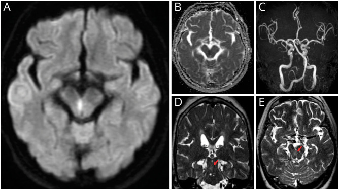
A 62-year-old woman presented with sudden-onset staggering gait and dysarthria. Neurologic examination revealed truncal and appendicular ataxia in all limbs and cerebellar dysarthria (Video 1), slight slowing of internal right eye rotation during leftward gaze, and left eye nystagmus on abduction. The deep tendon reflexes were normal. Diffusion-weighted imaging showed a high-signal midline lesion in the right midbrain (Figure). We diagnosed the patient with Wernekink commissure syndrome caused by midbrain infarction. Wernekink commissure syndrome is a rare syndrome of bilateral ataxia and oculomotor dysfunction caused by caudal paramedian midbrain lesions.1,2 The responsible vessel is considered the inferior paramedian mesencephalic artery, which originates from both the distal basilar artery and proximal posterior cerebral artery or the superior cerebellar artery.2 This lesion involves the superior cerebellar peduncles, their decussation, and the medial longitudinal fasciculi.2 Clinicians should consider midbrain lesions in patients with acute-onset bilateral ataxia.
Figure. Brain MRI.
(A, B) Axial brain MRI showed hyperintensity and hypointensity in the right paramedian midbrain on diffusion-weighted imaging and apparent diffusion coefficient map, respectively. (C) Magnetic resonance angiography showed no obvious major artery stenosis on day 1. (D, E) Axial and coronal T2-weighted images on day 27 showed a faint high-signal lesion at the same site (arrow). The inferior olivary nucleus showed no signal change.
Neurological examination showed cerebellar dysarthria, bilateral limb ataxia, and ataxic gait.Download Supplementary Video 1 (38.4MB, mp4) via http://dx.doi.org/10.1212/201706_Video_1
Acknowledgment
The authors thank Edanz (jp.edanz.com/ac) for editing a draft of this manuscript.
Appendix. Authors

Footnotes
Teaching slides links.lww.com/WNL/C532.
Video links.lww.com/WNL/C533.
Study Funding
The authors report no targeted funding.
Disclosure
The authors report no disclosures relevant to the manuscript. Go to Neurology.org/N for full disclosures.
References
- 1.Dong M, Wang L, Teng W, et al. . Wernekink commissure syndrome secondary to a rare 'V'-shaped pure midbrain infarction: a case report and review of the literature. Int J Neurosci. 2020;130(8), 826-833. [DOI] [PubMed] [Google Scholar]
- 2.Zhou C, Xu Z, Huang B, et al. . Caudal paramedian midbrain infarction: a clinical study of imaging, clinical features and stroke mechanisms. Acta Neurol Belg. 2021;121(2), 443-450. [DOI] [PubMed] [Google Scholar]
Associated Data
This section collects any data citations, data availability statements, or supplementary materials included in this article.
Supplementary Materials
Neurological examination showed cerebellar dysarthria, bilateral limb ataxia, and ataxic gait.Download Supplementary Video 1 (38.4MB, mp4) via http://dx.doi.org/10.1212/201706_Video_1


