

Abstract
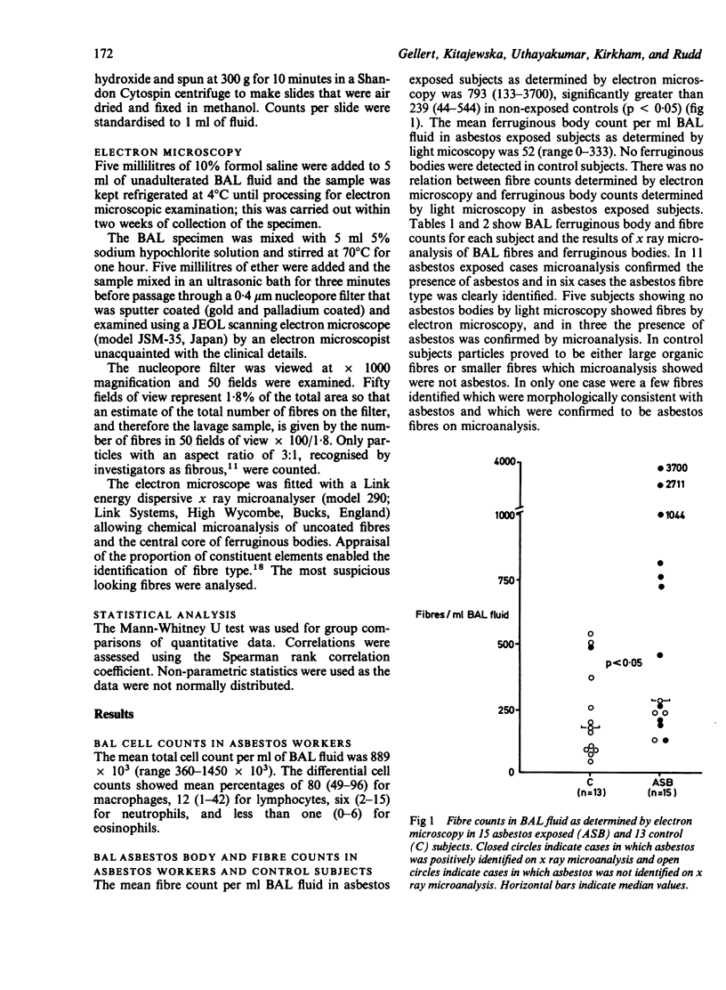
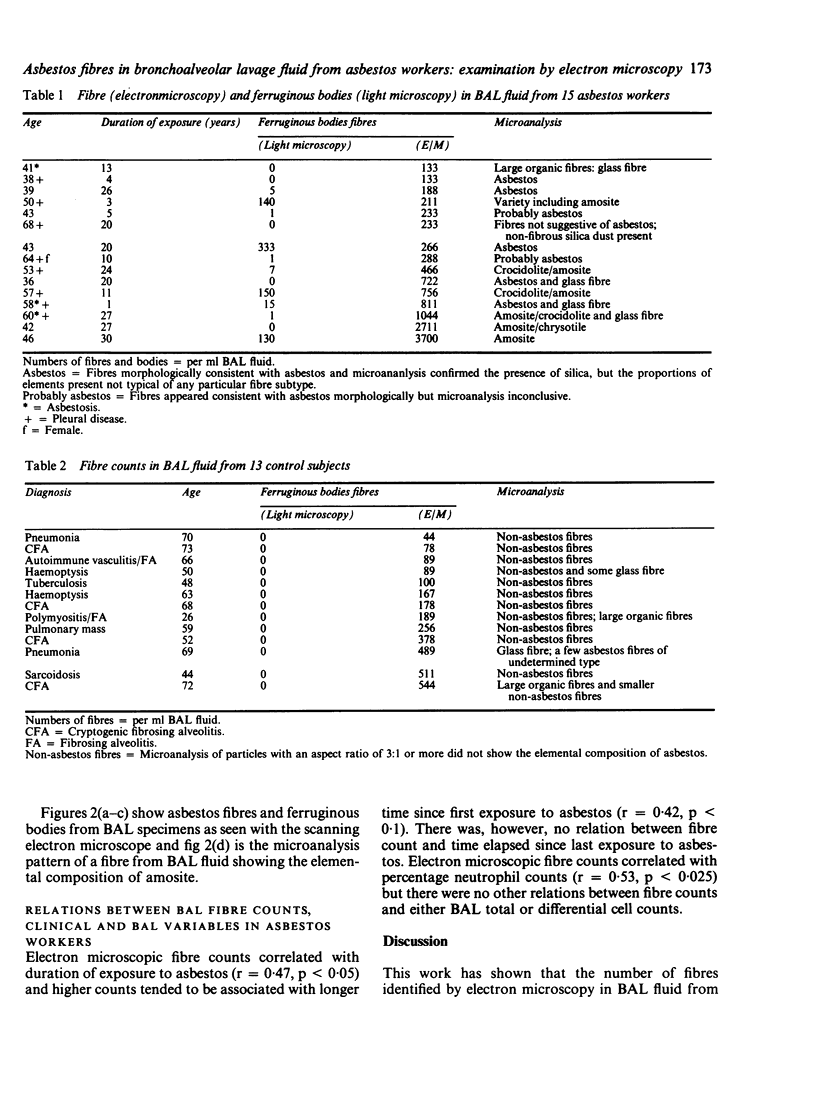
The uncoated and coated fibre load in bronchoalveolar lavage (BAL) fluid was assessed using light microscopy, scanning electron microscopy, and x ray microanalysis in 15 subjects with previous, unprotected exposure to asbestos, including three with clinical and radiological evidence of asbestosis, and in 13 urban dwelling control subjects with no known occupational exposure to asbestos. The mean ferruginous body count per ml BAL fluid in asbestos exposed subjects as determined by light microscopy was 52 (range 0-333). No ferruginous bodies were detected in control subjects. The mean fibre count per ml BAL fluid in asbestos exposed subjects as determined by electron microscopy was 793 (133-3700), significantly greater than 239 (44-544) in controls (p less than 0.05). Electron microscopic counts correlated with duration of previous exposure to asbestos (r = 0.47, p less than 0.05) and with percentage neutrophil counts (r = 0.53, p less than 0.025). There was no relation between electron microscopic fibre counts and light microscopic ferruginous body counts. In 11 asbestos exposed cases x ray microanalysis confirmed the presence of asbestos and in six the asbestos fibre type was clearly identified. Of five subjects showing no asbestos bodies by light microscopy, all showed fibres by electron microscopy, and in three cases the presence of asbestos was confirmed by microanalysis. Among control subjects, fibres were either large organic fibres or smaller particles which microanalysis showed were not asbestos. In only one control case were a few fibres identified which were confirmed as asbestos fibres on microanalysis. Electron microscopic examination of BAL fluid may confirm past exposure to asbestos and probably gives a crude quantitative estimate of asbestos load.
Full text
PDF

Images in this article
Selected References
These references are in PubMed. This may not be the complete list of references from this article.
- Ashcroft T., Heppleston A. G. The optical and electron microscopic determination of pulmonary asbestos fibre concentration and its relation to the human pathological reaction. J Clin Pathol. 1973 Mar;26(3):224–234. doi: 10.1136/jcp.26.3.224. [DOI] [PMC free article] [PubMed] [Google Scholar]
- Churg A., Warnock M. L. Analysis of the cores of asbestos bodies from members of the general population: patients with probable low-degree exposure to asbestos. Am Rev Respir Dis. 1979 Oct;120(4):781–786. doi: 10.1164/arrd.1979.120.4.781. [DOI] [PubMed] [Google Scholar]
- Churg A., Warnock M. L. Asbestos fibers in the general population. Am Rev Respir Dis. 1980 Nov;122(5):669–678. doi: 10.1164/arrd.1980.122.5.669. [DOI] [PubMed] [Google Scholar]
- De Vuyst P., Mairesse M., Gaudichet A., Dumortier P., Jedwab J., Yernault J. C. Mineralogical analysis of bronchoalveolar lavage fluid as an aid to diagnosis of "imported" pleural asbestosis. Thorax. 1983 Aug;38(8):628–629. doi: 10.1136/thx.38.8.628. [DOI] [PMC free article] [PubMed] [Google Scholar]
- Dodson R. F., Greenberg S. D., Williams M. G., Jr, Corn C. J., O'Sullivan M. F., Hurst G. A. Asbestos content in lungs of occupationally and nonoccupationally exposed individuals. JAMA. 1984 Jul 6;252(1):68–71. [PubMed] [Google Scholar]
- Dodson R. F., Williams M. G., Jr, McLarty J. W., Hurst G. A. Asbestos bodies and particulate matter in sputum from former asbestos workers. An ultrastructural study. Acta Cytol. 1983 Nov-Dec;27(6):635–640. [PubMed] [Google Scholar]
- Gellert A. R., Langford J. A., Uthayakumar S., Rudd R. M. Bronchoalveolar lavage and clearance of 99m-Tc-DTPA in asbestos workers without evidence of asbestosis. Br J Dis Chest. 1985 Jul;79(3):251–257. [PubMed] [Google Scholar]
- Gellert A. R., Langford J. A., Winter R. J., Uthayakumar S., Sinha G., Rudd R. M. Asbestosis: assessment by bronchoalveolar lavage and measurement of pulmonary epithelial permeability. Thorax. 1985 Jul;40(7):508–514. doi: 10.1136/thx.40.7.508. [DOI] [PMC free article] [PubMed] [Google Scholar]
- Gylseth B., Mowé G., Wannag A. Fibre type and concentration in the lungs of workers in an asbestos cement factory. Br J Ind Med. 1983 Nov;40(4):375–379. doi: 10.1136/oem.40.4.375. [DOI] [PMC free article] [PubMed] [Google Scholar]
- Johnson N. F., Lincoln J. L., Wills H. A. Analysis of fibres recovered from lung tissue. Lung. 1984;162(1):37–47. doi: 10.1007/BF02715626. [DOI] [PubMed] [Google Scholar]
- Pooley F. D. Electron microscope characteristics of inhaled chrysotile asbestos fibre. Br J Ind Med. 1972 Apr;29(2):146–153. doi: 10.1136/oem.29.2.146. [DOI] [PMC free article] [PubMed] [Google Scholar]
- Rogers A. J. Determination of mineral fibre in human lung tissue by light microscopy and transmission electron microscopy. Ann Occup Hyg. 1984;28(1):1–12. doi: 10.1093/annhyg/28.1.1. [DOI] [PubMed] [Google Scholar]
- Warnock M. L., Prescott B. T., Kuwahara T. J. Numbers and types of asbestos fibers in subjects with pleural plaques. Am J Pathol. 1982 Oct;109(1):37–46. [PMC free article] [PubMed] [Google Scholar]