Abstract
Transcriptional changes in Rett syndrome (RTT) are assumed to directly correlate with steady-state mRNA levels, but limited evidence in mice suggests that changes in transcription can be compensated by post-transcriptional regulation. We measure transcription rate and mRNA half-life changes in RTT patient neurons using RATEseq, and re-interpret nuclear and whole-cell RNAseq from Mecp2 mice. Genes are dysregulated by changing transcription rate or half-life and are buffered when both change. We utilized classifier models to predict the direction of transcription rate changes and find that combined frequencies of three dinucleotides are better predictors than CA and CG. MicroRNA and RNA-binding Protein (RBP) motifs are enriched in 3ʹUTRs of genes with half-life changes. Nuclear RBP motifs are enriched on buffered genes with increased transcription rate. We identify post-transcriptional mechanisms in humans and mice that alter half-life or buffer transcription rate changes when a transcriptional modulator gene is mutated in a neurodevelopmental disorder.
Subject terms: Developmental disorders, RNA decay, Transcriptional regulatory elements, Disease model, Data integration
Rett syndrome (RTT) is a neurodevelopmental disorder caused by mutations in the transcriptional modulator MECP2. Here, the authors measured transcription rate and mRNA half-life changes in RTT patient-derived neurons to show transcription rate buffered by mRNA half-life changes.
Introduction
Rett syndrome (RTT) is a neurodevelopmental disorder in girls caused by damaging mutations in the methyl CpG-binding protein 2 (MECP2)1. Most evidence indicates that MECP2 regulates transcription globally after binding to methylated (m)CG dinucleotides in immature neurons and mCA dinucleotides in adult neurons2–8. Specifically, recent models support a role for MECP2 binding to mCA/mCGs in the gene-body that can: 1) slow RNA polymerase II elongation5 2) inhibit transcription initiation by looping interactions between the promoter and high-density MECP2-bound gene-bodies9 or 3) inhibit transcription initiation by microsatellite interruptions of nucleosome binding that impact intragenic enhancer activation10.
The number or fraction of mCA/mCG and gene-length have been primarily used to interpret transcription rate dysregulation in RTT mouse models5,9. However, the informative power of these DNA features is limited, and it is still challenging to anticipate a priori which genes are transcriptionally up- or down-regulated in RTT, raising questions about whether other sequence features might also participate in transcription dysregulation mediated by the loss of MECP211. Recent developments in machine learning techniques have revealed unsuspected DNA and RNA-sequence features associated with gene regulatory programs12,13. Employing these techniques in the RTT context could help explain the molecular mechanisms of MECP2 function and uncover other DNA sequence features important for MECP2 function.
Genome-wide analyses of steady-state mRNA levels and transcription rate changes in RTT models have demonstrated a global dysregulation of gene expression5,9,14–18. It is uniformly acknowledged that the magnitude of mRNA steady-state level changes is surprisingly small3,4,14,18,19. In 2017, Johnson et al. 17 used GRO-Seq for nascent RNA and RNA-Seq of chromatin, nuclear, and cytoplasmic subcellular fractions to reveal the small steady-state alterations in the Mecp2-null mouse brain is the result of a previously unsuspected post-transcriptional regulatory mechanism. They proposed that large transcription rate changes are compensated by reciprocally adjusting mRNA half-life, and they provided initial support for the role of two RNA-binding proteins (RBPs). In particular, they examined the enrichment of 12 RBP binding sites in subsets of mRNAs and identified HuR-(ELAVL1) cis-acting elements in the 3ʹUTR with extended mRNA stability, or AGO2 cis-acting elements with reduced stability to implicate the action of unknown micro(mi)RNAs17. Their model of post-transcriptional regulation is similar to transcription buffering where RBPs present in the nucleus tag nascent mRNAs and shuttle with them to the cytoplasm. The RBPs then modify half-life to buffer transcription rate changes that preserve steady-state levels (reviewed by Hartenian E et al.20). These results have not been independently tested in mouse and it is unknown whether the mechanism is conserved in human RTT neurons. Therefore, it is important to experimentally measure the direction and magnitude of half-life changes in human MECP2-null neurons. Moreover, the post-transcriptional mechanism has not been studied by systematic enrichment analysis of all known miRNA and RBP cis-acting elements of the post-transcriptionally regulated mRNAs.
Here, we simultaneously investigate the potential role of sequence features mediating transcriptional dysregulation in RTT and expand on the post-transcriptional findings of Johnson et al. in human isogenic induced Pluripotent Stem Cell (iPSC)-derived RTT neurons. We use RNA-approach to equilibrium-sequencing (RATEseq) to measure transcription rate and half-life changes and employed machine learning to uncover sequence features underlying these changes in human and mouse RTT models. In parallel, we compare our human neuron findings to the high-confidence RNA-seq from subcellular fractionations of Mecp2 mutant mouse brains9. We find that transcription rate changes in both human and mouse datasets are best predicted by combinations of three dinucleotide frequencies in gene-bodies that include the expected CA/CG motifs, but are most accurate if they also include other dinucleotides. We discover extensive half-life changes that identify: 1) a gene set with exclusive mRNA stability dysregulation (half-life only) and no associated transcription rate changes; and 2) a larger buffered gene set in which half-life regulation compensates for transcription rate changes that fully offset or minimize mRNA steady-state changes. We demonstrate a global absolute downregulation of miRNA levels, that corresponds with a global absolute half-life increase in RTT neurons. We find individual enriched miRNA binding-sites in the half-life only gene set but very few in the buffered gene set. RBP-binding sites were enriched in the 3ʹUTRs of half-life only genes, and distinct sites were also enriched in buffered genes with increased transcription rate. Overall, we propose that transcription rate increases in MECP2 neurons are subject to surveillance by RBPs that post-transcriptionally regulate RNA half-life. We find that the buffering of transcription rate changes by half-life changes is a conserved feature of RTT models which minimize the steady-state changes in mRNA levels.
Results
Transcription rate changes in RTT neurons do not always alter mRNA steady states
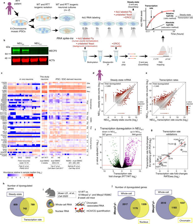
To simultaneously measure direct changes in transcription rate and mRNA half-life in RTT neurons, we performed RATEseq on human cortical neurons derived from WT (NEUWT) and MECP2-Null (NEURTT) isogenic patient-derived iPSCs21 (Fig. 1a). Conventional RATEseq measurements of absolute half-life rely on the 4sU saturation curve. In addition, half-life fold-change can be derived by a second method that calculates a ratio between the steady-state value and the transcription rate of nascent RNA measured at early time points of 0.5 and 1 hour (Fig. 1a). We use the term steady-state here in a restricted context to refer to the equilibrium between transcription rate and half-life in the whole cells at 24 hours.
Fig. 1. Changes in transcription rate in RTT neurons do not automatically result in altered mRNA steady states.
a Schematics of experimental outline for simultaneous quantification of transcription rate, mRNA half-life, and steady-state mRNA level. An isogenic pair of human WT and MECP2-null iPSC-derived cortical neurons were pulse-labeled with 4sU, and at designated time-points total RNA was harvested. 4sU-labeled Drosophila melanogaster (fly) and unlabeled Saccharomyces cerevisiae (yeast), and ERCC spike-in RNAs were added as indicated and used as pull-down efficiency, non-specific binding, library preparation, and sequencing controls. Steady-state mRNA levels were quantified from an aliquot of the 24 hour time point (non-biotinylated and unprocessed). This experiment was repeated for a total of two replicates. b Typical western blot showing the presence of MECP2 protein in two replicates of the NEUWT and absence in NEURTT neurons. Uncropped western blot provided as a Source Data file. c Heatmap of mRNA abundance of cortical neuronal patterning and synaptic marker genes. The heatmap compares the mRNA abundance in the steady-state neuron samples generated in this study compared to neurons from previously published studies. The mRNA abundance in each replicate is measured relative to the sample median. d, e Scatter-plots depicting genome-wide changes in steady-state and transcription rate. This experiment was repeated for a total of two independent replicates. f Volcano plot showing genes with increased or decreased transcription rate in MECP2-null neurons (NEURTT). g Transcription rate fold-changes determined by RATEseq (X-axis, standard error of the log2 fold-change estimated by DESeq2 from 4 biological replicates) were validated using an alternative approach. Neurons were incubated with 5-ethylnyl uridine (EU) and quantified following Click-it reaction and qRT-PCR (Y-axis, standard error of the log2 fold-change derived from qRT-PCR experiments using 3 biological replicates, n = 3) of genes selected to cover a large spectrum of fold-changes including genes with no changes. h overlap of genes altered at transcription rate and/or steady-state in human NEURTT. i Summary of samples from Boxer et al. re-analyzed in our study. j Overlap of genes with altered mRNA abundances in the nucleus or chromatin (transcription proxy) and whole-cell (steady-state) in the brains of Mecp2 y/- mouse model. Panels a, d, and i were created with BioRender.com.
In brief, iPSCs were differentiated into cortical neurons using a dual-SMAD protocol (Fig. S1A) and the non-neuronal cells were depleted based on the presence of specific surface proteins using MACS22,23. 4sU RNA labeling was performed and samples taken from 0.5 to 24 hours (except for the 1-hour NEURTT samples). Cellular viability was consistently high as expected24 (Fig. S1B, C). The steady-state was determined using 24-hour input samples that were treated with 4sU but without biotinylation and pulldown. Multiple RNA spike-ins (Fig. 1a) controlled for 4sU pulldown efficiency, background contamination, equivalent cell numbers, and sequencing quality25, and indicate the high quality of the RNA samples and sequencing data (Fig. S1D–J). 3ʹend RNA-seq (QuantSeq) was utilized to quantitatively map 3ʹUTR isoform diversity to better understand the role of miRNAs and RBP-binding sites in half-life regulation and buffering of transcription dysregulation. Western blots of separate batches of the cells confirmed the absence of MECP2 in the NEURTT samples (Fig. 1b). Efficient differentiation into mixed cortical neurons was confirmed by analyzing the steady-state mRNA abundance of a panel of neuronal marker genes (Fig. 1c) and comparing it to previously published transcriptomics of pluripotent stem cell-derived neurons of equivalent age or neurons collected from human fetal neocortex26–28. We first calculated the half-life using the 4sU saturation curve method25 and deduced the transcription rate using the 0.5-hour time-point (plus a pseudoreplicate from the 1-hour time point for the NEUWT, see methods) by calculating the number of newly synthesized mRNAs over 30 mins. Importantly, the measurement of transcription rate fold-changes between NEUWT and NEURTT were highly comparable using only the 0.5-hour replicates or the 0.5 plus 1-hour time-point pseudoreplicates (Fig. S1K). However, the saturation curve-derived half-life measurements failed for most genes that had low transcription rates (Fig. S1L, M) and these are genes that may be repressed by MECP2 in neurons. We, therefore, calculated the relative half-life fold-changes using the ratio method, and these findings were similar to the saturation curve values for genes that it had determined with high confidence (Fig. S1N). Having shown the improved utility of the ratio method for the genes of most interest, we proceeded to next examine changes in transcription rate and how they correlate with changes in the steady-state.
As expected, we found widespread dysregulation of transcription rate and steady-state in the NEURTT neurons (Figs. 1d–f, S1O, Supplementary Data 1). An independent 5-Ethynyl Uridine (EU) metabolic incorporation assay followed by qRT-PCR experimentally validated transcription rate changes of specific genes measured using both 0.5-hour or 0.5 and 1-hour time-points (Figs. 1g and S1P). Reassuringly, a comparison of our transcription rate datasets with a MECP2 ChIP-seq in mouse brain revealed that the genes with the highest changes in TR were more enriched for MECP2-binding (Fig. S1Q), and most transcriptionally upregulated genes displayed lower basal transcription rate in the WT controls (Fig. S1R)9. Importantly, we found that approximately half of the transcription rate dysregulated genes in NEURTT were not altered at the steady-state mRNA level (Fig. 1h).
Given the relevance of these findings to disease mechanisms, we validated the discrepancy between transcription rate and steady-state changes in an orthogonal in vivo RTT system. We re-analyzed the high-confidence datasets from Boxer et al. 9 that sequenced nuclear and chromatin-associated mRNA abundance as a proxy for transcriptional dysregulation and the whole-cell fractions from cortical forebrain samples of WT, Mecp2 y/-, and point-mutant Mecp2 R306C adult mice (Fig. 1i). As shown previously in the context of in vitro-derived neurons, sequencing of nuclear RNA fractions is highly correlated to the transcription activity measured using GRO-seq17. Our analysis shows a similar pattern where approximately half of the genes transcriptionally dysregulated in the nucleus are not altered at the whole cell level that we define as the steady-state (Fig. 1i, j). This is consistent with the human neuron RATEseq findings that transcription rate dysregulation in the absence of MECP2 does not automatically result in altered steady-state mRNA levels, and that unaltered steady-state mRNA level does not automatically mean there is no change in transcription rate.
The direction of transcription rate changes in RTT neurons is predicted by gene-body dinucleotide frequencies
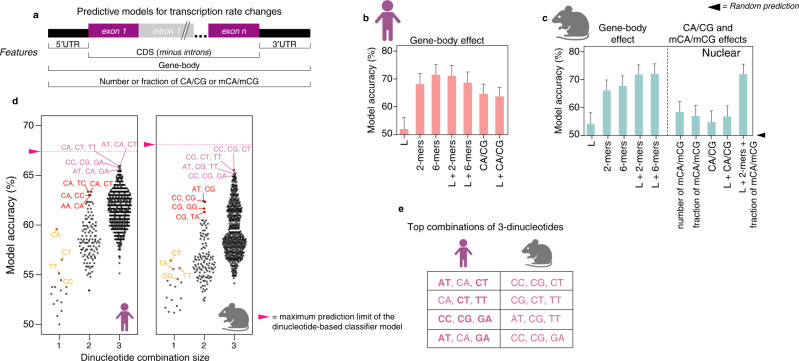
The high fraction or number of mCA/mCG in longer genes have been associated with the small magnitude upregulation of these genes in Mecp2 mouse models3,7,9. However, consistent with Johnson et al. 17, our analyses did not show a gene-body length effect in genes dysregulated at the transcription rate or steady-state (Fig. S2A). The contribution of other sequence features that predict which genes will be transcriptionally dysregulated in RTT models has not been systematically evaluated in vivo11. To learn sequence features of the differentially expressed genes that are relevant for the direction of transcriptional shifts in our immature RTT neurons, we trained a classifier model using our measurements of genome-wide transcription rate changes in NEURTT (Figs. 2a and S2B). As anticipated by the lack of correlation between gene-body length and transcription rate changes (Fig. S2A), this model also resulted in random predictions based on gene-body length (including introns) (Figs. 2b and S2C, D). In contrast, frequencies of dinucleotides in the gene-body produced high prediction accuracies similarly found when using either the coding sequence (CDS) or 3ʹUTR (Fig. S2C, D). The prediction accuracies of CA/CG in gene-bodies were lower than predictions based on any combination of dinucleotides even when combined with gene-body length. This supports a prominent role for additional gene-body dinucleotide combinations in modulating transcription rates (Figs. 2b and S2C, D).
Fig. 2. The direction of transcription rate changes in RTT neurons is predicted by three combined gene-body dinucleotide frequencies.
a Random forest classifier for prediction of gene-body sequence features relevant for transcription rate fold-changes in human NEURTT and in cortical brain samples of the mouse Mecp2 y/-. Percent accuracy (Y-axis) of transcription rate fold-change predictions in human NEURTT (b) or mouse Mecp2 y/- (c) considering different gene-body sequence features. L = gene-body length; 2-mers and 6-mers= 2 and 6-nucleotide sequence elements, respectively; CA/CG = number of CA or CG di-nucleotides; mCA/mCG= methylated CA and CGs; + sign denotes combinations of two or more sequence features. Error bars depict 95% CI and are estimated from approximately 400 genes for human and 500 for mice. Log2 fold-change for each gene is derived from 4 independent replicates (30 minutes and 1 h) for human and 10 replicates for mice. d Top combinations of dinucleotides contributing to the full prediction accuracy described in b and c. Accuracy increases to the maximum when a specific combination of three dinucleotides is used. Similar behavior is observed between human (left) and mouse (right) datasets. Each point (accuracy) is estimated from approximately 400 genes for human and 500 for mice. The log2 fold-change for each gene is derived from 4 independent replicates (30 minutes and 1 h) for human and 10 replicates for mice. e Top combinations of three dinucleotides contributing to the full prediction model. Predictive di-nucleotides conserved between human and mouse are denoted in bold fonts in the human column. Panels b, c, d, and e were created with BioRender.com.
To test the combined dinucleotide-based predictive model in an orthogonal in vivo system, we trained a classifier model on the data from Boxer et al. which also includes cytosine methylation quantification (Fig. 1i)9. In the adult mouse brain, our model captured gene-body length and mCA fraction as predicting whether a gene is transcriptionally up-regulated in the absence of Mecp2 (Fig. S2E, F). However, these models do not discern whether a gene is transcriptionally down-regulated, nor discriminate up- versus down-regulated genes (Fig. S2E, F). In contrast, the gene-body dinucleotide frequencies captured the direction of most transcriptional dysregulation in the Mecp2 y/- mouse, with lower accuracy predicted by the CDS and UTR separately, an effect that was independent of gene-body length (Figs. 2c and S2G–I). Surprisingly, combining CA/CG frequencies had less predictive accuracy than combinations of remaining dinucleotides (2-mers) independent of their methylation status (Figs. 2c, S2G–I). To investigate which dinucleotide or combinations thereof were responsible for the high predictive accuracy of transcription rate changes in RTT neurons, we repeated the classifier model considering single or multiple dinucleotide combinations. The classifier found that specific combinations of three dinucleotides reached the predictive accuracy of the full model for both human and mouse even though the specific combinations of three dinucleotides were different across species (Fig. 2d, e). Taken together, the classifier models of fetal stage human neurons and adult mouse brain indicate that combined frequencies of three dinucleotides that include non-CA/non-CG dinucleotides contribute to the direction of transcription rate modulation mediated by MECP2.
Half-life fold-changes in RTT neurons directly alter the steady-state or buffer transcription rate
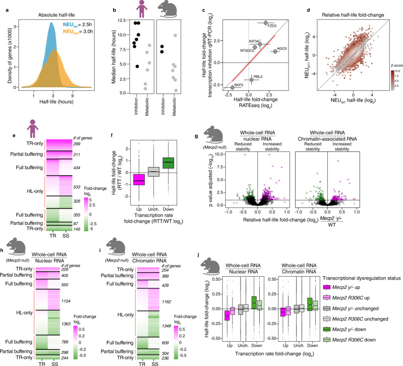
To measure changes in absolute half-life in hours we estimated the time required to reach half of the steady-state mRNA abundance from the 4sU saturation curves (saturation method, Fig. 1a). This absolute RATEseq half-life measurement revealed widespread changes in mRNA stabilities that caused a global absolute increase in the median half-life from 2.5 to 3.0 hours in NEURTT (and an increase in the mean half-life from 2.9 hours in NEUWT to 4.6 hours in NEURTT) (Figs. 3a, S1E–H, and Supplementary Data 2). The global half-life values for neurons correlate well with published median half-lives from other cell types (Fig. 3b, and Supplementary Data 3). These measurements were independently validated on specific genes using a pulse treatment with actinomycin D to induce transcription inhibition followed by qRT-PCR (Fig. 3c). Other orthogonal 4sU pulse-chase methods for measuring half-life have high reproducibility compared to RATEseq29. Using the ratio method to measure relative half-life fold change, we found approximately 860 (0.5 h plus 1 h time-points) or 957 (0.5 h time-point only) genes (~35% or 30% of all genes with altered half-life, respectively) with significantly increased or decreased mRNA half-life but with unchanged transcription rate, termed half-life only (HL-only), leading directly to changes in the steady-state levels (Figs. 3d, e and S3A, B. See also Fig. 1h). The class of RNAs and their identities were highly comparable when we used incorporation rate data from either 0.5 h or 0.5 h plus 1 h to calculate transcription rates (Fig. S3B, C, and Supplementary Data 4) supporting the robustness of our calculation methods. These analyses show that a significant fraction of genes dysregulated at the steady-state level were exclusively driven by changes in mRNA stability.
Fig. 3. Widespread changes in mRNA half-life directly alter steady-state abundance or buffer transcription rate changes in human and mouse RTT models.
a Global increase in the absolute half-life in NEURTT. Median half-life increases to 3 hours in NEURTT. b Median mRNA half-life, as measured in previous studies in human and mouse models, is higher than 1 hour (see also Supplementary Data 3). Black and grey dots: average half-life measured using transcription inhibition or metabolic incorporation methods, respectively. c Half-life changes determined by RATEseq (X-axis, saturation curve 2 biological replicates) were validated using transcription inhibition. Selected genes covering a wide range of fold-changes were quantified by qRT-PCR (Y-axis, 3 independent biological replicates). Error bars= spread of half-life log2 fold-changes measurement calculated from 50% CI of each genotype. d Global relative half-life changes in NEURTT. e Number of genes with changes in transcription rate only, partially or fully buffered by half-life changes, and genes whose change in steady-state are caused by altered half-life only (altered transcription rate= log2 fold-change p-adjusted value smaller than 0.1). Genes were defined with transcription rate only changes when the difference between steady-state and transcription rate log2 fold-change was less than 25% of transcription rate log2 fold-change. f Most genes with increased (magenta) or decreased (green) transcription rate show decreased or increased half-life, respectively, and genes with unchanged transcription rate do not display significant changes in half-life. Transcriptional changes derived from data represented in Fig. 1f. g Global changes in relative half-life in brains of the Mecp2 y/- mouse estimated from nuclear (left) or chromatin-associated RNAs (right) (horizontal line= FDR 0.1). h, i Number of genes with changes in nuclear or chromatin-associated RNA abundances (transcription rate proxies) partially or fully buffered by mRNA stability mechanisms, and genes whose change in steady-state are caused by altered half-life only. j Mecp2 y/- and 306C mouse models display the reciprocal behaviour in half-life regulation relative to transcription rate. Fold changes derived from data represented in h, i (10 biological replicates). TR = transcription rate, HL = half-life, SS = steady-state. For f and j, up and low hinges are 25th and 75th percentiles. Up and low whiskers are 1.5*IQR (inter-quartile range) above and below the corresponding hinges. Notches are 1.58*IQR / sqrt(n) which matches 95% CI for median comparison. Panels b, e, g, h, i, and j were created with BioRender.com.
We then explored the impact on steady-state mRNA levels when both half-life and transcription rate were changed. We found that in these cases, half-life moved in the opposite direction to the transcription rate and decreased the net change in steady-state levels (Fig. 3e–f). We found that 789 genes exhibited full buffering where the RNA half-life entirely offset the transcription rate change resulting in no steady-state change. A further 298 genes showed half-life changes that partially counteracted the transcription rate changes modulating steady-state changes. The contribution of half-life to the steady-state level changes (Fig. S3D) and buffering (Fig. S3E) was independent of the fold-change or FDR thresholds chosen. None of the measured changes correlate with gene-body or processed transcript lengths (Figs. S2A and S3F). Highly similar proportions of buffered and half-life only genes in NEURTT were observed when calculations used the 0.5 h time point only on genes whose transcription rates were altered by at least 4-fold (Fig. S3B). Moreover, genes with high transcription rates were buffered by changes in half-life, showing that the buffering effect was not induced by detection noise from low abundance transcripts (Fig. S3G). The buffering effect was also observed when we used the saturation method (Fig. S3H, I). Altogether, these data indicate that the buffering of transcriptional dysregulation by opposite changes in mRNA half-life is a robust result not sensitive to the calculation method (ratio vs. saturation), signal-to-noise ratios, or the number of time points. We found ~28% of all genes (n = 444) with altered steady-state expression to be exclusively dysregulated at the transcription rate level (TR-only, Fig. 3e), underscoring the role of post-transcriptional regulation of mRNA stability in directly altering or buffering the RTT transcriptome.
Proxy values support half-life shifts and transcription buffering in Mecp2 mouse models
Given the importance of finding global changes in half-life in the NEURTT neurons and its implications for interpreting steady-state level dysregulation typically found in RTT models, we investigated whether these results were reproducible in vivo. To determine proxies of half-life changes in the mouse brain in RTT models we re-analyzed the data from Boxer et al. for whole-cell, nuclear, and chromatin-associated RNAs9. We accomplished this by comparing the abundance of transcribed genes in the nucleus and chromatin fractions as proxies for transcription rate as previously demonstrated17, to the whole-cell fraction that includes mRNA undergoing decay in the cytoplasm17 that we define as the steady-state (see methods). By applying the proxy value in the ratio method, we calculated relative half-life fold-changes. From the 5032 genes reported by Boxer et al. to be differentially regulated in Mecp2 y/- mice, we found a similar pattern of widespread fold-changes in half-life (Fig. 3g, Supplementary Data 5). We also found similar proportions of HL-only genes (Fig. 3h, i), and the extent of full or partial buffering in both mouse models (Figs. 3j and S4A. See also Fig. 1h, j). In line with our RTT human neuron findings, the contribution of half-life to the steady-state level changes (Fig. S4B) and buffering (Fig. S4C) was also independent of the fold-change or FDR thresholds chosen.
Despite the conserved relationship that half-life and transcription rate or their proxies have on steady-state gene expression in human and mouse RTT models, we found minimal overlap in the identities of genes dysregulated in each species (Fig. S4D, E). Furthermore, minimal overlap was also observed between the Mecp2 y/- and Mecp2 R306C mouse models as already observed by Boxer et al. (Fig. S4F, G). Overall, our results consistently identify steady-state level changes driven by half-life only without any measurable transcription rate shift in both human and mouse RTT models. Importantly, the majority of all half-life shifts fully or partially buffer Mecp2-mediated transcription rate dysregulation, and only a small number of genes have increased steady-state changes due to combined half-life and transcription rate changes in the same direction. Finally, similar to the human findings, we only find 473 genes (13% of total genes altered at steady-state, which excludes the full buffered group) with transcription rate only dysregulation as measured in the nuclear fraction. Altogether, our findings demonstrate a pattern of half-life shifts and transcription buffering that is conserved in RTT mouse and human models.
Cis-acting elements in the 3ʹUTRs are highly associated with half-life changes
Our findings indicate that RNA half-life is a critical regulatory layer defining steady-state RNA levels in RTT models. We therefore considered several potential mechanisms underlying how RNA half-life is controlled in RTT neurons: 1) alternative polyadenylation; 2) alternative-splicing; 3) codon usage integrating translation elongation to RNA stability; 4) sequence composition of 3ʹUTR and gene-bodies; and 5) enrichment of miRNA binding-sites and RBP cis-acting elements in the 3ʹUTR between buffered and non-buffered genes. Initially we investigated the contribution of mRNA isoforms by mapping and quantification of 3ʹUTR alternative poly-Adenylation events that showed no difference in the frequency of poly-Adenylation site usage in NEURTT (Fig. S5A–C). Moreover, measurement of 3ʹUTR (NEUWT vs. NEURTT) and alternatively-spliced mRNA isoforms (mouse Boxer et al.) indicated that all mRNA isoforms display the half-life buffering effect to the same degree (Fig. S5D–F). These analyses argue against changes in mRNA isoform usage participating in the half-life shifts in RTT models.
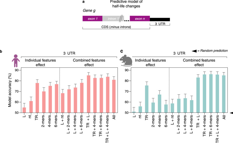
Next, we created a classifier predictive model to estimate the effect of codons and sequence composition on the direction of changes to mRNA half-life (Fig. 4a). Overall 3ʹUTR length and nucleotide frequency had no predictive value for classifying increased or decreased half-life changes (Fig. 4b). Transcription rate changes were anti-correlated with half-life leading to high predictive accuracy. These results underscore the unidirectional and reciprocal link between transcription rate dysregulation and compensatory mRNA stability control in NEURTT. Dinucleotide frequencies in the 3ʹUTR offered significant prediction accuracy on whether the half-life was increased or decreased. Increasing the size of the tested k-mers from dinucleotides to 4-mers and 6-mers to encompass potential cis-acting elements improved the prediction accuracy of half-life (Figs. 4b and S5G), equivalent to transcription rate alone. A combination of transcription rate and 6-mers showed no further improvement in accuracy. Curiously, we found that classifier features in the CDS mirror that of the 3ʹUTR models also offering significant prediction accuracies for the half-life changes (Fig. S5H, I), highlighting a significant sequence composition correlation between CDSs and 3ʹUTRs (Fig. S5J). In contrast, comparison of the prediction models for in-frame codons (3-nt sequences) indicates that codon optimality has no effect on half-life changes in NEURTT (Fig. S5I). Importantly, while the predictive accuracy of k-mers is lower in mouse, the classifier model predictions are upheld in the Mecp2 mouse model. These findings show a reproducible effect of transcription rate on half-life, thereby implicating a conserved buffering mechanism through cis-acting elements impacting half-life (Figs. 4c, S5K–M).
Fig. 4. Cis-acting elements in the 3ʹUTR are highly associated with half-life changes.
a Random forest classifier for prediction of mRNA sequence features relevant for half-life fold-change in human NEURTT and in cortical brain samples of the mouse Mecp2 y/-. Percent accuracy (Y-axis) of half-life fold-change predictions in human NEURTT (b) from 2 replicates or mouse Mecp2 y/- (c) from 10 replicates considering different mRNA sequence features. >80% prediction accuracies can be achieved with the features tested, and indicate that 3ʹUTRs contain sequence elements relevant for half-life changes in both humans and mice. L = gene-body length; nt= nucleotide sequence; 2-mers, 4-mers, and 6-mers= 2, 4, and 6-nucleotide sequence elements, respectively; TR = transcription rate; All= all features considered at the same time; + sign denotes combinations of two or more sequence features. Error bars depict 95% CI. Panels b and c were created with BioRender.com.
miRNA and RBP cis-acting elements correlate with half-life changes in RNA stability exclusive genes
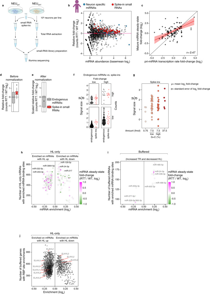
To examine a possible role of miRNAs in mRNA half-life regulation, we first performed small RNA-seq with a spike-in strategy to normalize cell numbers and inform on relative and absolute changes in miRNA abundance between the isogenic human NEUWT and NEURTT (Fig. 5a). These results showed that the steady-state levels of miRNAs changed by as much as 4-fold up or down (Fig. 5b, Supplementary Data 6). Interestingly, most changes in miRNA steady-state abundance were captured in the RATEseq data by shifts in the transcription rate of miRNA genes (Fig. 5c, Supplementary Data 6). These findings indicate that transcription dysregulation of miRNA genes drives the changes in miRNA abundance, in addition to miRNA maturation processing as indicated previously30,31. Moreover, after normalization against a pool of small spike-in RNAs, the scaled absolute fold-change of most miRNAs was decreased in NEURTT (Fig. 5d–g). This global decrease in miRNA abundance in NEURTT is in line with the global absolute upshift of median half-life (Fig. 3a).
Fig. 5. miRNA and RBP cis-acting elements correlate with half-life changes in RNA stability exclusive genes.
a Small RNA-seq to quantify absolute changes in human miRNA abundances. Total RNA was extracted from the same number of NEUWT and NEURTT neurons, to which we added the same mass of a small RNA spike-in mixture. This spike-in mixture contains small RNA molecules covering a wide range of sequence composition and molar concentrations. This experiment was repeated for a total of two independent replicates (n = 2). b Changes in miRNA abundances in the NEURTT. X-axis= basal abundance of each mature miRNA detected in the NEUWT, Y-axis= fold-change in the NEURTT. Blue dots, neuronal-specific miRNAs accumulate at abundances higher than the mean in both NEUWT and NEURTT. Red dots, abundance of each small RNA spike-in added on a per-cell basis and used for library preparation control and absolute quantification of miRNA abundance. c Pearson’s correlation of the transcription rate fold-changes between steady-state mature miRNA levels and primary miRNA (pri-miRNA) in NEUWT and NEURTT showing a significant correlation between both (r = 0.47, p val 4.3−7), indicating many changes in mature miRNA steady-state levels are caused by changes in their transcription rate. Error bars= 95% CI of the linear model. d DESeq2-calculated miRNA level fold-change before and after spike-in normalization (e). The absolute abundance of miRNAs in NEURTT is reduced for most miRNAs. Up and low hinges= 25th and 75th percentiles. Up and low whiskers= 1.5 *IQR (inter-quartile range) above and below the corresponding hinges. Notches= 1.58 *IQR/sqrt(n) matching 95% CI for median comparison. f–g The precision of fold-change measurement is quantified as a ratio of log2(fold-change) and its standard error. Comparison with endogenous miRNAs and number of detected counts (f) or comparison between small RNA spike-in across molar concentrations and G + C content (g). miRNA-binding sites enriched in half-life only (h) or in combination with transcription rate up and half-life down (i). Y-axis= the number of genes containing miRNA-binding sites found, and X-axis= enrichment levels. Color represents the miRNA steady-state fold-change in the NEURTT measured in b. j 7-mers known to be targeted by RBPs enriched in the HL-only group based on the RNAcompete database33. TR = transcription rate, HL = half-life. Panels a and b were created with BioRender.com.
To investigate the role of these miRNA changes in the regulation of half-life in NEURTT, we performed motif enrichment analysis of miRNA-binding sites present in the TargetScan database32. This analysis identified multiple potential miRNA-binding sites as enriched in up to 800 genes in the HL-only group of mRNAs whose steady-state level changes are exclusively directed by half-life changes (Fig. 5h, Supplementary Data 7). In contrast, we found significantly fewer miRNA-binding sites sequences enriched in the group of buffered mRNAs with increased transcription rate and decreased half-life and many of these show no change in the miRNA abundance (Fig. 5i, Supplementary Data 7). We did not find miRNA sites enriched in the opposite group with decreased transcription rate and increased half-life (Supplementary Data 7). Overall, these data indicate that the reduced miRNA abundance contributes to the regulation of HL-only genes in NEURTT. However, very few individual miRNAs correlate with buffering, although combinatorial effects of multiple miRNAs cannot be excluded.
We then performed an unbiased search for the enrichment of 174 RBP cis-acting elements, as described in the RNACompete database33, in the 3ʹUTR of mRNAs with altered half-life in NEURTT. We found hundreds of enriched RBP targets in the group of HL-only genes (Fig. 5j, Supplementary Data 7). This result includes the RBP HuR (ELAVL1) whose different motifs are enriched in 300-900 mRNAs with increased half-life in NEURTT. Our results suggest that 3ʹUTR-directed miRNA and RBP regulation best explain the HL-only gene set changes.
Only RBP cis-acting elements are enriched in buffered genes with increased transcription rate and decreased half-life
Having excluded miRNAs as playing a substantial role in mRNA buffering, we explored a role for the 174 RBP cis-acting elements in the buffered group of mRNAs. We observed that no RBP cis-acting elements were enriched for genes with decreased transcription rate and increased half-life (Fig. 6a, right panel), and only a handful were depleted. This result suggests that RBPs are not involved in regulating transcripts with decreased transcription rate for half-life stabilization. In contrast, we found numerous RBP elements that were enriched in 100-200 buffered genes with increased transcription rate and decreased half-life (Fig. 6a, left panel and Supplementary Data 7). To identify RBPs that plausibly regulate nascent mRNAs, we aggregated all the buffering enriched RBPs with reported cellular localizations, and found that 51 were nuclear and/or able to shuttle to the cytoplasm whereas only 18 were reported to be cytoplasmic only (Fig. 6b). To examine roles of specific RBPs, we first noted that ELAVL1 elements are depleted in >150 genes in this set, demonstrating that it is only enriched in the HL-only gene set. In contrast, NOVA2 and ZFP36 were enriched in more than 200 buffered genes and these RBPs bind premature mRNA co-transcriptionally34,35. Additionally, PTBP1 and multiple arginine-serine rich (SRSF) splicing factors that have been shown to participate in transcriptional buffering36 were enriched in >150 genes. Further support for a directional role by NOVA2 and SRSFs is that they were depleted in the buffered genes with decreased transcription rate and increased half-life (Fig. 6a). Our results reveal specific sets of RBP motifs associated with half-life regulation of buffered genes with increased transcription rate in RTT neurons.
Fig. 6. RBP cis-acting elements are enriched in buffered genes with increased TR and decreased half-life.
a 7-mers known to be targeted by RBPs enriched in the group of buffered mRNAs. A different set of RBP cis-acting elements was found enriched compared to the HL-only group (Fig. 5j). b Cellular distribution of the RBPs enriched in mRNAs with increased transcription rate and decreased half-life showing that these are predominantly nuclear.
Discussion
Our analysis of a dataset of transcription rate and half-life changes in human neurons and independent re-analyses of mouse RTT models demonstrate that most transcription rate changes in the absence of MECP2 are buffered by post-transcriptional regulation of mRNA stability. We used RATEseq to simultaneously measure transcription rate and half-life changes in human neuron samples. We complemented this approach by applying the subcellular fractionation strategy employed by Johnson et al. to the mouse resource dataset of Boxer et al. The consistent findings from both methods and model systems unambiguously show that post-transcriptional regulation is a modifier of transcription rate dysregulation in RTT, and that it is a conserved mechanism shared by human and mouse neurons. We provide evidence for changes in steady-state levels driven solely by half-life only shifts, and large transcription rate shifts that are entirely offset at the steady-state level by half-life mechanisms that we refer to as buffering. These observations have major implications for interpreting RNA-Seq results in RTT and potentially other neurodevelopmental disorders, or in diseases of other tissues caused by mutations in genes that modulate transcription like MECP2. The existence of transcriptional buffering mechanisms in mammals raises a cautionary note for interpreting RNA-Seq steady-state results in the general context of transcriptional regulation. It also argues in favour of more widely prioritizing methods that directly measure nascent transcription or account for mRNA stability. Moreover, we extend the limited search by Johnson et al. who found two RBP cis-acting elements enriched in mRNAs with altered half-life by discovering hundreds of RBP cis-acting elements in the half-life only gene set. We also demonstrate the enrichment of miRNA-binding sites in the HL-only gene set. The buffered gene set with increased transcription rate likely describes the genes transcriptionally repressed by MECP237. In this group, we identified a restricted subset of mostly nuclear or shuttling-capable RBPs whose cis-acting elements are highly enriched. Our findings thus reveal numerous candidate RBPs potentially involved in buffering the increased transcription rate of their mRNA targets. This mechanism may act as a network to coordinate mRNA degradation in healthy neurons and to compensate for transcription rate dysregulation in RTT neurons.
It is important to note that, while we found transcriptional buffering conserved between the human and mouse RTT models, the identity of genes dysregulated at both transcription and mRNA stability between species was considerably different. It is conceivable that the differences in gene identities between ours (human) and the Boxer et al. (mouse) datasets originate from the analysis of different cell types (in vitro-derived human neurons vs. mouse whole brains), or from the maturation stage differences between the studies. A high degree of variability between the dysregulated genes in the RTT mouse models has also been observed previously38. While the nature of this variability is, at this point, obscure, comparative studies have shown agreement on a core set of dysregulated genes38. Nonetheless, this indicates a high level of flexibility of the buffering mechanism.
Our computational methods were focused on defining cis-acting elements that are relevant for transcription rate or half-life regulation. At the DNA level, our classifier model discovered that the direction of transcription rate shifts in human and mouse RTT models are best predicted by combinations of three dinucleotides that include one or both of the canonical MECP2-binding sites CA and CG, together with other dinucleotides such as AT. The MECP2 AT-hook domain contributes to low-affinity transient interactions with AT-rich DNA that influence the dynamics of MECP2 binding to local high affinity methylated DNA sites39. Tellingly, the classifier predictions are low when modelling only CA/CG dinucleotide frequencies without accounting for low-affinity sites. These unbiased findings from machine learning algorithms indicate that the gene-body frequencies of other dinucleotides like AT are important and are a conserved mechanism defining the direction of transcription rate changes in RTT neuronal models. We speculate they may act by transient recruitment of MECP2 influencing its local binding dynamics to nearby methylated DNA. We confirmed a role of gene-body length in transcription rate regulation but only in the adult mouse neuron resource of Boxer et al. However, neither our classifier model on the human neuron RATEseq dataset nor the adult mouse dataset from Johnson et al. support this observation. It is possible that gene-body length contributes less to transcription rate regulation in fetal stage neurons derived from iPSC in which only mCG modifications are expected to be present7,40, or that it requires the power of ten replicate samples used in the Boxer et al. resource to be detected.
With regards to half-life only regulation, to define which miRNA-binding sites to investigate we first used our human neuron miRNA dataset to identify the miRNAs that are altered in NEURTT. These results confirm the reported miRNA changes in RTT mouse models, although the RATEseq dataset shows that intergenic pri-miRNA transcription rate is altered in RTT adding another dimension to the known miRNA processing alterations in mouse30 and human neurons31. Through the use of spike-in scaling in the miRNA dataset, we deduced a global absolute downregulation of miRNAs in NEURTT that account for the global absolute increase in mRNA half-life of ~0.5 hours. Superimposed on the increased global half-life effect were individual HL-only genes which our classifier models revealed strong enrichment of miRNA-binding sites consistent with their known role in mRNA instability. Many RBP cis-acting elements including ELAVL1 (HuR) were enriched in the HL-only gene set. While Johnson et al. reported HuR and AGO2 cis-acting element enrichments in buffered gene sets in the mouse using the subcellular fractionation approach, our RATEseq results and unbiased search of RBP cis-acting elements point to a role for miRNAs and ELAVL1 in mRNA stability in human neurons rather than in the buffering mechanism itself.
While we eliminated several possible buffering mechanisms, one limitation is that we were unable to test a potential role of mRNA methylation modifications or poly(A)-tail length regulation on the targeted mRNAs with altered transcription rate41. We speculate that these mechanisms could also participate in the buffering mechanisms, particularly in the gene set with decreased transcription rate and increased half-life that we found was not associated with enrichment of either miRNA-binding sites or RBP cis-acting elements. The simplest mechanism for buffered genes with increased transcription rate is that the RBPs bind nascent mRNA in the nucleus and are transported to the cytoplasm where they tag the transcript for stabilization. We speculate that the nuclear RBPs are limiting in neurons, and if transcription rate increases then the proportion of tagged mRNA falls, leading to relatively more degradation in the cytoplasm and decreased half-life. A more complex variation on this model is that the concentration of some RBPs themselves may also change in RTT, and this may increase or decrease their ability to stabilize their target mRNAs. To distinguish these models, it will be necessary to determine which RBPs are changed at the protein level in RTT using proteomics of neuronal nuclei and then individually testing their impact through gain- or loss-of-function assays on the mRNA targets.
Equivalent loss-of-function experiments have already been described in humans with neurodevelopmental disorders caused by mutations in RBP genes such as NOVA235. The impact of these RBPs on buffering could be established using existing or new iPSC or mouse models. In fact, global transcription rate and RBP concentrations will inevitably be altered during the course of neurodevelopment, suggesting that it would be valuable to define the buffered gene sets in iPSC and their progeny Neural Progenitor Cells (NPC) relative to the final neurons described here. We and others18,23 have previously reported translational regulation changes in RTT neurons in both ribosomal loading and protein stability implemented through alterations of E3-ubiquitin ligase protein levels. These findings emphasize that buffering in RTT and potentially other disorders likely operates at both the mRNA and protein levels.
Methods
iPSC cultures and neuronal differentiation
iPSC lines #37 (WT) and #20 (isogenic MECP2-null) from a female patient were previously described21. Both cell lines were generated and cultured under the approval of the SickKids Research Ethics Board and the Canadian Institutes of Health Research Stem Cell Oversight Committee. iPSC lines were cultured in 5% CO2 on BD hESC-qualified matrigel (BD) in mTeSR medium (STEMCELL Technologies). Cultures were passaged using ReLeSR (STEMCELL Technologies) following the manufacture’s instruction every 6-7 days. For neuronal induction, iPSCs were aggregated as Embryoid Bodies (EBs) in low-attachment dishes in N2 media containing laminin (1 ml/ml) with 10 mM SB431542, 2 mM DSM, and 1x penicillin-streptomycin changed daily. After 7 days, EBs were plated on poly-L-ornithine + laminin-coated dishes and grown in N2 media + laminin (1 ml/ml). After 7 days, neural rosettes were manually picked and transferred to poly-L-ornithine + laminin-coated wells. After 7 days, neural rosettes were picked a second time, digested with Accutase and plated on poly-L-ornithine + laminin-coated wells. Resulting NPCs were grown as a monolayer and split every 5-7 days in NPC media containing DMEM/F12, N2, B27, 1x non-essential amino acid (NEAA), 2 mg/ml Heparin, 1 mg/ml laminin. To generate neurons, NPCs were plated on poly-L-ornithine + laminin-coated plates at a density of 106 cells per 10 cm dish and cultured for 3 weeks in neural differentiation medium (Neurobasal, N2, B27, 1 mg/ml laminin, 1x penicillin-streptomycin, 10 ng/ml BDNF, 10 ng/ml GDNF, 200 mM ascorbic acid, and 10 mM cAMP).
Neuronal enrichment using MACS
Neuronal cultures were enriched for all experiments to exclude contaminating glia and NPCs present after differentiation. Enrichment of 3-week old neuronal cultures was made as described earlier23,42. 3-week old heterogeneous neuronal cultures were enriched by a negative selection strategy using antibodies against surface markers CD44 (Biotin Mouse Anti-Human CD44, 1:1000 dilution, BD Biosciences, Clone G44-26; Cat# 555477) and CD184 (Biotin Mouse Anti-Human CD184, 1:1000 dilution, BD Biosciences, Clone 12G5; Cat# 555973) recognizing NPCs, glial progenitors and astrocytes43 using magnetic-activated cell sorting (MACS® - Miltenyi Biotec). After enrichment, neurons were re-seeded onto Matrigel-coated 6-well plates, cultured in neural differentiation medium, and allowed to recover for one extra week, for a total of 4 weeks neuronal differentiation.
Western blots
Cells were washed in ice-cold PBS and total protein extracted in radioimmune precipitation assay (RIPA) buffer (25 mM Tris-HCl, pH7.6, 150 mM NaCl, 1% Nonidet P-40, 1% sodium deoxycholate, and 0.1% SDS). Equivalent protein mass was loaded on SDS-PAGE and transferred to Hybond ECL (GE HealthCare) nitrocellulose membrane. Antibodies MECP2 (Rabbit Anti-MECP2, 1:1000 dilution, Millipore, Cat# 07-013; RRID:AB_2144004), and Beta Actin (Mouse Anti-bActin, 1:5000 dilution, Sigma-Aldrich, Cat# A5441; RRID:AB_476744) were used. Near-Infra Red-conjugated secondary antibodies (IRDye 800CW Donkey anti-Mouse IgG, Cat# 926-32212, and IRDye 680RD Donkey anti-Rabbit IgG, Cat# 926-68073, 1:25000 dilution for both, LI-COR) were used and membranes scanned using LI-COR Odyssey CLx scanner according to manufacturer’s instructions. Acquired images were analyzed using ImageStudio v5.2.5.
4sU metabolic labeling of Neurons and RNA extractions
When enriched neuronal cultures reached 4-week of differentiation, media was replaced with neuronal differentiation media supplemented with 100 μM 4sU (Sigma-Aldrich) reconstituted in DMSO. Neurons were harvested at 0.5, 1, 4, 8, and 24 h after the addition of 4sU (except for the MECP2-null line where the time-point 1 h was omitted from both replicates due to low differentiation yields). Metabolic labeling was designed such that all time points were collected together. After incorporation, cells were quickly washed twice with ice-cold 1× PBS Total RNA and scraped into ice-cold 1.5 ml Eppendorf tubes. Cells were collected by spinning at 1000 g for 5 min at 4 °C and cell pellets were resuspended in 1 mL of Trizol (Thermo Fisher Scientific). Total RNA was extracted according to manufacturer instructions. The steady-state sample was prepared from a 5 μg aliquot of the 24 h time-point added with 0.5 μg of both 4sU labeled and unlabeled spike-in RNAs. Neuronal viability in the presence of 100 μM 4sU was monitored up to 24 h of treatment on parallel cultures by using Trypan blue staining and live/dead cell counting.
Biotinylation and pull down of 4sU-labeled RNAs
50 µg of total neuronal RNA was mixed with 5 µg unlabeled yeast RNA and 5 µg 4sU-labeled S2 fly RNA in a total volume of 120 µL. 1 mg/mL HPDP-biotin (ThermoFisher Scientific) was reconstituted in dimethylformamide by shaking at 37 °C for 30 min at 300 RPM. 120 µL of 2.5× citrate buffer (25 mM citrate, pH 4.5, 2.5 mL EDTA) and 60 µL of 1 mg/mL HPDP-biotin were added to the premixed RNA sample for each time point. The solution was incubated at 37 °C for 2 h at 300 RPM on an Eppendorf ThermoMixer F1.5 in the dark. Samples were extracted twice with acid phenol, pH 4.5, and once with chloroform. RNA was precipitated with 18 µL 5 M NaCl, 750 µL 100% ethanol, and 2 µL GlycoBlue (Invitrogen) overnight at −20 °C. Precipitated RNA was pelleted for 30 min at 21,000 g at 4 °C. The RNA pellet was resuspended in 200 µL of 1× wash buffer (10 mM Tris-HCl, pH 7.4, 50 mM NaCl, 1 mM EDTA). Biotinylated RNA was purified using the µMACS Streptavidin microbeads system (Miltenyi Biotec). 50 µL Miltenyi beads per sample were pre-blocked with 48 µL 1× wash buffer and 2 µL yeast tRNA (Invitrogen), rotating for 20 min at room temperature. µMACS microcolumns were washed 1× with 100 µL nucleic acid equilibration buffer (Miltenyi Biotec), followed by 5× washes with 100 µL 1× wash buffer. Beads were applied to microcolumns in 100 µL aliquots and again washed 5× with 100 µL 1× wash buffer. Beads were demagnetized and eluted off the column with 2× 100 µL 1× wash buffer, and columns were placed back on the magnetic stand. A total of 200 µL beads was mixed with each sample of biotinylated RNA and rotated at room temperature for 20 min. Samples were applied to the microcolumns in 100 µL aliquots, washed 3× with 400 µL wash A buffer (10 mM Tris-HCl, pH 7.4, 6 M urea, 10 mM EDTA) prewarmed to 65 °C, and washed 3× with 400 µL wash B buffer (10 mM Tris-HCl, pH 7.4, 1 M NaCl, 10 mM EDTA). RNA was eluted with 5× 100 µL of 1× wash buffer supplemented with 0.1 M DTT, and flow-through was collected in a tube. Purified RNA was precipitated with 30 µL 5 M NaCl, 2 µL GlycoBlue, and 1 mL 100% ethanol, incubated at −20 °C overnight. Samples were spun at 21,000 g at 4 °C for 30 min and resuspended in 20 µL water. RNA quality was assessed by running 3 µL of samples on a 1.5% agarose gel.
Transcription rate measurement using EU
Transcription rate measurements were validated by an alternative method using the metabolic incorporation of 5-ethynyl uridine (5-EU) followed by quantifying mRNA levels by qRT-PCR. NEUWT and NEURTT were incubated with 0.5 mM 5-EU (ThermoFisher) for 30 min. Total RNA was extracted and processed using Click-iT Nascent RNA Capture Kit (ThermoFisher) according to the manufacturer’s instructions. The captured RNAs were used as a template for cDNA synthesis, followed by qRT-PCR to quantify mRNA level (for the primer list, please see supplementary information file). Genes were chosen to cover a wide range of transcription rate changes determined by RATE-seq.
Half-life measurements using transcription inhibition
Half-life measurements were validated by an alternative method using transcription inhibition followed by quantifying mRNA levels by qRT-PCR. 10 μg/mL actinomycin D (Sigma-Aldrich) was added to NEUWT and NEURTT. RNAs were isolated at 1 h, 3 h, and 6 h time points using the RNeasy Plus kit (QIAGEN). The RNAs were used as a template for cDNA synthesis followed by qRT-PCR to quantify mRNA levels (for the primer list, please see supplementary information file). Genes were chosen to cover a wide range of half-life changes as determined by RATE-seq. Fold-changes for all time points were calculated relative to the 0-hour time point. Then, data was fit with lm function from R (formula = log(Fold-change, 2) ~ Time + 0). The coefficient of “Time” term measures degradation rate . And a half-life is derived as ln(2)/ . Finally, the confidence intervals are estimated with the confint function (stats package).
cDNA synthesis and qRT-PCR
cDNAs were synthesized using SuperScript III reverse transcriptase (ThermoFisher) with random hexamer primers according to the manufacturer’s instructions. For qRT-PCR, we used SYBR Select PCR Master Mix (ThermoFisher). Fold-changes were calculated by the ΔΔCt methods using Glyceraldehyde 3-phosphate dehydrogenase (GAPDH) and 18 S as housekeeping genes, averaged between technical and subsequently biological replicates to achieve an average fold difference (for the primer list, please see supplementary information file).
miRNA extraction and spike-in strategy
To calculate relative and absolute differences in the miRNA population in NEUWT and NEURTT, small RNAs were extracted from two replicates of both lines using the same number of cells followed by the addition of a set of spike-in RNAs. Small RNAs were extracted from 500,000 neurons of each line using the SPLIT RNA extraction Kit (Lexogen) according to the manufacturer’s instructions. A set of 52 RNA spike-ins (QIAseq miRNA Library QC Spike-Ins – Qiagen) that spanned a wide range of concentrations were added to the recovered RNAs according to the manufacturer’s instructions. Sequencing libraries were made using the Small RNA library preparation kit NEBNext (NEB) according to the manufacturer’s instructions. Sequencing was performed on the Illumina HiSeq 2500 using the Rapid Run mode. Datasets can be accessed from GEO using the access number GSE191168.
Library preparation and RNA-sequencing
RNA-seq libraries were prepared for each time-point and steady-state sample using the QuantSeq 3’ mRNA-Seq Library Prep Kit FWD for Illumina (Lexogen) automated on the NGS WorkStation (Agilent) at The Centre for Applied Genomics (TCAG) according to the manufacturer’s instructions. PCR cycle numbers were determined using the PCR Add-on Kit for Illumina (Lexogen). All steady-state samples were processed with 250 ng of total RNA input. To minimize variability between time-points within a batch, RNA samples were processed with the same total RNA input with a minimum of 100 ng of total RNA used. Each sample was spiked-in with ERCC RNA Spike-In Control Mix 1 (Ambion) according to the manufacturer’s instructions prior to the start of library preparation. Library quality and quantity were measured at TCAG prior to sequencing with Bioanalyzer (Agilent) and KAPA qPCR (Roche). Sequencing was also performed at TCAG on the Illumina HiSeq 2500 with single-end 100 bp read length yielding 40 to 50 million reads. Datasets can be accessed from GEO using the access number GSE191168.
Processing of raw sequencing reads
Processing starts with trimming of reads in 4 steps using cutadapt version 1.1044. First, we removed adapters exactly at the 3‘-end of the reads (-a AGATCGGAAGAGCACACGTCTGAACTCCAGTCAX -O 4 -e 0.1 --minimum-length 25). Second, we removed internal or long stretches of adapter (-a AGATCGGAAGAGCACACGTCTGAACTCCAGTCA -O 30 -e 0.18 --minimum-length 25). Third, we trimmed low-quality bases at the 3‘-end of the reads (-q 20 -O 4). Finally, we removed poly-A tail at the 3‘-end of the reads (-a AAAAAAAAAAAAAAAAAAAAAAAAAAAAAAX -O 4 -e 0 –minimum-length 25).
Generation of custom hybrid genome index and reads alignment with STAR
We generated a custom genome index to accommodate the quantification of yeast, fly, and ERCC spike-in RNA. Annotations (gencode version 29, flybase version all-r6.22, saccharomyces_cerevisiae.gff from yeastgenome.org, custom for ERCC) and genomes (hg38, dm6, sacCer3, ERCC from ThermoFisher) for all species and ERCC were combined and then processed with STAR version 2.6.0c (--sjdbOverhang 100). Finally, reads are aligned to hybrid genome with STAR version 2.6.0c (default settings)45.
Quantification of RNA abundance
Poly-A sites were obtained from PolyA_DB version 3 and converted to hg38 coordinates with liftOver (UCSC)46,47. Reads with MAPQ < 2 are filtered out. Finally, usage of poly-A sites was defined as a sum of reads whose 3‘-ends are falling within 20 bp upstream and 10 bp downstream of the poly-A sites. The sum was counted with a custom Python script using pybedtools, pysam, pypiper48–50. Annotation of pri-miRNA transcripts structures was downloaded from Mendel lab51. Each transcript was matched to miRNA gene based on overlaps with GENCODE annotated pre-miRNA coordinates52. Then, pri-miRNA poly-A sites overlapping mRNA or lncRNA poly-A sites from PolyA_DB were removed. Finally, usage of pri-miRNA poly-A sites was quantified with featureCounts (strandSpecific=1, read2pos = 3 from Rsubread package)53. The abundance of mature miRNAs was quantified with mirdeep2 pipeline54. Reads were preprocessed and collapsed with mapper.pl script (-e -h -j -k AGATCGGAAGAGCACA -l 18 -m -v) and quantified with quantifier.pl script, using hairpin and mature sequences obtained from miRbase55.
Heatmap comparison of neurons
Counts of each replicate are normalized relative to the median of the sample and log-transformed. Transformed values are visualized for the NPC and Glia markers in addition to a list of neuron-specific genes obtained from Zaslavsky et al. 56.
Normalization of human read counts with fly spike-ins
First, reads are assigned as originating from either human, fly, yeast or ERCC, based on alignment to hybrid genome. Then, human raw counts are divided by the sum of all fly spike-in raw counts. Since both human and fly RNAs are 4sU-labeled, this normalization to fly spike-ins reconstructs the fraction of 4sU-labeled human RNA at each time point.
Spike-in RNA usage clarification
Fly spike-ins were used to normalize human counts for all time points (0.5-hour to 24-hour) to assist with transcription rate and absolute half-life calculations. Yeast spike-ins are only used as quality control for contamination in the pull-downs as presented in Fig. S1. ERCC spike-ins are only used to control for sequencing quality as presented in Fig. S1. First, it directly estimates sequencing error magnitude, excluding biological variation. Second, it shows the capacity of 3‘-end QuantSeq to reconstruct molar concentrations. Note that the steady-state sample is not normalized with spike-ins and is separate from the 24-hour time point sample.
RNA dynamics in 4sU incorporation experiment
Let Y(t) be human normalized counts at time t, YSS be human normalized counts at steady-state, transcription rate, and RNA degradation rate. Then, Y(t) changes over time t as follows:
| 1 |
| 2 |
Then, 4sU incorporation kinetics is a solution to above equation, when Y(t)=0 at time t = 0:
| 3 |
In general, cell division dilutes 4sU labeled transcripts. This effect can be accounted for by using instead of just . However, contribution is absent for our experiment since neurons are post-mitotic. Then, at early time points ():
| 4 |
Note that is equal to transcription rate . This means that slope of around t = 0 measures transcription rate. This property is used in the analysis described below.
Transcription rate and half-life measurements
The transcription rate was estimated from 0.5-hour and 1-hour time points for WT neurons and only from 0.5-hour time point for RTT neurons. This estimate assumes that RNA degradation is negligible for most genes at time points before 1 hour (see Supplementary Data 1 for previously published mRNA half-life measurements using different cell models and techniques). First, human normalized counts at 1-hour time point are divided by 2 to create a new approximate replicate at 0.5-hour time point. Division by 2 is done to account for a twice longer period of transcription in a 1-hour time point sample. To clarify, the addition of an approximate replicate was motivated by the reduction in the standard error of log2FC estimated with DESeq2. To compare the transcription rate between cell types using DESeq2, human normalized counts are further quantile normalized between replicates of the same cell type with normalize.quantiles (preprocessCore package)57,58. Half-life was estimated in 2 separate ways: fit of the 4sU saturation curve and the ratio of steady-state to transcription rate. For the 4sU saturation curve method, the half-life is estimated in a 2-step procedure. First, normalized counts are fit with nls (nonlinear least squares from stats package) to approximate the true number of counts Y at each timepoint. Then, in a second pass, normalized counts are fit again with nls, but now correcting for the increase in variance using weights set as 1/Y. Confidence intervals are estimated with confint function (stats package). For the ratio method, the half-life is estimated with DESeq2 using raw human counts from 0.5-hour, 1-hour and steady-state samples (design = ~ assay). The assay is a 2-level factor one for transcription rate and one for steady-state. 0.5-hour and 1-hour samples correspond to the transcription rate.
A caveat of using a pseudo-replicate
In brief, the transcription rate measurement aims to quantify a fold-change between genotypes from the replicates. A true replicate incorporates all sources of variation, including pipetting or plate handling. All time points of our RATEseq experiment are grown on the same multi-well plate. Thus, 0.5 h & 1 h samples account only for inter-well but not for inter-plate variation. Such a replicate is a pseudo-replicate. The inclusion of a pseudo-replicate may artificially reduce variance and introduce bias.
The shift in average mRNA half-life between genotypes
To describe the global absolute mRNA half-life shift, we filtered out unreliable genes from the saturation curve method. First, both gene transcription rate and steady-state should be above the bottom 10% quantile. Second, the ratio of 50% confidence interval and estimate of the half-life should be below 0.75. Then, the mean and median half-life was calculated from the filtered set of genes.
Processing of mouse datasets
Mouse data for whole-cell, nuclear and chromatin RNA-seq was downloaded from GSE128178. Mouse Mecp2 ChIP-seq was downloaded from GSE139509. Differential expression analysis for nuclear and chromatin RNA-seq was downloaded from the supplementary materials of the Boxer et al. study9. The abundance of 3ʹUTR isoforms for all samples is estimated using the QAPA standard pipeline59. Half-life was estimated as a ratio between whole-cell counts and nuclear or chromatin counts using the interaction term approach in DESeq2 (design = ~ celltype + batch + assay + celltype:assay). Assay factor encodes whole-cell vs nuclear or chromatin samples. Coefficient of the celltype:assay term is used to measure the log2 fold-change in half-life between cell types. Before the DESeq2 run, we filter out genes with a sum of counts in replicates less than 20 in a pair of compared cell types.
Random forest prediction of up and down-regulated genes in transcription rates and mRNA half-life
Fold-changes in human half-life in log scale between cell types A and B were estimated as follows:
| 5 |
There, and were average and standard error of the half-life in cell type A, estimated by DESeq2 as a ratio from steady-state and transcription rate replicates. Z-score of a fold-change was used as a measure of accuracy.
Features of the classifier are frequencies of k-mers in 3ʹUTR, coding sequence or gene-body, calculated using oligonucleotideFrequency (Biostrings package)60. In addition, the methylation status of CA and CG in the gene-body was added for mouse analysis from GSE139509. Predicted variable denotes genes that are either up or down-regulated in transcription rate or half-life. Thresholds for the human data were:
TRup:
TRdown:
HLup:
HLdown:
The half-life for the mouse was either from nuclear or chromatin. Thresholds for the mouse data were:
TRup:
TRdown:
TRnot:
HLup:
HLdown:
Data was split into 75% and 25% for training and test sets. The classifier is trained using randomForest (randomForest package)61. Precision-recall and receiver operator characteristic curves were obtained with evalmod (precrec package)62.
Transite analysis of miRNAs and RBPs
Genes were split into up- or down-regulated according to their transcription rate and half-life fold-change. Then, we performed multiple comparisons of 3ʹUTR sequences between groups of genes using run_kmer_tsma (transite package)63. Groups of compared genes:
Foreground: TRdown and HLup Background: TRdown
Foreground: TRup and HLdown Background: TRup
Foreground: TRnot and HLdown Background: TRnot
These comparisons were performed for all transite RNA binding protein motifs and for TargetScan seed sequences32. TargetScan analysis includes 439 human miRNA-seed sequences with family conservation scores of 0,1,2 that were selected from miR_Family_Info.txt (TargetScan website). Definitions of up- and down-regulated genes:
TRup:
TRdown:
TRnot:
HLup:
HLdown:
The results of transite analysis were further processed with a custom script for multiple hypothesis correction. Motifs with a low number of sites detected in both background and foreground were removed from the analysis. A separate threshold for the number of sites was chosen for each transite analysis. A threshold was determined from a requirement for p-values distribution to be unimodal and enriching at p = 0. The distribution of p-values with unfiltered sites is bimodal with peaks at both p = 0 and p = 1.
Reporting summary
Further information on research design is available in the Nature Portfolio Reporting Summary linked to this article.
Supplementary information
Description of Additional Supplementary Files
Acknowledgements
This study was funded by grants from the Canadian Institutes of Health Research (CIHR; PJT-148746, PJT-168905, and ERARE Team ERT 161303 to J.E.); the Canada First Research Excellence Fund (Medicine by Design Cycle I: J.E.); the Col. Harland Sanders Rett Syndrome Research Fund at the University of Toronto (J.E.); the Ontario Brain Institute (POND Network: J.E.); and John Evans Leaders Fund/Canada Foundation for Innovation (JELF/CFI: J.E); Canada Research Chairs Program (M.D.W. and J.E.); Early Researcher Award from the Ontario Ministry of Research and Innovation (M.D.W.); Genome Canada Disruptive Innovation in Genomics Grant (M.D.W to support K.E.Y.); NIH grant R35GM128680 and the University of Colorado RNA Bioscience Initiative (O.R.); David Steven Cant Scholarship (M.M.). We thank the Centre for Applied Genomics (TCAG) at SickKids for RNA sequencing, and Brian Kalish for comments on the manuscript. Some figure panels were created with BioRender.com.
Source data
Author contributions
Conceptualization, D.C.R., M.M., A.N., O.S.R., and J.E.; Investigation, D.C.R., M.M., A.N., K.Y., W.W., A.P., and J.L.; Software, M.M., and A.N.; Writing—original draft, D.C.R., M.M., and J.E.; Writing—Review and Editing, D.C.R., M.M., A.N., K.Y., P.P., O.S.R., M.D.W., and J.E.; Visualization, D.C.R., and M.M.; Supervision and Funding Acquisition, O.S.R., M.D.W., and J.E.
Peer review
Peer review information
Nature Communications thanks In-Hyun Park and the other, anonymous, reviewer(s) for their contribution to the peer review of this work. Peer reviewer reports are available.
Data availability
The data that support this study are available from the corresponding author upon reasonable request. The original sequencing data generated in the course of this study can be accessed from GEO using the access number GSE191168. The mouse data reanalyzed in this study can be downloaded from GSE128178 (whole-cell, nuclear and chromatin RNA-seq) and GSE139509 for MECP2 ChIP-seq. The external data used on the heatmap comparison of neurons can be downloaded from GSE38805 (neocortex) and GSE86985 (iPSC-derived dual-SMAD neurons) and GSE112732 (iPSC-derived dual-SMAD NGN2 neurons). Source data are provided with this paper.
Code availability
The computational code used in this manuscript is available at Github – https://github.com/JellisLab/stabilome-rett64 under 10.5281/zenodo.7537735.
Competing interests
The authors declare no competing interests.
Footnotes
Publisher’s note Springer Nature remains neutral with regard to jurisdictional claims in published maps and institutional affiliations.
These authors contributed equally: Deivid C. Rodrigues, Marat Mufteev.
Supplementary information
The online version contains supplementary material available at 10.1038/s41467-023-37339-6.
References
- 1.Amir RE, et al. Rett syndrome is caused by mutations in X-linked MECP2, encoding methyl-CpG-binding protein 2. Nat. Genet. 1999;23:185–188. doi: 10.1038/13810. [DOI] [PubMed] [Google Scholar]
- 2.Guo JU, et al. Distribution, recognition and regulation of non-CpG methylation in the adult mammalian brain. Nat. Neurosci. 2014;17:215–222. doi: 10.1038/nn.3607. [DOI] [PMC free article] [PubMed] [Google Scholar]
- 3.Gabel HW, et al. Disruption of DNA-methylation-dependent long gene repression in Rett syndrome. Nature. 2015;522:89–93. doi: 10.1038/nature14319. [DOI] [PMC free article] [PubMed] [Google Scholar]
- 4.Lagger S, et al. MeCP2 recognizes cytosine methylated tri-nucleotide and di-nucleotide sequences to tune transcription in the mammalian brain. Plos Genet. 2017;13:e1006793. doi: 10.1371/journal.pgen.1006793. [DOI] [PMC free article] [PubMed] [Google Scholar]
- 5.Cholewa-Waclaw J, et al. Quantitative modelling predicts the impact of DNA methylation on RNA polymerase II traffic. Proc. Natl Acad. Sci. 2019;116:14995–15000. doi: 10.1073/pnas.1903549116. [DOI] [PMC free article] [PubMed] [Google Scholar]
- 6.Sperlazza MJ, Bilinovich SM, Sinanan LM, Javier FR, Williams DC. Structural Basis of MeCP2 Distribution on Non-CpG Methylated and Hydroxymethylated DNA. J. Mol. Biol. 2017;429:1581–1594. doi: 10.1016/j.jmb.2017.04.009. [DOI] [PMC free article] [PubMed] [Google Scholar]
- 7.Lister R, et al. Global epigenomic reconfiguration during mammalian brain development. Science. 2013;341:1237905. doi: 10.1126/science.1237905. [DOI] [PMC free article] [PubMed] [Google Scholar]
- 8.Kriaucionis S, Heintz N. The nuclear DNA base 5-Hydroxymethylcytosine is present in Purkinje neurons and the brain. Science. 2009;324:929–930. doi: 10.1126/science.1169786. [DOI] [PMC free article] [PubMed] [Google Scholar]
- 9.Boxer LD, et al. MeCP2 represses the rate of transcriptional initiation of highly methylated long genes. Mol. Cell. 2020;77:294–309.e9. doi: 10.1016/j.molcel.2019.10.032. [DOI] [PMC free article] [PubMed] [Google Scholar]
- 10.Ibrahim A, et al. MeCP2 is a microsatellite binding protein that protects CA repeats from nucleosome invasion. Science. 2021;372:eabd5581. doi: 10.1126/science.abd5581. [DOI] [PubMed] [Google Scholar]
- 11.Kinde B, Gabel HW, Gilbert CS, Griffith EC, Greenberg ME. Reading the unique DNA methylation landscape of the brain: Non-CpG methylation, hydroxymethylation, and MeCP2. Proc. Natl Acad. Sci. 2015;112:6800–6806. doi: 10.1073/pnas.1411269112. [DOI] [PMC free article] [PubMed] [Google Scholar]
- 12.Kelley DR, et al. Sequential regulatory activity prediction across chromosomes with convolutional neural networks. Genome Res. 2018;28:739–750. doi: 10.1101/gr.227819.117. [DOI] [PMC free article] [PubMed] [Google Scholar]
- 13.Agarwal V, Shendure J. Predicting mRNA abundance directly from genomic sequence using deep convolutional neural networks. Cell Rep. 2020;31:107663. doi: 10.1016/j.celrep.2020.107663. [DOI] [PubMed] [Google Scholar]
- 14.Tudor M, Akbarian S, Chen RZ, Jaenisch R. Transcriptional profiling of a mouse model for Rett syndrome reveals subtle transcriptional changes in the brain. Proc. Natl Acad. Sci. 2002;99:15536–15541. doi: 10.1073/pnas.242566899. [DOI] [PMC free article] [PubMed] [Google Scholar]
- 15.Chahrour M, et al. MeCP2, a key contributor to neurological disease, activates and represses transcription. Science. 2008;320:1224–1229. doi: 10.1126/science.1153252. [DOI] [PMC free article] [PubMed] [Google Scholar]
- 16.Ben-Shachar S, Chahrour M, Thaller C, Shaw CA, Zoghbi HY. Mouse models of MeCP2 disorders share gene expression changes in the cerebellum and hypothalamus. Hum. Mol. Genet. 2009;18:2431–2442. doi: 10.1093/hmg/ddp181. [DOI] [PMC free article] [PubMed] [Google Scholar]
- 17.Johnson BS, et al. Biotin tagging of MeCP2 in mice reveals contextual insights into the Rett syndrome transcriptome. Nat. Med. 2017;23:1203–1214. doi: 10.1038/nm.4406. [DOI] [PMC free article] [PubMed] [Google Scholar]
- 18.Li Y, et al. Global transcriptional and translational repression in human-embryonic-stem-cell-derived Rett syndrome neurons. Cell Stem Cell. 2013;13:446–458. doi: 10.1016/j.stem.2013.09.001. [DOI] [PMC free article] [PubMed] [Google Scholar]
- 19.Kinde B, Wu DY, Greenberg ME, Gabel HW. DNA methylation in the gene body influences MeCP2-mediated gene repression. Proc. Natl Acad. Sci. 2016;113:15114–15119. doi: 10.1073/pnas.1618737114. [DOI] [PMC free article] [PubMed] [Google Scholar]
- 20.Hartenian E, Glaunsinger BA. Feedback to the central dogma: cytoplasmic mRNA decay and transcription are interdependent processes. Crit. Rev. Biochem Mol. 2019;54:1–14. doi: 10.1080/10409238.2019.1679083. [DOI] [PMC free article] [PubMed] [Google Scholar]
- 21.Cheung AYL, et al. Isolation of MECP2-null Rett Syndrome patient hiPS cells and isogenic controls through X-chromosome inactivation. Hum. Mol. Genet. 2011;20:2103–2115. doi: 10.1093/hmg/ddr093. [DOI] [PMC free article] [PubMed] [Google Scholar]
- 22.Djuric U, et al. Spatiotemporal proteomic profiling of human cerebral development*. Mol. Cell Proteom. 2017;16:1548–1562. doi: 10.1074/mcp.M116.066274. [DOI] [PMC free article] [PubMed] [Google Scholar]
- 23.Rodrigues DC, et al. Shifts in ribosome engagement impact key gene sets in neurodevelopment and ubiquitination in Rett syndrome. Cell Rep. 2020;30:4179–4196.e11. doi: 10.1016/j.celrep.2020.02.107. [DOI] [PubMed] [Google Scholar]
- 24.Friedel CC, Dölken L. Metabolic tagging and purification of nascent RNA: implications for transcriptomics. Mol. Biosyst. 2009;5:1271–1278. doi: 10.1039/b911233b. [DOI] [PubMed] [Google Scholar]
- 25.Lugowski A, Nicholson B, Rissland OS. Determining mRNA half-lives on a transcriptome-wide scale. Methods. 2018;137:90–98. doi: 10.1016/j.ymeth.2017.12.006. [DOI] [PubMed] [Google Scholar]
- 26.Yao Z, et al. A Single-Cell Roadmap of Lineage Bifurcation in Human ESC Models of Embryonic Brain Development. Cell Stem Cell. 2017;20:120–134. doi: 10.1016/j.stem.2016.09.011. [DOI] [PMC free article] [PubMed] [Google Scholar]
- 27.Nehme R, et al. Combining NGN2 Programming with Developmental Patterning Generates Human Excitatory Neurons with NMDAR-Mediated Synaptic Transmission. Cell Rep. 2018;23:2509–2523. doi: 10.1016/j.celrep.2018.04.066. [DOI] [PMC free article] [PubMed] [Google Scholar]
- 28.Fietz SA, et al. Transcriptomes of germinal zones of human and mouse fetal neocortex suggest a role of extracellular matrix in progenitor self-renewal. Proc. Natl Acad. Sci. 2012;109:11836–11841. doi: 10.1073/pnas.1209647109. [DOI] [PMC free article] [PubMed] [Google Scholar]
- 29.Boileau E, Altmüller J, Vries ISN, Dieterich C. A comparison of metabolic labeling and statistical methods to infer genome-wide dynamics of RNA turnover. Brief. Bioinform. 2021;22:bbab219. doi: 10.1093/bib/bbab219. [DOI] [PMC free article] [PubMed] [Google Scholar]
- 30.Cheng T-L, et al. MeCP2 Suppresses Nuclear MicroRNA Processing and Dendritic Growth by Regulating the DGCR8/Drosha Complex. Dev. Cell. 2014;28:547–560. doi: 10.1016/j.devcel.2014.01.032. [DOI] [PubMed] [Google Scholar]
- 31.Mellios N, et al. MeCP2-regulated miRNAs control early human neurogenesis through differential effects on ERK and AKT signaling. Mol. Psychiatr. 2018;23:1051–1065. doi: 10.1038/mp.2017.86. [DOI] [PMC free article] [PubMed] [Google Scholar]
- 32.Agarwal V, Bell GW, Nam J-W, Bartel DP. Predicting effective microRNA target sites in mammalian mRNAs. eLife. 2015;4:101. doi: 10.7554/eLife.05005. [DOI] [PMC free article] [PubMed] [Google Scholar]
- 33.Ray D, et al. A compendium of RNA-binding motifs for decoding gene regulation. Nature. 2013;499:172–177. doi: 10.1038/nature12311. [DOI] [PMC free article] [PubMed] [Google Scholar]
- 34.Tu Y, et al. Tristetraprolin-RNA interaction map reveals a novel TTP-RelB regulatory network for innate immunity gene expression. Mol. Immunol. 2020;121:59–71. doi: 10.1016/j.molimm.2020.02.004. [DOI] [PubMed] [Google Scholar]
- 35.Saito Y, et al. Differential NOVA2-Mediated Splicing in Excitatory and Inhibitory Neurons Regulates Cortical Development and Cerebellar Function. Neuron. 2019;101:707–720.e5. doi: 10.1016/j.neuron.2018.12.019. [DOI] [PMC free article] [PubMed] [Google Scholar]
- 36.Zhang X, et al. Cell-Type-Specific Alternative Splicing Governs Cell Fate in the Developing Cerebral Cortex. Cell. 2016;166:1147–1162.e15. doi: 10.1016/j.cell.2016.07.025. [DOI] [PMC free article] [PubMed] [Google Scholar]
- 37.Lyst MJ, et al. Rett syndrome mutations abolish the interaction of MeCP2 with the NCoR/SMRT co-repressor. Nat. Neurosci. 2013;16:898–902. doi: 10.1038/nn.3434. [DOI] [PMC free article] [PubMed] [Google Scholar]
- 38.Sanfeliu A, Kaufmann WE, Gill M, Guasoni P, Tropea D. Transcriptomic Studies in Mouse Models of Rett Syndrome: A Review. Neuroscience. 2019;413:183–205. doi: 10.1016/j.neuroscience.2019.06.013. [DOI] [PubMed] [Google Scholar]
- 39.Piccolo FM, et al. MeCP2 nuclear dynamics in live neurons results from low and high affinity chromatin interactions. Elife. 2019;8:e51449. doi: 10.7554/eLife.51449. [DOI] [PMC free article] [PubMed] [Google Scholar]
- 40.Hawkins RD, et al. Distinct epigenomic landscapes of pluripotent and lineage-committed human cells. cell stem cell. 2010;6:479–491. doi: 10.1016/j.stem.2010.03.018. [DOI] [PMC free article] [PubMed] [Google Scholar]
- 41.Slobodin B, et al. Transcription Dynamics Regulate Poly(A) Tails and Expression of the RNA Degradation Machinery to Balance mRNA Levels. Mol. Cell. 2020;78:434–444.e5. doi: 10.1016/j.molcel.2020.03.022. [DOI] [PubMed] [Google Scholar]
- 42.Rodrigues DC, Mufteev M, Ellis J. Quantification of mRNA ribosomal engagement in human neurons using parallel translating ribosome affinity purification (TRAP) and RNA sequencing. Star. Protoc. 2021;2:100229. doi: 10.1016/j.xpro.2020.100229. [DOI] [PMC free article] [PubMed] [Google Scholar]
- 43.Yuan SH, et al. Cell-Surface Marker Signatures for the Isolation of Neural Stem Cells, Glia and Neurons Derived from Human Pluripotent Stem Cells. PloS one. 2011;6:e17540–16. doi: 10.1371/journal.pone.0017540. [DOI] [PMC free article] [PubMed] [Google Scholar]
- 44.Martin M. Cutadapt removes adapter sequences from high-throughput sequencing reads. EMBnet. J. 2011;17:10–12. doi: 10.14806/ej.17.1.200. [DOI] [Google Scholar]
- 45.Dobin A, et al. STAR: ultrafast universal RNA-seq aligner. Bioinforma. (Oxf., Engl.) 2013;29:15–21. doi: 10.1093/bioinformatics/bts635. [DOI] [PMC free article] [PubMed] [Google Scholar]
- 46.Karolchik D, et al. The UCSC Table Browser data retrieval tool. Nucleic Acids Res. 2004;32:D493–D496. doi: 10.1093/nar/gkh103. [DOI] [PMC free article] [PubMed] [Google Scholar]
- 47.Wang R, Nambiar R, Zheng D, Tian B. PolyA_DB 3 catalogs cleavage and polyadenylation sites identified by deep sequencing in multiple genomes. Nucleic Acids Res. 2018;46:D315–D319. doi: 10.1093/nar/gkx1000. [DOI] [PMC free article] [PubMed] [Google Scholar]
- 48.Dale RK, Pedersen BS, Quinlan AR. Pybedtools: a flexible Python library for manipulating genomic datasets and annotations. Bioinformatics. 2011;27:3423–3424. doi: 10.1093/bioinformatics/btr539. [DOI] [PMC free article] [PubMed] [Google Scholar]
- 49.Danecek P, et al. Twelve years of SAMtools and BCFtools. Gigascience. 2021;10:giab008. doi: 10.1093/gigascience/giab008. [DOI] [PMC free article] [PubMed] [Google Scholar]
- 50.Sheffield NC, Stolarczyk M, Reuter VP, Rendeiro AF. Linking big biomedical datasets to modular analysis with Portable Encapsulated Projects. Gigascience. 2021;10:giab077. doi: 10.1093/gigascience/giab077. [DOI] [PMC free article] [PubMed] [Google Scholar]
- 51.Chang T-C, Pertea M, Lee S, Salzberg SL, Mendell JT. Genome-wide annotation of microRNA primary transcript structures reveals novel regulatory mechanisms. Genome Res. 2015;25:1401–1409. doi: 10.1101/gr.193607.115. [DOI] [PMC free article] [PubMed] [Google Scholar]
- 52.Frankish A, et al. GENCODE 2021. Nucleic Acids Res. 2020;49:gkaa1087. doi: 10.1093/nar/gkaa1087. [DOI] [PMC free article] [PubMed] [Google Scholar]
- 53.Liao Y, Smyth GK, Shi W. The R package Rsubread is easier, faster, cheaper and better for alignment and quantification of RNA sequencing reads. Nucleic Acids Res. 2019;47:e47–e47. doi: 10.1093/nar/gkz114. [DOI] [PMC free article] [PubMed] [Google Scholar]
- 54.Friedländer MR, Mackowiak SD, Li N, Chen W, Rajewsky N. miRDeep2 accurately identifies known and hundreds of novel microRNA genes in seven animal clades. Nucleic Acids Res. 2012;40:37–52. doi: 10.1093/nar/gkr688. [DOI] [PMC free article] [PubMed] [Google Scholar]
- 55.Kozomara A, Birgaoanu M, Griffiths-Jones S. miRBase: from microRNA sequences to function. Nucleic Acids Res. 2019;47:D155–D162. doi: 10.1093/nar/gky1141. [DOI] [PMC free article] [PubMed] [Google Scholar]
- 56.Zaslavsky K, et al. SHANK2 mutations associated with autism spectrum disorder cause hyperconnectivity of human neurons. Nat. Neurosci. 2019;22:556–564. doi: 10.1038/s41593-019-0365-8. [DOI] [PMC free article] [PubMed] [Google Scholar]
- 57.Love MI, Huber W, Anders S. Moderated estimation of fold change and dispersion for RNA-seq data with DESeq2. Genome Biol. 2014;15:550. doi: 10.1186/s13059-014-0550-8. [DOI] [PMC free article] [PubMed] [Google Scholar]
- 58.Bolstad, B. preprocessCore: A collection of pre-processing functions. R package version 1.56.0. (https://github.com/bmbolstad/preprocessCore, 2021).
- 59.Ha KCH, Blencowe BJ, Morris Q. QAPA: a new method for the systematic analysis of alternative polyadenylation from RNA-seq data. Genome Biol. 2018;19:45. doi: 10.1186/s13059-018-1414-4. [DOI] [PMC free article] [PubMed] [Google Scholar]
- 60.Pagès, H., Aboyoun, P., Gentleman, R. & DebRoy., S. Biostrings: Efficient manipulation of biological strings. R package version 2.62.0. (https://bioconductor.org/packages/Biostrings, 2021).
- 61.Liaw A, Wiener M. Classification and Regression by randomForest. R. N. 2002;3:18–22. [Google Scholar]
- 62.Saito T, Rehmsmeier M. Precrec: fast and accurate precision–recall and ROC curve calculations in R. Bioinformatics. 2017;33:145–147. doi: 10.1093/bioinformatics/btw570. [DOI] [PMC free article] [PubMed] [Google Scholar]
- 63.Krismer K, et al. Transite: A Computational Motif-Based Analysis Platform That Identifies RNA-Binding Proteins Modulating Changes in Gene Expression. Cell Rep. 2020;32:108064. doi: 10.1016/j.celrep.2020.108064. [DOI] [PMC free article] [PubMed] [Google Scholar]
- 64.Mufteev, M. Buffering of transcription rate by mRNA half-life is a conserved feature of Rett syndrome models. GitHub (2023) 10.5281/zenodo.7537735. [DOI] [PMC free article] [PubMed]
Associated Data
This section collects any data citations, data availability statements, or supplementary materials included in this article.
Supplementary Materials
Description of Additional Supplementary Files
Data Availability Statement
The data that support this study are available from the corresponding author upon reasonable request. The original sequencing data generated in the course of this study can be accessed from GEO using the access number GSE191168. The mouse data reanalyzed in this study can be downloaded from GSE128178 (whole-cell, nuclear and chromatin RNA-seq) and GSE139509 for MECP2 ChIP-seq. The external data used on the heatmap comparison of neurons can be downloaded from GSE38805 (neocortex) and GSE86985 (iPSC-derived dual-SMAD neurons) and GSE112732 (iPSC-derived dual-SMAD NGN2 neurons). Source data are provided with this paper.
The computational code used in this manuscript is available at Github – https://github.com/JellisLab/stabilome-rett64 under 10.5281/zenodo.7537735.


