Abstract
Extended loop extrusion across the immunoglobulin heavy-chain (Igh) locus facilitates VH-DJH recombination following downregulation of the cohesin-release factor Wapl by Pax5, resulting in global changes in the chromosomal architecture of pro-B cells. Here, we demonstrate that chromatin looping and VK-JK recombination at the Igk locus were insensitive to Wapl upregulation in pre-B cells. Notably, the Wapl protein was expressed at a 2.2-fold higher level in pre-B cells compared with pro-B cells, which resulted in a distinct chromosomal architecture with normal loop sizes in pre-B cells. High-resolution chromosomal contact analysis of the Igk locus identified multiple internal loops, which likely juxtapose VK and JK elements to facilitate VK-JK recombination. The higher Wapl expression in Igμ-transgenic pre-B cells prevented extended loop extrusion at the Igh locus, leading to recombination of only the 6 most 3’ proximal VH genes and likely to allelic exclusion of all other VH genes in pre-B cells. These results suggest that pro-B and pre-B cells with their distinct chromosomal architectures use different chromatin folding principles for V gene recombination, thereby enabling allelic exclusion at the Igh locus, when the Igk locus is recombined.
Subject terms: VDJ recombination, Gene regulation in immune cells, Humoral immunity
V gene recombination at the immunoglobulin heavy chain locus (Igh) is facilitated by extended loop extrusion. In this study, the authors find that, unlike Igh, the κ light chain locus does not involve extended loop extrusion but instead involves multiple, short-range loops for V gene combination.
Introduction
V(D)J recombination at the antigen receptor loci depends on the three-dimensional chromosomal architecture, which is organized in multiple layers as revealed by high-resolution genome-wide chromosome conformation capture (Hi-C) experiments1–3. At the megabase scale, the chromosomes consist of compartments that reflect the segregation of transcriptionally active (type A) and inactive (type B) chromatin4,5. At the next level, the topologically associated domains (TADs) with a median size below 1 megabase constitute contiguous regions with a high frequency of intradomain DNA interactions that are thought to mediate the communication between promoters and enhancers2,6,7. The generation of TADs and their chromatin loops depends on the ring-shaped cohesin complex8–10, which is enriched at the DNA-bound zinc finger protein CTCF in the genome11,12. Chromatin loops, which are generated by loop extrusion13,14, are predominantly anchored by pairs of convergent CTCF-binding elements (CBEs) that are bound by CTCF in an orientation-dependent manner5. Cohesin, in association with Nipbl, functions as the loop extrusion factor15,16, which continuously extrudes a chromatin loop until the process is halted by CTCF at the base of a loop2. As the cohesin-release factor Wapl determines the residence time of cohesin on chromatin17,18, its loss leads to a strong increase in loop size, demonstrating that Wapl restricts chromatin loop extension10,19,20.
Humoral immunity to foreign pathogens depends on the generation of a diverse antigen receptor repertoire by V(D)J recombination, which assembles the variable regions of immunoglobulin (Ig) genes from variable (V), diversity (D) and joining (J) segments during B cell development21–23. The recombination of Ig genes is sequentially regulated within the B cell lineage, as the Ig heavy-chain (Igh) locus undergoes rearrangements in early B cell development prior to the light-chain genes (Igk, Igl) in pre-B cells21,22. Moreover, DH-JH rearrangements at the Igh locus are initiated in lymphoid progenitors, followed by VH-DJH recombination in committed pro-B cells21,22. The mouse Igh locus spans 2.8 Mb and is composed of a 0.26-Mb long 3’ proximal region (containing the DH, JH and CH gene segments) and of a distal 2.44-Mb long VH gene cluster24,25. Given the large size of the VH gene cluster, contraction of the entire Igh locus is required to juxtapose distantly located VH genes next to the 3′ proximal DJH-rearranged gene segment, which facilitates VH-DJH recombination in committed pro-B cells26–31.
The transcription factor Pax5, which is essential for Igh locus contraction27,30, was recently shown to promote prolonged chromatin loop extrusion across the entire Igh locus by downregulating the expression of the cohesin-release factor Wapl in pro-B cells20. By binding to the Wapl promoter, Pax5 recruits the Polycomb repressive complex 2 (PRC2), which leads to fourfold repression of Wapl and, consequently, to an increased residence time of cohesin on chromatin. This, in turn, causes global changes in the chromosomal architecture, as the number and length of chromatin loops are significantly increased, while the compartments are weakened in pro-B cells20. Prolonged loop extrusion and VH gene recombination across the entire Igh locus strictly depend on the following two features of the Igh locus. First, all CBEs in the VH gene cluster are present in the same forward direction25 and are thus in convergent orientation to the reverse CBEs at the 3’ end of the Igh locus, thereby facilitating loop formation3,20. Consequently, the inversion of CBEs in the VH gene cluster was shown to interfere with loop formation and VH gene rearrangements20,32. Second, all VH genes have the same forward orientation, which facilitates convergent alignment of the recognition signal sequences (RSS) of VH genes and the DJH-rearranged segment by loop extrusion in the 3′ proximal RAG+ recombination center23,33 prior to RAG-mediated deletional VH-DJH recombination. As a consequence, the inversion of VH genes was shown to interfere with VH-DJH recombination in pro-B cells20,32.
Upon successful Igh rearrangement in pro-B cells and subsequent transition to the pre-B cell stage, the Igk light-chain locus undergoes VK-JK recombination at a high frequency in small pre-B cells21, although Igk rearrangements can be detected at a low level already in pro-B cells34. The Igk locus, with a size of 3.2 Mb, is even larger than the Igh locus and contains 92 functional VK genes, four functional JK elements, and one CK region25 (Supplementary Fig. 1a). Contraction of the Igk locus takes place in small pre-B cells28 and was later shown to be initiated already in pro-B cells35. Importantly, the Igk locus differs in two key aspects from the Igh locus. First, most CBEs, which are largely located in the central region of the VK gene cluster, are present in the same reverse orientation as the two CBEs of the Cer element located upstream of the JK elements at the 3′ end of the Igk locus25,36 (Supplementary Fig. 1a), which may not favor loop formation by loop extrusion across the Igk locus. Second, 59 of the functional VK genes, which are mainly located in the central VK gene region, are also present in reverse orientation25 (Supplementary Fig. 1a), which leads to inversional VK-JK recombination23. Based on the known function of loop extrusion in controlling Igh recombination20,32, the reverse orientation of both the CBEs and VK genes in the Igk locus appears to be incompatible with a role of extended loop extrusion in aligning the convergent RSS sequences of all VK and JK elements prior to RAG-mediated recombination.
Here, we studied the role of Wapl expression in controlling VK-JK recombination in small pre-B cells. VDJ-seq analysis revealed that the VK genes across the Igk locus rearranged at a similar frequency in both Wapl∆P1,2/∆P1,2 and control Wapl+/+ pre-B cells, although Wapl∆P1,2/∆P1,2 pre-B cells exhibited a 3-fold higher expression of Wapl due to deletion of the Pax5-binding site P1 in the Wapl promoter20. Likewise, the chromosomal architecture was also similar in Wapl∆P1,2/∆P1,2 and Wapl+/+ pre-B cells, indicating that both VK-JK recombination and the chromosomal architecture in pre-B cells are largely insensitive to Wapl expression changes. In contrast to the equally low Wapl mRNA levels in pro-B and pre-B cells20, we unexpectedly discovered that the Wapl protein was expressed at a 2.2-fold higher level in pre-B cells compared with pro-B cells, which resulted in a distinct chromosomal architecture with smaller loop sizes in pre-B cells compared with the pro-B cell architecture characterized by extended loops. High-resolution mapping of interactions in pre-B cells revealed that the contraction of the Igk locus28 is caused by the formation of multiple internal loops, which likely juxtaposes VK and JK elements to facilitate VK-JK recombination. Notably, the higher Wapl expression in Igμ-transgenic pre-B cells interfered with extended loop extrusion at the Igh locus, leading to recombination of only the 6 most 3’ proximal VH genes and likely to the allelic exclusion of all other VH genes in pre-B cells. Together, our data demonstrate that the Igh and Igk loci use distinct folding principles for V gene recombination due to the different chromosomal architectures of pro-B and pre-B cells.
Results
Efficient VK gene recombination across the Igk locus in pre-B cells with high Wapl expression
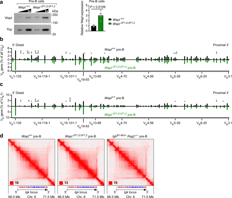
The starting point for our study was the previous finding that Wapl mRNA is fourfold downregulated in both pro-B and pre-B cells compared with uncommitted progenitors and mature B cells20. This observation raised the question of whether Wapl repression is also essential for VK gene recombination at the Igk locus as recently shown for the Igh locus20. To address this issue, we took advantage of the Wapl∆P1,2/∆P1,2 mouse, which lacks the functional Pax5-binding site P1 in the Wapl promoter as well as a hypersensitive region downstream of Wapl, containing a second Pax5-binding site (P2) that is not required for Wapl expression in early B cells20. As Wapl mRNA expression is known to be increased fourfold in Wapl∆P1,2/∆P1,2 pre-B cells compared with wild-type pre-B cells20, we next analyzed the expression of the Wapl protein by immunoblot analysis of ex vivo sorted Wapl∆P1,2/∆P1,2 and control Wapl+/+ pre-B cells (CD19+B220+IgM–IgD–Kit–CD25+) from the bone marrow (Supplementary Fig. 2a). Consistent with the observed Wapl mRNA increase, the Wapl protein was threefold more highly expressed in Wapl∆P1,2/∆P1,2 pre-B cells relative to Wapl+/+ pre-B cells (Fig. 1a). As elevated Wapl expression can lower the residence time of cohesin on chromatin20, the observed Wapl increase may lead to impaired loop extrusion and thus potential defects in VK-JK rearrangement in Wapl∆P1,2/∆P1,2 pre-B cells. Unexpectedly however, systematic analysis of VK-JK recombination by the VDJ-seq method37 revealed that individual VK genes along the entire Igk locus rearranged at a largely similar frequency in Wapl∆P1,2/∆P1,2 and Wapl+/+ pre-B cells (Fig. 1b and Supplementary Data 1a), regardless of their forward or reverse orientation (Supplementary Fig. 1b). Even the most distal VK genes (VK2-137, VK1-135) at the 5’ end of the Igk locus recombined at similar frequencies in both pre-B cell types. Differences in recombination frequency were primarily observed for the most 3’ proximal VK genes (VK3-2 to VK3-12) and some central VK genes (VK4-79 to VK10-96) that rearranged more efficiently in Wapl∆P1,2/∆P1,2 pre-B cell, while certain distal VK genes (VK9-120 to VK11-133) rearranged better in Wapl+/+ pre-B cells (Fig. 1b and Supplementary Fig. 1c).
Fig. 1. Efficient VK-JK rearrangements and long-range interactions across the Igk locus upon high Wapl expression in Wapl∆P1,2/∆P1,2 pre-B cells.
a Wapl expression in ex vivo sorted pre-B cells from the bone marrow of Wapl+/+ (black) and Wapl∆P1,2/∆P1,2 (green) mice, as determined by immunoblot analysis of threefold serially diluted whole-cell extracts with antibodies detecting Wapl or the TATA-binding protein (Tbp; loading control). Marker proteins of the indicated size (in kilodaltons, kDa) are shown to the right. The quantification of four independent immunoblot experiments is indicated to the right. Statistical data are shown as mean values with SEM and were analyzed with the Student’s t-test (unpaired and two-tailed). b, c VK gene recombination analysis of ex vivo sorted Wapl+/+ and Wapl∆P1,2/∆P1,2 pre-B cells, as determined by VDJ-seq experiments37. The VDJ-seq data obtained with Wapl+/+ (black) and Wapl∆P1,2/∆P1,2 (green) pre-B cells are shown in the upper and lower part, respectively. The recombination frequency of each VK gene is indicated as a percentage of all VK-JK (b) or only the VK-JK1 (c) rearrangements and is shown as mean value with SEM based on four independent VDJ-seq experiments for each pre-B cell type. The different VK genes (horizontal axis) are aligned according to their position in the Igk locus25 (Supplementary Data 1a). Statistical data were analyzed by multiple t-tests (unpaired and two-tailed) with Holm–Sidak correction; *P < 0.05, **P < 0.01, ***P < 0.001, ****P < 0.0001. d Hi-C contact matrices of the Igk region on chromosome 6, which were determined for ex vivo sorted Wapl+/+, Wapl∆P1,2/∆P1,2, and IghB1-8hi/+ Rag2–/– pre-B cells. The orientation and annotation of the Igk locus are shown. The intensity of each pixel represents the normalized number of contacts between a pair of loci5. The maximum intensity is indicated in the lower left of each panel (red square). The following resolution of the Hi-C data was calculated as described in Methods; 6.65 kb (Wapl+/+ pre-B cells), 4.25 kb (Wapl∆P1,2/∆P1,2 pre-B cells), and 11.8 kb (IghB1-8hi/+ Rag2–/– pre-B cells). One of two Hi-C experiments performed with pre-B cells of each genotype is shown. The VDJ-seq and Hi-C-seq data are further described in Supplementary Data 3. Source data are provided in the Source Data file.
The majority of primary VK-JK rearrangements is known to involve the 5′ most JK1 element, while the downstream JK2, JK4 and JK5 elements are mainly used for secondary VK-JK recombination occurring during receptor editing38,39. The overall frequencies of VK rearrangements involving the 4 functional JK elements were equivalent between Wapl∆P1,2/∆P1,2 and Wapl+/+ pre-B cells (Supplementary Fig. 1d) and were comparable to published data40,41. Moreover, the recombination frequencies of individual VK genes involving the JK1 or JK5 element were also largely similar in Wapl∆P1,2/∆P1,2 and Wapl+/+ pre-B cells (Fig. 1c and Supplementary Fig. 1e), further indicating that a threefold increase of Wapl expression had only a minimal effect on VK-JK recombination across the Igk locus.
We next used Hi-C sequencing5 to study the long-range interactions at the Igk locus in ex vivo sorted pre-B cells from the bone marrow of Wapl∆P1,2/∆P1,2 and Wapl+/+ mice (Supplementary Fig. 2a). The Hi-C contact map of the Igk locus in Wapl+/+ pre-B cells revealed that the Igk 3′ region, containing the JK elements and Igk enhancers, interacted with sequences across the entire Igk locus and that the large Igk TAD consisted of sub-TADs (Fig. 1d), as previously reported in ref. 42. Surprisingly, the Hi-C contact maps of Wapl∆P1,2/∆P1,2 and Wapl+/+ pre-B cells were comparable, which was confirmed by the quantification of the Hi-C interaction frequencies at the Igk locus (Supplementary Fig. 3a–c). Hence, the Igk locus has a similar TAD structure in both pre-B cell types (Fig. 1d). We next performed Hi-C analysis with IghB1-8hi/+ Rag2–/– pre-B cells, which were generated in the absence of V(D)J recombination due to RAG2 loss by skipping the pro-B cell stage through the expression of the functionally pre-rearranged Igh gene B1-8hi (ref. 43). Notably, the Hi-C contact map of IghB1-8hi/+ Rag2–/– pre-B cells was similar to those of Wapl+/+ and Wapl∆P1,2/∆P1,2 pre-B cells (Fig. 1d and Supplementary Fig. 3a–c), which indicated that the Hi-C stripe extending from the 3′ end across the entire Igk locus is caused by long-range interactions rather than by VK-JK rearrangements. We conclude, therefore, that increased Wapl expression in Wapl∆P1,2/∆P1,2 pre-B cells had a minimal effect on long-range interactions and VK gene rearrangements at the Igk locus, which is in marked contrast to the exquisite sensitivity of Igh VH-DJH recombination to elevated Wapl levels in Wapl∆P1,2/∆P1,2 pro-B cells20.
Increased Wapl expression minimally affects the chromosomal architecture of pre-B cells
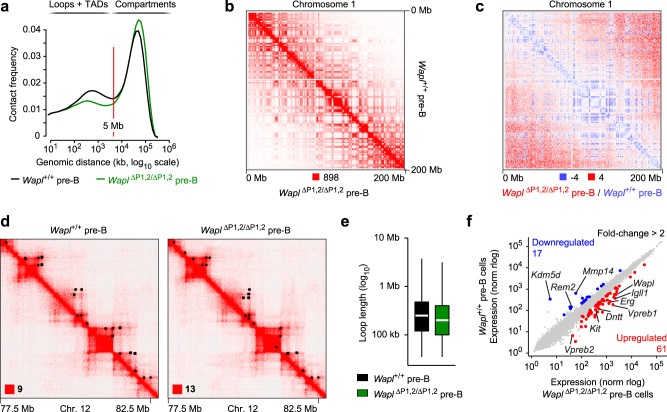
We next analyzed the chromosomal architecture of the entire genome in Wapl+/+ and Wapl∆P1,2/∆P1,2 pre-B cells by interrogating the Hi-C data. Analysis of all identified sequence contacts within the genome revealed that the frequencies of intrachromosomal contacts up to a distance of 5 Mb, which largely generate chromatin loops within TADs5, were moderately decreased in Wapl∆P1,2/∆P1,2 pre-B cells compared with Wapl+/+ pre-B cells (Fig. 2a). Moreover, the frequencies of contacts over very large distances (>10 Mb), which largely reflect chromosomal compartments4, were modestly increased in Wapl∆P1,2/∆P1,2 pre-B cells relative to Wapl+/+ pre-B cells (Fig. 2a). Consistent with this finding, visual inspection of the Hi-C contact map of chromosome 1 revealed a well-defined checkerboard pattern for both Wapl∆P1,2/∆P1,2 and Wapl+/+ pre-B cells (Fig. 2b). Modest differences are best highlighted by a differential contact map, which displays the difference in contact frequencies between Wapl+/+ and Wapl∆P1,2/∆P1,2 pre-B cells (Fig. 2c). This analysis confirmed a relative enrichment of short-range interactions in the TADs of Wapl+/+ pre-B cells, while long-range interactions in the compartment range were increased in Wapl∆P1,2/∆P1,2 pre-B cells. However, these minimal differences had no apparent effect on TAD structures and chromatin looping in Wapl∆P1,2/∆P1,2 pre-B cells, as shown for a zoomed-in region on chromosome 12 (Fig. 2d). This finding is consistent with the observed minor decrease of the median loop length from 250 kb in Wapl+/+ pre-B cells to 200 kb in Wapl∆P1,2/∆P1,2 pre-B cells (Fig. 2e and Supplementary Fig. 4a) and with the minimal difference in loop numbers determined for the two pre-B cell types (Supplementary Fig. 4b). Together, these data indicate that a threefold increase of Wapl protein expression had a relatively minor effect on the chromosomal architecture of pre-B cells in marked contrast to the strong effects elicited by a similar increase of Wapl protein expression in pro-B cells20.
Fig. 2. Minor differences in the chromosomal architecture of Wapl+/+ and Wapl∆P1,2/∆P1,2 pre-B cells.
a Frequency distribution of intrachromosomal contacts as a function of the genomic distance using logarithmically increased genomic distance bins, determined by Hi-C analysis of ex vivo sorted Wapl+/+ (black) and Wapl∆P1,2/∆P1,2 (green) pre-B cells using HOMER (see Methods). b Hi-C contact matrices of chromosome 1, determined for ex vivo sorted Wapl+/+ and Wapl∆P1,2/∆P1,2 pre-B cells and plotted at a 500-kb bin resolution with Juicebox74. The intensity of each pixel represents the normalized number of contacts between a pair of loci5. The maximum pixel intensity is indicated below (red square). c Differential Hi-C contact matrix of chromosome 1 displaying the difference in pixel intensity between Wapl+/+ pre-B and Wapl∆P1,2/∆P1,2 pre-B cells as the ratio (Wapl∆P1,2/∆P1,2)/(Wapl+/+). More interactions (blue) in the TAD range were observed for Wapl+/+ pro-B cells, while more interactions (red) in the compartment range were detected for Wapl∆P1,2/∆P1,2 pre-B cells. d Hi-C contact matrices of a zoomed-in region on chromosome 12 (mm9; 77,500,000–82,500,000), shown for Wapl+/+ and Wapl∆P1,2/∆P1,2 pre-B cells. Black dots indicate loop anchors identified with Juicebox. The maximum pixel intensity is indicated in the lower left of the panel (red square). e Distribution of the loop length (in kb). White lines indicate the median and boxes represent the middle 50% of the data. Whiskers denote all values of the 1.5× interquartile range. The median loop length is 250 kb in Wapl+/+ pre-B cells (black) and 200 kb in Wapl∆P1,2/∆P1,2 pre-B cells (green). f Scatterplot of gene expression differences between Wapl∆P1,2/∆P1,2 and Wapl+/+ pre-B cells isolated from the bone marrow of the respective mice. Genes upregulated (red) or downregulated (blue) in Wapl∆P1,2/∆P1,2 pre-B cell compared with Wapl+/+ pre-B cells were analyzed with DESeq2 and defined by an expression difference of >2-fold, an adjusted P value of <0.05 and a TPM value of >5 in at least one of the two pre-B cell types (Supplementary Data 2). Two RNA-seq experiments were performed with ex vivo sorted pre-B cells of each genotype.
To explore whether the observed architectural changes affect gene expression, we analyzed ex vivo sorted Wapl∆P1,2/∆P1,2 and Wapl+/+ pre-B cells by RNA-sequencing, which identified 61 upregulated and 17 downregulated genes with an expression difference of >2-fold in Wapl∆P1,2/∆P1,2 pre-B cells relative to Wapl+/+ pre-B cells (Fig. 2f and Supplementary Data 2). The differentially expressed genes code for proteins of distinct functional classes, including several surface proteins, signal transducers, and metabolic enzymes (Supplementary Fig. 4c and Supplementary Data 2). Notably, the genes encoding the surrogate light chains VpreB1, VpreB2, and Igλ (Igll1), the surface receptor Kit, and the terminal deoxynucleotidyl transferase (Dntt) were expressed at a higher level in Wapl∆P1,2/∆P1,2 pre-B cells compared with Wapl+/+ pre-B cells (Fig. 2f), where these genes are normally downregulated in response to pre-B cell receptor signaling44. The same expression analysis of Wapl∆P1,2/∆P1,2 and Wapl+/+ pro-B cells previously identified a higher number of differentially expressed genes, as 161 genes were upregulated and 159 genes were downregulated upon increased Wapl expression in pro-B cells20. There was, however, only a small overlap between the differentially expressed genes in pro-B and pre-B cells, with 16 upregulated and eight downregulated genes being present in both datasets (Supplementary Fig. 4d). These data, therefore, indicated that increased Wapl protein expression had a smaller effect on both differential gene expression and genomic architecture in pre-B cells relative to pro-B cells.
Wild-type pro-B and pre-B cells strongly differ in their chromosomal architecture
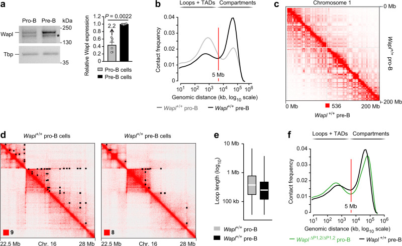
We next investigated the Wapl protein levels in ex vivo sorted pro-B and pre-B cells of Wapl+/+ mice by immunoblot analysis, which revealed that Wapl was expressed at a 2.2-fold higher level in pre-B cells compared with pro-B cells (Fig. 3a). This result was unexpected, as the Wapl mRNA is expressed at the same low level in pro-B and pre-B cells and is increased 4-fold only in immature and mature B cells20 (Supplementary Fig. 4e). The discrepancy between Wapl mRNA and Wapl protein expression in pre-B cells may indicate that the Wapl mRNA is either more efficiently translated in pre-B cells or that the Wapl protein is stabilized by a yet unknown posttranslational mechanism in pre-B cells. As predicted by the 2.2-fold difference in Wapl protein expression between wild-type pro-B and pre-B cells, the chromosomal architecture differed considerably between the two cell types, as indicated by the significantly lower frequencies of intrachromosomal contacts in the TAD range (<5 Mb) and the strongly increased frequencies of long-distance contacts in the compartment range (>10 Mb) in Wapl+/+ pre-B cells compared with Wapl+/+ pro-B cells (Fig. 3b). In this context, it is important to note that lower Wapl expression is known to cause a breakdown of compartmentalization, as longer loops interfere with the compartment structure10,19. Consistent with this finding, visual inspection of the Hi-C contact map of chromosome 1 revealed a well-defined checkerboard pattern (Fig. 3c), caused by increased compartmentalization (Supplementary Fig. 4f), in Waplhigh Wapl+/+ pre-B cells relative to Wapllow Wapl+/+ pro-B cells. Moreover, a zoomed-in region on chromosome 16 revealed that the long extension of loops in Wapl+/+ pro-B cells was not detected in Wapl+/+ pre-B cells (Fig. 3d), which is consistent with the observed decrease of the median loop length from 375 kb in Wapl+/+ pro-B cells20 to 250 kb in Wapl+/+ pre-B cells (Fig. 3e) and a 1.8-fold decrease in loop numbers from Wapl+/+ pro-B cells to Wapl+/+ pre-B cells (Supplementary Fig. 4g). Importantly, these findings revealed an exquisite sensitivity of loop formation on Wapl dosage, as a 2.2-fold increase of Wapl protein expression from Wapl+/+ pro-B to Wapl+/+ pre-B cells resulted in drastic changes of the chromosomal architecture and loop length, while an additional 3-fold increase of Wapl protein expression from Wapl+/+ pre-B cells to Wapl∆P1,2/∆P1,2 pre-B cells had only a minimal effect on chromosomal architecture and loop length (Supplementary Fig. 4h).
Fig. 3. Wild-type pro-B and pre-B cells strongly differ in their chromosomal architecture.
a Wapl protein expression in ex vivo sorted Rag2–/– pro-B (gray) and wild-type pre-B (black) cells from the bone marrow, as determined by immunoblot analysis of whole-cell extracts with antibodies detecting Wapl or Tbp (loading control). Marker proteins of the indicated size (in kilodaltons, kDa) are shown to the right. A non-specific band is indicated by an asterisk. One of 5 independent experiments is shown, and the quantification of all immunoblot experiments is indicated to the right. Statistical data are shown as mean values with SEM and were analyzed with the Student’s t-test (unpaired and two-tailed). b Comparison of the frequency distribution of intrachromosomal contacts between pro-B and pre-B cells of Wapl+/+ mice. The frequency distribution of intrachromosomal contacts was determined as a function of the genomic distance using logarithmically increased genomic distance bins, based on the Hi-C data of Wapl+/+ pre-B cells (black, this study) and published Hi-C data of short-term cultured Wapl+/+ pro-B cells (gray)20. c Hi-C contact matrices of chromosome 1, determined for Wapl+/+ pro-B and Wapl+/+ pre-B cells, were plotted at a 500-kb bin resolution with Juicebox74. The maximum pixel intensity is indicated below (red square). d Hi-C contact matrices of a zoomed-in region on chromosome 16 (mm9; 22,500,000–28,000,000), shown for Wapl+/+ pro-B and pre-B cells. Black dots indicate loop anchors identified with Juicebox74. e Distribution of the loop length (in kb) in Wapl+/+ pro-B (gray) and pre-B (black) cells. White lines indicate the median and boxes represent the middle 50% of the data. Whiskers denote all values of the 1.5× interquartile range. The median loop length is 375 kb in Wapl+/+ pro-B cells and 250 kb in Wapl+/+ pre-B cells. f Comparison of the frequency distribution of intrachromosomal contacts between Wapl+/+ pre-B cells (black) and Wapl∆P1,2/∆P1,2 pro-B cells (green)20, as described in (b). Source data are provided in the Source Data file.
Notably, Wapl∆P1,2/∆P1,2 pro-B cells and Wapl+/+ pre-B cells exhibited a similar chromosomal architecture, which was manifested by their comparable contact frequency distributions of intrachromosomal contacts (Fig. 3f), similar patterns of A- and B-type compartmentalization (Supplementary Fig. 5a, b) and comparable loop numbers and lengths (Supplementary Fig. 5c, d). The observed similarities are consistent with a similar Wapl protein increase in Wapl∆P1,2/∆P1,2 pro-B cells (threefold) and Wapl+/+ pre-B cells (2.2-fold) compared with Wapl+/+ pro-B cells (Fig. 3a)20. Together these data, therefore, demonstrate that wild-type pro-B and pre-B cells strongly differ in their chromosomal architecture, which is likely caused by the observed 2.2-fold difference in Wapl protein expression between the two cell types.
VK gene recombination across the Igk locus in both Waplhigh and Wapllow pro-B cells
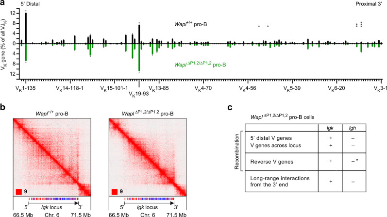
As pro-B cells have a distinct chromosomal architecture, already undergo contraction of the Igk locus35 and rearrange VK genes at a low frequency34,41, we next investigated the VK gene recombination pattern by VDJ-seq in ex vivo sorted pro-B cells (CD19+B220+IgM–IgD–Kit+CD25–) from the bone marrow of Wapl+/+ and Wapl∆P1,2/∆P1,2 mice (Supplementary Fig. 2b). Notably, VK gene recombination was observed along the entire Igk locus and involved the different JK elements at a similar frequency in both pro-B cell types, regardless of low (Wapl+/+) or high (Wapl∆P1,2/∆P1,2) Wapl expression and independent of the VK gene orientation (Fig. 4a and Supplementary Fig. 6a–c). The VK gene recombination pattern was quite similar between Wapl+/+ and Wapl∆P1,2/∆P1,2 pro-B cells (Fig. 4a). However, the most 3′ proximal VK genes (VK3-4 to VK3-12) and central VK genes (VK4-86 to VK10-95) rearranged more efficiently in Wapl∆P1,2/∆P1,2 pro-B cells compared with Wapl+/+ pro-B cells (Supplementary Fig. 6b) similar to the observed increased recombination frequency of these VK genes in Wapl∆P1,2/∆P1,2 pre-B cells relative to Wapl+/+ pre-B cells (Supplementary Fig. 1c). Notably, a direct comparison of the VK gene recombination pattern between Wapl+/+ pro-B and Wapl+/+ pre-B cells revealed that the VK genes in the 3′ half of the Igk locus up to the VK4-79 gene rearranged better in Wapl+/+ pre-B cells, while the more distal VK genes recombined more efficiently in Wapl+/+ pro-B cells (Supplementary Fig. 6d), as previously described41. Importantly, the most distal VK genes (VK2-137, VK1-135), which are present in forward orientation at the 5’ end of the Igk locus, were still able to recombine in Wapl∆P1,2/∆P1,2 pro-B cells (Fig. 4a), while distal VH genes (also in forward orientation) at the 5’ end of the Igh locus fail to recombine in the very same pro-B cells20. These data, therefore, suggest that the mechanism of chromatin folding at the Igk and Igh loci must be fundamentally different in pro-B cells.
Fig. 4. VK-JK recombination at the Igk locus in Waplhigh and Wapllow pro-B cells.
a VK gene recombination analysis of ex vivo sorted Wapl+/+ (Wapllow; black) and Wapl∆P1,2/∆P1,2 (Waplhigh; green) pro-B cells, as determined by VDJ-seq experiments. The recombination frequency of each VK gene is indicated as a percentage of all VK-JK rearrangement events and is shown as a mean value with SEM based on six independent VDJ-seq experiments for each pro-B cell type. The different VK genes (horizontal axis) are aligned according to their position in the Igk locus25 (Supplementary Data 1a). Statistical data were analyzed by multiple t-tests (unpaired and two-tailed) with Holm–Sidak correction; *P < 0.05, **P < 0.01, ***P < 0.001. b Hi-C contact matrices of the Igk region on chromosome 6 based on published Hi-C data of short-term cultured Wapl+/+ and Wapl∆P1,2/∆P1,2 pro-B cells20. The orientation and annotation of the Igk locus are shown. The resolution of the Hi-C data was 6.7 and 7.25 kb for the Wapl+/+ and Wapl∆P1,2/∆P1,2 pro-B cells, respectively. c Differences in V gene recombination and long-range interactions between the Igh and Igk locus in Wapl∆P1,2/∆P1,2 pro-B cells, based on the data shown in Fig. 4 and published data20. The loss of recombination of VH genes upon their inversion (indicated by an asterisk) in the Igh locus was analyzed in IghV8-8-inv/V8-8-inv and Igh∆890/∆890 pro-B cells20 and upon inversion of the entire VH gene cluster in v-Abl immortalized pro-B cells32. Source data are provided in the Source Data file.
Hi-C contact maps revealed long-range interactions from the Igk 3′ region across the entire Igk locus in Wapl+/+ pro-B cells (Fig. 4b) similar to Wapl+/+ pre-B cells (Fig. 1d). The structures of the sub-TADs were, however, less well defined in Wapl+/+ pro-B cells compared with Wapl∆P1,2/∆P1,2 pro-B cells as the low Wapl expression in Wapl+/+ pro-B cells resulted in a significant extension of loops within the Igk TAD in these cells (Fig. 4b). Consequently, the sub-TAD structures in the VK gene cluster differed between Wapl+/+ and Wapl∆P1,2/∆P1,2 pro-B cells, as shown by quantification of the Hi-C interaction frequencies at the Igk locus (Supplementary Fig. 3d, f). Surprisingly, the long-range interactions from the Igk 3’ region along the Igk locus were still formed in Wapl∆P1,2/∆P1,2 pro-B cells, although at a lower frequency compared with Wapl+/+ pro-B cells (Fig. 4b and Supplementary Fig. 3d, e). Finally, a direct comparison of the Hi-C contact maps of the Igk and Igh loci in Wapl∆P1,2/∆P1,2 pro-B cells by visual inspection highlighted the fact that long-range interactions from the 3’ end were observed across the Igk locus but were absent along the Igh locus in these Waplhigh pro-B cells (Supplementary Fig. 6e).
In summary, the V gene rearrangements at the Igk and Igh loci differ in three fundamental aspects in Wapl∆P1,2/∆P1,2 pro-B cells. First, V genes rearrange across the entire Igk locus in these Waplhigh pro-B cells in marked contrast to the Igh locus (Fig. 4a, c). Second, reverse-oriented V genes undergo rearrangements at the Igk locus but fail to recombine in the context of the Igh locus in pro-B cells (Fig. 4c and Supplementary Fig. 6a). Third, long-range interactions from the 3′ end occur only at the Igk locus but not at the Igh locus in Wapl∆P1,2/∆P1,2 pro-B cells (Fig. 4b, c). We conclude therefore that a different chromatin folding principle must operate at the Igk locus to promote VK recombination as opposed to the extended loop extrusion model that explains the convergent alignment of RSS sequences of the VH genes and DJH-rearranged element prior to VH-DJH recombination at the Igh locus20.
The VK gene region contracts in pre-B cells by folding into multiple different loops
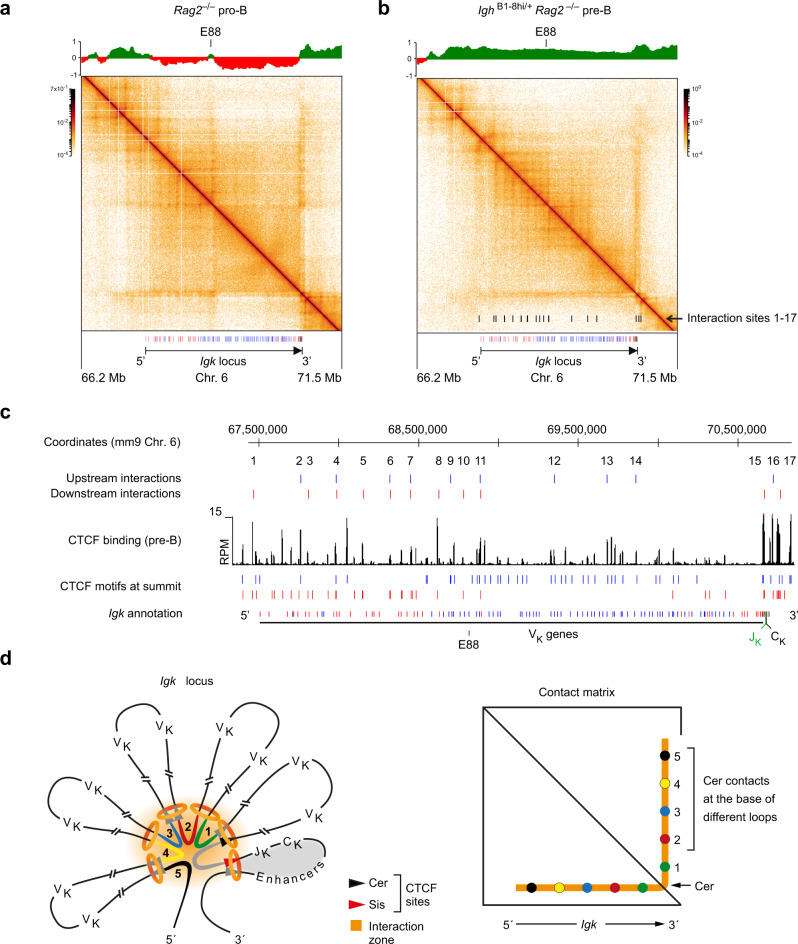
The recently developed Micro-C method, which relies on micrococcal nuclease digestion of fixed chromatin, facilitates genome-wide analysis of the fine-scale chromatin organization at nucleosomal resolution45,46. We next employed Micro-C analysis to study the chromatin folding along the Igk locus at high resolution in ex vivo sorted Rag2–/– pro-B and IghB1-8hi/+ Rag2–/– pre-B cells (Fig. 5a, b and Supplementary Fig. 7a, b). Notably, the Micro-C contact matrices at the Igk locus differed significantly from each other in pro-B and pre-B cells. In pro-B cells, the Igk locus consisted of two different TADs, which were present in distinct regions of less accessible compartment B that were separated by a small stretch of transcriptionally active compartment A (Fig. 5a) centered at the E88 enhancer42. In contrast, compartment A was present throughout the entire Igk locus in pre-B cells (Fig. 5b). As the interactions from the 3′ end across the Igk locus appeared to differ between pro-B and pre-B cells, we quantified the interaction frequencies along this stripe (Supplementary Fig. 7c). Notably, the interaction frequencies were specifically increased in the distal half of the Igk locus in Rag2–/– pro-B cells compared with IghB1-8hi/+ Rag2–/– pre-B cells (Supplementary Fig. 7c), which may explain the observed preferential VK gene usage in the distal Igk region in Rag2+/+ pro-B cells relative to Rag2+/+ pre-B cells (Supplementary Figs. 6d, 7d).
Fig. 5. High-resolution mapping of loop interactions at the Igk locus in Rag2-deficient pro-B and pre-B cells.
a, b Contact matrices of the Igk region (on chromosome 6) in ex vivo sorted Rag2–/– pro-B cells (a) and IghB1-8hi/+ Rag2–/– pre-B cells (b). The interaction data were determined by Micro-C analysis45,46 and are displayed at a 10-kb bin resolution with the HiGlass visualization tool81. Each dot on the contact matrix represents the contact intensity between a pair of nucleosomes according to the density scale shown. White lines denote regions that could not be mapped due to too low contact density. The annotation and orientation of the Igk locus are shown below. The PC1 (eigenvector) values, which define the compartments A (green) and B (red), are shown above the contact matrices together with the location of the E88 enhancer42. The locations of the contact sites that generate the different stripes at the Igk locus are indicated as black bars at the bottom of the contact matrix of IghB1-8hi/+ Rag2–/– pre-B cells (b). c Location of the mapped loop anchors at the Igk locus in IghB1-8hi/+ Rag2–/– pre-B cells. The anchor sites of loops facing upstream (blue) or downstream (red) in the Igk locus of pre-B cells were determined with the Cross-score program, as described in detail in Supplementary Fig. 7, and are shown above the CTCF ChIP-seq track and the annotation of the forward (red)- and reverse (blue)-oriented CBEs. d Schematic diagram explaining the looping organization of the Igk locus. Due to the presence of forward and reverse CBEs along the VK gene cluster, multiple different loops are formed, which leads to the collision of cohesin rings (orange) in response to ongoing loop extrusion. As a consequence, a transient interaction zone (orange) is formed that juxtaposes DNA sequences (1–5) at the base of these loops next to DNA sequences (gray) of the Cer region, which facilitates their crosslinking and defines specific interactions along the stripe emanating from the Cer region in the Micro-C data. Due to the high Wapl expression in pre-B cells, loops constantly turn over so that new loops present different DNA sequences in the interaction zone (see Supplementary Movie 1), which results in a contiguous stripe consisting of all possible interactions along the VK gene cluster. The orientation of CBEs in the VK gene region is indicated by gray arrowheads, while the CBEs at Cer and Sis are shown in black and red, respectively. The relatively stable “regulatory” loop containing the JK, CK, and Igk enhancer elements is indicated by gray shading.
Interestingly, a high degree of substructure was observed in the 5′ distal and central regions of the VK gene cluster in pre-B cells, while the density of interactions between these substructures and the stripe emanating from the 3′ proximal Cer region was quite low in these cells (Fig. 5b). To map the interactions causing the observed substructures at the Igk locus, we analyzed the Micro-C data with the Cross-score algorithm that quantifies the frequency of upstream and downstream long-distance contacts for each genomic site, thus measuring its ability to anchor genomic loops (Supplementary Fig. 7a, b, Methods). Peak calling on the upstream and downstream Cross-score profiles identified at least 17 peaks, which colocalized with the observed stripes in the Micro-C pattern of the Igk locus (Supplementary Fig. 7b). Notably, the Cross-score peaks mapped to CBEs of matching orientation (i.e., peaks in the downstream Cross-score profile matched forward CBEs, and vice versa), suggesting that these peaks located at the interaction stripes are caused by CTCF-mediated anchoring of cohesin loops (Fig. 5c and Supplementary Fig. 7a). These data, therefore, demonstrate that the forward and reverse CBEs are responsible for the formation of multiple different loops along the VK gene region. Interestingly, a deep gap was observed in both Cross-score interaction profiles at the very 3’ end of the Igk locus (Supplementary Fig. 7b). This gap indicates a relatively high degree of contact insulation between the VK gene region and the “regulatory” loop, which contains the JK, CK, and Igk enhancer elements and is likely formed between the upstream Sis element and first downstream CBE at the Igk 3’ end (Fig. 5d and Supplementary Fig. 1a).
Our finding that the pre-B cells with their elevated Wapl expression level can only form loops with a median size of 250 kb (Fig. 3e) raises the question of how to explain the long-range interactions from the Cer region across the entire 3.2-Mb long VK gene cluster. In this context, it is important to note that continuous loop extrusion can lead to the collision of loops14,47. Hence, the folding of the large VK gene region into multiple different loops likely leads to the collision of cohesin rings at the base of these loops (Fig. 5d). We, therefore, hypothesize that the collision of loops results in the formation of a transient interaction zone that juxtaposes DNA sequences at the base of these loops next to the DNA sequences of the Cer region. This, in turn, facilitates crosslinking of these DNA sequences, thus defining specific interactions along the stripe originating from the Cer region in the Micro-C data (Fig. 5d). Due to high Wapl expression in pre-B cells, loops constantly turn over so that new loops present different DNA sequences in the interaction zone (see Supplementary Movie 1), which results in a contiguous stripe consisting of all possible interactions along the VK gene cluster that can be detected in the large population of one million pre-B cells analyzed. Hence, this model could explain how contraction of the Igk locus through the formation of multiple intervening loops can bring a 5′ distant VK gene near the loop base into close vicinity of the 3′ proximal JK elements to facilitate VK-JK recombination in pre-B cells (Fig. 7).
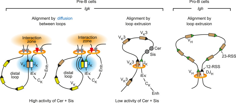
Fig. 7. Different folding principles leading to convergent alignment of RSS sequences at the Igk and Igh loci.
Schematic diagrams depict the different folding principles leading to a convergent alignment of RSS sequences during V gene recombination at the Igk and Igh loci. The convergent orientation of the 12-RSS (recognition signal sequence with a 12-bp spacer, black arrowhead) of the VK genes or DH segment and the 23-RSS (with a 23-bp spacer, green arrowhead) of the JK element or VH genes is essential for mediating RAG-dependent cleavage and recombination21. Cohesin (orange)-mediated loop extrusion across the entire Igh locus is responsible for the convergent alignment of the RSS sequences of VH genes and the DJH-rearranged segment prior to RAG-mediated cleavage and recombination in pro-B cells (right)20. Similarly, the forward-oriented members of the most 3’ proximal VK3 gene family could be convergently aligned with a JK element by loop extrusion under conditions where the insulating activity of Cer and Sis is reduced or lost (middle). All other VK genes can undergo convergent alignment with a JK element only by local diffusion62 (blue), once these elements are brought into close proximity at the interaction zone (orange), which is generated by the collision of multiple loops leading to contraction of the VK gene region (left). Enh, 3′Eκ and Edκ enhancers.
High density of long-range interactions across the entire VH gene cluster in pro-B cells
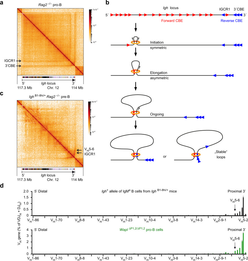
The Micro-C pattern at the Igh locus in Rag2–/– pro-B cells was dominated by two strong stripes emanating from the IGCR1 and 3′ CBEs (Fig. 6a and Supplementary Fig. 8a), which are known to act as loop anchors for long-range interactions across the entire Igh locus20,30. Notably, there was a high and relatively uniform density of long-range interactions across the entire VH gene cluster in pro-B cells, while only weak substructures could be detected (Fig. 6a) in marked contrast to the situation observed at the Igk locus in pre-B cells (Fig. 5b). The high density of interactions is best explained by the presence of 125 forward-oriented CBEs in the VH gene cluster and reverse-oriented CBEs at the IGCR1 and 3′CBE elements at the 3’ end of the Igh locus (Fig. 6b), which facilitate loop extrusion across the entire Igh locus in pro-B cells20. Loop extrusion likely initiates at random positions in the VH gene cluster and initially proceeds in a symmetrical manner, until the cohesin ring interacts with a CTCF protein bound to the next upstream forward CBE, which leads to stabilized binding of cohesin at this site48. Thereafter, asymmetric loop extrusion reels the DNA of the downstream Igh regions into the loop, until it is halted by a CTCF protein bound to a reverse CBE in convergent orientation at the IGCR1 or 3′CBE elements5,49. As predicted by this extended loop extrusion model, all the different sequences of the Igh locus should transiently interact and thus be cross-linked during loop extrusion in individual cells of the large pro-B cell population analyzed, which likely explains the observed high density of long-range interactions across the entire VH gene cluster.
Fig. 6. High density of interactions across the VH gene cluster in pro-B cells and decontraction of the Igh locus in pre-B cells.
a Contact matrix of the Igh region (on chromosome 12) in ex vivo sorted Rag2–/– pro-B cells. The interaction data were generated by Micro-C analysis and are displayed at a 10-kb bin resolution with the HiGlass visualization tool81. Each dot on the contact matrix represents the contact intensity between a pair of nucleosomes, as displayed according to the density scale shown. White lines denote regions that could not be mapped due to low contact density. The annotation and orientation of the Igh locus is shown below. The stripes corresponding to interactions from the 3’CBE or IGCR1 region are indicated. b Schematic diagram explaining the chromatin looping at the Igh locus in pro-B cells. The forward CBEs (red arrowheads) in the VH gene region are shown together with the reverse CBEs (blue arrowheads) at the IGCR1 and 3’CBE elements. Loop extrusion, upon random initiation in the VH gene cluster, proceeds first in a symmetrical manner, until the cohesin ring (orange) interacts with a CTCF protein bound to the next upstream forward CBE, which leads to stabilized binding of cohesin at this site48. Thereafter, asymmetric loop extrusion reels the DNA of the downstream Igh regions into the loop, until it is halted by a CTCF protein bound to a reverse CBE in convergent orientation at the IGCR1 or 3’CBE elements5. c Contact matrix of the Igh region in ex vivo sorted IghB1-8hi/+ Rag2–/– pre-B cells. See a for an explanation. The position of the IGCR1 element is indicated together with the location of the VH5-6 gene. d Upper part: VDJ-seq data obtained with sorted immature IgMa B cells from IghB1-8hi/+ (black) mice (Supplementary Fig. 8e). Only the VH gene reads originating from the Igh+ allele are shown, as the reads of the 5′ distal VH1-72 (B1-8hi) gene originating from the IghB1-8hi allele were eliminated together with the reads that mapped to the related VH1–53 gene exhibiting high sequence similarity to the VH1–72 gene. The recombination frequency of each VH gene is indicated as a percentage of all VDJH and DJH rearrangements and is shown as a mean value with SEM based on eight independent VDJ-seq experiments. The different VH genes (horizontal axis) are aligned according to their position in the Igh locus25 (Supplementary Data 1b). d Lower part: The published VH gene recombination pattern of Wapl∆P1,2/∆P1,2 pro-B cells20 is shown as reference data in green. The recombination frequency of the VH5-2 (VH81X) gene was 1.5% (1 Igh+ allele) in IgMa IghB1-8hi/+ B cells and 3.5% (2 Igh+ alleles) in Wapl∆P1,2/∆P1,2 pro-B cells. Source data are provided in the Source Data file.
Loss of long-range loops at the Igh locus due to increased Wapl expression in pre-B cells
As pro-B and pre-B cells significantly differ in their chromosomal architectures, we next analyzed the interaction pattern at the Igh locus in IghB1-8hi/+ Rag2–/– pre-B cells by visual inspection of Micro-C and Hi-C analyses (Fig. 6c and Supplementary Fig. 8b–d). Interestingly, the long-range interactions from the IGCR1 element were lost in these pre-B cells. Moreover, the interactions from the 3′CBE region were also strongly reduced throughout the VH gene cluster in IghB1-8hi/+ Rag2–/– pre-B cells but were still efficiently formed up to the position of the VH5-6 gene (Fig. 6c and Supplementary Fig. 8b, d) similar to the Igh interaction pattern observed in Wapl∆P1,2/∆P1,2 pro-B cells20. In the absence of long-range interactions, multiple substructures suggestive of internal looping were observed at the VH gene cluster in IghB1-8hi/+ Rag2–/– pre-B cells in contrast to Rag2–/– pro-B cells (Fig. 6a, c and Supplementary Fig. 8a–d). We, therefore, conclude that extended loop extrusion across the entire Igh locus in pro-B cells largely suppresses the formation of internal loops within the VH gene region.
By analyzing immature B cells of an Igμ-transgenic mouse strain, we previously demonstrated that only the most 3′ proximal VH genes of the Igh locus escape allelic exclusion in pre-B cells28. As a similar situation may exist in IghB1-8hi/+ pre-B cells, we tested this hypothesis by analyzing the VH-DJH recombination pattern in immature B cells from the bone marrow of IghB1-8hi/+ mice by VDJ-seq analysis. As shown in Supplementary Fig. 8e, the majority of immature IghB1-8hi/+ B cells expressed IgMa from the IghB1-8hi allele (of 129 origin), while a minor fraction expressed IgMb from the Igh+ allele (of C57BL/6 origin), possibly due to recombination-mediated inactivation of the IghB1-8hi gene in early B cell development50,51. VDJ-seq analysis of sorted immature IgMa B cells from IghB1-8hi/+ mice revealed that the wild-type Igh+ allele gave rise to efficient recombination of only the six most 3’ proximal VH genes up to the VH5-6 gene (Fig. 6d). Notably, the recombination pattern of these six VH genes in immature IgMa IghB1-8hi/+ B cells strongly resembled that of Wapl∆P1,2/∆P1,2 pro-B cells20 (Fig. 6d). Moreover, the recombination frequency of the VH5-2 (VH81X) gene was similar in IgMa IghB1-8hi/+ B cells (1.5% for 1 Igh+ allele) and Wapl∆P1,2/∆P1,2 pro-B cells (3.5% for 2 Igh+ allele) (Fig. 6d).
Together, these data indicate that the increased Wapl expression in pre-B cells causes the loss of long-range interactions across the VH gene cluster, which likely explains the previously described decontraction of the Igh locus, recombination of only the 6 most 3’ proximal VH genes and allelic exclusion of all other VH genes in pre-B cells28.
Discussion
How the Igk locus undergoes VK-JK recombination is still poorly understood, as its organization fundamentally differs from that of the other three antigen receptor loci. The V genes and their associated CBEs at the Igh, Tcrb (T cell receptor β), and Tcra/d (T cell receptor α/δ) loci are oriented in the same forward direction, while the CBEs in their 3′ proximal domain are present in reverse orientation25, which is compatible with loop extrusion across the entire locus20. In contrast, about half of all VK genes and CBEs are present in reverse orientation in the Igk locus, which results in inversional VK-JK recombination at a high frequency. Moreover, only the Igk locus undergoes RAG-mediated recombination between V genes, which additionally shapes the VK repertoire by deletion of the intervening VK genes52. Here, we have shown that VK-JK recombination in pro-B and pre-B cells is insensitive to high Wapl expression in marked contrast to VH-DJH recombination at the Igh locus in pro-B cells20, which is consistent with a recent report indicating that VK-JK recombination is also minimally affected upon Wapl degradation in in vitro cultured v-Abl-immortalized pre-B cell lines32. Our Hi-C and Micro-C analyses furthermore demonstrated that the contraction of the Igk locus is brought about by the formation of multiple internal loops within the VK gene cluster, which juxtaposes distal VK genes and proximal JK elements next to each other. Based on these data, we propose that the contraction of the Igk locus by many internal loops promotes diffusion-mediated alignment of the RSS sequences of distal VK genes and proximal JK elements to facilitate RAG-mediated cleavage and recombination (Fig. 7).
Unexpectedly, we discovered that pro-B and pre-B cells strongly differ in their chromosomal architectures with regard to their compartment structures and loop formation, although both cell types express the same low level of Wapl mRNA20 (Supplementary Fig. 4e). The observed 2.2-fold increase of Wapl protein expression in pre-B cells, possibly due to enhanced protein translation or stabilization, likely causes these global changes in chromosomal architecture, consistent with our previous finding that a 1.7- to 1.9-fold increase of Wapl expression in Wapl∆P1/∆P1 and Wapl∆P1,2/+ pro-B cells, respectively, abolishes VH gene recombination across the Igh locus due to drastic architectural changes20. Interestingly, a further threefold increase of Wapl protein expression in Wapl∆P1,2/∆P1,2 pre-B cells had only a minimal effect on the chromosomal organization (Supplementary Fig. 4h). Hence, the entire chromosomal architecture is exquisitely sensitive to a small change of Wapl protein expression during the pro-B-to-pre-B cell transition, but thereafter is quite insensitive to any further increase in Wapl concentration.
The exquisite Wapl dosage dependence of the chromosomal architecture is contrasted by the insensitivity of VK-JK recombination to Wapl protein changes in pro-B and pre-B cells. VK rearrangements are known to efficiently occur across the entire 3.2-Mb Igk locus in pre-B cells, although these cells are only able to form chromatin loops with a medium size of 0.25 Mb, which rules out prolonged loop extrusion as a basis for VK-JK recombination. Instead, we show here by high-resolution Micro-C analysis that the presence of many reverse CBEs is responsible for folding the VK gene region into multiple internal loops that are formed between convergent forward and reverse CBEs. The distance shortening induced by the multiple loops likely accounts for the previously observed contraction of the Igk locus in pre-B cells28. This folding principle invariably leads to the collision of loops14,47 that likely results in the formation of a transient interaction zone, where distant DNA sequences at the base of the loops are juxtaposed next to proximal DNA sequences of the Cer region, thus explaining the observed long-range interactions across the Igk locus in pre-B cells (Fig. 5d). VK-JK rearrangements are known to occur at a low frequency in pro-B cells34,41 due to STAT5-mediated suppression of Igk activation in response to IL-7 signaling in pro-B cells53,54. Here, we have shown that the long-range interaction pattern and compartment structure at the Igk locus also differ between pro-B and pre-B cells. While the entire Igk locus is encompassed by the transcriptionally active compartment A in pre-B cells, it is present in the less accessible compartment B in pro-B cells. Compartment B is interrupted only by a small stretch of compartment A located at the B cell-specific E88 enhancer that is known to primarily activate the recombination of adjacent VK genes in pro-B and pre-B cells42. The Igk locus contains only low levels of the poised histone mark H3K4me1 in pro-B cells in marked contrast to pre-B cells36, which is consistent with the presence of compartment B in pro-B cells and compartment A in pre-B cells.
The two elements Cer and Sis in the VK-JK intervening region play an important role in promoting the recombination of central and distal VK genes by suppressing excessive usage of the 3’ proximal VK genes55–57. The Cer element is essential for the contraction of the Igk locus56 and contains two reverse-oriented CBEs that facilitate interactions across the entire VK gene region (Fig. 5d), while the inversion of these CBEs promotes the recombination of proximal VK genes at the expense of central and distal VK genes58. The Sis element, whose deletion results in a less prominent overactivation of proximal VK gene recombination55, contains two forward-oriented CBEs25 (Supplementary Fig. 1a) that are able to participate in the formation of the “regulatory” loop containing the JK, CK, and Igk enhancer elements (Fig. 5d). Notably, the function of Cer and Sis appears to be only essential for establishing a balanced VK gene usage across the VK gene cluster during the primary VK-JK1 recombination event, which simultaneously leads to the deletion or inversional 5’ translocation of these two elements. Notably, conditional loss of CTCF in pre-B cells increases the interactions of the 3′ proximal VK genes with the downstream Igk enhancers, which strongly promotes recombination of these proximal VK genes at the expense of central and distal VK genes59. Furthermore, a similar phenotype was seen upon double deletion of Cer and Sis57. Here, we also observed increased recombination of the 3’ proximal VK3 gene family in Wapl∆P1,2/∆P1,2 versus Wapl+/+ pre-B cells as well as in Wapl∆P1,2/∆P1,2 versus Wapl+/+ pro-B cells. Hence, the decreased residence time of cohesin on chromatin upon increased Wapl expression in Wapl∆P1,2/∆P1,2 cells may interfere with CTCF-mediated insulation of the 3′ proximal VK genes by Cer and Sis, thus leading to enhanced interactions of the proximal VK3 genes with the downstream Igk enhancers.
The orientation of the RSS sequences at the V, D, and J elements of all antigen receptor genes requires that they are convergently aligned in the 3′ proximal RAG+ recombination center prior to RAG-mediated cleavage and recombination33. Detailed molecular analysis of DH-JH recombination60 and VH-DJH recombination3,20,61 at the Igh locus has provided strong evidence for the convergent alignment of RSS sequences by loop extrusion (Fig. 7). Convergent alignment by loop extrusion prior to VH-DJH recombination requires, however, that all VH genes of the Igh locus are present in the same forward orientation, as a VH gene upon its inversion fails to recombine in pro-B cells20. As half of the VK genes are present in reverse orientation in the Igk locus, convergent RSS alignment by loop extrusion is impossible except for the forward-oriented members of the most 3′ proximal VK3 gene family, which may only be possible upon loss of the insulating activity of Cer and Sis (Fig. 7). We, therefore, hypothesize that the RSS sequences of VK and JK elements, which are brought into close proximity in the interaction zone by contraction of the VK gene region through multiple loops, are aligned by local diffusion62 prior to VK-JK recombination (Fig. 7).
We have previously demonstrated that the non-functionally rearranged Igh allele undergoes decontraction in response to pre-BCR signaling, which results in feedback inhibition of VH-DJH recombination except for the most 3’ proximal VH genes that escape allelic exclusion in pre-B cells28. However, the molecular mechanism causing Igh decontraction has remained elusive until to date. Here, we have shown by high-resolution Micro-C analysis that the long-range interactions from the IGCR1 region are lost in IghB1-8hi/+ Rag2–/– pre-B cells. Moreover, the interactions from the 3′ CBE region are strongly reduced beyond the location of the VH5-6 gene, similar to the interaction pattern observed in Wapl∆P1,2/∆P1,2 pro-B cells20. Notably, the loss of extended loop extrusion at the Igh locus in IghB1-8hi/+ pre-B cells led to recombination of only the six most 3′ proximal VH genes up to the VH5-6 gene and possibly to allelic exclusion of all other VH genes, which strongly resembles the VH gene recombination pattern of Wapl∆P1,2/∆P1,2 pro-B cells20. These data, therefore, suggest that the increased Wapl expression in pre-B cells may be the molecular cause for Igh decontraction and allelic exclusion in pre-B cells. Future genetic experiments aiming at the downregulation of Wapl expression in pre-B cells will be required to conclusively demonstrate an essential role of Wapl in the control of allelic exclusion at the Igh locus.
In summary, we have shown that pro-B and pre-B cells have distinct chromosomal architectures and that the recombination of V genes at the equally large Igh and Igk loci is facilitated by fundamentally different folding principles in pro-B and pre-B cells, respectively. As decontraction and allelic exclusion of the non-functionally rearranged Igh allele likely depend on increased Wapl expression in pre-B cells, it could be argued that the Igk locus had to assume a different organization and folding principle to undergo efficient VK-JK recombination under conditions of high Wapl expression.
Methods
Mice
The following mice were maintained on the C57BL/6 background: Wapl∆P1,2/∆P1,2 mice20, Rag2–/– mice63, and IghB1-8hi/B1-8hi mice43. Experimental and control mice were co-housed under standard pathogen-free conditions at a temperature of 22 °C and 55% humidity with a day cycle of 14 h light and 10 h dark and with unrestricted access to food and water. Cells were harvested from mice that were 4–5-week-old (VDJ-seq analysis), 5–6-week-old (Hi-C and Micro-C analysis), and 4–6-week-old (immunoblot analysis). Mice were euthanized by carbon dioxide inhalation. Both female and male mice were used at a similar ratio in this study. All mouse experiments were carried out according to valid project licenses, which were approved and regularly controlled by the Austrian Veterinary Authorities.
Antibodies and flow-cytometric analysis
The following monoclonal antibodies were used for flow-cytometric analysis of mouse bone marrow cells: B220/CD45R (RA3-6B2; BD; 1:200), CD19 (1D3; BD; 1:300), CD25/IL-2Rα (PC61; BD Pharmingen; 1:500), CD117/Kit (2B8; Invitrogen; 1:1000), IgD (11-26c, Invitrogen; 1:2000), IgM (II/41, Invitrogen; 1:300), IgMa (MA-69, BioLegend; 1:1000), and IgMb (AF6-78, BioLegend; 1:1000). The following antibodies were used for immunoblot or immuno-precipitation analyses: anti-Wapl (rabbit polyclonal Ab, A960; Peters laboratory), anti-Tbp (mouse mAb clone 3TF1-3G3; Active Motif), and anti-H3K27ac (rabbit polyclonal Ab, ab4729; Abcam).
B cell types in the bone marrow were defined as CD19+B220+IgM–IgD–Kit+CD25– pro-B cells, CD19+B220+IgM–IgD–Kit–CD25+ pre-B cells, and CD19+B220+IgMaIgD–Kit– immature B cells. Flow-cytometric experiments and cell sorting were performed on LSR Fortessa (BD Biosciences) and FACSAria III (BD Biosciences) machines, respectively, using the FACS Diva (8.0) software. Flowjo software (Treestar) was used for data analysis.
Protein extract preparation and immunoblot analysis
Ex vivo pro-B and pre-B cells were sorted from the bone marrow by flow cytometry, and whole-cell extracts were prepared, using 2x SDS-PAGE sample buffer containing β-mercaptoethanol. The proteins were denatured by boiling, separated by SDS-PAGE, and analyzed by immunoblot analysis. The signal intensity of protein bands was quantified using ImageJ software and normalized to that of the Tbp loading control.
VDJ-seq analysis
VDJ-seq analysis of recombination at the Igk and Igh loci was performed as described in ref. 37. Genomic DNA was extracted from ex vivo sorted pro-B, pre-B, and immature B cells. The DNA (2 μg) was sheared using the Bioruptor sonicator (Diagenode) and subjected to end-repair and A-tailing, followed by ligation of adapters containing 12 UMI sequences using the NEBNext Ultra II DNA library prep kit for Illumina (NEB). A primer extension step with biotinylated JK- or JH-specific primers generated the single-stranded DNA products that were captured using Dynabeads MyOne streptavidin T1 beads (Thermo Fisher Scientific) and PCR-amplified with nested JK- or JH-specific and adapter-binding primers37. The Illumina sequencing adapter primers, including the indexes for multiplexing of libraries, were added to the PCR products in a final PCR amplification step. Paired-end 300-bp sequencing was performed on a MiSeq (Illumina) sequencing instrument (Supplementary Data 3). The bioinformatic analysis of the VDJ-seq data was performed as described in detail37, and the resulting data were processed for display in the respective figures using R version 3.3.3.
cDNA preparation for RNA-sequencing
Total RNA from ex vivo sorted pre-B cells was isolated with the RNeasy Plus Mini Kit (Qiagen), and mRNA was purified by two rounds of poly(A) selection with the Dynabeads mRNA purification kit (Invitrogen). The mRNA was fragmented by heating at 94 °C for 3 min in a fragmentation buffer and cDNA was prepared as described in ref. 20.
Library preparation and Illumina deep sequencing
About 0.6–20 ng of cDNA or ChIP-precipitated DNA was used as starting material for the generation of sequencing libraries with the NEBNext Ultra II DNA library prep kit for Illumina (NEB). Alternatively, sequencing libraries were generated using the NEBNext End Repair/dA-Tailing Module and NEBNext Ultra Ligation Module (NEB) followed by amplification with the KAPA Real-Time Amplification kit (KAPA Biosystems). Cluster generation and sequencing were carried out using the Illumina HiSeq 2500 system with a read length of 50 nucleotides, according to the manufacturer’s guidelines.
Hi-C library preparation
Wapl+/+, Wapl∆P1,2/∆P1,2, and IghB1-8hi/+ Rag2–/– pre-B cells were isolated from the bone marrow by immunomagnetic enrichment with anti-CD19-MicroBeads (Miltenyi Biotec) and were subsequently sorted by flow cytometry as CD19+B220+IgM–IgD–Kit–CD25+ pre-B cells prior to Hi-C library preparation. Hi-C libraries were prepared from 2 × 10⁷ cells as described in detail in ref. 5 and were sequenced using the Illumina NextSeq system with a read length of 75 nucleotides in the paired-end mode, according to the manufacturer’s guidelines.
Micro-C library preparation
Pro-B cells from the bone marrow of Rag2–/– mice were isolated by immunomagnetic enrichment with anti-CD19-MicroBeads (Miltenyi Biotec) followed by flow-cytometric sorting as CD19+B220+IgM–IgD–Kit+CD25– cells, while IghB1-8hi/+ Rag2–/– pre-B cells were sorted as CD19+B220+IgM–IgD–Kit–CD25+ pre-B cells. Micro-C libraries45 were prepared from 1 × 10⁶ cells using the Dovetail Micro-C Kit (# 21006) according to the manufacturer’s user manual (https://dovetailgenomics.com/wp-content/uploads/2021/09/Dovetail%E2%84%A2-Micro-C-Kit-User-Guide-Version-1.2.pdf). Libraries were sequenced using the NovaSeq 6000 S4 system with a read length of 150 nucleotides in the paired-end mode, according to the manufacturer’s guidelines.
Bioinformatic analysis of CTCF peaks in the Igk locus
We identified CTCF peaks (here referred to as CTCF-binding elements; CBEs) in the Igk locus based on the published data of our CTCF antibody ChIP-seq experiment (GSM1145865) that was performed with short-term cultured Rag2–/– pro-B cells30. In addition, published CTCF ChIP-seq data of ex vivo sorted pre-B cells36 (GSM2973687) were used. Sequence reads were uniquely aligned to the mouse genome assembly version of July 2007 (NCBI37/mm9) using the Bowtie program version 1.0 (ref. 64). CTCF peaks were called by MACS 2.2.5 (ref. 65) and filtered for P values of <10−10 to obtain a total of 61,354 peaks in Rag2–/– pro-B cells and 36,225 peaks in pre-B cells. For the Igk locus (mm9 Chr. 6; 67,505,630-70,694,944), this resulted in a total of 71 peaks in pro-B and pre-B cells, with 48 common peaks, and 19 and 4 unique peaks in pro-B and pre-B cells, respectively. We subsequently split all 71 peaks using PeakSplitter66 to obtain a final list of peak summits. This resulted in 112 CBEs in pro-B cells and 53 CBEs in pre-B cells across the Igk locus (Supplementary Fig. 1a).
To enumerate all potential CTCF-binding sites in the Igk locus, we retrieved the repeat-masked mouse genome sequence (mm9) using EXONERATE67 and scanned the sequence region of the Igk locus with a CTCF motif derived from the summits of the top 300 CTCF peaks, using MEME68. The scanning was done with FIMO version 4.9.1 (ref. 69) by setting the P value threshold to <0.001, which resulted in 77 motifs in pro-B cells and 52 motifs in pre-B cells that were clearly assigned to a CBE within 100 bp of the peak summit (shown in Supplementary Fig. 1a). In case of ambiguity, we selected the motif with the higher score.
Analysis of RNA-seq data
The number of reads per gene was counted using the featureCounts version 1.5.0 (ref. 70) with default settings. Transcripts per million (TPM) values were calculated as described71. Differential gene expression between ex vivo sorted Wapl+/+ and Wapl∆P1,2/∆P1,2 pre-B cells (Fig. 2f and Supplementary Data 2) was analyzed using R version 3.3.3. and DESeq2 version 2.1.14.1. Regularized log transformations were computed with the blind option set to “FALSE”. Genes with an adjusted P value of <0.05, TPM (averaged for each genotype) of >5 at least in one of the two genotypes, and a fold-change of >2 were called as significantly differentially expressed. All transcripts of the V, D, and J gene segments at the Igh, Igk and Igl loci were eliminated from the list of significantly regulated genes, although the immunoglobulin and T cell receptor transcripts were included in all TPM calculations.
Processing, normalization, and resolution of Hi-C data
The HiCUP pipeline version 0.5.10 (ref. 72) with the scorediff parameter set to “10” was used to truncate, align and filter the reads by applying the following software versions: R 3.4.1 (https://www.r-project.org), Bowtie 2.2.9 (ref. 64), and SAMtools 1.4. (ref. 73). Contact matrix files have been produced with the Juicer tools 1.8.9 (ref. 74). The resolution of the Hi-C data has been calculated according to ref. 5 by using the script “calculate_map_resolution”. The following unique di-tags were generated; 411,290,986 (GSM6427693) and 105,703,179 (GSM6427695) with Wapl+/+ pre-B cells, 694,115,546 (GSM6427694) and 76,038,129 (GSM6427696) with Wapl∆P1,2/∆P1,2 pre-B cells as well as 452,767,106 (GSM6427697) with IghB1-8hi/+ Rag2–/– pre-B cells. The following maximal resolution of the Hi-C data was calculated; 6.65 kb (Wapl+/+ pre-B cells), 4.25 kb (Wapl∆P1,2/∆P1,2 pre-B cells), and 11.8 kb (IghB1-8hi/+ Rag2–/– pre-B cells). We also analyzed the different Hi-C datasets with the open2c distiller-nf pipeline [https://github.com/open2c/distiller-nf], normally used for Micro-C data analysis (see below), to be able to compare Hi-C and Micro-C data with each other, to calculate the compartmentalization scores for the saddle plot analysis (Supplementary Fig. 5b) and to analyze the contact frequencies across the Igk locus in detail (Supplementary Fig. 3). Both pipelines (HiCUP and distiller-nf) led to very similar contact matrices for the same cell type and genotype.
Analysis of intrachromosomal contact frequency (Hi-C)
Contact frequency distributions have been calculated using the makeTagDirectory command of HOMER 4.10.3 (ref. 75). The contact frequency plots shown in Fig. 2a and Fig. 3b, f are based on ~50 contact data points based on ~50 bins, whereby each bin is defined as 0.1 step on the log10 scale of genomic distance observed between the contact points. Each data point is thus the sum of all contact fraction values in the respective bin. The contact frequency plot is shown as a smoothened line of the ~50 contact data points plotted against the logarithmic (log10) genomic distance. Detailed contact frequencies across the Igk locus (Supplementary Fig. 3), as well as the saddle plots (Supplementary Fig. 5b), were analyzed and generated with Cooltools76 and Python 3.8.13 (ref. 77), based on the data obtained with the open2c distiller-nf pipeline. To this end, we followed the description in the Cooltools notebook to generate the contact frequency [P(s)] plots, their first derivative (slope) curves, as well as the saddle plots for the various cell types and genotypes.
Analysis of chromatin loops (Hi-C)
Intrachromosomal loops have been called with the HiCCUPS algorithm from the Juicer tools74, based on the contact matrix derived from both Hi-C replicates of the same cell type. For the Hi-C comparisons between Wapl+/+ and Wapl∆P1,2/∆P1,2 pre-B cells (Supplementary Fig. 4b), we down-sampled the aligned reads obtained with the Wapl∆P1,2/∆P1,2 pre-B cells to the same read number obtained with the Wapl+/+ pre-B cells. In contrast, the Hi-C analyses resulted in similar read numbers for Wapl+/+ pro-B and Wapl+/+ pre-B cells (Supplementary Fig. 4g) as well as for Wapl+/+ pre-B and Wapl∆P1,2/∆P1,2 pro-B cells (Supplementary Fig. 5d). The obtained loops identified by HiCCUPS were overlapped between the two cell types of each cell pair to be compared, using “bedtools intersect” from the BEDTools package version 2.27.1 (ref. 78), with command parameters “-wa -wb -f 0.9 -r”. A reciprocal minimal overlap of 90% was required for two loops to be called “common”, while all other loops were referred to as “unique”. The distributions of loop lengths were plotted with R.
Analysis of Hi-C contact correlations
Correlation coefficients between Hi-C contact maps were calculated with HiCRep.py79, by using the parameters “—binsize 25000 -h 10 0dBPMax 20000000 –bdownSample –excludeChr chrM chrX chrY”, and by using two Hi-C contact matrices to be compared as input in the cooler format.
Bioinformatic analysis of Micro-C data
The open2c distiller-nf pipeline [https://github.com/open2c/distiller-nf] was used to process Micro-C datasets. The pipeline essentially aligns all sequences to the reference genome (mm9), parses the alignments into interaction pairs, filters out PCR duplicates, and finally aggregates pairs into binned matrices of Micro-C interactions. The underlying methods are all based on the pairtools (https://github.com/open2c/pairtools) and cooler80.
The stripes of enriched contacts in Micro-C maps at 2 kb resolution were detected using the Cross-score algorithm (https://github.com/glab-vbc/cross-score). Briefly, for each genomic bin, Cross-score calculates the total frequency of all contacts made with its neighbors located in a given range of distances, by separately analyzing the contacts with upstream or downstream neighbors. This score reveals genomic bins that participate in long-range interactions at a higher-than-normal frequency. Since our goal was to detect genomic bins that anchor focal loops or contact stripes, we calculated the Cross-score values for interactions at distances between 30 kb and 1 Mb, a typical range of interactions formed by cohesin-mediated looping. Peaks in Cross-score profiles were called using the ggpmisc package (Pedro J. Aphalo, 2021; https://github.com/aphalo/ggpmisc).
Eigenvalue decomposition to calculate compartment signals was performed with the eigs-cis program from the cooltools package (https://zenodo.org/record/5214125#.YhjAXJYo_mE), using H3K27ac ChIP-seq data of ex vivo sorted Rag1Cre/Cre pro-B cells (GSM6427700; Supplementary Data 3) as active chromatin phasing data. For smoother visualization of interaction and compartment tracks, we imputed missing values (due to missing mapping information) by using the imputeTS R packages (Moritz, Steffen, and Bartz-Beielstein (2017) “imputeTS: Time Series Missing Value Imputation in R.” R Journal 9.1, doi: 10.32614/RJ-2017-009). All resulting files (multi-resolution cooler files, compartment scores, and interaction scores) were visualized using the HiGlass visualization tool81 (http://higlass.io).
Statistical analysis
Statistical analysis was performed with the GraphPad Prism 7 software. Two-tailed unpaired Student’s t-test analysis was used to assess the statistical significance of one observed parameter between two experimental groups. The statistical evaluation of the RNA-seq data is described above (Analysis of RNA-seq data).
Reporting summary
Further information on research design is available in the Nature Portfolio Reporting Summary linked to this article.
Supplementary information
Description of Additional Supplementary Files
Acknowledgements
We thank Karin Aumayr’s team for flow-cytometric sorting and Andreas Sommer’s team at the Vienna BioCenter Core Facilities for Illumina sequencing. This research was supported by Boehringer Ingelheim, the Austrian Research Promotion Agency (FFG-878286), the European Research Council (ERC) under the European Union’s Horizon 2020 research and innovation program (grant agreement No 740349 [M.B.] and No 1020558 [J.-M.P.]), the Human Frontier Science Program (grant RGP0057/2018 [J.-M.P.]), and the Vienna Science and Technology Fund (grant LS19-029 [J.-M.P.]).
Source data
Author contributions
L.H. performed most experiments; G.W. generated the Hi-C and Micro-C data; L.C. isolated and prepared RAG2-deficient pro-B and pre-B cells for Hi-C and Micro-C analyses; H.T. performed the VDJ-seq analysis of immature IghB1-8hi/+ B cells; M.J. performed all bioinformatic analyses; J.-M.P. provided supervision and advice on cohesin biology; A.G. provided advice on loop extrusion and Micro-C analysis; L.H. and M.B. planned the project, designed the experiments, and wrote the manuscript.
Peer review
Peer review information
Nature Communications thanks Anne Corcoran and the other, anonymous, reviewer(s) for their contribution to the peer review of this work. Peer reviewer reports are available.
Data availability
The RNA-seq, ChIP-seq, VDJ-seq, Hi-C, and Micro-C data reported in this study (Supplementary Data 3) are available at the Gene Expression Omnibus repository under the accession number GSE210289. Source data are provided with this paper.
Competing interests
The authors declare no competing interests.
Footnotes
Publisher’s note Springer Nature remains neutral with regard to jurisdictional claims in published maps and institutional affiliations.
These authors contributed equally: Gordana Wutz, Markus Jaritz.
Supplementary information
The online version contains supplementary material available at 10.1038/s41467-023-37994-9.
References
- 1.Dekker J, Mirny L. The 3D genome as moderator of chromosomal communication. Cell. 2016;164:1110–1121. doi: 10.1016/j.cell.2016.02.007. [DOI] [PMC free article] [PubMed] [Google Scholar]
- 2.Davidson IF, Peters J-M. Genome folding through loop extrusion by SMC complexes. Nat. Rev. Mol. Cell Biol. 2021;22:445–464. doi: 10.1038/s41580-021-00349-7. [DOI] [PubMed] [Google Scholar]
- 3.Zhang Y, Zhang X, Dai HQ, Hu H, Alt FW. The role of chromatin loop extrusion in antibody diversification. Nat. Rev. Immunol. 2022;22:550–566. doi: 10.1038/s41577-022-00679-3. [DOI] [PMC free article] [PubMed] [Google Scholar]
- 4.Lieberman-Aiden E, et al. Comprehensive mapping of long-range interactions reveals folding principles of the human genome. Science. 2009;326:289–293. doi: 10.1126/science.1181369. [DOI] [PMC free article] [PubMed] [Google Scholar]
- 5.Rao SS, et al. A 3D map of the human genome at kilobase resolution reveals principles of chromatin looping. Cell. 2014;159:1665–1680. doi: 10.1016/j.cell.2014.11.021. [DOI] [PMC free article] [PubMed] [Google Scholar]
- 6.Dixon JR, et al. Topological domains in mammalian genomes identified by analysis of chromatin interactions. Nature. 2012;485:376–380. doi: 10.1038/nature11082. [DOI] [PMC free article] [PubMed] [Google Scholar]
- 7.Nora EP, et al. Spatial partitioning of the regulatory landscape of the X-inactivation centre. Nature. 2012;485:381–385. doi: 10.1038/nature11049. [DOI] [PMC free article] [PubMed] [Google Scholar]
- 8.Rao SSP, et al. Cohesin loss eliminates all loop domains. Cell. 2017;171:305–320. doi: 10.1016/j.cell.2017.09.026. [DOI] [PMC free article] [PubMed] [Google Scholar]
- 9.Schwarzer W, et al. Two independent modes of chromatin organization revealed by cohesin removal. Nature. 2017;551:51–56. doi: 10.1038/nature24281. [DOI] [PMC free article] [PubMed] [Google Scholar]
- 10.Wutz G, et al. Topologically associating domains and chromatin loops depend on cohesin and are regulated by CTCF, WAPL, and PDS5 proteins. EMBO J. 2017;36:3573–3599. doi: 10.15252/embj.201798004. [DOI] [PMC free article] [PubMed] [Google Scholar]
- 11.Parelho V, et al. Cohesins functionally associate with CTCF on mammalian chromosome arms. Cell. 2008;132:422–433. doi: 10.1016/j.cell.2008.01.011. [DOI] [PubMed] [Google Scholar]
- 12.Wendt KS, et al. Cohesin mediates transcriptional insulation by CCCTC-binding factor. Nature. 2008;451:796–801. doi: 10.1038/nature06634. [DOI] [PubMed] [Google Scholar]
- 13.Sanborn AL, et al. Chromatin extrusion explains key features of loop and domain formation in wild-type and engineered genomes. Proc. Natl. Acad. Sci. USA. 2015;112:E6456–E6465. doi: 10.1073/pnas.1518552112. [DOI] [PMC free article] [PubMed] [Google Scholar]
- 14.Fudenberg G, et al. Formation of chromosomal domains by loop extrusion. Cell Rep. 2016;15:2038–2049. doi: 10.1016/j.celrep.2016.04.085. [DOI] [PMC free article] [PubMed] [Google Scholar]
- 15.Davidson IF, et al. DNA loop extrusion by human cohesin. Science. 2019;366:1338–1345. doi: 10.1126/science.aaz3418. [DOI] [PubMed] [Google Scholar]
- 16.Kim Y, Shi Z, Zhang H, Finkelstein IJ, Yu H. Human cohesin compacts DNA by loop extrusion. Science. 2019;366:1345–1349. doi: 10.1126/science.aaz4475. [DOI] [PMC free article] [PubMed] [Google Scholar]
- 17.Kueng S, et al. Wapl controls the dynamic association of cohesin with chromatin. Cell. 2006;127:955–967. doi: 10.1016/j.cell.2006.09.040. [DOI] [PubMed] [Google Scholar]
- 18.Tedeschi A, et al. Wapl is an essential regulator of chromatin structure and chromosome segregation. Nature. 2013;501:564–568. doi: 10.1038/nature12471. [DOI] [PMC free article] [PubMed] [Google Scholar]
- 19.Haarhuis JHI, et al. The cohesin release factor WAPL restricts chromatin loop extension. Cell. 2017;169:693–707. doi: 10.1016/j.cell.2017.04.013. [DOI] [PMC free article] [PubMed] [Google Scholar]
- 20.Hill L, et al. Wapl repression by Pax5 promotes V gene recombination by Igh loop extrusion. Nature. 2020;584:142–147. doi: 10.1038/s41586-020-2454-y. [DOI] [PMC free article] [PubMed] [Google Scholar]
- 21.Alt FW, Zhang Y, Meng F-L, Guo C, Schwer B. Mechanisms of programmed DNA lesions and genomic instability in the immune system. Cell. 2013;152:417–429. doi: 10.1016/j.cell.2013.01.007. [DOI] [PMC free article] [PubMed] [Google Scholar]
- 22.Jhunjhunwala S, van Zelm MC, Peak MM, Murre C. Chromatin architecture and the generation of antigen receptor diversity. Cell. 2009;138:435–448. doi: 10.1016/j.cell.2009.07.016. [DOI] [PMC free article] [PubMed] [Google Scholar]
- 23.Schatz DG, Swanson PC. V(D)J recombination: mechanisms of initiation. Annu. Rev. Genet. 2011;45:167–202. doi: 10.1146/annurev-genet-110410-132552. [DOI] [PubMed] [Google Scholar]
- 24.Johnston CM, Wood AL, Bolland DJ, Corcoran AE. Complete sequence assembly and characterization of the C57BL/6 mouse Ig heavy chain V region. J. Immunol. 2006;176:4221–4234. doi: 10.4049/jimmunol.176.7.4221. [DOI] [PubMed] [Google Scholar]
- 25.Proudhon C, Hao B, Raviram R, Chaumeil J, Skok JA. Long-range regulation of V(D)J recombination. Adv. Immunol. 2015;128:123–182. doi: 10.1016/bs.ai.2015.07.003. [DOI] [PMC free article] [PubMed] [Google Scholar]
- 26.Kosak ST, et al. Subnuclear compartmentalization of immunoglobulin loci during lymphocyte development. Science. 2002;296:158–162. doi: 10.1126/science.1068768. [DOI] [PubMed] [Google Scholar]
- 27.Fuxa M, et al. Pax5 induces V-to-DJ rearrangements and locus contraction of the immunoglobulin heavy-chain gene. Genes Dev. 2004;18:411–422. doi: 10.1101/gad.291504. [DOI] [PMC free article] [PubMed] [Google Scholar]
- 28.Roldán E, et al. Locus ‘decontraction’ and centromeric recruitment contribute to allelic exclusion of the immunoglobulin heavy-chain gene. Nat. Immunol. 2005;6:31–41. doi: 10.1038/ni1150. [DOI] [PMC free article] [PubMed] [Google Scholar]
- 29.Jhunjhunwala S, et al. The 3D structure of the immunoglobulin heavy-chain locus: implications for long-range genomic interactions. Cell. 2008;133:265–279. doi: 10.1016/j.cell.2008.03.024. [DOI] [PMC free article] [PubMed] [Google Scholar]
- 30.Medvedovic J, et al. Flexible long-range loops in the VH gene region of the Igh locus facilitate the generation of a diverse antibody repertoire. Immunity. 2013;39:229–244. doi: 10.1016/j.immuni.2013.08.011. [DOI] [PMC free article] [PubMed] [Google Scholar]
- 31.Ebert A, Hill L, Busslinger M. Spatial regulation of V-(D)J recombination at antigen receptor loci. Adv. Immunol. 2015;128:93–121. doi: 10.1016/bs.ai.2015.07.006. [DOI] [PubMed] [Google Scholar]
- 32.Dai H-Q, et al. Loop extrusion mediates physiological Igh locus contraction for RAG scanning. Nature. 2021;590:338–343. doi: 10.1038/s41586-020-03121-7. [DOI] [PMC free article] [PubMed] [Google Scholar]
- 33.Ji Y, et al. The in vivo pattern of binding of RAG1 and RAG2 to antigen receptor loci. Cell. 2010;141:419–431. doi: 10.1016/j.cell.2010.03.010. [DOI] [PMC free article] [PubMed] [Google Scholar]
- 34.Novobrantseva TI, et al. Rearrangement and expression of immunoglobulin light chain genes can precede heavy chain expression during normal B cell development in mice. J. Exp. Med. 1999;189:75–87. doi: 10.1084/jem.189.1.75. [DOI] [PMC free article] [PubMed] [Google Scholar]
- 35.Stadhouders R, et al. Pre-B cell receptor signaling induces immunoglobulin κ locus accessibility by functional redistribution of enhancer-mediated chromatin interactions. PLoS Biol. 2014;12:e1001791. doi: 10.1371/journal.pbio.1001791. [DOI] [PMC free article] [PubMed] [Google Scholar]
- 36.Loguercio S, Barajas-Mora EM, Shih H-Y, Krangel MS, Feeney AJ. Variable extent of lineage-specificity and developmental stage-specificity of cohesin and CCCTC-binding factor binding within the immunoglobulin and T cell receptor loci. Front. Immunol. 2018;9:425. doi: 10.3389/fimmu.2018.00425. [DOI] [PMC free article] [PubMed] [Google Scholar]
- 37.Chovanec P, et al. Unbiased quantification of immunoglobulin diversity at the DNA level with VDJ-seq. Nat. Protoc. 2018;13:1232–1252. doi: 10.1038/nprot.2018.021. [DOI] [PubMed] [Google Scholar]
- 38.Yamagami T, ten Boekel E, Andersson J, Rolink A, Melchers F. Frequencies of multiple IgL chain gene rearrangements in single normal or κL chain-deficient B lineage cells. Immunity. 1999;11:317–327. doi: 10.1016/S1074-7613(00)80107-7. [DOI] [PubMed] [Google Scholar]
- 39.Vettermann C, Timblin GA, Lim V, Lai EC, Schlissel MS. The proximal J kappa germline-transcript promoter facilitates receptor editing through control of ordered recombination. PLoS ONE. 2015;10:e0113824. doi: 10.1371/journal.pone.0113824. [DOI] [PMC free article] [PubMed] [Google Scholar]
- 40.Matheson LS, et al. Local chromatin features including PU.1 and IKAROS binding and H3K4 methylation shape the repertoire of immunoglobulin kappa genes chosen for V(D)J recombination. Front. Immunol. 2017;8:1550. doi: 10.3389/fimmu.2017.01550. [DOI] [PMC free article] [PubMed] [Google Scholar]
- 41.Kleiman E, Loguercio S, Feeney AJ. Epigenetic enhancer marks and transcription factor binding influence Vκ gene rearrangement in pre-B cells and pro-B cells. Front. Immunol. 2018;9:2074. doi: 10.3389/fimmu.2018.02074. [DOI] [PMC free article] [PubMed] [Google Scholar]
- 42.Barajas-Mora EM, et al. A B-cell-specific enhancer orchestrates nuclear architecture to generate a diverse antigen receptor repertoire. Mol. Cell. 2019;73:48–60. doi: 10.1016/j.molcel.2018.10.013. [DOI] [PMC free article] [PubMed] [Google Scholar]
- 43.Shih T-AY, Roederer M, Nussenzweig MC. Role of antigen receptor affinity in T cell-independent antibody responses in vivo. Nat. Immunol. 2002;3:399–406. doi: 10.1038/ni776. [DOI] [PubMed] [Google Scholar]
- 44.Herzog S, Reth M, Jumaa H. Regulation of B-cell proliferation and differentiation by pre-B-cell receptor signalling. Nat. Rev. Immunol. 2009;9:195–205. doi: 10.1038/nri2491. [DOI] [PubMed] [Google Scholar]
- 45.Hsieh T-HS, et al. Resolving the 3D landscape of transcription-linked mammalian chromatin folding. Mol. Cell. 2020;78:539–553. doi: 10.1016/j.molcel.2020.03.002. [DOI] [PMC free article] [PubMed] [Google Scholar]
- 46.Krietenstein N, et al. Ultrastructural details of mammalian chromosome architecture. Mol. Cell. 2020;78:554–565. doi: 10.1016/j.molcel.2020.03.003. [DOI] [PMC free article] [PubMed] [Google Scholar]
- 47.Costantino L, Hsieh TS, Lamothe R, Darzacq X, Koshland D. Cohesin residency determines chromatin loop patterns. eLife. 2020;9:e59889. doi: 10.7554/eLife.59889. [DOI] [PMC free article] [PubMed] [Google Scholar]
- 48.Li Y, et al. The structural basis for cohesin-CTCF-anchored loops. Nature. 2020;578:472–476. doi: 10.1038/s41586-019-1910-z. [DOI] [PMC free article] [PubMed] [Google Scholar]
- 49.Peters J-M. How DNA loop extrusion mediated by cohesin enables V(D)J recombination. Curr. Opin. Cell Biol. 2021;70:75–83. doi: 10.1016/j.ceb.2020.11.007. [DOI] [PubMed] [Google Scholar]
- 50.Taki S, Schwenk F, Rajewsky K. Rearrangement of upstream DH and VH genes to a rearranged immunoglobulin variable region gene inserted into the DQ52-JH region of the immunoglobulin heavy chain locus. Eur. J. Immunol. 1995;25:1888–1896. doi: 10.1002/eji.1830250715. [DOI] [PubMed] [Google Scholar]
- 51.Sonoda E, et al. B cell development under the condition of allelic inclusion. Immunity. 1997;6:225–233. doi: 10.1016/S1074-7613(00)80325-8. [DOI] [PubMed] [Google Scholar]
- 52.Shinoda K, et al. Intra-Vκ cluster recombination shapes the Ig kappa locus repertoire. Cell Rep. 2019;29:4471–4481. doi: 10.1016/j.celrep.2019.11.088. [DOI] [PMC free article] [PubMed] [Google Scholar]
- 53.Malin S, et al. Role of STAT5 in controlling cell survival and immunoglobulin gene recombination during pro-B cell development. Nat. Immunol. 2010;11:171–179. doi: 10.1038/ni.1827. [DOI] [PMC free article] [PubMed] [Google Scholar]
- 54.Mandal M, et al. Epigenetic repression of the Igk locus by STAT5-mediated recruitment of the histone methyltransferase Ezh2. Nat. Immunol. 2011;12:1212–1220. doi: 10.1038/ni.2136. [DOI] [PMC free article] [PubMed] [Google Scholar]
- 55.Xiang Y, Zhou X, Hewitt SL, Skok JA, Garrard WT. A multifunctional element in the mouse Igκ locus that specifies repertoire and Ig loci subnuclear location. J. Immunol. 2011;186:5356–5366. doi: 10.4049/jimmunol.1003794. [DOI] [PMC free article] [PubMed] [Google Scholar]
- 56.Xiang Y, Park S-K, Garrard WT. Vκ gene repertoire and locus contraction are specified by critical DNase I hypersensitive sites within the Vκ-Jκ intervening region. J. Immunol. 2013;190:1819–1826. doi: 10.4049/jimmunol.1203127. [DOI] [PMC free article] [PubMed] [Google Scholar]
- 57.Xiang Y, Park SK, Garrard WT. A major deletion in the Vκ-Jκ intervening region results in hyperelevated transcription of proximal Vκ genes and a severely restricted repertoire. J. Immunol. 2014;193:3746–3754. doi: 10.4049/jimmunol.1401574. [DOI] [PMC free article] [PubMed] [Google Scholar]
- 58.Kleiman E, Xu J, Feeney AJ. Cutting edge: proper orientation of CTCF sites in Cer is required for normal Jκ-distal and Jκ-proximal Vκ gene usage. J. Immunol. 2018;201:1633–1638. doi: 10.4049/jimmunol.1800785. [DOI] [PMC free article] [PubMed] [Google Scholar]
- 59.Ribeiro de Almeida C, et al. The DNA-binding protein CTCF limits proximal Vκ recombination and restricts κ enhancer interactions to the immunoglobulin κ light chain locus. Immunity. 2011;35:501–513. doi: 10.1016/j.immuni.2011.07.014. [DOI] [PubMed] [Google Scholar]
- 60.Zhang Y, et al. The fundamental role of chromatin loop extrusion in physiological V(D)J recombination. Nature. 2019;573:600–604. doi: 10.1038/s41586-019-1547-y. [DOI] [PMC free article] [PubMed] [Google Scholar]
- 61.Jain S, Ba Z, Zhang Y, Dai HQ, Alt FW. CTCF-binding elements mediate accessibility of RAG substrates during chromatin scanning. Cell. 2018;174:102–116. doi: 10.1016/j.cell.2018.04.035. [DOI] [PMC free article] [PubMed] [Google Scholar]
- 62.Khanna N, Zhang Y, Lucas JS, Dudko OK, Murre C. Chromosome dynamics near the sol-gel phase transition dictate the timing of remote genomic interactions. Nat. Commun. 2019;10:2771. doi: 10.1038/s41467-019-10628-9. [DOI] [PMC free article] [PubMed] [Google Scholar]
- 63.Shinkai Y, et al. RAG-2-deficient mice lack mature lymphocytes owing to inability to initiate V(D)J rearrangement. Cell. 1992;68:855–867. doi: 10.1016/0092-8674(92)90029-C. [DOI] [PubMed] [Google Scholar]
- 64.Langmead, B. Aligning short sequencing reads with Bowtie. Curr. Protoc. BioinformaticsChapter 11, Unit 11 7 (2010). [DOI] [PMC free article] [PubMed]
- 65.Zhang Y, et al. Model-based analysis of ChIP-Seq (MACS) Genome Biol. 2008;9:R137. doi: 10.1186/gb-2008-9-9-r137. [DOI] [PMC free article] [PubMed] [Google Scholar]
- 66.Salmon-Divon M, Dvinge H, Tammoja K, Bertone P. PeakAnalyzer: genome-wide annotation of chromatin binding and modification loci. BMC Bioinformatics. 2010;11:415. doi: 10.1186/1471-2105-11-415. [DOI] [PMC free article] [PubMed] [Google Scholar]
- 67.Slater GS, Birney E. Automated generation of heuristics for biological sequence comparison. BMC Bioinform. 2005;6:31. doi: 10.1186/1471-2105-6-31. [DOI] [PMC free article] [PubMed] [Google Scholar]
- 68.Bailey TL, Johnson J, Grant CE, Noble WS. The MEME suite. Nucleic Acids Res. 2015;43:W39–W49. doi: 10.1093/nar/gkv416. [DOI] [PMC free article] [PubMed] [Google Scholar]
- 69.Machanick P, Bailey TL. MEME-ChIP: motif analysis of large DNA datasets. Bioinformatics. 2011;27:1696–1697. doi: 10.1093/bioinformatics/btr189. [DOI] [PMC free article] [PubMed] [Google Scholar]
- 70.Liao Y, Smyth GK, Shi W. featureCounts: an efficient general purpose program for assigning sequence reads to genomic features. Bioinformatics. 2014;30:923–930. doi: 10.1093/bioinformatics/btt656. [DOI] [PubMed] [Google Scholar]
- 71.Wagner GP, Kin K, Lynch VJ. Measurement of mRNA abundance using RNA-seq data: RPKM measure is inconsistent among samples. Theory Biosci. 2012;131:281–285. doi: 10.1007/s12064-012-0162-3. [DOI] [PubMed] [Google Scholar]
- 72.Wingett S, et al. HiCUP: pipeline for mapping and processing Hi-C data. F1000Res. 2015;4:1310. doi: 10.12688/f1000research.7334.1. [DOI] [PMC free article] [PubMed] [Google Scholar]
- 73.Li H, et al. The sequence alignment/map format and SAMtools. Bioinformatics. 2009;25:2078–2079. doi: 10.1093/bioinformatics/btp352. [DOI] [PMC free article] [PubMed] [Google Scholar]
- 74.Durand NC, et al. Juicer provides a one-click system for analyzing loop-resolution Hi-C experiments. Cell Syst. 2016;3:95–98. doi: 10.1016/j.cels.2016.07.002. [DOI] [PMC free article] [PubMed] [Google Scholar]
- 75.Heinz S, et al. Simple combinations of lineage-determining transcription factors prime cis-regulatory elements required for macrophage and B cell identities. Mol. Cell. 2010;38:576–589. doi: 10.1016/j.molcel.2010.05.004. [DOI] [PMC free article] [PubMed] [Google Scholar]
- 76.Open2C, et al. Cooltools: enabling high-resolution Hi-C analysis in Python. Preprint at bioRxiv10.1101/2022.10.31.514564 (2022).
- 77.Van Rossum, G & Drake, F. L. Python 3 Reference Manual (CreateSpace, 2009).
- 78.Quinlan AR, Hall IM. BEDTools: a flexible suite of utilities for comparing genomic features. Bioinformatics. 2010;26:841–842. doi: 10.1093/bioinformatics/btq033. [DOI] [PMC free article] [PubMed] [Google Scholar]
- 79.Lin D, Sanders J, Noble WS. HiCRep.py: fast comparison of Hi-C contact matrices in Python. Bioinformatics. 2021;37:2996–2997. doi: 10.1093/bioinformatics/btab097. [DOI] [PMC free article] [PubMed] [Google Scholar]
- 80.Abdennur N, Mirny LA. Cooler: scalable storage for Hi-C data and other genomically labeled arrays. Bioinformatics. 2020;36:311–316. doi: 10.1093/bioinformatics/btz540. [DOI] [PMC free article] [PubMed] [Google Scholar]
- 81.Kerpedjiev P, et al. HiGlass: web-based visual exploration and analysis of genome interaction maps. Genome Biol. 2018;19:125. doi: 10.1186/s13059-018-1486-1. [DOI] [PMC free article] [PubMed] [Google Scholar]
Associated Data
This section collects any data citations, data availability statements, or supplementary materials included in this article.
Supplementary Materials
Description of Additional Supplementary Files
Data Availability Statement
The RNA-seq, ChIP-seq, VDJ-seq, Hi-C, and Micro-C data reported in this study (Supplementary Data 3) are available at the Gene Expression Omnibus repository under the accession number GSE210289. Source data are provided with this paper.


