Abstract
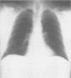
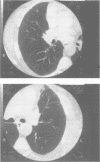
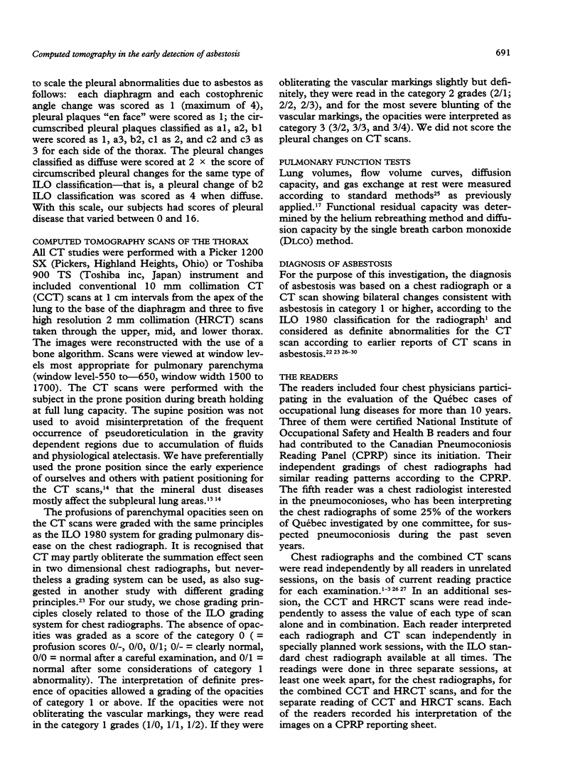
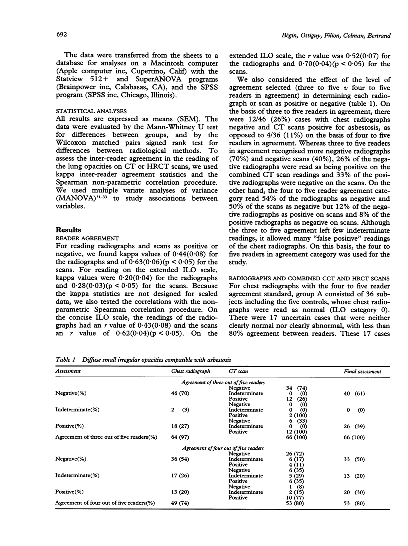
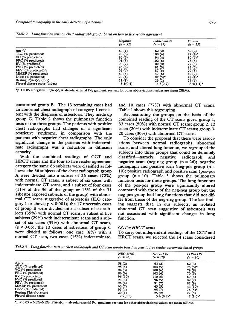
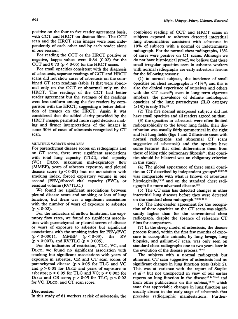
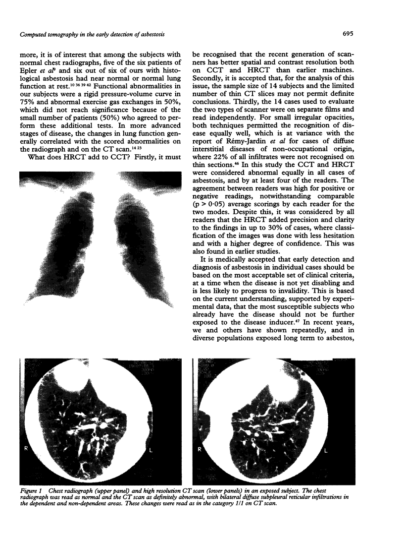
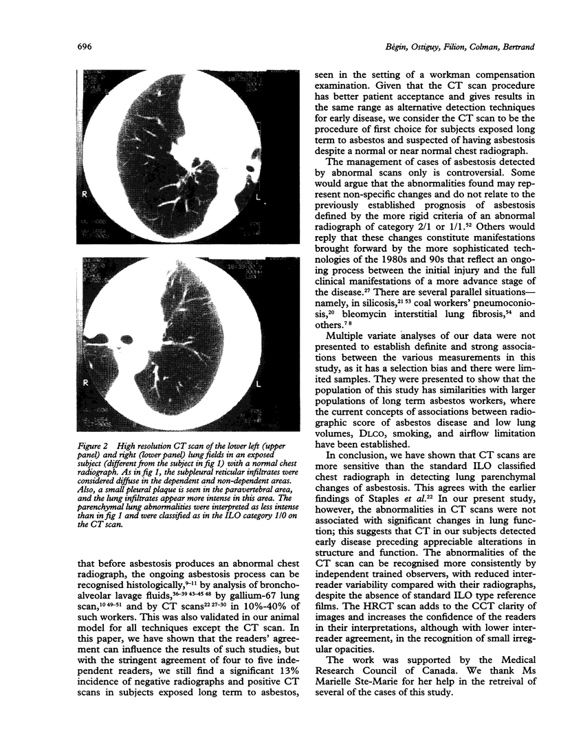
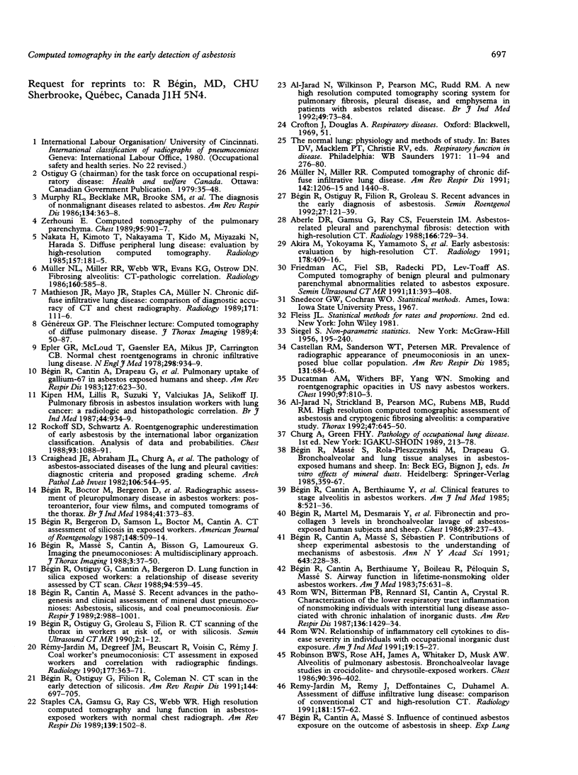
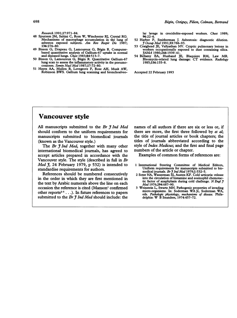
Computed tomography (CT; both conventional (CCT) and high resolution (HRCT)) scans of the thorax were evaluated to detect early asbestosis in 61 subjects exposed to asbestos dust in Québec for an average of 22(3) years and in five controls. The study was limited to consecutive cases with chest radiographs of the International Labour Organisation categories 0 or 1 determined independently. All subjects had a standard high kilovoltage posteroanterior and lateral chest radiograph, a set of 10-15 1 cm collimation CCT scans and a set of three to five 2 mm collimation HRCT scans in the upper, middle, and lower lung fields. Five experienced readers independently read each chest radiograph and sets of CT scans. On the basis of three to five readers agreeing for small opacities of the lung parenchyma, 12/46 (26%) negative chest radiographs were positive on CT scans, but 6/18 (33%) positive chest radiographs were negative on CT scan. On the basis of four to five readers agreeing on a chest radiograph, 36/66 (54%) subjects were normal (group A), 17/66 (26%) were indeterminate (group B), and 13/66 (20%) were abnormal (group C). By the combined readings of CCT and HRCT, 4/31 (13%) asbestos exposed subjects of group A were abnormal (p < 0.001), 6/17 (35%) of group B were abnormal, and in group C, 1/13 (8%) was normal, 2/13 were indeterminate, and 10/13 (77%) were abnormal. Separate readings of CCT and HRCT on distinct films in 14 subjects showed that all cases of asbestosis were abnormal on both CCT and HRCT. Inter-reader analyses by kappa statistics showed significantly better agreement for the readings of CT than the chest radiographs (p < 0.001), and for the reading of CCT than HRCT (p < 0.01). Thus CT scans of the thorax identifies significantly more irregular opacities consistent with the diagnosis of asbestosis than the chest radiograph (20 cases on CT scans v 13 on chest radiographs when four to five readers agreed, 13% of asbestos exposed subjects with normal chest radiographs or 21% of asbestos exposed subjects with normal or near normal chest radiographs. It decreased the number of indeterminate cases significantly from 17 on chest radiographs to 13 on CT scans. All cases of asbestosis detected only on CT scans were similarly seen on CCT and HRCT and did not have significant changes in lung function. The CT scans significantly reduced the inter-reader variability, despite the absence of ILO type reference films for these scans.
Full text
PDF
Images in this article
Selected References
These references are in PubMed. This may not be the complete list of references from this article.
- Aberle D. R., Gamsu G., Ray C. S., Feuerstein I. M. Asbestos-related pleural and parenchymal fibrosis: detection with high-resolution CT. Radiology. 1988 Mar;166(3):729–734. doi: 10.1148/radiology.166.3.3340770. [DOI] [PubMed] [Google Scholar]
- Akira M., Yokoyama K., Yamamoto S., Higashihara T., Morinaga K., Kita N., Morimoto S., Ikezoe J., Kozuka T. Early asbestosis: evaluation with high-resolution CT. Radiology. 1991 Feb;178(2):409–416. doi: 10.1148/radiology.178.2.1987601. [DOI] [PubMed] [Google Scholar]
- American Thoracic Society. Medical Section of the American Lung Association: The diagnosis of nonmalignant diseases related to asbestos. Am Rev Respir Dis. 1986 Aug;134(2):363–368. doi: 10.1164/arrd.1986.134.2.363. [DOI] [PubMed] [Google Scholar]
- Bellamy E. A., Husband J. E., Blaquiere R. M., Law M. R. Bleomycin-related lung damage: CT evidence. Radiology. 1985 Jul;156(1):155–158. doi: 10.1148/radiology.156.1.2408293. [DOI] [PubMed] [Google Scholar]
- Bisson G., Drapeau G., Lamoureux G., Cantin A., Rola-Pleszczynski M., Bégin R. Computer-based quantitative analysis of gallium-67 uptake in normal and diseased lungs. Chest. 1983 Nov;84(5):513–517. doi: 10.1378/chest.84.5.513. [DOI] [PubMed] [Google Scholar]
- Bisson G., Lamoureux G., Bégin R. Quantitative gallium 67 lung scan to assess the inflammatory activity in the pneumoconioses. Semin Nucl Med. 1987 Jan;17(1):72–80. doi: 10.1016/s0001-2998(87)80008-9. [DOI] [PubMed] [Google Scholar]
- Bégin R., Bergeron D., Samson L., Boctor M., Cantin A. CT assessment of silicosis in exposed workers. AJR Am J Roentgenol. 1987 Mar;148(3):509–514. doi: 10.2214/ajr.148.3.509. [DOI] [PubMed] [Google Scholar]
- Bégin R., Boctor M., Bergeron D., Cantin A., Berthiaume Y., Péloquin S., Bisson G., Lamoureux G. Radiographic assessment of pleuropulmonary disease in asbestos workers: posteroanterior, four view films, and computed tomograms of the thorax. Br J Ind Med. 1984 Aug;41(3):373–383. doi: 10.1136/oem.41.3.373. [DOI] [PMC free article] [PubMed] [Google Scholar]
- Bégin R., Cantin A., Berthiaume Y., Boileau R., Bisson G., Lamoureux G., Rola-Pleszczynski M., Drapeau G., Massé S., Boctor M. Clinical features to stage alveolitis in asbestos workers. Am J Ind Med. 1985;8(6):521–536. doi: 10.1002/ajim.4700080604. [DOI] [PubMed] [Google Scholar]
- Bégin R., Cantin A., Berthiaume Y., Boileau R., Péloquin S., Massé S. Airway function in lifetime-nonsmoking older asbestos workers. Am J Med. 1983 Oct;75(4):631–638. doi: 10.1016/0002-9343(83)90449-7. [DOI] [PubMed] [Google Scholar]
- Bégin R., Cantin A., Drapeau G., Lamoureux G., Boctor M., Massé S., Rola-Pleszczynski M. Pulmonary uptake of gallium-67 in asbestos-exposed humans and sheep. Am Rev Respir Dis. 1983 May;127(5):623–630. doi: 10.1164/arrd.1983.127.5.623. [DOI] [PubMed] [Google Scholar]
- Bégin R., Cantin A., Massé S. Recent advances in the pathogenesis and clinical assessment of mineral dust pneumoconioses: asbestosis, silicosis and coal pneumoconiosis. Eur Respir J. 1989 Nov;2(10):988–1001. [PubMed] [Google Scholar]
- Bégin R., Cantin A., Massé S., Sébastien P. Contributions of experimental asbestosis in sheep to the understanding of asbestosis. Ann N Y Acad Sci. 1991 Dec 31;643:228–238. doi: 10.1111/j.1749-6632.1991.tb24467.x. [DOI] [PubMed] [Google Scholar]
- Bégin R., Martel M., Desmarais Y., Drapeau G., Boileau R., Rola-Pleszczynski M., Massé S. Fibronectin and procollagen 3 levels in bronchoalveolar lavage of asbestos-exposed human subjects and sheep. Chest. 1986 Feb;89(2):237–243. doi: 10.1378/chest.89.2.237. [DOI] [PubMed] [Google Scholar]
- Bégin R., Massé S., Cantin A., Bisson G., Bergeron D. Imaging the pneumoconioses: a multidisciplinary approach. J Thorac Imaging. 1988 Oct;3(4):37–50. doi: 10.1097/00005382-198810000-00007. [DOI] [PubMed] [Google Scholar]
- Bégin R., Ostiguy G., Cantin A., Bergeron D. Lung function in silica-exposed workers. A relationship to disease severity assessed by CT scan. Chest. 1988 Sep;94(3):539–545. doi: 10.1378/chest.94.3.539. [DOI] [PubMed] [Google Scholar]
- Bégin R., Ostiguy G., Filion R., Groleau S. Recent advances in the early diagnosis of asbestosis. Semin Roentgenol. 1992 Apr;27(2):121–139. doi: 10.1016/0037-198x(92)90054-6. [DOI] [PubMed] [Google Scholar]
- Bégin R., Ostiguy G., Fillion R., Colman N. Computed tomography scan in the early detection of silicosis. Am Rev Respir Dis. 1991 Sep;144(3 Pt 1):697–705. doi: 10.1164/ajrccm/144.3_Pt_1.697. [DOI] [PubMed] [Google Scholar]
- Castellan R. M., Sanderson W. T., Petersen M. R. Prevalence of radiographic appearance of pneumoconiosis in an unexposed blue collar population. Am Rev Respir Dis. 1985 May;131(5):684–686. doi: 10.1164/arrd.1985.131.5.684. [DOI] [PubMed] [Google Scholar]
- Craighead J. E., Abraham J. L., Churg A., Green F. H., Kleinerman J., Pratt P. C., Seemayer T. A., Vallyathan V., Weill H. The pathology of asbestos-associated diseases of the lungs and pleural cavities: diagnostic criteria and proposed grading schema. Report of the Pneumoconiosis Committee of the College of American Pathologists and the National Institute for Occupational Safety and Health. Arch Pathol Lab Med. 1982 Oct 8;106(11):544–596. [PubMed] [Google Scholar]
- Craighead J. E., Vallyathan N. V. Cryptic pulmonary lesions in workers occupationally exposed to dust containing silica. JAMA. 1980 Oct 24;244(17):1939–1941. [PubMed] [Google Scholar]
- Ducatman A. M., Withers B. F., Yang W. N. Smoking and roentgenographic opacities in US Navy asbestos workers. Chest. 1990 Apr;97(4):810–813. doi: 10.1378/chest.97.4.810. [DOI] [PubMed] [Google Scholar]
- Epler G. R., McLoud T. C., Gaensler E. A., Mikus J. P., Carrington C. B. Normal chest roentgenograms in chronic diffuse infiltrative lung disease. N Engl J Med. 1978 Apr 27;298(17):934–939. doi: 10.1056/NEJM197804272981703. [DOI] [PubMed] [Google Scholar]
- Friedman A. C., Fiel S. B., Radecki P. D., Lev-Toaff A. S. Computed tomography of benign pleural and pulmonary parenchymal abnormalities related to asbestos exposure. Semin Ultrasound CT MR. 1990 Oct;11(5):393–408. [PubMed] [Google Scholar]
- Genereux G. P. The Fleischner lecture: computed tomography of diffuse pulmonary disease. J Thorac Imaging. 1989 Jul;4(3):50–87. [PubMed] [Google Scholar]
- Harber P., Smitherman J. Asbestosis: diagnostic dilution. J Occup Med. 1991 Jul;33(7):786–793. [PubMed] [Google Scholar]
- Hayes A. A., Mullan B., Lovegrove F. T., Rose A. H., Musk A. W., Robinson B. W. Gallium lung scanning and bronchoalveolar lavage in crocidolite-exposed workers. Chest. 1989 Jul;96(1):22–26. doi: 10.1378/chest.96.1.22. [DOI] [PubMed] [Google Scholar]
- Jarad N. A., Wilkinson P., Pearson M. C., Rudd R. M. A new high resolution computed tomography scoring system for pulmonary fibrosis, pleural disease, and emphysema in patients with asbestos related disease. Br J Ind Med. 1992 Feb;49(2):73–84. doi: 10.1136/oem.49.2.73. [DOI] [PMC free article] [PubMed] [Google Scholar]
- Mathieson J. R., Mayo J. R., Staples C. A., Müller N. L. Chronic diffuse infiltrative lung disease: comparison of diagnostic accuracy of CT and chest radiography. Radiology. 1989 Apr;171(1):111–116. doi: 10.1148/radiology.171.1.2928513. [DOI] [PubMed] [Google Scholar]
- Müller N. L., Miller R. R. Computed tomography of chronic diffuse infiltrative lung disease. Part 1. Am Rev Respir Dis. 1990 Nov;142(5):1206–1215. doi: 10.1164/ajrccm/142.5.1206. [DOI] [PubMed] [Google Scholar]
- Müller N. L., Miller R. R., Webb W. R., Evans K. G., Ostrow D. N. Fibrosing alveolitis: CT-pathologic correlation. Radiology. 1986 Sep;160(3):585–588. doi: 10.1148/radiology.160.3.3737898. [DOI] [PubMed] [Google Scholar]
- Nakata H., Kimoto T., Nakayama T., Kido M., Miyazaki N., Harada S. Diffuse peripheral lung disease: evaluation by high-resolution computed tomography. Radiology. 1985 Oct;157(1):181–185. doi: 10.1148/radiology.157.1.4034963. [DOI] [PubMed] [Google Scholar]
- Remy-Jardin M., Degreef J. M., Beuscart R., Voisin C., Remy J. Coal worker's pneumoconiosis: CT assessment in exposed workers and correlation with radiographic findings. Radiology. 1990 Nov;177(2):363–371. doi: 10.1148/radiology.177.2.2217770. [DOI] [PubMed] [Google Scholar]
- Remy-Jardin M., Remy J., Deffontaines C., Duhamel A. Assessment of diffuse infiltrative lung disease: comparison of conventional CT and high-resolution CT. Radiology. 1991 Oct;181(1):157–162. doi: 10.1148/radiology.181.1.1887026. [DOI] [PubMed] [Google Scholar]
- Robinson B. W., Rose A. H., James A., Whitaker D., Musk A. W. Alveolitis of pulmonary asbestosis. Bronchoalveolar lavage studies in crocidolite- and chrysotile-exposed individuals. Chest. 1986 Sep;90(3):396–402. doi: 10.1378/chest.90.3.396. [DOI] [PubMed] [Google Scholar]
- Rockoff S. D., Schwartz A. Roentgenographic underestimation of early asbestosis by International Labor Organization classification. Analysis of data and probabilities. Chest. 1988 May;93(5):1088–1091. doi: 10.1378/chest.93.5.1088. [DOI] [PubMed] [Google Scholar]
- Rom W. N., Bitterman P. B., Rennard S. I., Cantin A., Crystal R. G. Characterization of the lower respiratory tract inflammation of nonsmoking individuals with interstitial lung disease associated with chronic inhalation of inorganic dusts. Am Rev Respir Dis. 1987 Dec;136(6):1429–1434. doi: 10.1164/ajrccm/136.6.1429. [DOI] [PubMed] [Google Scholar]
- Soter N. A., Wasserman S. I., Austen K. F. Cold urticaria: release into the circulation of histamine and eosinophil chemotactic factor of anaphylaxis during cold challenge. N Engl J Med. 1976 Mar 25;294(13):687–690. doi: 10.1056/NEJM197603252941302. [DOI] [PubMed] [Google Scholar]
- Spurzem J. R., Saltini C., Rom W., Winchester R. J., Crystal R. G. Mechanisms of macrophage accumulation in the lungs of asbestos-exposed subjects. Am Rev Respir Dis. 1987 Aug;136(2):276–280. doi: 10.1164/ajrccm/136.2.276. [DOI] [PubMed] [Google Scholar]
- Staples C. A., Gamsu G., Ray C. S., Webb W. R. High resolution computed tomography and lung function in asbestos-exposed workers with normal chest radiographs. Am Rev Respir Dis. 1989 Jun;139(6):1502–1508. doi: 10.1164/ajrccm/139.6.1502. [DOI] [PubMed] [Google Scholar]
- Zerhouni E. Computed tomography of the pulmonary parenchyma. An overview. Chest. 1989 Apr;95(4):901–907. doi: 10.1378/chest.95.4.901. [DOI] [PubMed] [Google Scholar]
- al-Jarad N., Strickland B., Pearson M. C., Rubens M. B., Rudd R. M. High resolution computed tomographic assessment of asbestosis and cryptogenic fibrosing alveolitis: a comparative study. Thorax. 1992 Aug;47(8):645–650. doi: 10.1136/thx.47.8.645. [DOI] [PMC free article] [PubMed] [Google Scholar]


