Abstract
Autism spectrum disorder (ASD) has a multifactorial etiology. Major efforts are underway to understand the neurobiological bases of ASD and to develop efficacious treatment strategies. Recently, the use of cannabinoid compounds in children with neurodevelopmental disorders including ASD has received increasing attention. Beyond anecdotal reports of efficacy, however, there is limited current evidence supporting such an intervention and the clinical studies currently available have intrinsic limitations that make the interpretation of the findings challenging. Furthermore, as the mechanisms underlying the beneficial effects of cannabinoid compounds in neurodevelopmental disorders are still largely unknown, the use of drugs targeting the endocannabinoid system remains controversial. Here, we studied the role of endocannabinoid neurotransmission in the autistic-like traits displayed by the recently validated Fmr1-Δexon 8 rat model of autism. Fmr1-Δexon 8 rats showed reduced anandamide levels in the hippocampus and increased 2-arachidonoylglycerol (2-AG) content in the amygdala. Systemic and intra-hippocampal potentiation of anandamide tone through administration of the anandamide hydrolysis inhibitor URB597 ameliorated the cognitive deficits displayed by Fmr1-Δexon 8 rats along development, as assessed through the novel object and social discrimination tasks. Moreover, blockade of amygdalar 2-AG signaling through intra-amygdala administration of the CB1 receptor antagonist SR141716A prevented the altered sociability displayed by Fmr1-Δexon 8 rats. These findings demonstrate that anandamide and 2-AG differentially modulate specific autistic-like traits in Fmr1-Δexon 8 rats in a brain region-specific manner, suggesting that fine changes in endocannabinoid mechanisms contribute to ASD-related behavioral phenotypes.
Subject terms: Autism spectrum disorders, Working memory
Introduction
Autism spectrum disorder (ASD) defines a group of neurodevelopmental disorders characterized by disturbed patterns of social behavior and further differentiated by other features such as atypical or deficient communication skills and restricted or repetitive patterns of behavior [1]. Other comorbid features are often associated with these core symptoms, such as intellectual disability (ID) and aberrant sensitivity to sensory stimulation [2]. Multiple factors are involved in the pathogenesis of the disease, ranging from environmental to genetic and even to combinations of the two factors [3].
Fragile X syndrome (FXS) is the most common inherited cause of ASD and ID worldwide. It is caused by the silencing of the Fragile X Messenger Ribonucleoprotein 1 (FMR1) gene which has a key role in the development of synapses and in underlying learning and memory processes [4–7]. While clinically and genetically distinct, FXS and ASD share significant comorbidity, suggesting that there may be common molecular and/or cellular bases controlling the dysregulated neuronal networking and circuitry, leading to common aberrant patterns of behavior [8–10].
To date, there are no approved curative therapies for ASD; current approaches focus on helpful educational and behavioral strategies, together with off-label drugs that mitigate specific symptoms such as hyperactivity, anxiety and seizures [11]. Yet, for many patients, these off-label medications, when used alone or in combination, have suboptimal efficacy and tolerability and do not ameliorate the cognitive and social impairments associated with ASD.
The endocannabinoid system (ECS) is a major neuromodulatory system involved in the regulation of synaptic plasticity, emotional responses, social behavior and cognitive states [12–15], all of which are affected in ASD and FXS [16, 17]. It consists of endocannabinoids (primarily anandamide (AEA) and 2-arachidonoylglycerol (2-AG)), cannabinoid receptors and the enzymes responsible for endocannabinoid synthesis (e.g., NAPE-PLD, DAGL) and degradation (e.g., FAAH, MAGL) [18–21], together with a “non-canonical” extended signaling network [22]. Abnormalities in the ECS have been found both in patients with ASD [23–26] and in animal models of ASD [27–29]. Furthermore, pharmacological modulation of the ECS restored synaptic and/or behavioral functions in animal models of ASD, including animal models of FXS [30–34]. This evidence has increased the scientific and industry interest in cannabinoid compounds as a treatment for the core symptoms and comorbidities of ASD: thus, some non-psychoactive phytocannabinoids (i.e., cannabidiol (CBD) and cannabidivarin (CBVD)) are currently under clinical trial in children with neurodevelopmental disorders. However, the few clinical studies currently available provided sometimes controversial results [35–37] and they have intrinsic limitations (e.g., inconsistencies in availability, quality and purity of cannabinoid compounds, open label design and small sample size) that make the interpretation of the findings challenging. In addition, the underlying mechanisms remain largely unexplored. This points out the necessity of clarifying the role of the ECS in ASD using appropriate animal models to determine whether specific changes in brain endocannabinoid signaling differentially contribute to ASD-related behavioral phenotypes across the lifespan and to test which pharmacological approach targeting endocannabinoid neurotransmission (i.e., cannabinoid receptor agonists or antagonists, inhibitors of endocannabinoid biosynthesis or metabolism) may be promising for the core symptoms and comorbidities of ASD.
In the present work, we aimed at investigating the role of endocannabinoid neurotransmission in the autistic-like traits displayed by the recently validated Fmr1-Δexon 8 rat model of ASD [38, 39]. This rat model is not a null knock-out of Fmr1, but instead results in a gene product with a loss of exon 8, which encodes a domain within Fmr1 that is responsible for RNA-binding [39]. Such deletion is sufficient to cause FXS-like behavioral traits and abnormal synaptic plasticity [38–42], making the Fmr1-Δexon 8 rat a valuable tool to study common neurobiological aspects of both FXS and ASD [40]. Specifically, we assessed whether a brain region-specific alteration in the ECS occurs in Fmr1-Δexon 8 rats by measuring the levels of the main endocannabinoids (AEA and 2-AG) in the hippocampus, prefrontal cortex and amygdala, as these brain regions have been suggested to mediate ASD-related phenotype together with associated morphological and functional changes in patients [43–45] and in preclinical models of ASD [46, 47]. Furthermore, we performed biochemical experiments to detect changes in other main components of the ECS (i.e., enzymes modulating synthesis/degradation of AEA and 2-AG, and cannabinoid receptors) in Fmr1-Δexon 8 rats compared to wild-type controls. Based on these results, we tested the behavioral effects of systemic administration of the AEA hydrolysis inhibitor URB597 in the short-term recognition memory and social deficits displayed by Fmr1-Δexon 8 rats. Then, we explored the effects of intracranial modulation of the endocannabinoid tone to verify the specific brain regions and mechanisms mediating the role of either AEA or 2-AG in the altered behavioral phenotype displayed by Fmr1-Δexon 8 rats.
Materials and methods
Experimental design
Juvenile (35-day-old) and adult (80-day-old) wild-type (WT) and Fmr1-Δexon 8 rats were used and tested following the experimental design shown in Supplementary Fig. 1.
Animals
WT (Charles River Laboratories, Italy) and Fmr1-Δexon 8 male and female rats (Horizon Discovery, formerly SAGE Labs, USA) on a Sprague-Dawley background were mated overnight. Pregnant rats were individually housed in Macrolon cages (40 (length) × 26 (width) × 20 (height) cm), under controlled conditions (temperature 20–21 °C, 55–65% relative humidity and 12/12 h light cycle with lights on at 07:00 h). Newborn litters found up to 17:00 h were considered to be born on that day (postnatal day (PND) 0). On PND 1, the litters were culled to eight animals (six males and two females), to reduce any litter size-induced variability in the growth and development of pups during the postnatal period. On PND 21, the pups were weaned and housed in groups of three (same sex and same genotype) and tested across development. One male pup per litter from different litters per treatment group was randomly used in each experiment. Sample size (n) is indicated in the figure legends and was based on our previous experiments and power analysis performed with the software G*Power. Potential outliers within each data set were calculated using the GraphPad Prism 8 software (Grubbs’ method). Scoring of the behavioral experiments was done in blind conditions using the Observer 3.0 software (Noldus, The Netherlands). Moreover, the operators for stereotaxic surgery, training/testing and scoring were different (i.e., the researchers who performed surgery did not test or score the animals and vice versa). The experiments were approved by the Italian Ministry of Health (Rome, Italy) and performed in agreement with the ARRIVE (Animals in Research: Reporting In Vivo Experiments) guidelines [48], the guidelines of the Italian Ministry of Health (D.L. 26/14) and the European Community Directive 2010/63/EU.
Biochemical analyses
Brain samples collection
Rats were decapitated and their brains were rapidly removed from the skull on a cold plate. The hippocampus, prefrontal cortex and amygdala were dissected by hand under microscopic control within 2 min and stored at −80 °C for endocannabinoid levels analysis at PND 35 [49–51]. qPCR experiments were performed in hippocampus and amygdala samples collected at PNDs 35 and 80; protein levels were assessed in samples collected at PND 35.
Endocannabinoid extraction and quantification
Brain tissues were weighed and placed into borosilicate glass tubes with 2 ml of acetonitrile and 5 pmol of [2H8] AEA and 5 nmol of [2H8] 2-AG for extraction and homogenized with a glass rod. Tissues were sonicated for 30 min on ice water and incubated overnight at −20 °C to precipitate proteins, then centrifuged at 1500 × g to remove particulates. Supernatants were transferred to a new glass tube and evaporated to dryness under N2 gas. The samples were reconstituted in 300 μl of acetonitrile and dried again under N2 gas. Lipid extracts were suspended in 200 μl of acetonitrile and stored at −80 °C until analysis. Analysis of AEA and 2-AG was performed by liquid chromatography tandem mass spectrometry analysis as previously detailed [52].
Quantitative PCR (qPCR) analysis
Total RNA isolation was performed by using Total RNA purification Kit (Norgen-Biotek Corp., Canada). RNA was retrotranscribed into complementary DNA (cDNA) using Oligo(dt) primer and SuperScript II Reverse Transcriptase system (Invitrogen, Thermo Fisher Scientific, USA). cDNA was amplified using SYBR Green Supermix (Bio-Rad, USA) in AriaMx RTPCR system (Agilent, USA), for 2 min at 95 °C, followed by 40 cycles (15 s at 95 °C and 30 s at 60 °C). Primer sequences used for gene expression analysis are provided in Supplementary Table 1. β-actin was used as reference gene. Data were analyzed using the 2−ΔΔCt method [53], and results are expressed as fold changes relative to the correspondent WT group.
Western blot analysis
FAAH activity assay
FAAH activity was assessed by performing a 96-well fluorimetric assay (Cat. ab252895, Abcam, UK). Hippocampal samples were rapidly sonicated in ice-cold FAAH assay buffer, followed by centrifugation at 10,000 × g, 4 °C and collection of supernatants. Protein concentration was measured by a colorimetric Breadford assay. FAAH hydrolytic activity of test sample was evaluated by measuring the production of the 7-amino-4-methylcoumarin (AMC) in kinetic mode for 60 min at 37 °C. The fluorescent output (Excitation/Emission = 360/465 nm) was recorded each 120 s for 30 cycles. Values are expressed as fold change on the average of the WT control group.
Surgical procedures
Surgery
Rats were anesthetized with a mixture of Zoletil® (50 mg/kg, i.p.) and Rompun® (7 mg/kg, i.p.) and placed in a stereotaxic frame (2biological Instruments, Italy). Two stainless-steel 24-gauge cannulae (Cooper’s Needleworks, UK) were implanted bilaterally above the CA1 region of the dorsal hippocampus [coordinates: AP, −3.3 mm; ML, ±1.7 mm; DV, −2.7 mm] or above the basolateral amygdala [coordinates: AP, −1.9 mm; ML, ±4.6 mm; DV, −7.5 mm]. Coordinates for each brain region were based on our previous studies [54, 55] and on the atlas of Paxinos and Watson (2014). Stylets (Cooper’s Needleworks, UK) were inserted into each cannula to maintain patency. After surgery, rats were individually housed and allowed to recover for 4 days. On the fifth day, they were re-housed in groups of three with their original cage mates. Behavioral testing began 1 week after surgery.
Drug and infusion procedures
URB597 (Sigma-Aldrich, Italy) was dissolved in 5% Tween 80/5% polyethylene glycol/saline and administered intraperitoneally (i.p.) at the dose of 0.1 mg/kg 2 h before testing in the novel object recognition, three-chamber and social discrimination tests. Drug doses and pre-treatment intervals were based on our previous findings [56] and on literature data in order to have a maximum augmentation of AEA levels [57–62]. Solutions were administered in a volume of 2 ml/kg in juvenile rats and 1 ml/kg in adult rats. For intracranial experiments, URB597 and the CB1 cannabinoid receptor antagonist/inverse agonist SR141716A (Sigma-Aldrich, Italy) were dissolved in 5% Tween 80/5% polyethylene glycol/saline. URB597 was infused in the hippocampus of juvenile and adult rats at the dose of 10 ng per side [50]. SR141716A was infused in the basolateral amygdala of juvenile rats at the dose of 0.1 μg per side. This dose did not alter the behavior per se and antagonized the effects of systemic URB597 treatment on social play [55]. Bilateral infusions of the drugs or the corresponding vehicle were made using 30-gauge injection needles (Bilaney, Germany) connected to 10 μl Hamilton microsyringes by polyethylene (PE-20) tubing. The injection needles protruded 1.5 mm beyond the cannula tips, and a 0.5 μl (for hippocampus) or 0.2 μl (for basolateral amygdala) injection volume per hemisphere was infused over 60 s using a syringe pump (Harvard Apparatus, USA). The injection needles remained within the guide cannulae for 60 s following drug infusion to facilitate diffusion and to prevent backflow of drug along the cannula track. Infusion of URB597 occurred immediately after training session (30 min before testing) in the novel object and social discrimination tests. Infusion of SR141716A occurred immediately before testing in the three-chamber test.
Histological confirmation of injection sites
Injection sites were verified as previously described [55, 63]. After testing, animals were sacrificed and microinjected with black ink over 60 s through the guide cannulae. Animals were immediately decapitated and their brains removed. Slices (20 μm thick) were collected throughout the forebrain and analyzed under a dissecting microscope for the location of the infusion sites according to the atlas of Paxinos and Watson (2014). Only samples with bilateral needle tracks terminating into the target area and no damage to the target tissues were included in the final analysis (Supplementary Fig. 2).
Behavioral test
Novel object recognition test
The test was performed as previously described [38, 64]. On the training trial, each rat was individually placed into an open-field arena containing two identical objects (A1 and A2) for 5 min. Thirty minutes later, one copy of the familiar object (A3) and a new object (B) were placed in the same location as during the training trial. Each rat was placed in the apparatus for 5 min, and the time spent exploring each object was recorded. The discrimination index was calculated as the difference in time spent by each animal exploring the novel compared with the familiar object divided by the total time spent exploring both objects [65, 66] in percentage. Exploration was scored when the animal was sniffing or touching the object with the nose and/or forepaws.
Three-chamber test
The test was performed as previously described [59, 67]. Each rat was individually allowed to explore a three-chamber apparatus for 10 min and then confined in the central compartment. An unfamiliar stimulus animal was confined in a cage located in one chamber of the apparatus, while the cage in the other chamber was left empty. Both doors to the side chambers were then opened, allowing the experimental animal to explore the apparatus for 10 min. The time spent in social approach (sniffing the stimulus animal) and the time spent exploring the empty chamber were scored. The discrimination index was calculated as the difference in time spent by each animal sniffing the cage with the stimulus animal compared with the empty cage divided by the total time spent exploring both cages in percentage.
Social discrimination test
The test was performed as previously described [49, 59]. Adult animals were isolated for 7 days before testing. The test consisted of a learning trial and a retrieval trial, separated by a 30 min inter-trial interval. During the learning trial, a juvenile unfamiliar rat was introduced into the home cage of the experimental rat for 5 min. The time spent by the experimental rat investigating (sniffing, allogrooming and following) the juvenile was measured. Thirty minutes after, the juvenile used in the learning trial was returned to the same adult’s cage together with a novel juvenile. The time spent by the adult exploring the novel and the familiar juveniles was monitored for 5 min. The discrimination index was calculated as the difference in time spent by each animal exploring the novel compared with the familiar juvenile divided by the total time spent exploring both juveniles in percentage.
Statistical analysis
Data are expressed as mean ± SEM. To assess the effects of the genotype in the biochemical experiments, data were analyzed by unpaired two sample Student’s t tests. To assess the effects of URB597 or SR141716A in the behavioral experiments, data were analyzed by two-way ANOVA, with genotype and treatments as factors. The Tukey’s post hoc test was used for individual group comparisons. The accepted value for significance was set at p < 0.05. Data were analyzed using GraphPad Prism 8.
Results
Fmr1-Δexon 8 rats showed brain region-specific changes in AEA and 2-AG levels, together with discrete alterations in the main endocannabinoid enzymes
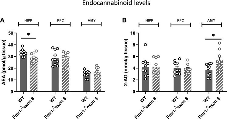
Juvenile Fmr1-Δexon 8 rats showed reduced AEA levels in the hippocampus (t = 2.684, df = 17, p < 0.05, Fig. 1A) and increased 2-AG levels in the amygdala (t = 2.604, df = 17, p < 0.05, Fig. 1B), while no differences in AEA and 2-AG levels were found in the prefrontal cortex of Fmr1-Δexon 8 rats compared to WT animals (AEA; t = 0.427, df = 17, p = n.s; 2-AG, t = 0.303; df = 17; p = n.s., Fig. 1A, B).
Fig. 1. AEA and 2-AG levels were altered in a region-specific manner in the brain of Fmr1-Δexon 8 rats.
Compared to WT animals, Fmr1-Δexon 8 rats showed reduced levels of AEA in the hippocampus (HIPP) (A) and increased levels of 2-AG into the amygdala (AMY) (B), while no differences in AEA and 2-AG levels were found in the prefrontal cortex (PFC) (A, B) (WT = 10; Fmr1-Δexon 8 = 9 animals per group). Data represent means ± SEM, *p < 0.05 vs WT group (Student’s t test).
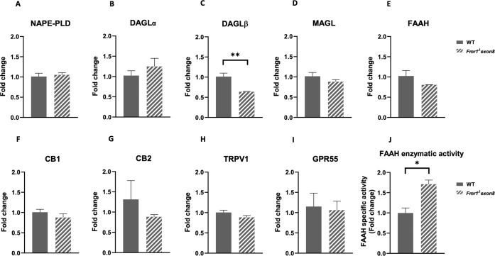
Based on these results, we further characterized the ECS in the hippocampus (Fig. 2) and amygdala (Fig. 3) of WT and Fmr1-Δexon 8 rats. In the hippocampus, we found a significant reduction in the expression of the enzyme DAGLβ, involved in the synthesis of 2-AG (t = 4.261, df = 6, p < 0.01; Fig. 2C) in juvenile Fmr1-Δexon 8 rats compared to WT controls; other enzymes involved in endocannabinoid synthesis were spared, such as NAPE-PLD (t = 0.432, df = 6, p = n.s.; Fig. 2A) and DAGLα (t = 0.962, df = 6, p = n.s.; Fig. 2B). The 2-AG degrading enzyme MAGL (t = 1.268, df = 6, p = n.s.; Fig. 2D) and the AEA catabolizing enzyme FAAH (t = 1.634, df = 6, p = n.s.; Fig. 2E) were also spared. No significant differences were found in the mRNA expression levels of CB1 (t = 1.107, df = 6, p = n.s.; Fig. 2F), CB2 (t = 0.780, df = 5, p = n.s.; Fig. 2G), TRPV1 (t = 1.683, df = 6, p = n.s.; Fig. 2H) and GPR55 (t = 0.221, df = 6, p = n.s.; Fig. 2I) receptors. Interestingly, FAAH activity was increased in the hippocampus of juvenile Fmr1-Δexon 8 rats compared to WT controls (t = 4.403, df = 4, p < 0.05; Fig. 2J). This suggests that the decreased levels of AEA found in the hippocampus of juvenile Fmr1-Δexon 8 rats (Fig. 1A) could be due to an increased activity of FAAH. Western blot analyses of the protein levels of the main cannabinoid targets are in line with qPCR results (DAGLα: t = 2.216, df = 6, p = 0.068; Supplementary Fig. 3A; MAGL: t = 0.891, df = 6, p = n.s.; Supplementary Fig. 3B; FAAH: t = 1.826, df = 6, p = n.s.; Supplementary Fig. 3C; CB1: t = 0.461, df = 6, p = n.s.; Supplementary Fig. 3D; CB2: t = 0.503, df = 6, p = n.s.; Supplementary Fig. 3E). Consistent data were obtained by qPCR analysis of the main endocannabinoid targets at adulthood, with only a significant decrease in DAGLβ expression (t = 3.008, df = 6, p < 0.05; Supplementary Fig. 4C) in Fmr1-Δexon 8 rats compared to WT controls; the other main cannabinoid enzymes NAPE-PLD (t = 0.481, df = 6, p = n.s.; Supplementary Fig. 4A), DAGLα (t = 1.579, df = 5, p = n.s.; Supplementary Fig. 4B), MAGL (t = 0.942, df = 6, p = n.s.; Supplementary Fig. 4D), FAAH (t = 1.845, df = 6, p = n.s.; Supplementary Fig. 4E) and the CB1 (t = 1.418, df = 6, p = n.s.; Fig. 4F), CB2 (t = 0.199, df = 6, p = n.s.; Supplementary Fig. 4G), TRPV1 (t = 0.085, df = 6, p = n.s.; Fig. 4H) and GPR55 (t = 0.124, df = 6, p = n.s.; Supplementary Fig. 4I) receptors were spared.
Fig. 2. qPCR analysis of the main components of the ECS in the hippocampus of juvenile Fmr1-Δexon 8 rats.
Fold induction of NAPE-PLD (A), DAGLα (B), DAGLβ (C), MAGL (D), FAAH (E), CB1 (F), CB2 (G), TRPV1 (H), GPR55 (I) gene expression in the hippocampus of Fmr1-Δexon 8 rats and WT animals, evaluated at PND 35, together with FAAH enzymatic activity (J) (WT = 3–4; Fmr1-Δexon 8 = 3–4 animals per group). Data represent mean ± SEM, *p < 0.05 and **p < 0.01 vs WT group (Student’s t test). NAPE-PLD, N-acylphosphatidylethanolamine-phospholipid D; DAGL, diacylglycerol lipase; MAGL, monoacylglycerol lipase; FAAH, fatty acid amide hydrolase; CB1, cannabinoid receptor 1; CB2, cannabinoid receptor 2; TRPV1, transient receptor potential vanilloid 1; GPR55, G protein-coupled receptor 55.
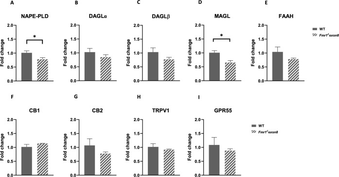
Fig. 3. qPCR analysis of the main components of the ECS in the amygdala of juvenile Fmr1-Δexon 8 rats.
Fold induction of NAPE-PLD (A), DAGLα (B), DAGLβ (C), MAGL (D), FAAH (E), CB1 (F), CB2 (G), TRPV1 (H), GPR55 (I) gene expression in the amygdala of Fmr1-Δexon 8 rats and WT animals, evaluated at PND 35 (WT = 3–4; Fmr1-Δexon 8 = 3–4 animals per group). Data represent mean ± SEM, *p < 0.05 vs WT group (Student’s t test).
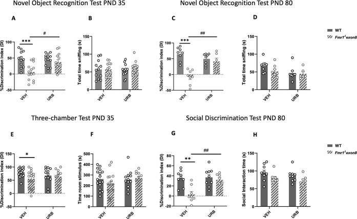
Fig. 4. Potentiation of AEA tone through systemic administration of URB597 rescued the impaired object and social discrimination abilities of Fmr1-Δexon 8 rats.
Systemic administration of URB597 (0.1 mg/kg, 2 h prior testing) rescued the altered discrimination index of both juvenile and adult Fmr1-Δexon 8 rats (A, C) without altering their total time spent in object exploration (B, D) (PND 35: WT-VEH = 12, WT-URB = 14, Fmr1-Δexon 8-VEH = 14, Fmr1-Δexon 8-URB = 13; PND 80: WT-VEH = 8, WT-URB = 8, Fmr1-Δexon 8-VEH = 8, Fmr1-Δexon 8-URB = 6 animals per group). In the three-chamber test, systemic administration of URB597 (0.1 mg/kg, 2 h prior testing) did not rescue but only attenuated the impaired sociability of juvenile Fmr1-Δexon 8 rats (E), as no significant differences were found between Fmr1-Δexon 8 rats treated with URB597 and WT rats treated with vehicle (WT-VEH = 17, WT-URB = 12, Fmr1-Δexon 8-VEH = 16, Fmr1-Δexon 8-URB = 12 animals per group). No differences among experimental groups were found in the time spent in the stimulus room (F). In the social discrimination test, URB597 normalized the impaired discrimination index of Fmr1-Δexon 8 adult rats (G) without altering the time spent in general social exploration by both WT and Fmr1-Δexon 8 rats (H) (WT-VEH = 9, WT-URB = 9, Fmr1-Δexon 8-VEH = 8, Fmr1-Δexon 8-URB = 7 animals per group). Data represent means ± SEM, *p < 0.05, **p < 0.01, ***p < 0.001 vs WT-VEH group, #p < 0.05, ##p < 0.01 vs Fmr1-Δexon 8-VEH group (two-way ANOVA with Tukey’s post hoc test).
In the amygdala of juvenile Fmr1-Δexon 8 rats, we found a significant reduction in the expression of the enzymes NAPE-PLD (t = 2.506, df = 6, p < 0.05; Fig. 3A) and MAGL (t = 3.380, df = 6, p < 0.05; Fig. 3D), whereas DAGLα (t = 1.119, df = 6, p = n.s.; Fig. 3B), DAGLβ (t = 1.536, df = 6, p = n.s.; Fig. 3C) and FAAH (t = 1.423, df = 6, p = n.s.; Fig. 3E) were spared. No changes in the expression levels of CB1 (t = 1.307, df = 6, p = n.s.; Fig. 3F), CB2 (t = 1.185, df = 6, p = n.s.; Fig. 3G), TRPV1 (t = 0.865, df = 6, p = n.s.; Fig. 3H), GPR55 (t = 0.735, df = 6, p = n.s.; Fig. 3I) receptors were found. These data correlate with the results in the protein levels of the main components of the ECS (DAGLα: t = 0.019, df = 6, p = n.s.; Supplementary Fig. 3F; MAGL: t = 1.283, df = 5, p = n.s.; Supplementary Fig. 3G; FAAH: t = 2.033, df = 6, p = 0.09; Supplementary Fig. 3H; CB1: t = 1.202, df = 6, p = n.s.; Supplementary Fig. 3I; CB2: t = 0.241, df = 6, p = n.s.; Supplementary Fig. 3J). At adulthood, we found a significant reduction in DAGLβ (t = 6.054, df = 5, p < 0.01; Supplementary Fig. 4L) and TRPV1 (t = 5.707, df = 6, p < 0.01; Supplementary Fig. 4Q) expression in Fmr1-Δexon 8 rats compared to WT controls; the other main cannabinoid enzymes NAPE-PLD (t = 1.812, df = 6, p = n.s.; Supplementary Fig. 4J), DAGLα (t = 1.403, df = 6, p = n.s.; Supplementary Fig. 4K), MAGL (t = 0.084, df = 6, p = n.s.; Supplementary Fig. 4M), FAAH (t = 0.959, df = 6, p = n.s.; Supplementary Fig. 4N) and CB1 (t = 0.608, df = 6, p = n.s.; Supplementary Fig. 4O), CB2 (t = 1.031, df = 5, p = n.s.; Supplementary Fig. 4P), and GPR55 (t = 1.331, df = 5, p = n.s.; Supplementary Fig. 4R) receptors were spared.
These results further confirm a discrete dysregulation of the brain ECS in Fmr1-Δexon 8 rats across development.
Potentiation of AEA tone through systemic administration of URB597 restored the impaired novel object and social discrimination abilities of Fmr1-Δexon 8 rats
Systemic administration of URB597 (0.1 mg/kg, i.p., 2 h before testing) rescued the altered discrimination index of juvenile and adult Fmr1-Δexon 8 rats in the novel object recognition task (PND 35: F(genotype) 1,49 = 12.39, p < 0.001; F(treatment) 1,49 = 4.289, p < 0.05; F(genotype × treatment) 1,49 = 6.141, p < 0.05, Fig. 4A; PND 80; F(genotype) 1,26 = 24.36, p < 0.001; F(treatment) 1,26 = 4.269, p < 0.05; F(genotype × treatment) 1,26 = 15.49, p < 0.001, Fig. 4C) without altering the total time spent in object exploration (PND 35: F(genotype) 1,49 = 0.537, p = n.s.; F(treatment) 1,49 = 0.047, p = n.s.; F(genotype × treatment) 1,49 = 0.451, p = n.s., Fig. 4B; PND 80: F(genotype) 1,26 = 1.881, p = n.s.; F(treatment) 1,26 = 4.411, p < 0.05; F(genotype × treatment) 1,26 = 1.136, p = n.s., Fig. 4D).
To explore whether potentiation of AEA tone also restored the deficits in sociability displayed by Fmr1-Δexon 8 rats [38], we performed a three-chamber test at PND 35 following systemic administration of URB597 (0.1 mg/kg, i.p., 2 h before testing). Fmr1-Δexon 8 displayed altered sociability, as they showed a lower discrimination index during testing; this deficit was mitigated but not fully antagonized when Fmr1-Δexon 8 rats were treated with URB597 (F(genotype) 1,53 = 4.252, p < 0.05; F(treatment) 1,53 = 0.128, p = n.s.; F(genotype × treatment) 1,53 = 2.212, p = n.s., Fig. 4E), as no differences were found between Fmr1-Δexon 8 rats treated with either URB597 or vehicle (p = n.s). URB597 did not affect the time spent in the stimulus room (F(genotype) 1,53 = 0.542, p = n.s.; F(treatment) 1,53 = 1.209, p = n.s.; F(genotype × treatment) 1,53 = 1.544, p = n.s., Fig. 4F).
Conversely, at adulthood, URB597 (0.1 mg/kg, i.p., 2 h before testing) rescued the impaired discrimination index of Fmr1-Δexon 8 rats while performing the social discrimination test (F(genotype) 1,29 = 11.36, p < 0.01; F(treatment) 1,29 = 7.489, p < 0.05; F (genotype × treatment) 1,29 = 6.073, p < 0.05, Fig. 4G), without altering the total time spent in general social exploration of WT and Fmr1-Δexon 8 rats (F(genotype) 1,29 = 6.113, p < 0.05; F(treatment) 1,29 = 1.968, p = n.s; F(genotype × treatment) 1,29 = 0.001, p = n.s, Fig. 4H). These results indicate that potentiation of AEA tone through systemic administration of URB597 restored the impaired novel object and social discrimination abilities of Fmr1-Δexon 8 rats, but only attenuated their impaired sociability in the three-chamber test.
Intra-hippocampal potentiation of AEA tone rescued the impaired novel object and social discrimination abilities of Fmr1-Δexon 8 rats
To evaluate whether the reduced levels of AEA found in the hippocampus of Fmr1-Δexon 8 rats contributed to their impaired short-term object recognition memory, we bilaterally infused the AEA hydrolysis inhibitor URB597 in the hippocampus of juvenile and adult Fmr1-Δexon 8 and WT animals immediately after the training phase of the novel object recognition test. URB597 rescued the altered discrimination index of Fmr1-Δexon 8 rats (PND 35: F(genotype) 1,43 = 8.772, p < 0.01; F(treatment) 1,43 = 7.517, p < 0.01; F(genotype × treatment) 1,43 = 6.831, p < 0.05, Fig. 5A; PND 80; F(genotype) 1,25 = 6.154 p < 0.05; F(treatment) 1,25 = 14.48, p < 0.001; F(genotype × treatment) 1,25 = 32.93, p < 0.001, Fig. 5C), without altering the total time spent exploring the objects during the test phase (PND 35: F(genotype) 1,43 = 0.563, p = n.s; F(treatment) 1,43 = 0.218, p = n.s.; F(genotype × treatment) 1,43 = 1.051, p = n.s., Fig. 5B; PND 80: F(genotype) 1,25 = 0.209, p = n.s.; F(treatment) 1,25 = 6.313, p < 0.05; F(genotype × treatment) 1,25 = 0.153, p = n.s., Fig. 5D).
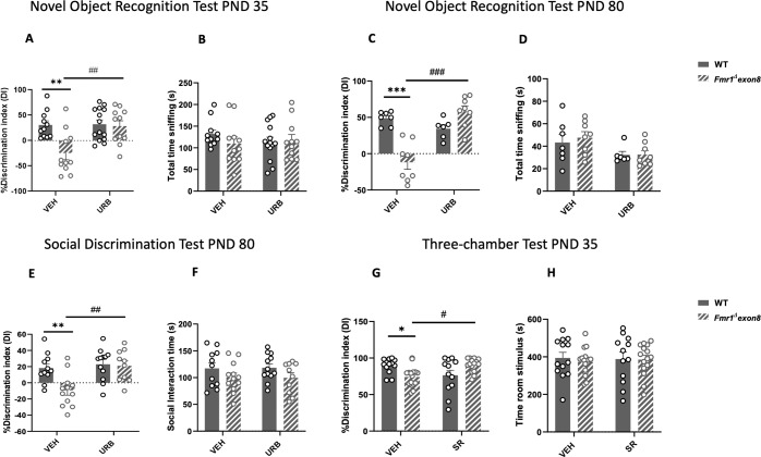
Fig. 5. Intra-hippocampal potentiation of AEA tone rescued the impaired object and social discrimination abilities of Fmr1-Δexon 8 rats, while blockade of CB1 cannabinoid receptors in the amygdala restored their altered sociability.
The bilateral infusion of URB597 (10 ng/0.5 μl) in the hippocampus rescued the altered discrimination index of Fmr1-Δexon 8 juvenile and adult rats in the novel object recognition test (A, C), without altering the total time spent in object exploration by both juvenile and adult WT and Fmr1-Δexon 8 rats (B, D) (PND 35: WT-VEH = 11, WT-URB = 14, Fmr1-Δexon 8-VEH = 11, Fmr1-Δexon 8-URB = 11; PND 80: WT-VEH = 7, WT-URB = 6, Fmr1-Δexon 8-VEH = 8, Fmr1-Δexon 8-URB = 8 animals per group). In the social discrimination task, hippocampal infusion of URB597 (10 ng/0.5 μl) normalized the impaired discrimination index of Fmr1-Δexon 8 adult rats (E) without altering the time spent in social exploration by both WT and Fmr1-Δexon 8 rats (F) (WT-VEH = 11, WT-URB = 12, Fmr1-Δexon 8-VEH = 10, Fmr1-Δexon 8-URB = 12 animals per group). Bilateral infusion of SR141716A (0.1 μg/0.2 μl) in the amygdala rescued the atypical sociability of Fmr1-Δexon 8 rats normalizing their discrimination index (G). Conversely, no differences among groups were found in the time spent in the stimulus room (H) (WT-VEH = 13, WT-URB = 12, Fmr1-Δexon 8-VEH = 16, Fmr1-Δexon 8-URB = 15 animals per group). Data represent means ± SEM, *p < 0.05, **p < 0.01, ***p < 0.001 vs WT-VEH group, #p < 0.01, ##p < 0.01, ###p < 0.001 vs Fmr1-Δexon 8-VEH group (two-way ANOVA with Tukey’s post hoc test).
Intra-hippocampal infusion of URB597 also normalized the impaired discrimination index of Fmr1-Δexon 8 adult rats in the social discrimination task (F(genotype) 1,41 = 6.448, p < 0.05; F(treatment) 1,41 = 9.220, p < 0.01; F(genotype × treatment) 1,41 = 4.902, p < 0.05, Fig. 5E), without affecting the total time spent by the animals in general social exploration (F(genotype) 1,41 = 4.074, p = n.s.; F(treatment) 1,41 = 0.001, p = n.s.; F(genotype × treatment) 1,41 = 0.012, p = n.s., Fig. 5F). These results indicate that AEA in the hippocampus is specifically involved in the short-term cognitive deficits displayed by Fmr1-Δexon 8 rats in the novel object and social discrimination tasks.
CB1 cannabinoid receptors in the amygdala modulate sociability in Fmr1-Δexon 8 rats
Based on the evidence that Fmr1-Δexon 8 rats showed abnormal 2-AG levels in the amygdala and that pharmacological/genetic blockade of CB1 receptors normalized some FXS-like traits in the mouse model of FXS [31, 68], we hypothesized that blocking CB1 receptors into the amygdala could restore the impaired sociability displayed by Fmr1-Δexon 8 rats in the three-chamber test possibly interfering with 2-AG signaling. In line with this possibility, intra-amygdala infusion of SR141716A immediately before the test phase rescued the atypical sociability of Fmr1-Δexon 8 rats by normalizing their discrimination index (F(genotype) 1,52 = 0.036, p = n.s.; F(treatment) 1,52 = 0.159, p = n.s.; F(genotype × treatment) 1,52 = 13.26, p < 0.001, Fig. 5G), without affecting the time spent in the stimulus room (F(genotype) 1,52 = 0.211, p = n.s.; F(treatment) 1,52 = 0.014, p = n.s.; F(genotype × treatment) 1,52 = 0.123, p = n.s., Fig. 5H).
Discussion
Our results revealed region-specific changes in endocannabinoid levels in the brain of Fmr1-Δexon 8 rats, i.e., reduced levels of AEA in the hippocampus and increased levels of 2-AG into the amygdala, together with discrete alterations in the expression of the enzymes modulating synthesis/degradation of AEA and 2-AG, reinforcing the idea that brain region-specific changes in endocannabinoid signaling contribute to the altered molecular, functional and behavioral profile that characterize this genetic rat model of ASD. Systemic injection and intra-hippocampal infusion of the AEA hydrolysis inhibitor URB597 rescued the aberrant cognitive performance of Fmr1-Δexon 8 rats in the novel object and social discrimination tasks, suggesting that the hippocampus is an important brain area where reduced AEA tone contributes to the short-term cognitive deficits displayed by Fmr1-Δexon 8 rats. Conversely, blockade of CB1 cannabinoid receptors in the amygdala by the antagonist/inverse agonist SR141716A rescued the altered sociability of Fmr1-Δexon 8 rats in the three-chamber test, indicating that the amygdala is likely a prominent site of action for 2-AG to underlie the altered sociability of Fmr1-Δexon 8 rats.
We have shown that Fmr1-Δexon 8 rats display cognitive, communicative and social impairments [38], suggesting the validity of this animal model in mimicking the key behavioral deficits that characterize FXS and some of the core and comorbid features of non-syndromic ASD. Converging evidence suggested a region-specific alteration of the ECS in the brain of Fmr1 KO mice [31, 32, 34, 69]. For instance, alterations in mGluR5 and DAGLα coupling led to disruption of mGluR5-mediated production of 2-AG in the ventral striatum and prefrontal cortex of Fmr1 KO mice [32, 70, 71], together with decreased 2-AG levels in the hippocampus [34]. On the other hand, pioneering studies in Fmr1 KO mice reported no differences in brain levels of 2-AG and AEA or in the hippocampal expression of several components of the ECS [69, 72]. For the first time, we here found that Fmr1-Δexon 8 rats displayed reduced basal AEA levels in the hippocampus and increased 2-AG levels into the amygdala, suggesting that an unbalance of endocannabinoid signaling in selected brain regions such as the hippocampus and amygdala could drive ASD-like behavioral features in these animals. Interestingly, Fmr1-Δexon 8 rats showed an increase in the hydrolytic activity of FAAH compared to WT controls at juvenile age. This led us to hypothesize that the decreased AEA levels in the hippocampus of juvenile Fmr1-Δexon 8 rats could be due to an increased FAAH activity. Concerning the amygdala, we found a decrease in the expression of the enzyme MAGL, that could correlate to the increased levels of 2-AG we found in the amygdala of juvenile Fmr1-Δexon rats. Interestingly, these discrete alterations in the main endocannabinoid synthesis/degradation enzymes appeared consistent across development.
The hippocampus and amygdala have a key role in the modulation of cognitive performance and emotional states, which are profoundly impaired in FXS patients [7, 73–76]. We found that increasing AEA levels via systemic administration of the selective FAAH inhibitor URB597 corrected the impaired short-term memory in the novel object recognition task in juvenile and adult Fmr1-Δexon 8 rats, as well as their impaired social discrimination in the social discrimination test. These tasks allow to assess the discriminative abilities of rodents respectively referred to object and social recognition and are based on the innate preference of rodents for exploring objects or congeners instead of re-exploring something they have already been exposed to, referred to as familiar. The ability to recognize familiar objects and social conspecifics in rodents relies on hippocampal integrity [77–80], thus suggesting the critical role of this brain region in recognition memory. Notably, in the social discrimination experiments, we tested adult rats after 7 days of individual housing, which is much shorter than the prolonged social isolation periods causing a dysregulation of the ECS [81–84]. Compared to its beneficial effects on short-term object and social recognition deficits, we found that URB597 only attenuated the impaired sociability displayed by juvenile Fmr1-Δexon 8 rats in the three-chamber task. For the first time, these results suggest that AEA is specifically involved in the short-term memory (for either a novel object or a social partner) deficits displayed by Fmr1-Δexon 8 rats, thus suggesting the potential of AEA signaling as an endogenous cannabinoid target for the short-term recognition memory deficits observed in this genetic rat model of ASD. Since juvenile Fmr1-Δexon 8 rats displayed reduced AEA levels in the hippocampus, together with increased FAAH activity, we hypothesized that intra-hippocampal infusion of URB597 would restore AEA levels and in turn rescue the short-term cognitive deficits shown by these animals. In line with this, hippocampal infusion of URB597 rescued the impaired short-term memory of Fmr1-Δexon 8 rats in the novel object and social discrimination tasks, thus supporting the idea that their alteration of object and social recognition involves hippocampal AEA signaling. Although we did not directly measure the levels of AEA following URB597 administration, extensive literature evidence shows that pharmacological inhibition of FAAH enhances AEA levels and reverses the behavioral deficits observed in different animal models of ASD [67, 85–91]. Thus, it is reasonable to hypothesize that URB597 administration had beneficial effects in Fmr1-Δexon 8 rats by increasing AEA levels. The hypothesis that dysregulated AEA signaling in the hippocampus could account for the altered object and social discrimination displayed by Fmr1-Δexon 8 rats is supported by several studies indicating that AEA exerts an overall modulatory effect in hippocampal-mediated cognitive processes in both physiological and pathological conditions [50, 61, 92–96]. It should be noted, however, that we here investigated only a short-term form of memory (i.e., with a 30 min inter-trial interval); therefore, testing the effects of URB597 on potential long-term memory deficits in Fmr1-Δexon 8 rats remains an interesting point that deserves further investigation.
As for the putative mechanisms involved, the positive effect of intra-hippocampal infusion of URB597 in Fmr1-Δexon 8 rats likely relates to an association between the ECS and FMRP loss, as it has previously been shown in the hippocampus of Fmr1 KO mice [97]. Thus, it is tempting to speculate that the loss of FMRP function in Fmr1-Δexon 8 rats could produce diminished CB1-mediated signaling driven by compromised hippocampal AEA, resulting in short-term recognition memory impairment. Despite there is consistent evidence suggesting that CB1 cannabinoid receptors mediate the beneficial effects of FAAH inhibitors in animal models of ASD and FXS [67, 88, 91, 98, 99], the putative mechanisms involved still remain to be fully elucidated. For instance, our results did not show any significant difference in the expression of the main cannabinoid receptors (i.e., CB1, CB2, TRPV1, GPR55) at the juvenile age, except for TRPV1 expression in the adult hippocampus. Since AEA also activates TRPV1 receptors [100], one can argue that the effects of URB597 could be mediated by this receptor, at least at adulthood. However, this remains an intriguing issue that needs to be further addressed.
Persistent deficits in social communication and social interaction are common key features of both ASD and FXS [7] and we have recently shown (and here confirmed) that Fmr1-Δexon 8 rats display altered sociability in the three-chamber task [38]. Together with AEA, the other main endocannabinoid 2-AG has a significant role in social behavior [101, 102]. Interestingly, increasing 2-AG levels augments social play in rats [63, 103, 104], while reducing 2-AG levels decreases acquisition of social conditioned place preference in mice [105]. Previous work has demonstrated that activation of CB1 cannabinoid receptors by endocannabinoids, including 2-AG, has a functional role in social behavior via modulation of both nucleus accumbens signaling and basolateral amygdala-nucleus accumbens activity [55, 63, 105, 106]. Based on the finding that juvenile Fmr1-Δexon 8 rats showed abnormal 2-AG levels in the amygdala, together with a reduction in the expression of MAGL enzyme, and on evidence showing that pharmacological or genetic blockade of CB1 cannabinoid receptors normalized some traits of FXS [31, 68], we hypothesized that blocking CB1 receptors into the amygdala could restore the impaired sociability displayed by Fmr1-Δexon 8 rats possibly interfering with 2-AG signaling. Interestingly, we found that infusion of the CB1 antagonist/inverse agonist SR141716A in the basolateral amygdala of Fmr1-Δexon 8 rats rescued their atypical performance in the three-chamber test. This supports the hypothesis that excessive 2-AG signaling in the amygdala, possibly due to the decreased expression of MAGL enzyme as revealed by the qPCR experiments, contributes to the impaired social function seen in these rats. In line with our findings, recent evidence showed that basolateral amygdala-nucleus accumbens circuit activation reduces sociability and it is highly regulated by 2-AG-mediated endocannabinoid signaling [107]: thus, optogenetic activation of the basolateral amygdala-nucleus accumbens glutamatergic circuit decreases sociability in the three-chamber task, increases social avoidance and reduces social interaction seeking in Shank3 mutant mice displaying ASD-like behaviors, via 2-AG signaling. These findings reinforce the idea that region-specific alterations in brain endocannabinoid signaling affect different facets of the behavioral profile of this genetic rat model of ASD, and further support FXS-related dysfunction in the amygdala [108–110], moving beyond the idea of the pathogenesis of FXS based primarily on studies focused in the hippocampus and neocortex.
Overall, our results indicate the multimodal role of AEA and 2-AG signaling in the autistic-like traits observed in a genetic model of autism based on FMR1 deletion in rats. We hypothesize that impairment in AEA signaling in the hippocampus, possibly as a consequence of an increased FAAH activity, mediates short-term novel object and social discrimination deficits in Fmr1-Δexon 8 rats, whereas exaggerated 2-AG signaling in the amygdala underlies their altered sociability through excessive activation of CB1 cannabinoid receptors. These findings not only provide clarification of the underlying molecular mechanisms of AEA and 2-AG in ASD-related socio-cognitive impairment, but also indicate that any assessment of potential pharmacological manipulations targeting endocannabinoid neurotransmission in ASD should consider that these compounds may have brain region- and behaviorally-specific effects.
Supplementary information
Author contributions
SS and AM performed, analyzed, and contributed to the design of the behavioral experiments. MM and MNH performed and analyzed the biochemical experiments to measure endocannabinoid levels. EC, AR, AF, and FA performed the qPCR and Western blotting experiments. EC, VB, and PC contributed to the behavioral experiments. SS, AM, and VT wrote the paper. VT supervised the project, designed the experiments, and wrote, revised, and edited the paper. All authors contributed to the paper and approved the submitted version.
Funding
This work was supported by Autism Speaks grant #11690 (AM and VT), MIUR PRIN 2017 grant # SXEXT5 (VT), Regione Lazio “Gruppi di ricerca 2020” grant # PROT. A0375-2020-36550 (VT) and operating funds from the Canadian Institutes of Health Research (MNH).
Competing interests
The authors declare no competing interests.
Footnotes
Publisher’s note Springer Nature remains neutral with regard to jurisdictional claims in published maps and institutional affiliations.
These authors contributed equally: Sara Schiavi, Antonia Manduca.
Supplementary information
The online version contains supplementary material available at 10.1038/s41386-022-01454-7.
References
- 1.American Psychiatric Association. Diagnostic and statistical manual of mental disorders - 5th ed. (DSM-5). Washington, DC: American Psychiatric Association; 2013.
- 2.Lai MC, Lombardo MV, Baron-Cohen S. Autism. Lancet. 2014;383:896–910. doi: 10.1016/S0140-6736(13)61539-1. [DOI] [PubMed] [Google Scholar]
- 3.Jeste SS, Geschwind DH. Disentangling the heterogeneity of autism spectrum disorder through genetic findings. Nat Rev Neurol. 2014;10:74–81. doi: 10.1038/nrneurol.2013.278. [DOI] [PMC free article] [PubMed] [Google Scholar]
- 4.Maurin T, Zongaro S, Bardoni B. Fragile X Syndrome: from molecular pathology to therapy. Neurosci Biobehav Rev. 2014;46:242–55. doi: 10.1016/j.neubiorev.2014.01.006. [DOI] [PubMed] [Google Scholar]
- 5.Song FJ, Barton P, Sleightholme V, Yao GL, Fry-Smith A. Screening for fragile X syndrome: a literature review and modelling study. Health Technol Assess. 2003;7:1–106. doi: 10.3310/hta7160. [DOI] [PubMed] [Google Scholar]
- 6.Hernandez RN, Feinberg RL, Vaurio R, Passanante NM, Thompson RE, Kaufmann WE. Autism spectrum disorder in fragile X syndrome: a longitudinal evaluation. Am J Med Genet Part A. 2009;149A:1125–37. doi: 10.1002/ajmg.a.32848. [DOI] [PMC free article] [PubMed] [Google Scholar]
- 7.Hagerman RJ, Berry-Kravis E, Hazlett HC, Bailey DB, Jr., Moine H, Kooy RF, et al. Fragile X syndrome. Nat Rev Dis Prim. 2017;3:17065. doi: 10.1038/nrdp.2017.65. [DOI] [PubMed] [Google Scholar]
- 8.Telias M. Molecular Mechanisms of Synaptic Dysregulation in Fragile X Syndrome and Autism Spectrum Disorders. Front Mol Neurosci. 2019;12:51. doi: 10.3389/fnmol.2019.00051. [DOI] [PMC free article] [PubMed] [Google Scholar]
- 9.Tran SS, Jun HI, Bahn JH, Azghadi A, Ramaswami G, Van Nostrand EL, et al. Widespread RNA editing dysregulation in brains from autistic individuals. Nat Neurosci. 2019;22:25–36. doi: 10.1038/s41593-018-0287-x. [DOI] [PMC free article] [PubMed] [Google Scholar]
- 10.Fyke W, Velinov M. FMR1 and Autism, an Intriguing Connection Revisited. Genes. 2021;12:1218. [DOI] [PMC free article] [PubMed]
- 11.Castagnola S, Bardoni B, Maurin T. The Search for an Effective Therapy to Treat Fragile X Syndrome: Dream or Reality? Front Synaptic Neurosci. 2017;9:15. doi: 10.3389/fnsyn.2017.00015. [DOI] [PMC free article] [PubMed] [Google Scholar]
- 12.Araque A, Castillo PE, Manzoni OJ, Tonini R. Synaptic functions of endocannabinoid signaling in health and disease. Neuropharmacology. 2017;124:13–24. doi: 10.1016/j.neuropharm.2017.06.017. [DOI] [PMC free article] [PubMed] [Google Scholar]
- 13.Segev A, Korem N, Mizrachi Zer-Aviv T, Abush H, Lange R, Sauber G, et al. Role of endocannabinoids in the hippocampus and amygdala in emotional memory and plasticity. Neuropsychopharmacol: Off Publ Am Coll Neuropsychopharmacol. 2018;43:2017–27. doi: 10.1038/s41386-018-0135-4. [DOI] [PMC free article] [PubMed] [Google Scholar]
- 14.Maldonado R, Cabanero D, Martin-Garcia E. The endocannabinoid system in modulating fear, anxiety, and stress. Dialogues Clin Neurosci. 2020;22:229–39. doi: 10.31887/DCNS.2020.22.3/rmaldonado. [DOI] [PMC free article] [PubMed] [Google Scholar]
- 15.Wei D, Allsop S, Tye K, Piomelli D. Endocannabinoid Signaling in the Control of Social Behavior. Trends Neurosci. 2017;40:385–96. doi: 10.1016/j.tins.2017.04.005. [DOI] [PMC free article] [PubMed] [Google Scholar]
- 16.Bagni C, Zukin RS. A Synaptic Perspective of Fragile X Syndrome and Autism Spectrum Disorders. Neuron. 2019;101:1070–88. doi: 10.1016/j.neuron.2019.02.041. [DOI] [PMC free article] [PubMed] [Google Scholar]
- 17.Takumi T, Tamada K, Hatanaka F, Nakai N, Bolton PF. Behavioral neuroscience of autism. Neurosci Biobehav Rev. 2020;110:60–76. doi: 10.1016/j.neubiorev.2019.04.012. [DOI] [PubMed] [Google Scholar]
- 18.Castillo PE, Younts TJ, Chavez AE, Hashimotodani Y. Endocannabinoid signaling and synaptic function. Neuron. 2012;76:70–81. doi: 10.1016/j.neuron.2012.09.020. [DOI] [PMC free article] [PubMed] [Google Scholar]
- 19.Ohno-Shosaku T, Kano M. Endocannabinoid-mediated retrograde modulation of synaptic transmission. Curr Opin Neurobiol. 2014;29:1–8. doi: 10.1016/j.conb.2014.03.017. [DOI] [PubMed] [Google Scholar]
- 20.Di Marzo V, Bifulco M, De Petrocellis L. The endocannabinoid system and its therapeutic exploitation. Nat Rev Drug Discov. 2004;3:771–84. doi: 10.1038/nrd1495. [DOI] [PubMed] [Google Scholar]
- 21.Piomelli D. The molecular logic of endocannabinoid signalling. Nat Rev Neurosci. 2003;4:873–84. doi: 10.1038/nrn1247. [DOI] [PubMed] [Google Scholar]
- 22.Cristino L, Bisogno T, Di Marzo V. Cannabinoids and the expanded endocannabinoid system in neurological disorders. Nat Rev Neurol. 2020;16:9–29. doi: 10.1038/s41582-019-0284-z. [DOI] [PubMed] [Google Scholar]
- 23.Aran A, Eylon M, Harel M, Polianski L, Nemirovski A, Tepper S, et al. Lower circulating endocannabinoid levels in children with autism spectrum disorder. Mol Autism. 2019;10:2. doi: 10.1186/s13229-019-0256-6. [DOI] [PMC free article] [PubMed] [Google Scholar]
- 24.Karhson DS, Krasinska KM, Dallaire JA, Libove RA, Phillips JM, Chien AS, et al. Plasma anandamide concentrations are lower in children with autism spectrum disorder. Mol Autism. 2018;9:18. doi: 10.1186/s13229-018-0203-y. [DOI] [PMC free article] [PubMed] [Google Scholar]
- 25.Smith DR, Stanley CM, Foss T, Boles RG, McKernan K. Rare genetic variants in the endocannabinoid system genes CNR1 and DAGLA are associated with neurological phenotypes in humans. PloS ONE. 2017;12:e0187926. doi: 10.1371/journal.pone.0187926. [DOI] [PMC free article] [PubMed] [Google Scholar]
- 26.De Pol M, Kolla NJ. Endocannabinoid markers in autism spectrum disorder: a scoping review of human studies. Psychiatry Res. 2021;306:114256. doi: 10.1016/j.psychres.2021.114256. [DOI] [PubMed] [Google Scholar]
- 27.Pietropaolo S, Marsicano G. The role of the endocannabinoid system as a therapeutic target for autism spectrum disorder: Lessons from behavioral studies on mouse models. Neurosci Biobehav Rev. 2022;132:664–78. doi: 10.1016/j.neubiorev.2021.11.031. [DOI] [PubMed] [Google Scholar]
- 28.Carbone E, Manduca A, Cacchione C, Vicari S, Trezza V. Healing autism spectrum disorder with cannabinoids: a neuroinflammatory story. Neurosci Biobehav Rev. 2021;121:128–43. doi: 10.1016/j.neubiorev.2020.12.009. [DOI] [PubMed] [Google Scholar]
- 29.Zamberletti E, Rubino T, Parolaro D. Therapeutic potential of cannabidivarin for epilepsy and autism spectrum disorder. Pharmacol Ther. 2021;226:107878. doi: 10.1016/j.pharmthera.2021.107878. [DOI] [PubMed] [Google Scholar]
- 30.Hurley MJ, Deacon RMJ, Chan AWE, Baker D, Selwood DL, Cogram P. Reversal of behavioural phenotype by the cannabinoid-like compound VSN16R in fragile X syndrome mice. Brain: J Neurol. 2022;145:76–82. doi: 10.1093/brain/awab246. [DOI] [PMC free article] [PubMed] [Google Scholar]
- 31.Busquets-Garcia A, Gomis-Gonzalez M, Guegan T, Agustin-Pavon C, Pastor A, Mato S, et al. Targeting the endocannabinoid system in the treatment of fragile X syndrome. Nat Med. 2013;19:603–7. doi: 10.1038/nm.3127. [DOI] [PubMed] [Google Scholar]
- 32.Jung KM, Sepers M, Henstridge CM, Lassalle O, Neuhofer D, Martin H, et al. Uncoupling of the endocannabinoid signalling complex in a mouse model of fragile X syndrome. Nat Commun. 2012;3:1080. doi: 10.1038/ncomms2045. [DOI] [PMC free article] [PubMed] [Google Scholar]
- 33.Neuhofer D, Lassalle O, Manzoni OJ. Muscarinic M1 Receptor Modulation of Synaptic Plasticity in Nucleus Accumbens of Wild-Type and Fragile X Mice. ACS Chem Neurosci. 2018;9:2233–40. doi: 10.1021/acschemneuro.7b00398. [DOI] [PubMed] [Google Scholar]
- 34.Wang W, Cox BM, Jia Y, Le AA, Cox CD, Jung KM, et al. Treating a novel plasticity defect rescues episodic memory in Fragile X model mice. Mol Psychiatry. 2018;23:1798–806. doi: 10.1038/mp.2017.221. [DOI] [PMC free article] [PubMed] [Google Scholar]
- 35.Heussler H, Cohen J, Silove N, Tich N, Bonn-Miller MO, Du W, et al. A phase 1/2, open-label assessment of the safety, tolerability, and efficacy of transdermal cannabidiol (ZYN002) for the treatment of pediatric fragile X syndrome. J Neurodev Disord. 2019;11:16. doi: 10.1186/s11689-019-9277-x. [DOI] [PMC free article] [PubMed] [Google Scholar]
- 36.Tartaglia N, Bonn-Miller M, Hagerman R. Treatment of Fragile X Syndrome with Cannabidiol: A Case Series Study and Brief Review of the Literature. Cannabis Cannabinoid Res. 2019;4:3–9. doi: 10.1089/can.2018.0053. [DOI] [PMC free article] [PubMed] [Google Scholar]
- 37.Berry-Kravis EHR, Budimirovic D, Erickson C, Heussler H, Tartaglia N, Cohen J, et al. A Pivotal Study of ZYN002 Cannabidiol (CBD) Transdermal Gel in Children and Adolescents With Fragile X Syndrome. Biological Psychiatry. 2021;89:S226–S7.
- 38.Schiavi S, Carbone E, Melancia F, Buzzelli V, Manduca A, Campolongo P, et al. Perinatal supplementation with omega-3 fatty acids corrects the aberrant social and cognitive traits observed in a genetic model of autism based on FMR1 deletion in rats. Nutritional Neurosci. 2022;25:898–911. doi: 10.1080/1028415X.2020.1819107. [DOI] [PubMed] [Google Scholar]
- 39.Golden CEM, Breen MS, Koro L, Sonar S, Niblo K, Browne A, et al. Deletion of the KH1 Domain of Fmr1 Leads to Transcriptional Alterations and Attentional Deficits in Rats. Cereb Cortex. 2019;29:2228–44. doi: 10.1093/cercor/bhz029. [DOI] [PMC free article] [PubMed] [Google Scholar]
- 40.Hamilton SM, Green JR, Veeraragavan S, Yuva L, McCoy A, Wu Y, et al. Fmr1 and Nlgn3 knockout rats: novel tools for investigating autism spectrum disorders. Behav Neurosci. 2014;128:103–9. doi: 10.1037/a0035988. [DOI] [PubMed] [Google Scholar]
- 41.Till SM, Asiminas A, Jackson AD, Katsanevaki D, Barnes SA, Osterweil EK, et al. Conserved hippocampal cellular pathophysiology but distinct behavioural deficits in a new rat model of FXS. Hum Mol Genet. 2015;24:5977–84. doi: 10.1093/hmg/ddv299. [DOI] [PMC free article] [PubMed] [Google Scholar]
- 42.Schiavi S, Carbone E, Melancia F, di Masi A, Jarjat M, Brau F, et al. Phosphodiesterase 2A inhibition corrects the aberrant behavioral traits observed in genetic and environmental preclinical models of Autism Spectrum Disorder. Transl Psychiatry. 2022;12:119. doi: 10.1038/s41398-022-01885-2. [DOI] [PMC free article] [PubMed] [Google Scholar]
- 43.Amaral DG, Schumann CM, Nordahl CW. Neuroanatomy of autism. Trends Neurosci. 2008;31:137–45. doi: 10.1016/j.tins.2007.12.005. [DOI] [PubMed] [Google Scholar]
- 44.Ha S, Sohn IJ, Kim N, Sim HJ, Cheon KA. Characteristics of Brains in Autism Spectrum Disorder: Structure, Function and Connectivity across the Lifespan. Exp Neurobiol. 2015;24:273–84. doi: 10.5607/en.2015.24.4.273. [DOI] [PMC free article] [PubMed] [Google Scholar]
- 45.Hashem S, Nisar S, Bhat AA, Yadav SK, Azeem MW, Bagga P, et al. Genetics of structural and functional brain changes in autism spectrum disorder. Transl Psychiatry. 2020;10:229. doi: 10.1038/s41398-020-00921-3. [DOI] [PMC free article] [PubMed] [Google Scholar]
- 46.Fereshetyan K, Chavushyan V, Danielyan M, Yenkoyan K. Assessment of behavioral, morphological and electrophysiological changes in prenatal and postnatal valproate induced rat models of autism spectrum disorder. Sci Rep. 2021;11:23471. doi: 10.1038/s41598-021-02994-6. [DOI] [PMC free article] [PubMed] [Google Scholar]
- 47.Sosa-Diaz N, Bringas ME, Atzori M, Flores G. Prefrontal cortex, hippocampus, and basolateral amygdala plasticity in a rat model of autism spectrum. Synapse. 2014;68:468–73. doi: 10.1002/syn.21759. [DOI] [PubMed] [Google Scholar]
- 48.Kilkenny C, Browne WJ, Cuthill IC, Emerson M, Altman DG. Improving bioscience research reporting: the ARRIVE guidelines for reporting animal research. PLoS Biol. 2010;8:e1000412. doi: 10.1371/journal.pbio.1000412. [DOI] [PMC free article] [PubMed] [Google Scholar]
- 49.Schiavi S, Iezzi D, Manduca A, Leone S, Melancia F, Carbone C, et al. Reward-Related Behavioral, Neurochemical and Electrophysiological Changes in a Rat Model of Autism Based on Prenatal Exposure to Valproic Acid. Front Cell Neurosci. 2019;13:479. doi: 10.3389/fncel.2019.00479. [DOI] [PMC free article] [PubMed] [Google Scholar]
- 50.Morena M, Roozendaal B, Trezza V, Ratano P, Peloso A, Hauer D, et al. Endogenous cannabinoid release within prefrontal-limbic pathways affects memory consolidation of emotional training. Proc Natl Acad Sci USA. 2014;111:18333–8. doi: 10.1073/pnas.1420285111. [DOI] [PMC free article] [PubMed] [Google Scholar]
- 51.Gray JM, Vecchiarelli HA, Morena M, Lee TT, Hermanson DJ, Kim AB, et al. Corticotropin-releasing hormone drives anandamide hydrolysis in the amygdala to promote anxiety. J Neurosci: Off J Soc Neurosci. 2015;35:3879–92. doi: 10.1523/JNEUROSCI.2737-14.2015. [DOI] [PMC free article] [PubMed] [Google Scholar]
- 52.Qi M, Morena M, Vecchiarelli HA, Hill MN, Schriemer DC. A robust capillary liquid chromatography/tandem mass spectrometry method for quantitation of neuromodulatory endocannabinoids. Rapid Commun Mass Spectrom. 2015;29:1889–97. doi: 10.1002/rcm.7277. [DOI] [PubMed] [Google Scholar]
- 53.Schmittgen TD, Livak KJ. Analyzing real-time PCR data by the comparative C(T) method. Nat Protoc. 2008;3:1101–8. doi: 10.1038/nprot.2008.73. [DOI] [PubMed] [Google Scholar]
- 54.Santori A, Morena M, Hill MN, Campolongo P. Hippocampal 2-Arachidonoyl Glycerol Signaling Regulates Time-of-Day- and Stress-Dependent Effects on Rat Short-Term Memory. Int J Mol Sci. 2020;21:7316. [DOI] [PMC free article] [PubMed]
- 55.Trezza V, Damsteegt R, Manduca A, Petrosino S, Van Kerkhof LW, Pasterkamp RJ, et al. Endocannabinoids in amygdala and nucleus accumbens mediate social play reward in adolescent rats. J Neurosci: Off J Soc Neurosci. 2012;32:14899–908. doi: 10.1523/JNEUROSCI.0114-12.2012. [DOI] [PMC free article] [PubMed] [Google Scholar]
- 56.Manduca A, Servadio M, Campolongo P, Palmery M, Trabace L, Vanderschuren LJ, et al. Strain- and context-dependent effects of the anandamide hydrolysis inhibitor URB597 on social behavior in rats. Eur Neuropsychopharmacol: J Eur Coll Neuropsychopharmacol. 2014;24:1337–48. doi: 10.1016/j.euroneuro.2014.05.009. [DOI] [PubMed] [Google Scholar]
- 57.Kathuria S, Gaetani S, Fegley D, Valino F, Duranti A, Tontini A, et al. Modulation of anxiety through blockade of anandamide hydrolysis. Nat Med. 2003;9:76–81. doi: 10.1038/nm803. [DOI] [PubMed] [Google Scholar]
- 58.Fegley D, Gaetani S, Duranti A, Tontini A, Mor M, Tarzia G, et al. Characterization of the fatty acid amide hydrolase inhibitor cyclohexyl carbamic acid 3’-carbamoyl-biphenyl-3-yl ester (URB597): effects on anandamide and oleoylethanolamide deactivation. J Pharmacol Exp Ther. 2005;313:352–8. doi: 10.1124/jpet.104.078980. [DOI] [PubMed] [Google Scholar]
- 59.Melancia F, Schiavi S, Servadio M, Cartocci V, Campolongo P, Palmery M, et al. Sex-specific autistic endophenotypes induced by prenatal exposure to valproic acid involve anandamide signalling. Br J Pharmacol. 2018;175:3699–712. doi: 10.1111/bph.14435. [DOI] [PMC free article] [PubMed] [Google Scholar]
- 60.Morena M, Berardi A, Colucci P, Palmery M, Trezza V, Hill MN, et al. Enhancing Endocannabinoid Neurotransmission Augments The Efficacy of Extinction Training and Ameliorates Traumatic Stress-Induced Behavioral Alterations in Rats. Neuropsychopharmacol: Off Publ Am Coll Neuropsychopharmacol. 2018;43:1284–96. doi: 10.1038/npp.2017.305. [DOI] [PMC free article] [PubMed] [Google Scholar]
- 61.Morena M, De Castro V, Gray JM, Palmery M, Trezza V, Roozendaal B, et al. Training-Associated Emotional Arousal Shapes Endocannabinoid Modulation of Spatial Memory Retrieval in Rats. J Neurosci: Off J Soc Neurosci. 2015;35:13962–74. doi: 10.1523/JNEUROSCI.1983-15.2015. [DOI] [PMC free article] [PubMed] [Google Scholar]
- 62.Piomelli D, Tarzia G, Duranti A, Tontini A, Mor M, Compton TR, et al. Pharmacological profile of the selective FAAH inhibitor KDS-4103 (URB597) CNS Drug Rev. 2006;12:21–38. doi: 10.1111/j.1527-3458.2006.00021.x. [DOI] [PMC free article] [PubMed] [Google Scholar]
- 63.Manduca A, Lassalle O, Sepers M, Campolongo P, Cuomo V, Marsicano G, et al. Interacting Cannabinoid and Opioid Receptors in the Nucleus Accumbens Core Control Adolescent Social Play. Front Behav Neurosci. 2016;10:211. doi: 10.3389/fnbeh.2016.00211. [DOI] [PMC free article] [PubMed] [Google Scholar]
- 64.Manduca A, Bara A, Larrieu T, Lassalle O, Joffre C, Laye S, et al. Amplification of mGlu5-Endocannabinoid Signaling Rescues Behavioral and Synaptic Deficits in a Mouse Model of Adolescent and Adult Dietary Polyunsaturated Fatty Acid Imbalance. J Neurosci: Off J Soc Neurosci. 2017;37:6851–68. doi: 10.1523/JNEUROSCI.3516-16.2017. [DOI] [PMC free article] [PubMed] [Google Scholar]
- 65.Bara A, Manduca A, Bernabeu A, Borsoi M, Serviado M, Lassalle O, et al. Sex-dependent effects of in utero cannabinoid exposure on cortical function. eLife. 2018;7:e36234. [DOI] [PMC free article] [PubMed]
- 66.Borsoi M, Manduca A, Bara A, Lassalle O, Pelissier-Alicot AL, Manzoni OJ. Sex Differences in the Behavioral and Synaptic Consequences of a Single in vivo Exposure to the Synthetic Cannabimimetic WIN55,212-2 at Puberty and Adulthood. Front Behav Neurosci. 2019;13:23. doi: 10.3389/fnbeh.2019.00023. [DOI] [PMC free article] [PubMed] [Google Scholar]
- 67.Servadio M, Melancia F, Manduca A, di Masi A, Schiavi S, Cartocci V, et al. Targeting anandamide metabolism rescues core and associated autistic-like symptoms in rats prenatally exposed to valproic acid. Transl Psychiatry. 2016;6:e902. doi: 10.1038/tp.2016.182. [DOI] [PMC free article] [PubMed] [Google Scholar]
- 68.Gomis-Gonzalez M, Busquets-Garcia A, Matute C, Maldonado R, Mato S, Ozaita A. Possible Therapeutic Doses of Cannabinoid Type 1 Receptor Antagonist Reverses Key Alterations in Fragile X Syndrome Mouse Model. Genes. 2016;7:56. [DOI] [PMC free article] [PubMed]
- 69.Zhang L, Alger BE. Enhanced endocannabinoid signaling elevates neuronal excitability in fragile X syndrome. J Neurosci: Off J Soc Neurosci. 2010;30:5724–9. doi: 10.1523/JNEUROSCI.0795-10.2010. [DOI] [PMC free article] [PubMed] [Google Scholar]
- 70.Neuhofer D, Henstridge CM, Dudok B, Sepers M, Lassalle O, Katona I, et al. Functional and structural deficits at accumbens synapses in a mouse model of Fragile X. Front Cell Neurosci. 2015;9:100. doi: 10.3389/fncel.2015.00100. [DOI] [PMC free article] [PubMed] [Google Scholar]
- 71.Martin HGS, Lassalle O, Manzoni OJ. Differential Adulthood Onset mGlu5 Signaling Saves Prefrontal Function in the Fragile X Mouse. Cereb Cortex. 2017;27:5592–602. doi: 10.1093/cercor/bhw328. [DOI] [PubMed] [Google Scholar]
- 72.Maccarrone M, Rossi S, Bari M, De Chiara V, Rapino C, Musella A, et al. Abnormal mGlu 5 receptor/endocannabinoid coupling in mice lacking FMRP and BC1 RNA. Neuropsychopharmacol: Off Publ Am Coll Neuropsychopharmacol. 2010;35:1500–9. doi: 10.1038/npp.2010.19. [DOI] [PMC free article] [PubMed] [Google Scholar]
- 73.Gallagher M, Chiba AA. The amygdala and emotion. Curr Opin Neurobiol. 1996;6:221–7. doi: 10.1016/S0959-4388(96)80076-6. [DOI] [PubMed] [Google Scholar]
- 74.Opitz B. Memory function and the hippocampus. Front Neurol Neurosci. 2014;34:51–9. doi: 10.1159/000356422. [DOI] [PubMed] [Google Scholar]
- 75.Weston CSE. Four Social Brain Regions, Their Dysfunctions, and Sequelae, Extensively Explain Autism Spectrum Disorder Symptomatology. Brain Sci. 2019;9:130. [DOI] [PMC free article] [PubMed]
- 76.Hernandez LM, Rudie JD, Green SA, Bookheimer S, Dapretto M. Neural signatures of autism spectrum disorders: insights into brain network dynamics. Neuropsychopharmacol: Off Publ Am Coll Neuropsychopharmacol. 2015;40:171–89. doi: 10.1038/npp.2014.172. [DOI] [PMC free article] [PubMed] [Google Scholar]
- 77.Stackman RW, Jr., Cohen SJ, Lora JC, Rios LM. Temporary inactivation reveals that the CA1 region of the mouse dorsal hippocampus plays an equivalent role in the retrieval of long-term object memory and spatial memory. Neurobiol Learn Mem. 2016;133:118–28. doi: 10.1016/j.nlm.2016.06.016. [DOI] [PMC free article] [PubMed] [Google Scholar]
- 78.Barker GR, Warburton EC. When is the hippocampus involved in recognition memory? J Neurosci: Off J Soc Neurosci. 2011;31:10721–31. doi: 10.1523/JNEUROSCI.6413-10.2011. [DOI] [PMC free article] [PubMed] [Google Scholar]
- 79.Warburton EC, Brown MW. Neural circuitry for rat recognition memory. Behav Brain Res. 2015;285:131–9. doi: 10.1016/j.bbr.2014.09.050. [DOI] [PMC free article] [PubMed] [Google Scholar]
- 80.Okuyama T, Kitamura T, Roy DS, Itohara S, Tonegawa S. Ventral CA1 neurons store social memory. Science. 2016;353:1536–41. doi: 10.1126/science.aaf7003. [DOI] [PMC free article] [PubMed] [Google Scholar]
- 81.Zamberletti E, Piscitelli F, Cadeddu F, Rubino T, Fratta W, Fadda P, et al. Chronic blockade of CB(1) receptors reverses startle gating deficits and associated neurochemical alterations in rats reared in isolation. Br J Pharmacol. 2012;167:1652–64. doi: 10.1111/j.1476-5381.2012.02095.x. [DOI] [PMC free article] [PubMed] [Google Scholar]
- 82.Zamberletti E, Vigano D, Guidali C, Rubino T, Parolaro D. Long-lasting recovery of psychotic-like symptoms in isolation-reared rats after chronic but not acute treatment with the cannabinoid antagonist AM251. Int J Neuropsychopharmacol. 2012;15:267–80. doi: 10.1017/S1461145710001185. [DOI] [PubMed] [Google Scholar]
- 83.Robinson SA, Loiacono RE, Christopoulos A, Sexton PM, Malone DT. The effect of social isolation on rat brain expression of genes associated with endocannabinoid signaling. Brain Res. 2010;1343:153–67. doi: 10.1016/j.brainres.2010.04.031. [DOI] [PubMed] [Google Scholar]
- 84.Malone DT, Kearn CS, Chongue L, Mackie K, Taylor DA. Effect of social isolation on CB1 and D2 receptor and fatty acid amide hydrolase expression in rats. Neuroscience. 2008;152:265–72. doi: 10.1016/j.neuroscience.2007.10.043. [DOI] [PMC free article] [PubMed] [Google Scholar]
- 85.Su T, Yan Y, Li Q, Ye J, Pei L. Endocannabinoid System Unlocks the Puzzle of Autism Treatment via Microglia. Front Psychiatry. 2021;12:734837. doi: 10.3389/fpsyt.2021.734837. [DOI] [PMC free article] [PubMed] [Google Scholar]
- 86.Zamberletti E, Gabaglio M, Parolaro D. The Endocannabinoid System and Autism Spectrum Disorders: Insights from Animal Models. Int J Mol Sci. 2017;18:1916. [DOI] [PMC free article] [PubMed]
- 87.Qin M, Zeidler Z, Moulton K, Krych L, Xia Z, Smith CB. Endocannabinoid-mediated improvement on a test of aversive memory in a mouse model of fragile X syndrome. Behav Brain Res. 2015;291:164–71. doi: 10.1016/j.bbr.2015.05.003. [DOI] [PMC free article] [PubMed] [Google Scholar]
- 88.Wei D, Dinh D, Lee D, Li D, Anguren A, Moreno-Sanz G, et al. Enhancement of Anandamide-Mediated Endocannabinoid Signaling Corrects Autism-Related Social Impairment. Cannabis Cannabinoid Res. 2016;1:81–9. doi: 10.1089/can.2015.0008. [DOI] [PMC free article] [PubMed] [Google Scholar]
- 89.Kerr DM, Gilmartin A, Roche M. Pharmacological inhibition of fatty acid amide hydrolase attenuates social behavioural deficits in male rats prenatally exposed to valproic acid. Pharmacol Res. 2016;113:228–35. doi: 10.1016/j.phrs.2016.08.033. [DOI] [PubMed] [Google Scholar]
- 90.Wu HF, Lu TY, Chu MC, Chen PS, Lee CW, Lin HC. Targeting the inhibition of fatty acid amide hydrolase ameliorate the endocannabinoid-mediated synaptic dysfunction in a valproic acid-induced rat model of Autism. Neuropharmacology. 2020;162:107736. doi: 10.1016/j.neuropharm.2019.107736. [DOI] [PubMed] [Google Scholar]
- 91.Doenni VM, Gray JM, Song CM, Patel S, Hill MN, Pittman QJ. Deficient adolescent social behavior following early-life inflammation is ameliorated by augmentation of anandamide signaling. Brain Behav Immun. 2016;58:237–47. doi: 10.1016/j.bbi.2016.07.152. [DOI] [PMC free article] [PubMed] [Google Scholar]
- 92.Akirav I. The role of cannabinoids in modulating emotional and non-emotional memory processes in the hippocampus. Front Behav Neurosci. 2011;5:34. doi: 10.3389/fnbeh.2011.00034. [DOI] [PMC free article] [PubMed] [Google Scholar]
- 93.Marsicano G, Lafenetre P. Roles of the endocannabinoid system in learning and memory. Curr Top Behav Neurosci. 2009;1:201–30. doi: 10.1007/978-3-540-88955-7_8. [DOI] [PubMed] [Google Scholar]
- 94.Zimmermann T, Bartsch JC, Beer A, Lomazzo E, Guggenhuber S, Lange MD, et al. Impaired anandamide/palmitoylethanolamide signaling in hippocampal glutamatergic neurons alters synaptic plasticity, learning, and emotional responses. Neuropsychopharmacol: Off Publ Am Coll Neuropsychopharmacol. 2019;44:1377–88. doi: 10.1038/s41386-018-0274-7. [DOI] [PMC free article] [PubMed] [Google Scholar]
- 95.Basavarajappa BS, Nagre NN, Xie S, Subbanna S. Elevation of endogenous anandamide impairs LTP, learning, and memory through CB1 receptor signaling in mice. Hippocampus. 2014;24:808–18. doi: 10.1002/hipo.22272. [DOI] [PMC free article] [PubMed] [Google Scholar]
- 96.Robledo-Menendez A, Vella M, Grandes P, Soria-Gomez E. Cannabinoid control of hippocampal functions: the where matters. FEBS J. 2022;289:2162–75. [DOI] [PubMed]
- 97.Straiker A, Min KT, Mackie K. Fmr1 deletion enhances and ultimately desensitizes CB(1) signaling in autaptic hippocampal neurons. Neurobiol Dis. 2013;56:1–5. doi: 10.1016/j.nbd.2013.04.002. [DOI] [PMC free article] [PubMed] [Google Scholar]
- 98.Gould GG, Seillier A, Weiss G, Giuffrida A, Burke TF, Hensler JG, et al. Acetaminophen differentially enhances social behavior and cortical cannabinoid levels in inbred mice. Prog neuro-Psychopharmacol Biol Psychiatry. 2012;38:260–9. doi: 10.1016/j.pnpbp.2012.04.011. [DOI] [PMC free article] [PubMed] [Google Scholar]
- 99.Onaivi ES, Benno R, Halpern T, Mehanovic M, Schanz N, Sanders C, et al. Consequences of cannabinoid and monoaminergic system disruption in a mouse model of autism spectrum disorders. Curr Neuropharmacol. 2011;9:209–14. doi: 10.2174/157015911795017047. [DOI] [PMC free article] [PubMed] [Google Scholar]
- 100.Ross RA. Anandamide and vanilloid TRPV1 receptors. Br J Pharmacol. 2003;140:790–801. doi: 10.1038/sj.bjp.0705467. [DOI] [PMC free article] [PubMed] [Google Scholar]
- 101.Shonesy BC, Parrish WP, Haddad HK, Stephenson JR, Baldi R, Bluett RJ, et al. Role of Striatal Direct Pathway 2-Arachidonoylglycerol Signaling in Sociability and Repetitive Behavior. Biol Psychiatry. 2018;84:304–15. doi: 10.1016/j.biopsych.2017.11.036. [DOI] [PMC free article] [PubMed] [Google Scholar]
- 102.Fyke W, Alarcon JM, Velinov M, Chadman KK. Pharmacological inhibition of the primary endocannabinoid producing enzyme, DGL-alpha, induces autism spectrum disorder-like and co-morbid phenotypes in adult C57BL/J mice. Autism research: official journal of the International Society for. Autism Res. 2021;14:1375–89. doi: 10.1002/aur.2520. [DOI] [PubMed] [Google Scholar]
- 103.Manduca A, Morena M, Campolongo P, Servadio M, Palmery M, Trabace L, et al. Distinct roles of the endocannabinoids anandamide and 2-arachidonoylglycerol in social behavior and emotionality at different developmental ages in rats. Eur Neuropsychopharmacol: J Eur Coll Neuropsychopharmacol. 2015;25:1362–74. doi: 10.1016/j.euroneuro.2015.04.005. [DOI] [PubMed] [Google Scholar]
- 104.Schiavi S, Manduca A, Segatto M, Campolongo P, Pallottini V, Vanderschuren L, et al. Unidirectional opioid-cannabinoid cross-tolerance in the modulation of social play behavior in rats. Psychopharmacology. 2019;236:2557–68. doi: 10.1007/s00213-019-05226-y. [DOI] [PubMed] [Google Scholar]
- 105.Wei D, Lee D, Li D, Daglian J, Jung KM, Piomelli D. A role for the endocannabinoid 2-arachidonoyl-sn-glycerol for social and high-fat food reward in male mice. Psychopharmacology. 2016;233:1911–9. doi: 10.1007/s00213-016-4222-0. [DOI] [PMC free article] [PubMed] [Google Scholar]
- 106.Manduca A, Servadio M, Damsteegt R, Campolongo P, Vanderschuren LJ, Trezza V. Dopaminergic Neurotransmission in the Nucleus Accumbens Modulates Social Play Behavior in Rats. Neuropsychopharmacol: Off Publ Am Coll Neuropsychopharmacol. 2016;41:2215–23. doi: 10.1038/npp.2016.22. [DOI] [PMC free article] [PubMed] [Google Scholar]
- 107.Folkes OM, Baldi R, Kondev V, Marcus DJ, Hartley ND, Turner BD, et al. An endocannabinoid-regulated basolateral amygdala-nucleus accumbens circuit modulates sociability. J Clin Investig. 2020;130:1728–42. doi: 10.1172/JCI131752. [DOI] [PMC free article] [PubMed] [Google Scholar]
- 108.Suvrathan A, Chattarji S. Fragile X syndrome and the amygdala. Curr Opin Neurobiol. 2011;21:509–15. doi: 10.1016/j.conb.2011.04.005. [DOI] [PubMed] [Google Scholar]
- 109.Fernandes G, Mishra PK, Nawaz MS, Donlin-Asp PG, Rahman MM, Hazra A, et al. Correction of amygdalar dysfunction in a rat model of fragile X syndrome. Cell Rep. 2021;37:109805. doi: 10.1016/j.celrep.2021.109805. [DOI] [PubMed] [Google Scholar]
- 110.Hessl D, Rivera S, Koldewyn K, Cordeiro L, Adams J, Tassone F, et al. Amygdala dysfunction in men with the fragile X premutation. Brain: J Neurol. 2007;130:404–16. doi: 10.1093/brain/awl338. [DOI] [PubMed] [Google Scholar]
Associated Data
This section collects any data citations, data availability statements, or supplementary materials included in this article.


