Abstract
Background
Capnocytophaga canimorsus is a Gram-negative bacillus commensal of the oral cavities of dogs and cats that can cause human infection after a bite or scratch. Cardiovascular manifestations have included endocarditis, heart failure, acute myocardial infarction, mycotic aortic aneurysm and prosthetic aortitis.
Case summary
A 37-year-old male presented septic manifestations, ST-segment alterations on the electrocardiogram and troponin rise, 3 days after a dog bite. N-terminal brain natriuretic peptide was elevated and transthoracic echocardiography revealed mild diffuse left ventricular (LV) hypokinesia. Coronary computed tomography angiography showed normal coronary arteries. Two aerobic blood cultures grew Capnocytophaga canimorsus. On Day 5, cardiovascular magnetic resonance (CMR), showed all diagnostic criteria of acute myocarditis, including focal areas of subepicardial oedema in the LV inferolateral wall, early hyperenhancement, nodular or linear foci of late gadolinium enhancement, increased T2-times and extracellular volume fraction. The outcome was favourable with amoxicillin.
Discussion
Four cases of myocardial infarction caused by Capnocytophaga canimorsus had been reported and coronary angiography showed normal coronary arteries in 3 cases. Herein, we report a case of documented acute myocarditis associated with Capnocytophaga canimorsus infection. Myocarditis was demonstrated by comprehensive CMR revealing all established diagnostic criteria. Acute myocarditis should be ruled out in patients with Capnocytophaga canimorsus infection and a clinical presentation of “acute myocardial infarction”, especially in those with unobstructed coronary arteries.
Keywords: Acute myocarditis, Cardiovascular magnetic resonance, Case report, Outcome, Capnocytophaga canimorsus
Learning points.
In case of septic presentation few days after a dog bite or scratch, Capnocytophaga canimorsus acute myocarditis should be ruled out even in the absence of chest pain. In such clinical scenario, 12-lead electrocardiogram, high sensitivity-troponin, and transthoracic echocardiography should be performed.
Early comprehensive cardiac magnetic resonance is key for the differential diagnosis between acute myocarditis (modified Lake Louise Criteria) and acute coronary syndromes.
Because biopsy is invasive and hampered by sampling errors, current recommendations encourage myocardial biopsy in the most severe patients with haemodynamic compromise, heart failure, or ventricular arrhythmias.
Introduction
A clinical scenario of acute myocardial infarction with unobstructed coronary arteries can occur with Capnocytophaga canimorsus associated sepsis.1 Because of diffuse ST elevation on electrocardiogram (ECG), lack or regional wall motion abnormalities, and normal invasive coronary angiography, some authors argued that the diagnosis of acute myocarditis was more likely.2,3 In most cases, acute myocarditis is caused by a viral infection. It may be due to Capnocytophaga canimorsus, a rod first described by Bobo and Newton, after a dog bite or scratch.4,5 We herein report the case of a Bobo–Newton acute myocarditis documented by comprehensive cardiovascular magnetic resonance (CMR) imaging.6
Timeline
Case
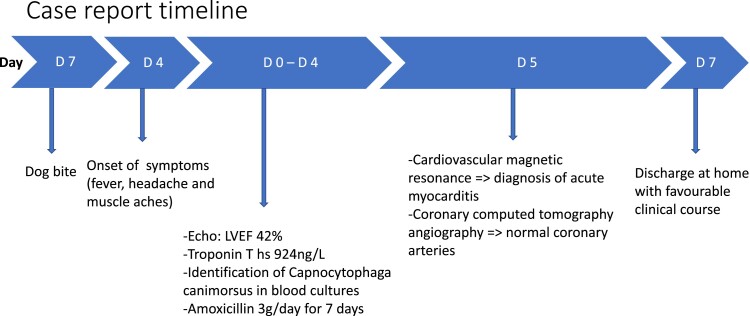
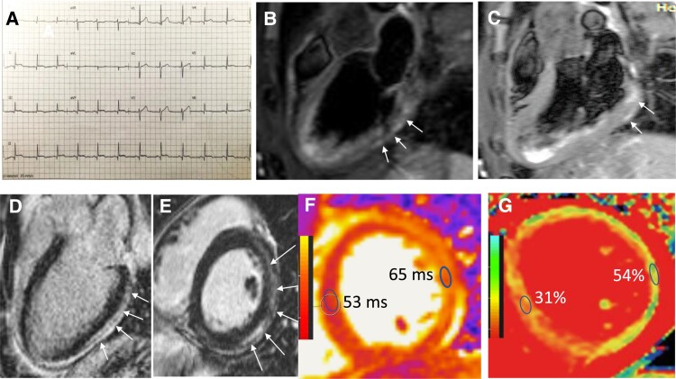
A 37-year-old man was admitted with a 4-day history of fever, headache and myalgia. He complained of epigastralgia, nausea, and diarrhoea. His past medical history consisted only of active smoking. He had no background of splenic disorders and alcohol excess and was not immunocompromised. He reported no cardiac symptoms. Three days before the beginning of symptoms, his right hand had been bitten by his dog. On admission, his temperature was 38.2°C, his heart rate 130 b.p.m., and his blood pressure 118/72 mmHg. Pulse oxygen saturation was 100% on room air. The clinical examination was unremarkable, apart from a bite wound on his right hand. There was no rash, no sign of cellulitis, or soft tissue necrosis. Laboratory tests showed an intense inflammatory status: C-reactive protein, 417 mg/L, normal range 0–5 mg/L; white blood cell count, 9.45 G/L, normal range 3.77–9.05 G/L (neutrophils 91%, lymphocytes 3.5%); procalcitonin, 8.42 μg/L, normal range 0–0.5 μg/L; thrombocytopaenia (platelets, 29 G/L, normal range 177–379 G/L); and acute kidney injury (creatinine, 189 μmol/L, normal range 45–84 μmol/L, ×3 vs. baseline) and abnormal liver enzymes (aspartate transaminase [AST] × 2 upper reference limit, alanine transaminase [ALT] × 3 upper reference limit). Markers of myocardial injury included elevated high sensitivity-troponin T (peak 1129 ng/L on Day 2, upper reference limit 50 ng/L) and N-terminal brain natriuretic peptide (1127 pg/mL, upper reference limit 500 pg/mL). Twelve-lead electrocardiogram showed a mild diffuse ST-segment elevation with micro-Q waves in inferior and lateral leads (Figure 1, panel A). Transthoracic echocardiography revealed mild global left ventricular (LV) hypokinesia with a decrease in LV ejection fraction (42%) and preserved right ventricular function. There was no pericardial effusion. Blood cultures were drawn, and the patient was treated with parenteral cefotaxime (3 g per day).
Figure 1.
Bobo–Newton (Capnocytophaga canimorsus) acute myocarditis. (A) Twelve-lead electrocardiogram showing mild diffuse ST-segment elevation with micro-Q waves in inferior and lateral leads. (B) Diastolic phase T2-weighted short-tau inversion-recovery image in the left ventricular three-chamber longitudinal view showing subepicardial nodular areas of hypersignal in the inferolateral wall, indicating focal oedema (arrows). (C) Diastolic frame extracted from T1-weighted spin echo sequence in the left ventricular three-chamber longitudinal view acquired 1 min. after the injection of 0.1 mm gadolinium chelates demonstrates early enhancement of the inferolateral wall, indicative of hyperaemia (arrows). (D) Late gadolinium-enhanced image of the left ventricular long-axis and short-axis (E) views acquired during diastole shows nodular and linear areas of hyper-enhancement in the subepicardium of the inferolateral and anterolateral walls (arrows).6 (F) T2 map of the mid short-axis basal view of the left ventricular showing an increased T2 relaxation time in the inferolateral and anterolateral walls, indicating the presence of focal oedema. (G) Extracellular volume map of the mid short-axis basal view of the left ventricular showing an increased extracellular volume in the inferolateral and anterolateral walls (54% vs. 31% in the septal wall). ECG, electrocardiogram; LV, left ventricular; LGE, late gadolinium-enhanced; ECV, extracellular volume.
After 45 and 47 h, the two aerobic blood cultures grew slender, fusiform, Gram-negative rods. Capnocytophaga canimorsus was identified by Maldi-time of flight mass spectrometry.
Cefotaxime was changed to amoxicillin (3 g/day for 7 days) with a favourable clinical course and subsequent recovery of biological markers.
Because of ECG alterations and troponin elevation, coronary computed tomography angiography was performed and showed normal coronary arteries. Cardiovascular magnetic resonance was performed on Day 5 and showed a non-dilated LV (87 mL/m²) with normal global systolic function (LV ejection fraction 56%). T2-weighted short-tau inversion-recovery sequence showed focal areas of subepicardial oedema in the LV inferolateral wall (Figure 1, panel B). Cine-cardiovascular magnetic resonance acquired 1 min after the injection of 0.1 mm gadolinium chelates demonstrated early hyper-enhancement in the LV inferolateral wall, indicating hyperaemia (Figure 1, panel C). Late gadolinium-enhancement imaging in the subepicardium of inferolateral and anterolateral walls confirmed the diagnosis of acute myocarditis (Figure 1, panel D).6 T2- and extracellular volume (ECV) LV maps revealed increased T2 time (oedema) and ECV (interstitial space) in the inferolateral and anterolateral walls as compared to the septal wall (Figure 1, panels E and F). Although it is the gold standard for the diagnosis of acute myocarditis, endomyocardial biopsy was not performed because there was no haemodynamic deterioration, no heart failure, or arrhythmias, and because the Gram-negative rod bacteria was covered with specific antibiotic regimen.7 The patient was discharged on Day 7. He remains well at 1 year without further symptoms at the last clinical visit.
Discussion
Capnocytophaga canimorsus is a fastidious Gram-negative bacillus, commensal of the oral cavities of dogs and cats, first described by Bobo and Newton.4,5 Human infection can occur after a dog bite, lick, or scratch and can take several forms, such as septic shock, disseminated intravascular coagulation, meningitis, endocarditis, and eye infections.5 Mortality rate may reach 33% in case of septic shock.8 The main risk factors for a severe bacteraemia infection are prior splenectomy and alcoholism, but it can occur in immunocompetent patients.8 Cardiovascular manifestations of Capnocytophaga canimorsus infection include endocarditis,8 heart failure in patients with pre-existing cardiac conditions,8 acute myocardial infarction,1 mycotic abdominal aortic aneurysm, or prosthetic aortitis. In previous reports, the diagnosis of myocardial infarction was based on the occurrence of chest pain associated with significant ST-elevation on ECG and cardiac enzyme rise. Coronary angiography showed normal coronary arteries in three cases.1 The diagnosis of myocardial infarction has been disputed by some authors2,3 who raised the higher likelihood of acute myocarditis but without formal demonstration by cardiac imaging or endomyocardial biopsy. Herein, we report a case of documented Bobo–Newton acute myocarditis associated with Capnocytophaga canimorsus infection.4 Acute myocarditis was demonstrated by comprehensive CMR that showed the presence of all diagnostic criteria (oedema–hyperaemia-typical patterns of late gadolinium-enhancement) with sparing of the subendocardium and therefore no myocardial infarction.6 Because acute myocarditis can mimic acute myocardial infarction, the current report indicates that acute myocarditis should be ruled out in patients with Capnocytophaga canimorsus infection and a clinical presentation of ‘acute myocardial infarction’, especially in those with unobstructed coronary arteries. The pathophysiology of acute myocarditis remains unclear, as myocardial damage may be caused by toxic-infectious mechanisms (immune-mediated myocarditis with cytokines storm) or by the presence of the bacteria within the myocardium (infectious myocarditis).7
Supplementary Material
Acknowledgements
We are indebted to Prof. Jean Damien Ricard and Prof. Damien Roux for the help in reviewing the manuscript.
Slide sets: A fully edited slide set detailing this case and suitable for local presentation is available online as Supplementary data.
Consent: The authors confirm that written consent for submission and publication of this case report including images and associated text has been obtained from the patient in line with COPE guidance.
Funding: None declared.
Contributor Information
Camille Le Breton, Department of Cardiology, Centre Hospitalier de Longjumeau, 159 Rue du Président François Mitterrand, 91160 Longjumeau, France.
Bernard El Braidy, Department of Cardiology, Centre Hospitalier de Longjumeau, 159 Rue du Président François Mitterrand, 91160 Longjumeau, France.
Florence Durup, Department of Cardiology, Centre Hospitalier de Longjumeau, 159 Rue du Président François Mitterrand, 91160 Longjumeau, France.
Jérôme Garot, Institut Cardiovasculaire Paris Sud, Cardiovascular Magnetic Resonance Laboratory, Hôpital Privé Jacques Cartier, Ramsay Santé, 91300 Massy, France.
Lead author biography
 Camille Le Breton is a senior assistant in Cardiology, working in the Department of Cardiology of the Montsouris Hospital, Paris, France, and in the Groupe hospitalier Nord Essonne, Longjumeau, France. She completed her undergraduate degrees at Paris Descartes University and the Residency in the Assistance Publique–Hôpitaux de Paris (Paris, France). In 2020, she obtained her Medical Degree in Cardiology and critical care medicine. Besides general Cardiology, her areas of interest include infectious diseases. In 2021, she graduated in critical care for infectious diseases at Université de Paris, France.
Supplementary material
Supplementary material is available at European Heart Journal—Case Reports.
Data availability
The data underlying this article are available in the article and in its online Supplementary material.
References
- 1. Scharf C, Widmer U. Myocardial infarction after dog bite. Circulation 2000;102:713. [DOI] [PubMed] [Google Scholar]
- 2. Auer J, Berent R, Eber B. Myocardial infarction after dog bite. Circulation 2001;103:e95. [DOI] [PubMed] [Google Scholar]
- 3. Bogaty P. Myocardial infarction after dog bite. Circulation 2001;103:e95–e96. [DOI] [PubMed] [Google Scholar]
- 4. Bobo RA, Newton EJ. A previously undescribed gram-negative bacillus causing septicemia and meningitis. Am J Clin Pathol. 1976;65:564–569. [DOI] [PubMed] [Google Scholar]
- 5. Markewitz RDH, Graf T. Capnocytophaga canimorsus infection. N Eng J Med 2020;383:1167. doi: 10.1056/NEJMicm1916407. [DOI] [PubMed] [Google Scholar]
- 6. Ferreira VM, Schulz-Menger J, Holmvang G, Kramer CM, Carbone I, Sechtem U, et al. Cardiovascular magnetic resonance in nonischemic myocardial inflammation: expert recommendations. J Am Coll Cardiol 2018;72:3158–3176. [DOI] [PubMed] [Google Scholar]
- 7. Caforio AL, Pankuweit S, Arbustini E, Basso C, Gimeno-Blanes J, Felix SB, et al. Current state of knowledge on aetiology, diagnosis, management, and therapy of myocarditis: a position statement of the European Society of Cardiology working group on myocardial and pericardial diseases. Eur Heart J 2013;34:2636–2648, 2648a–2648d. [DOI] [PubMed] [Google Scholar]
- 8. Lion C, Escande F, Burdin JC. Capnocytophaga canimorsus infections in humans: review of the literature and cases report. Eur J Epidemiol 1996;12:521–533. [DOI] [PubMed] [Google Scholar]
Associated Data
This section collects any data citations, data availability statements, or supplementary materials included in this article.
Supplementary Materials
Data Availability Statement
The data underlying this article are available in the article and in its online Supplementary material.


