Abstract
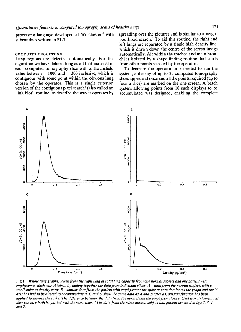
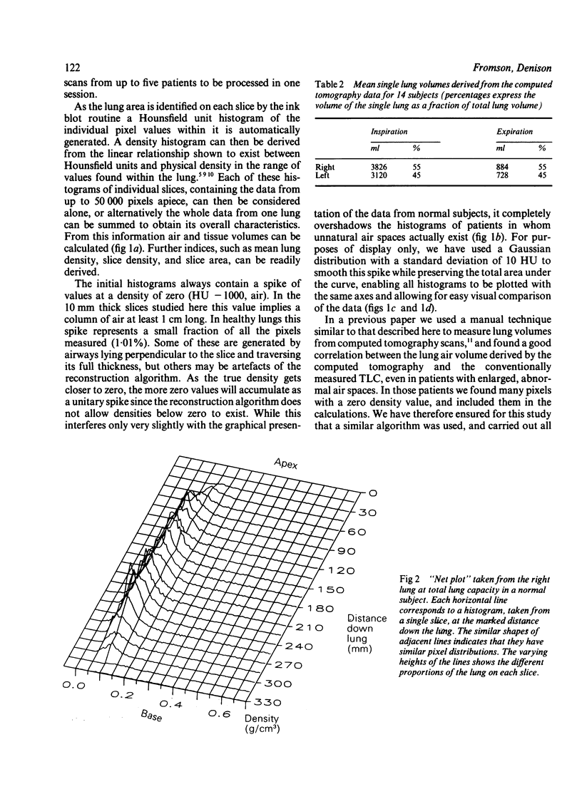
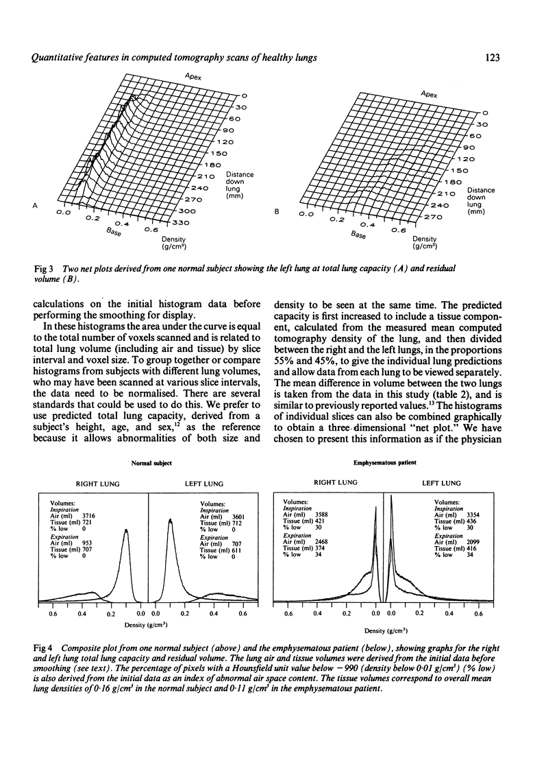
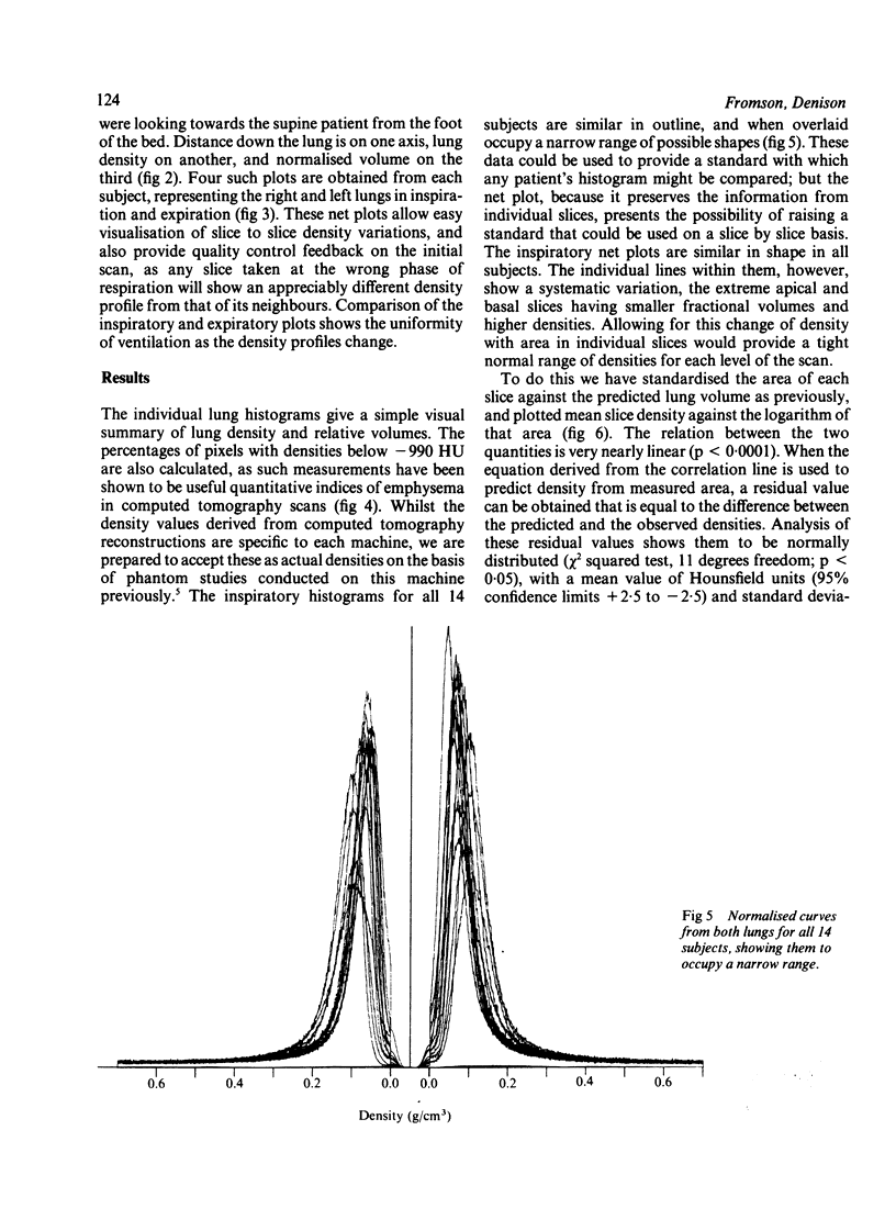
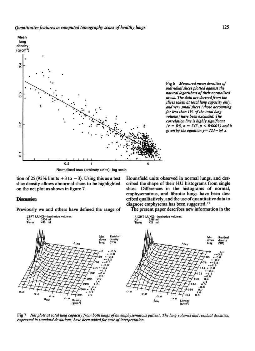
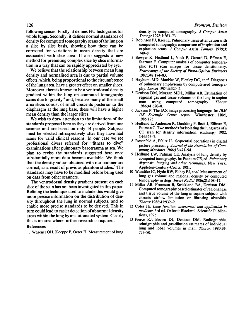
This study set out to determine whether quantitative features of lung computed tomography scans could be identified that would lead to a tightly defined normal range for use in assessing patients. Fourteen normal subjects with apparently healthy lungs were studied. A technique was developed for rapid and automatic extraction of lung field data from the computed tomography scans. The Hounsfield unit histograms were constructed and, when normalised for predicted lung volumes, shown to be consistent in shape for all the subjects. A three dimensional presentation of the data in the form of a "net plot" was devised, and from this a logarithmic relationship between the area of each lung slice and its mean density was derived (r = 0.9, n = 545, p less than 0.0001). The residual density, calculated as the difference between measured density and density predicted from the relationship with area, was shown to be normally distributed with a mean of 0 and a standard deviation of 25 Hounsfield units (chi 2 test: p less than 0.05). A presentation combining this residual density with the net plot is described.
Full text
PDF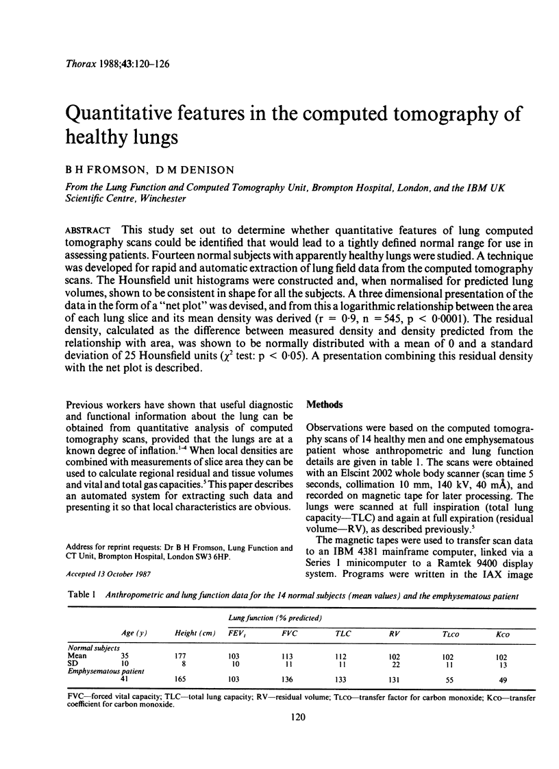
Selected References
These references are in PubMed. This may not be the complete list of references from this article.
- Denison D. M., Morgan M. D., Millar A. B. Estimation of regional gas and tissue volumes of the lung in supine man using computed tomography. Thorax. 1986 Aug;41(8):620–628. doi: 10.1136/thx.41.8.620. [DOI] [PMC free article] [PubMed] [Google Scholar]
- Hayhurst M. D., MacNee W., Flenley D. C., Wright D., McLean A., Lamb D., Wightman A. J., Best J. Diagnosis of pulmonary emphysema by computerised tomography. Lancet. 1984 Aug 11;2(8398):320–322. doi: 10.1016/s0140-6736(84)92689-8. [DOI] [PubMed] [Google Scholar]
- Hedlund L. W., Anderson R. F., Goulding P. L., Beck J. W., Effmann E. L., Putman C. E. Two methods for isolating the lung area of a CT scan for density information. Radiology. 1982 Jul;144(2):353–357. doi: 10.1148/radiology.144.2.7089289. [DOI] [PubMed] [Google Scholar]
- Millar A. B., Fromson B., Strickland B. A., Denison D. M. Computed tomography based estimates of regional gas and tissue volume of the lung in supine subjects with chronic airflow limitation or fibrosing alveolitis. Thorax. 1986 Dec;41(12):932–939. doi: 10.1136/thx.41.12.932. [DOI] [PMC free article] [PubMed] [Google Scholar]
- Pierce R. J., Brown D. J., Denison D. M. Radiographic, scintigraphic, and gas-dilution estimates of individual lung and lobar volumes in man. Thorax. 1980 Oct;35(10):773–780. doi: 10.1136/thx.35.10.773. [DOI] [PMC free article] [PubMed] [Google Scholar]
- Robinson P. J., Kreel L. Pulmonary tissue attenuation with computed tomography: comparison of inspiration and expiration scans. J Comput Assist Tomogr. 1979 Dec;3(6):740–748. [PubMed] [Google Scholar]
- Wandtke J. C., Hyde R. W., Fahey P. J., Utell M. J., Plewes D. B., Goske M. J., Fischer H. W. Measurement of lung gas volume and regional density by computed tomography in dogs. Invest Radiol. 1986 Feb;21(2):108–117. doi: 10.1097/00004424-198602000-00005. [DOI] [PubMed] [Google Scholar]
- Wegener O. H., Koeppe P., Oeser H. Measurement of lung density by computed tomography. J Comput Assist Tomogr. 1978 Jul;2(3):263–273. doi: 10.1097/00004728-197807000-00003. [DOI] [PubMed] [Google Scholar]


