Abstract
Streptococcus suis colonizes the upper respiratory tract of healthy pigs at high abundance but can also cause opportunistic respiratory and systemic disease. Disease-associated S. suis reference strains are well studied, but less is known about commensal lineages. It is not known what mechanisms enable some S. suis lineages to cause disease while others persist as commensal colonizers, or to what extent gene expression in disease-associated and commensal lineages diverge. In this study we compared the transcriptomes of 21 S . suis strains grown in active porcine serum and Todd–Hewitt yeast broth. These strains included both commensal and pathogenic strains, including several strains of sequence type (ST) 1, which is responsible for most cases of human disease and is considered to be the most pathogenic S. suis lineage. We sampled the strains during their exponential growth phase and mapped RNA sequencing reads to the corresponding strain genomes. We found that the transcriptomes of pathogenic and commensal strains with large genomic divergence were unexpectedly conserved when grown in active porcine serum, but that regulation and expression of key pathways varied. Notably, we observed strong variation of expression across media of genes involved in capsule production in pathogens, and of the agmatine deiminase system in commensals. ST1 strains displayed large differences in gene expression between the two media compared to strains from other clades. Their capacity to regulate gene expression across different environmental conditions may be key to their success as zoonotic pathogens.
Keywords: Streptococcus suis, transcriptomics, gene regulation, strain comparison, virulence, pathogen, commensal
Data Summary
The genome assemblies generated in this study are available under BioProject accession number PRJNA855487. The BioSample accession number of each strain is listed in Table S1 (available in the online version of this article). RNA sequencing data are available under BioProject accession number PRJNA863843.
Impact Statement.
Streptococcus suis is an abundant commensal colonizer of the porcine upper respiratory tract but also a zoonotic opportunistic pathogen. There is no cross-protective vaccine against S. suis , and S. suis disease is a major driver of antimicrobial usage and resistance in pig farms. Increased knowledge of S. suis gene expression and the mechanisms determining strain potential for invasive disease and commensal colonization could help us to better understand S. suis ecology and design management practices, vaccines, probiotics, or other treatments. This study adds to our knowledge of S. suis virulence and commensalism and suggests ways to rationally improve the design of future in vitro studies with respect to strain and media selection.
Introduction
Streptococcus suis is an opportunistic pathogen that can cause septicaemia and meningitis in pigs and humans, but also colonizes the upper respiratory tract of healthy pigs [1]. Different S. suis lineages appear to be specialized to different niches, being made up of either predominantly clinical strains isolated from necropsy (pathogenic clades) or non-clinical strains isolated from the oral cavity of pigs (commensal clades) [2]. Clinical and non-clinical strains are, however, found in all clades, showing that pathogenic lineages can colonize the oral cavity of asymptomatic pigs at low abundance and that commensal lineages can invade the host in certain circumstances. Recent microbiome studies have confirmed that commensal S. suis colonize the oral cavity of piglets at high abundance [3–5].
Limited research has been conducted on commensal S. suis and the mechanisms underlying their great success as piglet oral biofilm colonizers. Commensal S. suis clades have high genomic diversity and larger genome sizes compared to pathogenic clades [6], and production of secondary metabolites may aid them in antagonizing con- and heterospecific competitors [7]. Pathogens have conserved virulence-associated genes but reduced accessory genomes [6]. To successfully infect hosts, pathogens need to rapidly respond to different environments and shifts in nutrient availability and host immune responses. Firstly, they need to colonize the oral cavity to gain access to the host tissue and to persist in herds between outbreaks to ensure transmission. Secondly, they need to cross the host epithelium, enter the bloodstream, and survive and proliferate in host tissues and body fluids with varying nutrient availability while avoiding eradication by host defences. To cause meningitis, S. suis also crosses the blood–brain barrier. S. suis niche differentiation is poorly understood, and the exact combination of virulence-associated genes and gene–host–environment interactions necessary to cause invasive disease have not been established. It is possible that regulation of gene transcription by phase variation and master switches such as carbon catabolite repression are key to rapidly adapting to a changing environment [8–10].
Despite the great heterogeneity of S. suis lineages, most research has been focused on a few closely related and highly pathogenic strains. Transcriptomic studies have largely used sequence type (ST) 1 (serotype 2) strains associated with zoonotic disease, such as P1/7 and S10 [11–16]. This has led to knowledge of how the most common pathogenic S. suis clade adapts to the host but left other pathogenic and commensal clades understudied. It is not known how well results from the commonly studied strains translate to other clades, especially given the high functional redundancy of many virulence factors described for S. suis [17, 18]. A greater understanding of the species-wide S. suis transcriptome may yield insight into the differences between commensal and pathogenic S. suis clades and increase our understanding of S. suis ecology and evolution.
In this study we compare the transcriptomes of 21 S. suis strains from a wide phylogenetic background and different isolation sources, including not only clinical strains from pathogenic clades and non-clinical strains from commensal clades, but also clinical strains from commensal clades and non-clinical strains from pathogenic clades. We determined growth curves in Todd–Hewitt yeast broth (THY) and active porcine serum (APS), which contains complement and scarce amounts of essential metals [12], and extracted RNA during the exponential growth phase. We mapped RNA-seq data to the individual strain genomes and compared normalized sequencing coverage per gene. The resulting dataset allowed us to gain a better understanding of shared and niche-specific gene expression in commensal and pathogenic S. suis .
Methods
Strain selection
We selected 21 S. suis strains that are broadly representative of the species, including both clinical and non-clinical strains from different phylogenetic backgrounds (Table 1). Where available, we included closely related clinical and non-clinical strains for comparison (i.e. not only clinical strains from pathogenic clades and non-clinical strains from commensal clades, but also clinical strains from commensal clades and non-clinical strains from pathogenic clades). While the isolation source and phylogenetic clade of a strain gives an indication of its virulent potential, this has only been experimentally tested in vivo for a few strains. Strains P1/7 and S10 are known to be from a pathogenic clade and highly virulent, while strain T15 has been experimentally shown to have low virulence in pigs despite being from a pathogenic clade [19].
Table 1.
Strain overview. Shortened three-character name used in figures and tables, full original strain name, serotype, sequence type (ST), number of plasmids in genome assembly and strain metadata
|
Name |
Strain |
Country |
Serotype |
ST |
Clade type |
Isolation source |
Plasmids |
|---|---|---|---|---|---|---|---|
|
C01 |
M101999_C1 |
Spain |
8 |
nt |
Pathogenic |
Non-clinical |
1 |
|
C15 |
SS15055_N2_C15 |
Spain |
2 |
1 |
Pathogenic |
Non-clinical |
0 |
|
D11 |
DNS11 |
Denmark |
9 |
16 |
Pathogenic |
Systemic/brain |
1 |
|
D13 |
DNC13 |
Denmark |
nt |
nt |
Commensal |
Non-clinical |
2 |
|
D15 |
DNC15 |
Denmark |
16 |
nt |
Commensal |
Non-clinical |
0 |
|
D20 |
DNS20 |
Denmark |
nt |
nt |
Commensal |
Systemic/brain |
2 |
|
D43 |
DNR43 |
Denmark |
2 |
28 |
Pathogenic |
Respiratory |
1 |
|
D48 |
DNR48 |
Denmark |
8 |
nt |
Pathogenic |
Respiratory |
1 |
|
D49 |
DNC49 |
Denmark |
2 |
28 |
Pathogenic |
Non-clinical |
0 |
|
G69 |
DE609B |
Germany |
2 |
1 |
Pathogenic |
Systemic/brain |
0 |
|
I12 |
21435_1 |
Spain |
31 |
nt |
Commensal |
Non-clinical |
0 |
|
I27 |
21437_3 |
Spain |
4 |
nt |
Commensal |
Non-clinical |
0 |
|
L42 |
LSS42 |
UK |
16 |
nt |
Pathogenic |
Non-clinical |
0 |
|
P17 |
P1/7 |
UK |
2 |
1 |
Pathogenic |
Systemic/brain |
0 |
|
S10 |
S10 |
Netherlands |
2 |
1 |
Pathogenic |
Systemic/brain |
0 |
|
S11 |
M102942_S11 |
Spain |
9 |
123 |
Pathogenic |
Systemic/brain |
1 |
|
S20 |
M104300_S20 |
Spain |
2 |
1 |
Pathogenic |
Systemic/brain |
0 |
|
S26 |
M105052_S26 |
Spain |
19 |
nt |
Commensal |
Systemic/brain |
0 |
|
S40 |
M106471_S40 1 |
Spain |
30 |
nt |
Commensal |
Systemic/brain |
0 |
|
T15 |
T15 |
Netherlands |
2 |
19 |
Pathogenic |
Non-clinical |
2 |
|
X15 |
1521 251 |
Canada |
7 |
nt |
Pathogenic |
Systemic/brain |
0 |
NT, non-typeable.
This study aimed to assess the overall species transcriptome and compare groups of strains rather than focus on specific gene variants or individual strains. Thus, we prioritized RNA sequencing of a single sample from many different strains (biological replicates) rather than technical replicates of each strain. To benchmark the repeatability of our methods we performed RNA sequencing on three technical replicates of S10 grown in each medium, processed separately on different days.
Hybrid genome assembly
We created new Illumina–Nanopore hybrid genome assemblies for 19 of the 21 strains, as only strains P1/7 and S10 had complete genomes available. T15 had a circular genome assembly that lacked plasmids. We isolated DNA from overnight cultures using the PowerSoil DNA Isolation kit (Qiagen). Generation of short-read sequencing data for the strains from Germany, Canada and the UK has been previously reported [2, 3, 20]. Strains without available short-read sequencing data were 250 bp paired-end sequenced using an Illumina HiSeq 2500 instrument by MicrobesNG (Birmingham, UK). Nanopore sequencing was performed with the SQK-LSK109 Ligation Sequencing kit and Guppy Software v5.0.16 with high-accuracy base calling. Genomes were assembled using Unicyler v0.4.9 [21] with default settings. See Table S1 (available in the online version of this article) for genome statistics.
RNA extraction and sequencing
We sampled the strains in the mid exponential growth phase during growth in Todd–Hewitt yeast broth (THY) and active porcine serum (APS). THY is a rich laboratory medium used to rapidly grow streptococci to high density, and consists of several ingredients, including meat infusion, tryptone, glucose and yeast extract. Serum is extracted from blood by removing cells and clotting factors and although it contains high glucose concentrations essential minerals are scarce. For instance, iron is sequestered by host transferrin to limit bacterial growth in blood. Active serum is not heat-treated and may thus contain complement factors and antibodies binding to S. suis , as this pathogen is endemic on farms. S. suis has several mechanisms to evade complement activation and formation of the membrane attack complex, including the capsule, factor H-binding proteins and other less well understood mechanisms [17, 22].
Growth curves were started with an overnight culture of S. suis in THY and adjusted to OD600 0.5 by dilution in phosphate-buffered saline (PBS) and subsequently inoculated 1 : 10 in either THY or APS. For determination of growth curves the strains were grown in 96-well plates with 200 µl total volume per well and incubated at 37 °C with 5 % CO2. The plates were shaken to prevent sedimentation and measured with a SpectraMax M5 (Molecular Devices) every 30 min. Each strain–medium combination was grown in three separate plates with two technical replicates per plate.
For RNA isolation 10 ml cultures were grown in 15 ml Falcon tubes at 37 °C with 5 % CO2. All experiments were carried out using a single batch of THY and APS. Based on the growth curves we identified a time point where all strains were in the (mid-) exponential growth phase. To account for different growth in Falcon tubes compared to 96-well plates, we reduced the incubation time and confirmed that the culture was in the correct growth phase by measuring OD600 before RNA isolation. Cultures in APS were sampled at 95 min and THY cultures at 125 min. The APS and THY cultures were started at different times to enable RNA isolation at the same time. Cultures were pelleted by centrifugation and resuspended in QIAzol Lysis Reagent (Qiagen). After bead beating twice for 40 s with 0.1 mm silica beads (MP biomedicals) RNA was isolated using the miRNeasy kit (Qiagen). Trace DNA was digested using the DNase Max kit (Qiagen). RNA quantity and integrity were confirmed with nanodrop (Thermo Fisher) and TapeStation (Agilent). Library preparation was performed with the Illumina Stranded Total RNA Prep kit. rRNA was enzymatically depleted and the remaining RNA fragmented and translated to cDNA. The libraries were sequenced with 150 bp paired-end sequencing on the Illumina NovaSeq 6000 system with the Illumina NovaSeq 6000 SP reagent kit at iGenSeq (Institut du Cerveau, France).
Bioinformatics
The strain genomes were annotated with Prokka v1.14.5 [23]. We further identified antimicrobial resistance genes with Resfinder v4.1 [24] and biosynthetic gene clusters with antiSMASH v5.1.2 [25] and BiG-SCAPE v1.1.2 [26]. We used clinker [27] for gene cluster comparison. To compare the transcriptome between strains we grouped homologous and paralogous genes using Orthofinder v2.3.12 [28, 29], and the gene expression for paralogous genes was summed. This may in some cases result in the grouping of genes with different functions. The ‘orthogroups’ found by Orthofinder were given static names from the RefSeq locus tags of S. suis reference strains P1/7 (GCF_000091905.1) and D12 (GCF_000231905.1). GO terms for each Orthogroup were found with InterProScan v5.54–87.0 [30], and quantitative analysis of GO term expression was performed on the whole transcriptome, including accessory genes. The RNA sequencing data were adapter- and quality-trimmed with Trimmomatic v0.39 [31] before being mapped to the genome of the individual strain with Bowtie 2 [32]. FeatureCounts 2.0.1 [33] with default settings was used to count the number of reads mapping to each gene. Further analysis was carried out with R v4.1.3 [34].
Analysis
Combining the transcriptomes of different strains into a single analysis requires additional normalization compared to single-strain transcriptomic studies. In addition to variable sequencing depth per sample, strains vary in genome size and presence/absence and length of genes. We opted to use TPM read count normalization to facilitate comparison between strains. We identified differentially expressed genes using Wilcoxon rank sum test with false discovery rate (FDR) correction. Overall transcriptome difference was calculated separately using the whole-genome transcriptome (expression of accessory genes absent in strain set to 0) and the core genome transcriptome (using only core genes shared by all strains). To quantify and visualize overall transcriptome difference we used R package vegan [35] Bray–Curtis dissimilarity, principal component analysis (PCA) and redundancy analysis (RDA). To determine whether the transcriptome conservation differed significantly between groups, we used estimated marginal means on linear models with R function emmeans [36].
Results and discussion
Dataset description
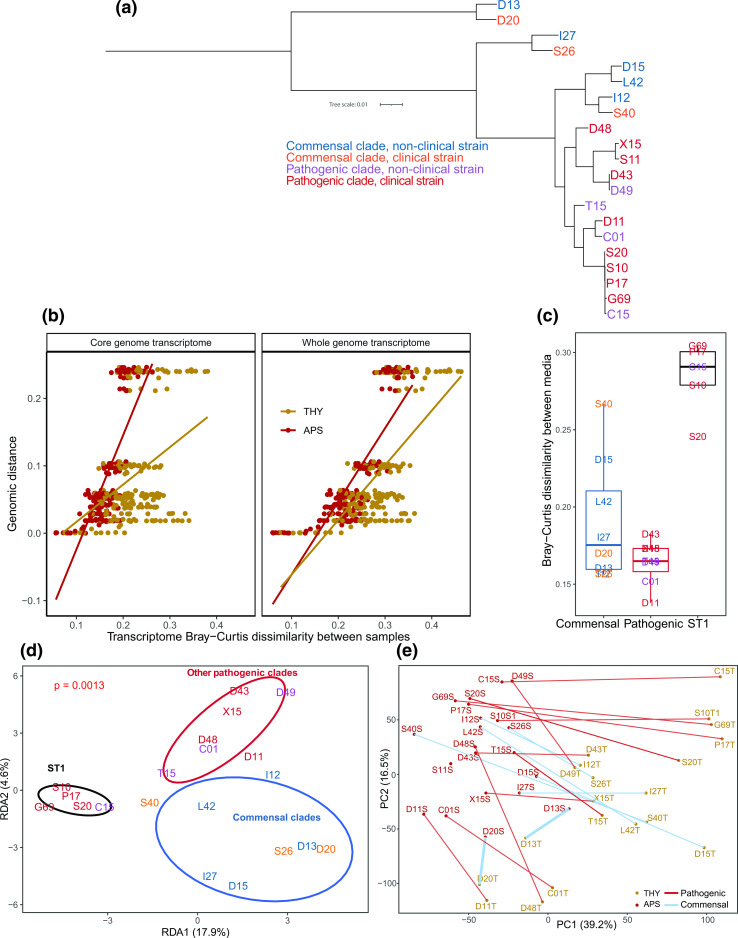
We sequenced the transcriptome of a genetically diverse set of 21 S. suis strains (Fig. 1a) in the mid-exponential growth phase in THY and APS. Overall, commensal and pathogenic strains grew equally well in both media (Fig. S1). All samples were successfully sequenced with a minimum of 20 million 150 bp paired-end reads (1114–1902× coverage). All samples were free from contamination, with >99.8 % reads mapping to the corresponding strain genome. One sample, S11 grown in THY, had excessively high expression of the arginine deiminase system (ADS; Fig. S2). ADS was previously described as being vital for S. suis growth in acidic medium [37, 38], and the high ADS expression may be linked to in vitro culturing and have limited in vivo relevance. Considering the excessive impact of this single gene cluster on the total transcriptome, we excluded the sample from the main analysis. Table S2 shows TPM values per orthogroup for all sequenced samples.
Fig. 1.
Strain phylogeny and overall transcriptome variation. (a) Core genome phylogenetic tree of the 21 strains included in the study, constructed by Orthofinder and STAG with default settings and mid-point rooted. While ST1 pathogenic strains such as P1/7 and S10 are closely related, commensal clades have high genomic diversity. (b) Transcriptome Bray–Curtis dissimilarity between strains correlates with genome phylogenetic distance. The transcriptome is more conserved in APS than in THY. Each point represents a pairwise comparison. Strains were only compared within the same medium. (c) Boxplot of core genome transcriptome Bray–Curtis dissimilarity between samples from each strain in the two media. (d) Redundancy analysis (RDA) on log2 (fold change) on the core genome transcriptome constrained by (ST1 vs other pathogenic clades vs commensal clades) showed that ST1 clade strains had a distinct transcriptome change between media. Ellipses represent 75 % confidence level. (e) Principal component analysis (PCA) showing core genome transcriptome separation between samples grown in THY and APS. The pairs of samples were well separated on PC1 in all strains except for strains D13 and D20. Each point (coloured by medium) is one sample, and the two samples of each strain are joined by lines coloured by clade type.
Comparison of S10 triplicate samples showed that our methods were highly reproducible, with a maximum pairwise Bray–Curtis dissimilarity of 0.1 between replicates. Distinct but very closely related strains within sequence type 1 (ST1) had similar pairwise dissimilarities, but this was expected, as their genomes are virtually identical. It does, however, show that small differences in gene expression between strains should be interpreted with care. For the remaining analysis only a single S10 sample from each medium was included.
Serum opacification
Three commensal strains increased the OD600 rapidly and without lag phase when grown in APS, reaching far higher OD600 values than the other strains (Fig. S1). This was not due to an increase of bacterial biomass, but opacification of the supernatant. Formation of large lipid particles by the protein opacification factor of serum (ofs, SSU_RS07445) has previously been described in S. suis , and the gene is known to occur in different variants with and without opacifying function [39, 40]. The three strains with strong opacification activity had high expression of ofs variants diverged from those previously described (Fig. S3). While the shorter ofs type-1 [40] variant found in ST1 is expressed at low levels and has limited opacification capacity, it is conserved in pathogenic clades and may have a function related to binding to host cells [39, 41].
Conservation of core genome expression in serum
Commensal clades are diverse, with long (core and whole-genome-based phylogeny) branch lengths between strains, while pathogenic clades, in particular the commonly researched ST1 clade, consist of closely related strains [6]. Our strain selection included both ST1 strains and a wider range of strains from other pathogenic and commensal clades. Strains D13 and D20 form an outgroup to the other strains in our dataset (Fig. 1a). The phylogenetic status of this ‘divergent’ outgroup clade is debated, as it can also be considered a separate species (see closely related strains in ‘clade 2’ [42]). Despite large genomic differences (<86 % ANI) to other included strains, their transcriptome was conserved in APS (measured in Bray–Curtis dissimilarity, Fig. 1b). Overall, the S. suis transcriptome was significantly more conserved in APS compared to THY, and in the expression of the core genome compared to the whole-genome transcriptome (estimated marginal means, P<0.01). Despite the large genomic differences between outgroup D13+D20 and the remaining strains, pairwise transcriptome Bray–Curtis dissimilarities to the other strains were overlapping with those between strains from different pathogenic clades. The 13 GO terms most expressed in THY were all more expressed in APS (Fig. S4). This suggests that the conserved APS transcriptome reflects upregulation of core physiological functions related to growth and cell division. This may be relevant in facilitating exponential growth during host invasion. See Table S3 for differences in expression of all GO terms and genes between clades and media.
Large transcriptome differences between media in ST1 strains
Transcriptome difference between the two media was largest for ST1 strains (Fig. 1c). Redundancy analysis (RDA) on log2 fold change of gene expression between THY and APS cultures constrained by clade type (ST1 vs other pathogenic clades vs commensal clades) also showed that regulation of gene expression in ST1 strains was distinct, while other pathogenic clades and commensals were more similar (Fig. 1d). The transcriptome of ST1 strains was, however, not strongly divergent from other clades in either medium. PCA on all samples (Fig. 1e) showed that the pairs of samples from each strain separated in similar directions on the PC1 and PC2 axis, except for the two outgroup commensal strains D13 and D20. ST1 strains showed stronger regulation of several genes. This included downregulation of SSU_RS07200 (SprT-like protein) and upregulation of SSU_RS02105 (cysteine synthase) in THY (Fig. S5). ST1 and other pathogenic S. suis are thought to regulate gene expression related to virulence factors via a phase-variable type I DNA methyltransferase system [9, 43]. However, this system is unlikely to explain the differences seen in the present study, as the THY and APS cultures only grew separately for a few generations. This is unlikely to be sufficient time for selection to drive change across the experimental population. We did not observe differences in growth rates of the strains, indicating that selection pressure for either phase variant was limited, and that phase variation did not occur. Moreover, non-ST1 strains with the same phase variation system had small transcriptome differences between media. These results suggest that ST1 strains may have additional undescribed mechanisms enabling strong regulation of gene expression in different environmental conditions.
High cps expression in serotype 2 strains
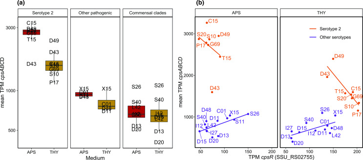
The S. suis polysaccharide capsule (CPS) exists in many variants (serotypes). Serotype 2, a capsule type with terminal sialic acid, is the most studied serotype due to its association with high (zoonotic) virulence [44–46]. In this study all ST1 strains were serotype 2, in addition to strains T15 and D43+D49. Genes involved in capsule production (cps) were upregulated in APS in most strains (Fig. 2a). Cps expression was high and strongly upregulated in APS in serotype 2 strains compared to other serotypes, although serotype 2 strains D43 and D49 appeared to have divergent regulation. D49 downregulated cps expression less, and D43 had higher cps expression in THY, opposite to other serotype 2 strains. Non-typeable strains D13 and D20 had the lowest cps expression, but apart from these and serotype 2 strains, cps expression overlapped between commensal (serotype 4, 16, 19, 30 and 31) and pathogenic clades (serotype 7, 8 and 9). The pathogenic clades had higher expression levels than the commensals, but this was only significant in APS (P=0.03) and not in THY (P=0.15).
Fig. 2.
Capsule gene cluster expression. (a) Mean expression of cpsABCD (SSU_RS02765-SSU_RS02780), the initial four genes in the cps gene cluster, which are shared by all strains. Grouped by serotype 2 (all from pathogenic clades), other pathogenic clade strains and commensal clade strains. (b) Scatterplot with regression lines comparing the mean expression of cpsABCD with the cpsR (SSU_RS02755) regulator. Only the serotype 2 strains had a negative correlation between cpsR and cps expression, although serotype 2 strains D43 and D49 also appeared to be regulated differently, as cpsABCD expression was similar in the two media.
Streptococcal cps expression has been linked to negative regulation by a protein (SSU_RS02755) variably named cpsR/gntR/orf2Y [47–49]. In Streptococcus pneumoniae , cpsR has been shown to interact with the cps promoter, dependent on glucose concentration, negatively controlling cps expression and CPS production [48]. We only found a negative correlation between cpsR and cps expression in serotype 2 strains (Fig. 2b). This, and the lack of cps regulation in the serotype 2 strains D43 and D49, may be due to variation in cpsR, which occurs as a distinct variant in all serotype 2 strains except D43 and D49. Although the D43 and D49 cpsR sequences contain arginine in position 17 characteristic for serotype 2 strains, all other sequence variation specific to this serotype is absent (Figs S6, S7). It is not clear how different combinations of serotypes and cps regulation may be beneficial in different niches.
High expression of the agmatine deiminase system in serum in commensal strains
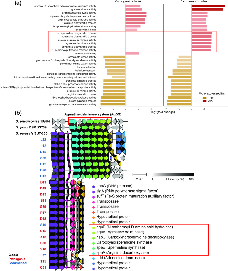
As one might expect, many of the genes and pathways most differentially expressed between THY and APS related to acquisition and metabolism of nutrients that are differentially present in the media (Fig. 3a, Table S3). Metal ion acquisition was upregulated in APS while trehalose import and metabolism were highly expressed in some strains in THY. All strains had high expression of the lac operon (downstream from SSU_RS04590) in THY, but this was strongly downregulated in APS, except in outgroup strain D13. This strain expressed the lac operon constitutively, possibly due to having an unusual, truncated, copy of encoding repressor lacR. It did, however, also carry a lac gene cluster copy more similar to other strains (Fig. S8). Lactose metabolism is likely to be important for S. suis during commensal colonization of pre-weaning piglets, and the fitness cost of constitutive lac operon expression is likely limited (D13 was collected from a piglet approximately 42 days old and thus appears able to persist post-weaning).
Fig. 3.
The accessory AgDS system has a large influence on transcriptome differences between THY and APS in commensals. (a) The 30 GO terms with the largest log2 fold changes between THY and APS cultures (average across all strains, GO terms with <10 mean TPM excluded). Several GO terms highly expressed in APS only occurred in commensal clades due to the presence/absence of the agmatine deiminase system, suggesting that this gene cluster may be important for commensal S. suis . GO terms associated with the agmatine deiminase system highlighted in red. (b) The AgDS gene cluster is only found in commensal clade strains and has high similarity to the AgDS of S. pneumoniae , S. porci and S. parasuis .
One of the largest overall differences between growth in THY and APS was in amino acid metabolism. The genomes of six of eight commensal strains, but no pathogens, encode an agmatine deiminase system (AgDS, SSUD12_RS06980-SSUD12_RS07005). In the strains in which it was present, AgDS was highly expressed and upregulated in APS compared to in THY. This gene cluster has not previously been described in S. suis but is known from S. pneumoniae [50] and Streptococcus mutans [51–53]. The S. suis AgDS share high similarity to the AgDS of S. pneumoniae reference strain TIGR4 (SP_RS04525-SP_RS04560), Streptococcus porci and Streptococcus parasuis (Fig. 3b). The S. mutans AgDS is diverged, with several indels and <50 % amino acid residue identity to the S. suis agmatine deiminase gene. In S. suis the AgDS appear to be linked to a set of unrelated genes, including DNA primase (Fig. 3b), and in some strains these make up a 11.5 kb genomic island flanked by transposases.
The arginine deiminase system (ADS/arcABC operon, SSU_RS03045-SSU_RS03060) was found in all strains and in contrast to AgDS it was more expressed in THY than in APS. Agmatine is formed upon decarboxylation of arginine, and agmatine and arginine metabolic pathways show considerable overlap. Both the arginine and agmatine deiminase system produce ammonia, increasing intracellular pH and tolerance to low pH. High ADS and AgDS expression may be linked to acidification of the medium during growth. ADS has been shown to be important for S. suis survival in acidic medium [37, 54]. AgDS is thought to be relevant to acidic stress in S. mutans [53], but does not appear to increase pH tolerance in S. pneumoniae [50]. It is possible that AgDS activity provides a competitive advantage to commensal S. suis by increasing intracellular pH. Agmatine released by competing taxa inhibits the growth of S. mutans [51], and a similar mechanism may apply to S. suis . Considering its variable presence, it may be relevant to both inter- and intra-species competition.
High expression of Mn2+-binding lipoprotein troA in pathogens
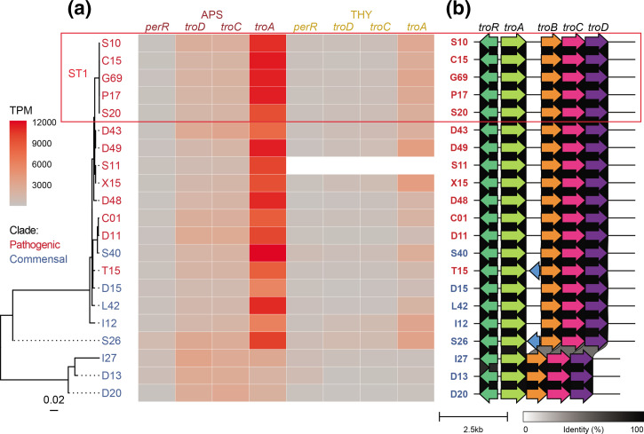
Transition metal ion homeostasis is vital to bacteria. While commensals sequester metal ions in competition with other microbes, pathogens must overcome host metal ion chelation during infection. Some lactic acid bacteria, including S. suis and S. pneumoniae , utilize a larger proportion of manganese relative to iron than most bacteria, and this may provide them with a competitive advantage in some niches [55–58]. We found that genes putatively involved in both iron (SSU_RS03155-SSU_RS03170) and manganese (SSU_RS09395-SSU_RS09415) scavenging were more expressed in APS than THY, although the manganese import gene cluster was five times more expressed than the iron import gene cluster. TroA, a putative scavenger protein for the troBCD ABC transport system [58–60], was also three–four times more expressed than the troBCD ABC transporter genes in the gene cluster (Fig. 4a). This indicates that expression of this gene is regulated separately.
Fig. 4.
Variation in the sequence and expression level of manganese import gene cluster troABCD and its regulators. (a) Regulator perR maximum-likelihood tree with heatmap of gene expression TPM values. Mn2+-binding lipoprotein troA was more expressed than troBCD and more expressed in APS compared to THY. Some commensal strains with diverged perR and troRABCD copies had reduced troA expression. (b) In addition to sequence variation to the other strains, strains I27, D13 and D20 shared a truncated troABCD gene cluster that lacked SSU_RS09410, a putative membrane protein variably annotated as a pseudogene (shown in blue for T15 and S26) or unannotated by Prokka in different strains, likely due to its short length and truncation.
TroA expression varied greatly between strains (232–4165 TPM in THY and 1899–12228 TPM in APS). Some commensals had reduced troA expression compared to ST1 strains despite having similar or higher troBCD expression (Fig. 4a). This may be due to sequence variation of previously described regulators and the gene cluster itself. S. suis troABCD expression has been reported to be repressed by dtxR family metalloregulator troR (SSU_RS09420) and fur family regulator perR (SSU_RS01575), depending on metal ion concentrations and oxidative stress [15, 60–62]. Additionally, mntE (SSU_RS05010) has been identified as a manganese efflux system [63]. Both troR and perR may contribute to troABCD upregulation in APS compared to THY. Variation in troA expression levels between strains grown in the same medium is unlikely to be caused by troR, because strains with identical variants varied greatly in expression. PerR and troA itself also had sequence variation, and the strains with the lowest troA expression had a truncated gene cluster structure (Figs 4b and S9). A region between troA and troB contains a putative oligopeptide transporter pseudogene in most strains, but this was deleted in the strains with the lowest troA expression, D13, D20, and I27 (Fig. 4b). The initial part of this region has higher expression than troA, and its conservation across most of the included S. suis clades suggests that it may have a function irrespective of (the length of) the longest predicted open reading frame.
Conclusions
We found that S. suis strains with large genomic divergence have unexpectedly conserved transcriptomes when grown in APS. More variation was observed in THY, and this should be considered when selecting growth medium for in vitro assays. Despite overall conservation, the transcriptome of the strains varied in key functions, including well-described regulatory mechanisms. In most clades the manganese import and capsule gene clusters may be regulated differently than described for ST1 strains. In general, ST1 strains displayed larger changes in gene expression between THY and APS cultures compared to other clades, and these differences in gene expression may help them adapt rapidly to environmental changes, most notably when changing between upper respiratory tract colonization and host invasion. The gene cluster encoding production of the capsule, which is key in avoiding the host complement attack complex, was among the genes most strongly upregulated and most highly expressed in ST1 strains.
Supplementary Data
Funding information
This research was financially supported by EU Horizon 2020 Programme Grant Agreement 727 966, funded under H2020-EU.3.2.1.1. S.F. is a PhD student funded by the Netherlands Centre for One Health.
Acknowledgements
We thank Maria Laura Ferrando and Isabela Fernandes de Oliveira for providing strains and Illumina sequencing data.
Author contribution
S.F., S.R. and M.J.B. conducted wet-lab experiments. S.F., S.R., G.M., P.B. and J.B. carried out bioinformatic processing and data analysis. J.W. provided supervision. All authors reviewed and edited the manuscript.
Conflicts of interest
The authors declare that there are no conflicts of interest.
Footnotes
Abbreviations: APS, active porcine serum; GO, gene ontology; ST, sequence type; THY, Todd-Hewitt Yeast Broth.
All supporting data, code and protocols have been provided within the article or through supplementary data files. Nine supplementary figures and three supplementary tables are available with the online version of this article.
References
- 1.Segura M, Aragon V, Brockmeier S, Gebhart C, Greeff A, et al. Update on Streptococcus suis research and prevention in the era of antimicrobial restriction: 4th international workshop on S. suis . Pathogens. 2020;9:374. doi: 10.3390/pathogens9050374. [DOI] [PMC free article] [PubMed] [Google Scholar]
- 2.Weinert LA, Chaudhuri RR, Wang J, Peters SE, Corander J, et al. Erratum: genomic signatures of human and animal disease in the zoonotic pathogen Streptococcus suis . Nat Commun. 2015;6:7272. doi: 10.1038/ncomms8272. [DOI] [PMC free article] [PubMed] [Google Scholar]
- 3.Fredriksen S, Neila-Ibáñez C, Hennig-Pauka I, Guan X, Dunkelberger J, et al. Streptococcus suis infection on European farms is associated with an altered tonsil microbiome and resistome. bioRxiv. doi: 10.1101/2022.08.01.500980. [DOI]
- 4.Murase K, Watanabe T, Arai S, Kim H, Tohya M, et al. Characterization of pig saliva as the major natural habitat of Streptococcus suis by analyzing oral, fecal, vaginal, and environmental microbiota. PLoS One. 2019;14:e0215983. doi: 10.1371/journal.pone.0215983. [DOI] [PMC free article] [PubMed] [Google Scholar]
- 5.Fredriksen S, Guan X, Boekhorst J, Molist F, van Baarlen P, et al. Environmental and maternal factors shaping tonsillar microbiota development in piglets. BMC Microbiol. 2022;22:224. doi: 10.1186/s12866-022-02625-8. [DOI] [PMC free article] [PubMed] [Google Scholar]
- 6.Murray GGR, Charlesworth J, Miller EL, Casey MJ, Lloyd CT, et al. Genome reduction is associated with bacterial pathogenicity across different scales of temporal and ecological divergence. Mol Biol Evol. 2021;38:1570–1579. doi: 10.1093/molbev/msaa323. [DOI] [PMC free article] [PubMed] [Google Scholar]
- 7.Liang Z, Wu H, Bian C, Chen H, Shen Y, et al. The antimicrobial systems of Streptococcus suis promote niche competition in pig tonsils. Virulence. 2022;13:781–793. doi: 10.1080/21505594.2022.2069390. [DOI] [PMC free article] [PubMed] [Google Scholar]
- 8.Ferrando ML, van Baarlen P, Orrù G, Piga R, Bongers RS, et al. Carbohydrate availability regulates virulence gene expression in Streptococcus suis . PLoS One. 2014;9:e89334. doi: 10.1371/journal.pone.0089334. [DOI] [PMC free article] [PubMed] [Google Scholar]
- 9.Atack JM, Weinert LA, Tucker AW, Husna AU, Wileman TM, et al. Streptococcus suis contains multiple phase-variable methyltransferases that show a discrete lineage distribution. Nucleic Acids Res. 2018;46:11466–11476. doi: 10.1093/nar/gky913. [DOI] [PMC free article] [PubMed] [Google Scholar]
- 10.Zhong X, Ma J, Bai Q, Zhu Y, Zhang Y, et al. Identification of the RNA-binding domain-containing protein RbpA that acts as a global regulator of the pathogenicity of Streptococcus suis serotype 2. Virulence. 2022;13:1304–1314. doi: 10.1080/21505594.2022.2103233. [DOI] [PMC free article] [PubMed] [Google Scholar]
- 11.Arenas J, Bossers-de Vries R, Harders-Westerveen J, Buys H, Ruuls-van Stalle LMF, et al. In vivo transcriptomes of Streptococcus suis reveal genes required for niche-specific adaptation and pathogenesis. Virulence. 2019;10:334–351. doi: 10.1080/21505594.2019.1599669. [DOI] [PMC free article] [PubMed] [Google Scholar]
- 12.Ferrando ML, Gussak A, Mentink S, Gutierrez MF, van Baarlen P, et al. Active human and porcine serum induce competence for genetic transformation in the emerging zoonotic pathogen Streptococcus suis . Pathogens. 2021;10:156. doi: 10.3390/pathogens10020156. [DOI] [PMC free article] [PubMed] [Google Scholar]
- 13.Koczula A, Jarek M, Visscher C, Valentin-Weigand P, Goethe R, et al. Transcriptomic analysis reveals selective metabolic adaptation of Streptococcus suis to porcine blood and cerebrospinal fluid. Pathogens. 2017;6:7. doi: 10.3390/pathogens6010007. [DOI] [PMC free article] [PubMed] [Google Scholar]
- 14.Ni H, Li M, Wang Q, Wang J, Liu X, et al. Inactivation of the htpsa gene affects capsule development and pathogenicity of Streptococcus suis . Virulence. 2020;11:927–940. doi: 10.1080/21505594.2020.1792080. [DOI] [PMC free article] [PubMed] [Google Scholar]
- 15.Zheng C, Wei M, Qiu J, Jia M, Zhou X, et al. TroR negatively regulates the TroABCD system and is required for resistance to metal toxicity and virulence in Streptococcus suis . Appl Environ Microbiol. 2021;87:e0137521. doi: 10.1128/AEM.01375-21. [DOI] [PMC free article] [PubMed] [Google Scholar]
- 16.Zhong X, Zhang Y, Zhu Y, Dong W, Ma J, et al. Identification of an autorepressing two-component signaling system that modulates virulence in Streptococcus suis serotype 2. Infect Immun. 2019;87:e00377-19. doi: 10.1128/IAI.00377-19. [DOI] [PMC free article] [PubMed] [Google Scholar]
- 17.Segura M, Fittipaldi N, Calzas C, Gottschalk M. Critical Streptococcus suis virulence factors: are they all really critical? Trends Microbiol. 2017;25:585–599. doi: 10.1016/j.tim.2017.02.005. [DOI] [PubMed] [Google Scholar]
- 18.Wang J, Liang P, Sun H, Wu Z, Gottschalk M, et al. Comparative transcriptomic analysis reveal genes involved in the pathogenicity increase of Streptococcus suis epidemic strains. Virulence. 2022;13:1455–1470. doi: 10.1080/21505594.2022.2116160. [DOI] [PMC free article] [PubMed] [Google Scholar]
- 19.Vecht U, Arends JP, van der Molen EJ, van Leengoed LA. Differences in virulence between two strains of Streptococcus suis type II after experimentally induced infection of newborn germ-free pigs. Am J Vet Res. 1989;50:1037–1043. [PubMed] [Google Scholar]
- 20.Hadjirin NF, Miller EL, Murray GGR, Yen PLK, Phuc HD, et al. Large-scale genomic analysis of antimicrobial resistance in the zoonotic pathogen Streptococcus suis . BMC Biol. 2021;19:191. doi: 10.1186/s12915-021-01094-1. [DOI] [PMC free article] [PubMed] [Google Scholar]
- 21.Wick RR, Judd LM, Gorrie CL, Holt KE. Unicycler: resolving bacterial genome assemblies from short and long sequencing reads. PLOS Comput Biol. 2017;13:e1005595. doi: 10.1371/journal.pcbi.1005595. [DOI] [PMC free article] [PubMed] [Google Scholar]
- 22.Deng S, Xu T, Fang Q, Yu L, Zhu J, et al. The surface-exposed protein SntA contributes to complement evasion in zoonotic Streptococcus suis . Front Immunol. 2018;9:1063. doi: 10.3389/fimmu.2018.01063. [DOI] [PMC free article] [PubMed] [Google Scholar]
- 23.Seemann T. Prokka: rapid prokaryotic genome annotation. Bioinformatics. 2014;30:2068–2069. doi: 10.1093/bioinformatics/btu153. [DOI] [PubMed] [Google Scholar]
- 24.Bortolaia V, Kaas RS, Ruppe E, Roberts MC, Schwarz S, et al. ResFinder 4.0 for predictions of phenotypes from genotypes. J Antimicrob Chemother. 2020;75:3491–3500. doi: 10.1093/jac/dkaa345. [DOI] [PMC free article] [PubMed] [Google Scholar]
- 25.Blin K, Shaw S, Steinke K, Villebro R, Ziemert N, et al. antiSMASH 5.0: updates to the secondary metabolite genome mining pipeline. Nucleic Acids Res. 2019;47:W81–W87. doi: 10.1093/nar/gkz310. [DOI] [PMC free article] [PubMed] [Google Scholar]
- 26.Navarro-Muñoz JC, Selem-Mojica N, Mullowney MW, Kautsar SA, Tryon JH, et al. A computational framework to explore large-scale biosynthetic diversity. Nat Chem Biol. 2020;16:60–68. doi: 10.1038/s41589-019-0400-9. [DOI] [PMC free article] [PubMed] [Google Scholar]
- 27.Gilchrist CLM, Chooi Y-H. Clinker & clustermap.js: Automatic generation of gene cluster comparison figures. Bioinformatics. 2021;37:2473–2475. doi: 10.1093/bioinformatics/btab007. [DOI] [PubMed] [Google Scholar]
- 28.Emms DM, Kelly S. OrthoFinder: phylogenetic orthology inference for comparative genomics. Genome Biol. 2019;20:238. doi: 10.1186/s13059-019-1832-y. [DOI] [PMC free article] [PubMed] [Google Scholar]
- 29.Emms DM, Kelly S. STAG: Species Tree inference from All Genes. Evol Biol. 2018 doi: 10.1101/267914. [DOI] [PMC free article] [PubMed] [Google Scholar]
- 30.Jones P, Binns D, Chang H-Y, Fraser M, Li W, et al. InterProScan 5: genome-scale protein function classification. Bioinformatics. 2014;30:1236–1240. doi: 10.1093/bioinformatics/btu031. [DOI] [PMC free article] [PubMed] [Google Scholar]
- 31.Bolger AM, Lohse M, Usadel B. Trimmomatic: a flexible trimmer for Illumina sequence data. Bioinformatics. 2014;30:2114–2120. doi: 10.1093/bioinformatics/btu170. [DOI] [PMC free article] [PubMed] [Google Scholar]
- 32.Langmead B, Trapnell C, Pop M, Salzberg SL. Ultrafast and memory-efficient alignment of short DNA sequences to the human genome. Genome Biol. 2009;10:R25. doi: 10.1186/gb-2009-10-3-r25. [DOI] [PMC free article] [PubMed] [Google Scholar]
- 33.Liao Y, Smyth GK, Shi W. featureCounts: an efficient general purpose program for assigning sequence reads to genomic features. Bioinformatics. 2014;30:923–930. doi: 10.1093/bioinformatics/btt656. [DOI] [PubMed] [Google Scholar]
- 34.R Core Team R: A Language and Environment for Statistical Computing. Vienna, Austria: R Foundation for Statistical Computing; 2022. [Google Scholar]
- 35.Oksanen J, Kindt R, Legendre P, O’Hara B, Stevens MHH, et al. The vegan package. Community ecology package. 2007;10:631–637. [Google Scholar]
- 36.Lenth RV. emmeans: Estimated Marginal Means, aka Least-Squares Means. 2022.
- 37.Fulde M, Willenborg J, Huber C, Hitzmann A, Willms D, et al. The arginine-ornithine antiporter ArcD contributes to biological fitness of Streptococcus suis . Front Cell Infect Microbiol. 2014;4:107. doi: 10.3389/fcimb.2014.00107. [DOI] [PMC free article] [PubMed] [Google Scholar]
- 38.Fulde M, Willenborg J, de Greeff A, Benga L, Smith HE, et al. ArgR is an essential local transcriptional regulator of the arcABC operon in Streptococcus suis and is crucial for biological fitness in an acidic environment. Microbiology. 2011;157:572–582. doi: 10.1099/mic.0.043067-0. [DOI] [PubMed] [Google Scholar]
- 39.Baums CG, Kaim U, Fulde M, Ramachandran G, Goethe R, et al. Identification of a novel virulence determinant with serum opacification activity in Streptococcus suis . Infect Immun. 2006;74:6154–6162. doi: 10.1128/IAI.00359-06. [DOI] [PMC free article] [PubMed] [Google Scholar]
- 40.Takamatsu D, Osaki M, Tharavichitkul P, Takai S, Sekizaki T. Allelic variation and prevalence of serum opacity factor among the Streptococcus suis population. J Med Microbiol. 2008;57:488–494. doi: 10.1099/jmm.0.47755-0. [DOI] [PubMed] [Google Scholar]
- 41.Courtney HS, Pownall HJ. The structure and function of serum opacity factor: a unique streptococcal virulence determinant that targets high-density lipoproteins. J Biomed Biotechnol. 2010;2010:956071. doi: 10.1155/2010/956071. [DOI] [PMC free article] [PubMed] [Google Scholar]
- 42.Baig A, Weinert LA, Peters SE, Howell KJ, Chaudhuri RR, et al. Whole genome investigation of a divergent clade of the pathogen Streptococcus suis . Front Microbiol. 2015;6:1191. doi: 10.3389/fmicb.2015.01191. [DOI] [PMC free article] [PubMed] [Google Scholar]
- 43.Atack JM, Tan A, Bakaletz LO, Jennings MP, Seib KL. Phasevarions of bacterial pathogens: methylomics sheds new light on old enemies. Trends Microbiol. 2018;26:715–726. doi: 10.1016/j.tim.2018.01.008. [DOI] [PMC free article] [PubMed] [Google Scholar]
- 44.Segura M, Calzas C, Grenier D, Gottschalk M. Initial steps of the pathogenesis of the infection caused by Streptococcus suis: fighting against nonspecific defenses. FEBS Lett. 2016;590:3772–3799. doi: 10.1002/1873-3468.12364. [DOI] [PubMed] [Google Scholar]
- 45.Van Calsteren M-R, Gagnon F, Lacouture S, Fittipaldi N, Gottschalk M. Structure determination of Streptococcus suis serotype 2 capsular polysaccharide. Biochem Cell Biol. 2010;88:513–525. doi: 10.1139/o09-170. [DOI] [PubMed] [Google Scholar]
- 46.Okura M, Auger J-P, Shibahara T, Goyette-Desjardins G, Van Calsteren M-R, et al. Capsular polysaccharide switching in Streptococcus suis modulates host cell interactions and virulence. Sci Rep. 2021;11:6513. doi: 10.1038/s41598-021-85882-3. [DOI] [PMC free article] [PubMed] [Google Scholar]
- 47.Smith HE, Damman M, van der Velde J, Wagenaar F, Wisselink HJ, et al. Identification and characterization of the cps locus of Streptococcus suis serotype 2: the capsule protects against phagocytosis and is an important virulence factor. Infect Immun. 1999;67:1750–1756. doi: 10.1128/IAI.67.4.1750-1756.1999. [DOI] [PMC free article] [PubMed] [Google Scholar]
- 48.Wu K, Xu H, Zheng Y, Wang L, Zhang X, et al. CpsR, a GntR family regulator, transcriptionally regulates capsular polysaccharide biosynthesis and governs bacterial virulence in Streptococcus pneumoniae . Sci Rep. 2016;6:29255. doi: 10.1038/srep29255. [DOI] [PMC free article] [PubMed] [Google Scholar]
- 49.Zhou Y, Nie R, Liu X, Kong J, Wang X, et al. GntR is involved in the expression of virulence in strain Streptococcus suis P1/7. FEMS Microbiol Lett. 2018;365 doi: 10.1093/femsle/fny091. [DOI] [PubMed] [Google Scholar]
- 50.Shah P, Nanduri B, Swiatlo E, Ma Y, Pendarvis K. Polyamine biosynthesis and transport mechanisms are crucial for fitness and pathogenesis of Streptococcus pneumoniae . Microbiology. 2011;157:504–515. doi: 10.1099/mic.0.042564-0. [DOI] [PubMed] [Google Scholar]
- 51.Griswold AR, Jameson-Lee M, Burne RA. Regulation and physiologic significance of the agmatine deiminase system of Streptococcus mutans UA159. J Bacteriol. 2006;188:834–841. doi: 10.1128/JB.188.3.834-841.2006. [DOI] [PMC free article] [PubMed] [Google Scholar]
- 52.Griswold AR, Chen Y-Y, Burne RA. Analysis of an agmatine deiminase gene cluster in Streptococcus mutans UA159. J Bacteriol. 2004;186:1902–1904. doi: 10.1128/JB.186.6.1902-1904.2004. [DOI] [PMC free article] [PubMed] [Google Scholar]
- 53.Liu Y, Zeng L, Burne RA. AguR is required for induction of the Streptococcus mutans agmatine deiminase system by low pH and agmatine. Appl Environ Microbiol. 2009;75:2629–2637. doi: 10.1128/AEM.02145-08. [DOI] [PMC free article] [PubMed] [Google Scholar]
- 54.Gruening P, Fulde M, Valentin-Weigand P, Goethe R. Structure, regulation, and putative function of the arginine deiminase system of Streptococcus suis . J Bacteriol. 2006;188:361–369. doi: 10.1128/JB.188.2.361-369.2006. [DOI] [PMC free article] [PubMed] [Google Scholar]
- 55.Johnston JW, Myers LE, Ochs MM, Benjamin WH, Briles DE, et al. Lipoprotein PsaA in virulence of Streptococcus pneumoniae: surface accessibility and role in protection from superoxide. Infect Immun. 2004;72:5858–5867. doi: 10.1128/IAI.72.10.5858-5867.2004. [DOI] [PMC free article] [PubMed] [Google Scholar]
- 56.Lisher JP, Giedroc DP. Manganese acquisition and homeostasis at the host-pathogen interface. Front Cell Infect Microbiol. 2013;3:91. doi: 10.3389/fcimb.2013.00091. [DOI] [PMC free article] [PubMed] [Google Scholar]
- 57.Niven DF, Ekins A, al-Samaurai AA. Effects of iron and manganese availability on growth and production of superoxide dismutase by Streptococcus suis . Can J Microbiol. 1999;45:1027–1032. doi: 10.1139/w99-114. [DOI] [PubMed] [Google Scholar]
- 58.Wichgers Schreur PJ, Rebel JMJ, Smits MA, van Putten JPM, Smith HE. TroA of Streptococcus suis is required for manganese acquisition and full virulence. J Bacteriol. 2011;193:5073–5080. doi: 10.1128/JB.05305-11. [DOI] [PMC free article] [PubMed] [Google Scholar]
- 59.Zheng B, Zhang Q, Gao J, Han H, Li M, et al. Insight into the interaction of metal ions with TroA from Streptococcus suis . PLoS One. 2011;6:e19510. doi: 10.1371/journal.pone.0019510. [DOI] [PMC free article] [PubMed] [Google Scholar]
- 60.Peng W, Yang X, Wang N, Gao T, Liu Z, et al. PerR-regulated manganese import contributes to oxidative stress defense in Streptococcus suis . Appl Environ Microbiol. 2022;88:e0008622. doi: 10.1128/aem.00086-22. [DOI] [PMC free article] [PubMed] [Google Scholar]
- 61.Zhang T, Ding Y, Li T, Wan Y, Li W, et al. A Fur-like protein PerR regulates two oxidative stress response related operons dpr and metQIN in Streptococcus suis . BMC Microbiol. 2012;12:85. doi: 10.1186/1471-2180-12-85. [DOI] [PMC free article] [PubMed] [Google Scholar]
- 62.Peng W, Yang X, Wang Y, Wang N, Li X, et al. Mn uptake system affects the virulence of Streptococcus suis by mediating oxidative stress. Vet Microbiol. 2022;272:109518. doi: 10.1016/j.vetmic.2022.109518. [DOI] [PubMed] [Google Scholar]
- 63.Xu J, Zheng C, Cao M, Zeng T, Zhao X, et al. The manganese efflux system MntE contributes to the virulence of Streptococcus suis serotype 2. Microb Pathog. 2017;110:23–30. doi: 10.1016/j.micpath.2017.06.022. [DOI] [PubMed] [Google Scholar]
Associated Data
This section collects any data citations, data availability statements, or supplementary materials included in this article.


