Abstract
Computationally designed multi-subunit assemblies have shown considerable promise for a variety of applications, including a new generation of potent vaccines. One of the major routes to such materials is rigid body sequence-independent docking of cyclic oligomers into architectures with point group or lattice symmetries. Current methods for docking and designing such assemblies are tailored to specific classes of symmetry and are difficult to modify for novel applications. Here we describe RPXDock, a fast, flexible, and modular software package for sequence-independent rigid-body protein docking across a wide range of symmetric architectures that is easily customizable for further development. RPXDock uses an efficient hierarchical search and a residue-pair transform (RPX) scoring method to rapidly search through multidimensional docking space. We describe the structure of the software, provide practical guidelines for its use, and describe the available functionalities including a variety of score functions and filtering tools that can be used to guide and refine docking results towards desired configurations.
Introduction
There has been considerable progress in the design of symmetric protein assemblies ranging from relatively small, cyclically symmetric proteins, to megadalton structures containing more than 100 subunits [1–11]. There are three widely used approaches for generating such materials: generation of backbone arrangements using parametric equations (primarily applied to helical bundles with cyclic symmetries such as coiled coils) [12–14]; rigid fusion of cyclic protein oligomers with their internal symmetry axes aligned with those of a desired symmetric architecture [6–8,15,16], and sequence-independent rigid body docking of cyclic oligomers such that their internal symmetry axes are aligned with those of a desired architecture followed by combinatorial sequence optimization at the newly generated protein-protein interface to drive assembly [3,4,17–22]. The third approach has the advantage of considerable generality since cyclic building blocks can be combined in a very wide variety of docked arrangements independent of the constraint of chain fusion accessibility. However, while many sequence-dependent docking methods exist for protein-protein interaction prediction [23–27], software for sequence-independent docking for protein design remains relatively underdeveloped. One challenge such methods face is that in the absence of sequence information, scoring of different docked arrangements is not straightforward. Fast Fourier Transform (FFT) docking methods can be used without sequence information for design applications, but the interatomic interactions are blurred out, and the results are generally not rotationally invariant [28]. The “slide-into-contact” tc_dock method [19] and derivatives thereof, which use a residue-pair transform (RPX) hashing method to approximate residue-residue interaction energies prior to explicit sequence design [17], have proven useful in the design of a wide variety of symmetric protein nanomaterials including cyclic homooligomers [17], dihedral assemblies [18], multi-component symmetric protein nanocages [1–4,19], one-dimensional fibers [20], two-dimensional layers [21,22], and three-dimensional crystals [29]. However, these methods have not been thoroughly documented, are computationally inefficient, and are difficult to modify for new applications.
We set out to develop a computationally efficient and readily customizable method for rigid body sequence-independent docking capable of pruning unproductive regions of the available search space to reduce time spent in computationally expensive downstream sequence design calculations. Here we describe the RPXDock software, which improves on the earlier tc_dock software in three major areas:
Generalizability: RPXDock unifies previous docking methods specific to particular architectures under a single framework that globally searches rigid body space, sampling the relevant rigid body degrees of freedom (DOFs) across multiple classes of symmetric and asymmetric architectures.
Extensibility: All the computationally expensive operations in RPXDock are written in C++ that the user interfaces via python. The lower-level libraries are interoperable and thoroughly covered by tests. The codebase is structured to encourage development of new user-defined constraints such that the top outputs are the highest quality docks that satisfy a given set of criteria. For example, newly implemented features allow biasing of the results towards particular interface sizes and protein termini geometry. Adding new docking architectures, score functions, or filters requires minimal updates to existing code.
Speed: RPXDock utilizes hierarchical decomposition of the underlying degrees of freedom paired with a matching hierarchy of RPX score functions to rapidly scan a full docking space at lower resolution; discard large, low-quality regions of the space; and refine docks in progressively higher-quality regions. As a result, RPXDock is very fast and computationally inexpensive, capable of explicitly evaluating millions of docked configurations in minutes. A typical docking trajectory involving two building blocks finishes in seconds to minutes, including overhead.
Prior to publication of this manuscript, RPXDock was used to successfully design cyclic oligomers [30], one-component nanocages [31], two-component nanocages [29], and even larger pseudo-symmetric nanomaterials, establishing its utility and generality. Here we provide a guide to using RPXDock to produce rigid body docks, prior to sequence design [32,33]. Additional technical descriptions of individual modules in the software are provided in the S1 Text.
Design and implementation
Overview of RPXDock general methodology
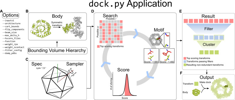
A visual outline of the software structure is provided in Fig 1. Users pass options into the dock.py application, which include required inputs such as Protein Data Bank (.pdb) files and the desired docking architecture, as well as other optional docking parameters described in detail in subsequent sections. A full list of command line options can be found in S1 Table and can be retrieved interactively using ‐‐help. The dock.py application interprets user-defined options and drives the machinery behind the docking algorithm. Input .pdb files are loaded using PyRosetta [34] as poses, then converted by the Body class into body objects. Various structural data are compiled from the input .pdb files, including transformable Bounding Volume Hierarchies (BVH) that index atomic coordinates. The Spec and Sampler classes define the DOFs of the target architecture and how they are to be broken down hierarchically. This space is traversed in the Search class, using a hierarchical search algorithm similar to branch-and-bound search [35]. During each iteration of the hierarchical search, each docked configuration, or transform, is evaluated by a residue-pair motif score [17] matched to the resolution of the search step, and then by a user-selected score function. Residue-pair motifs are identified by interacting pairs of backbone positions determined via the BVH data structures. Once the hierarchical search algorithm reaches its final resolution, the remaining docked configurations can be filtered with optional user-defined metrics. The filtered docked configurations are clustered based on redundancy among docked transforms and stored by the Result class as transformation matrices and scores in an xarray dataframe. The Result class can subsequently be used to re-apply a transformation matrix to the stored input pose, yielding a full-atom.pdb file.
Fig 1. General software structure of RPXDock.
A. User-defined inputs are given as options to the dock.py application. B. Within the application, input .pdb files are stored in the Body object as a PyRosetta pose. The Body class implements a Bounding Volume Hierarchy (BVH) for rapid operations on coordinates. C. The Spec and Sampler classes define the rigid-body DOFs the Body object is allowed to sample. D. Within the Search class, the Body object receives the DOFs as rigid body transforms (indicated as grid squares). Each transform is evaluated by the Motif and Score classes, which ranks the quality of residue-pair motifs at a given interface of a dock [17] and subsequently summarizes the residue-pair motif scores with additional interface quality metrics through a user-selected score function. The top scoring transforms are searched iteratively with higher resolution sampling and scoring in a hierarchical search algorithm. E. The final top scoring transforms from the search are fed into the Result class, which prunes the results using filter metrics and clusters the transforms based on backbone redundancy. F. The results are stored and output as transforms, which can be re-applied to the input Body object to generate a full-atom .pdb file of the resulting docked configuration.
Inputs and bodies
RPXDock uses the PyRosetta [34] pose module to load the atomic coordinates of input .pdb files and make secondary structure assignments via Define Secondary Structure of Proteins (DSSP) [36,37]. The PyRosetta pose is stored in the Body class as a Body object. Input .pdb files are provided to the dock.py application using the ‐‐inputs1 option. The input can be a path to a single .pdb (e.g., example.pdb) file, or a path with a wildcard (e.g., /path/to/files/*.pdb) can be supplied for multiple inputs. For multicomponent docking, additional inputs can be provided using the ‐‐inputs2 and ‐‐inputs3 option as necessary. For trajectories with multiple input lists provided to ‐‐inputs[n], each object in the list will be sampled against every other object in a partner list. The results for list inputs are batched and ranked together against one another. Thus, the “top” results may not include representatives from every input .pdb. If results from every input are desired, the user can either analyze the entire output list or execute each input or pair of inputs in separate RPXDock trajectories.
Bounding volume hierarchy (BVH)
The Body class implements a Bounding Volume Hierarchy (BVH) representation for efficient contact, sliding, clash checking, and determination of contacts for scoring [38]. As time taken for these operations scales with interface size, valuable compute time is saved by our implementation of BVH, which utilizes spheres rather than traditional bounding boxes for rotational invariance, allowing rigid body motions without recalculation. The BVH is first used to check for contacts, rapidly discarding configurations where bodies do not interact. During docking, clashing docks where the BVH intersect are removed. Lastly, BVH identifies all interacting pairs of residue stubs during scoring so that only interacting residues are evaluated. These operations are adjusted conservatively based on the resolution of the sampling, such that even large regions of the search space can be discarded as unlikely to contain favorable configurations.
Defining degrees of freedom (DOFs)
Sampling configurations of bodies is performed through a composable set of primitive samplers, including 1D, 2D, and 3D cartesian grids, 1D rotations, 2D directions, and 3D orientations. The space of orientations is modeled as the equivalent space of quaternions on a 3-sphere, and sampling is performed by subdividing the cells of a bitruncated 24-cell, a uniform 4D polytope that divides the 3-sphere uniformly into roughly cubic regions. This approach avoids the pitfalls of using Euler angles to represent 3D rotations. Streamlined combinations of these samplers are provided, such as rotation and translation on a symmetry axis, or a full 6D rigid body transformation, as well as a simple framework to create user-defined compositions and products of sampling spaces. All of these samplers and their combinations provide configurable resolutions, bounds, a hierarchy of nested sampling grids, and the ability to map indices between higher and lower resolutions.
Symmetric architectures
In symmetrical systems, the “architecture” defines the connectivity and allowed rigid-body kinematics, or movements, of the building blocks. RPXDock currently has built-in support for asymmetric, cyclic, stacking, dihedral, wallpaper (2D), and polyhedral group architectures. While the current release of dock.py does not support helical (1D) and crystal (3D) architectures, the components necessary for these protocols are available, and we plan to implement these in future builds of RPXDock. The desired architecture is specified per trajectory with the ‐‐architecture option using a keyword (Table 1).
Table 1. Keywords associated with each currently supported architecture.
| ‐‐architecture | Number of unique protein components supported | |
|---|---|---|
| Asymmetric | 2 | “ASYM” |
| Cyclic | 1 | “C[n]” where [n] = 1, 2, 3, …, n |
| Stacking | 2 | “AXLE_[n]” where [n] = 1, 2, 3, …, n, or “AXLE_1_[m]_[n]” where [m] and [n] correspond to the cyclic symmetries of the inputs and [n] ! = [m]. Currently supports up to [m] = 5 and [n] = 6. |
| Dihedral | 1 | “DX_X”, where X is the cyclic symmetry perpendicular to the dihedral plane and the oligomeric state of the input scaffold |
| 1 | “DX_2”, same as above, but the input oligomer is a dimer aligned to the dihedral plane | |
| Polyhedral group | 1 | “T2”, “T3”, “O2”, “O3”, “O4”, “I2”, “I3”, “I5” |
| 2 | “T32”, “T33”, “O32”, “O42”, “O43”, “I32”, “I52”, “I53” | |
| 3 | “T332”, “O432”, “I532” | |
| Wallpaper | 2, 3 | “P6_632”, “P6_63”, “P6_62”, “P6_32”, “P3_33”, “P4_42”, “P4_44” where “Px” describes the lattice symmetry and cyclic oligomer symmetries are listed after the underscore |
Input preparation
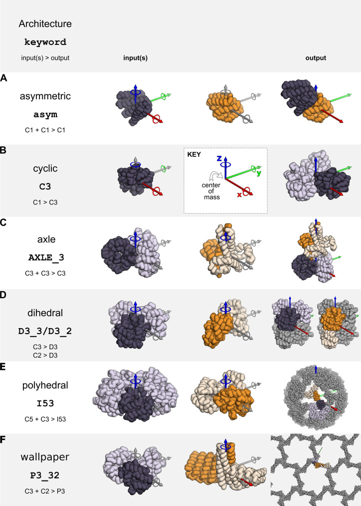
To dock two distinct monomers asymmetrically or to form cyclic oligomers, monomeric building blocks should have their center of mass at [0,0,0] (Fig 2A and 2B). RPXDock will not center the inputs by default, but the ‐‐recenter_input option can be passed to translate a monomeric building block such that its center of mass is at [0,0,0]. The final transform values reported are relative to the recentered pose, so it is recommended that inputs are pre-centered if the user plans to use these values.
Fig 2. Example inputs and docking output architectures currently supported by RPXDock.
X/Y/Z cartesian axes are shown in red, green, and blue respectively. Corresponding translational and rotational DOFs are sampled along and around these axes. Axes where DOFs are not sampled for an architecture are colored gray. A. Asymmetric docking samples 6 DOFs belonging to the first of two input monomers. B. Cyclic docking samples four DOFs belonging to an input monomer to generate a cyclic structure with its cyclic axis aligned to the Z axis. C-F. Oligomeric input structures must have their cyclic axis aligned to the Z axis and the input .pdb should only contain the asu (dark). Stacking, dihedral, polyhedral group, and wallpaper docking samples the rotational and translational DOF along the Z axis of the input cyclic oligomer, which is aligned during docking to the relevant rotational symmetry axes in the target architecture.
To form dihedral, stacking, wallpaper, and polyhedral group symmetries such as tetrahedral, octahedral, and icosahedral architectures, the input building blocks must be cyclic oligomers. The input .pdb files must be pre-aligned such that their internal rotational symmetry axes are aligned to the Z axis and the center of mass of the oligomer should be centered at [0,0,0] (Fig 2C and 2D). It is important to note that the input .pdb files should only contain the asymmetric unit (asu) of the cyclic oligomer rather than the full symmetric building block, as RPXDock will generate the symmetry-related chains. Currently, dihedral docking only supports one-component (i.e., homomeric) architectures; stacking supports two-component architectures; polyhedral group docking supports one-, two-, and three-component architectures; and wallpaper docking supports two- and three-component architectures.
Defining the search space
The search spaces for the supported architectures in RPXDock are either one-, two-, or three-body problems and the number of allowed DOFs sampled depends on the kinematics defined by the specified architecture. Two-body asymmetric docking technically allows all three rotational DOFs and all three translational DOFs (X, Y, and Z) per component, but in practice it is sufficient to hold one component static while sampling the other component against it (Fig 2A). Cyclic docking allows sampling of all three rotational DOFs but only one translational DOF (the radius), as sampling the remaining two cartesian DOFs results in identical final structures (Fig 2B). Each oligomeric component in stacking, dihedral, polyhedral group, wallpaper, and crystal architectures is aligned to a single rotational symmetry axis in the target architecture (the Z axis in the input .pdb) and is therefore limited to sampling one rotational and one translational DOF along that axis (Fig 2C–2F).
Each translational or rotational DOF is set by bounds in cartesian or angular space. Cartesian bounds can be set by ‐‐cart_bounds d1 d2 where the lower (d1) and upper (d2) bounds are distances in Ångstroms. The default values of d1 and d2 for symmetrical architectures are 0 and 500, limiting the search to only the positive direction of the space, as the reverse translational degrees of freedom are redundant when combined with the ‐‐flip_components option (see below). For asymmetrical docking scenarios, however, the default values are ‐500 and 500, allowing search in both directions. The larger this range is set, the longer the runtime and memory required. Thus, if the user has an idea of the desired search size, these values should be reduced as appropriate. Angular bounds are defined by the cyclic symmetry of the input component by default. For example, the angular bounds of a C3 input component are 0 and 120°. The final search space is defined by combining the DOF assignments and boundaries.
Restricting additional DOFs
For some docking problems, a user may want to restrict either one or all of the rotational or translational DOFs of their inputs during the search; for example, some docking problems require specific building blocks to be aligned to additional symmetry axes [29]. The rotational and/or translational DOFs can be turned off (‐‐fixed_rot,‐‐fixed_trans,‐‐fixed_components) or restricted to a user-defined range (‐‐fixed_wiggle). These are activated by listing which inputs should be fixed (0-delimited; e.g., ‐‐fixed_rot 1 to restrict the rotation DOF of all .pdb files provided in ‐‐inputs2, or ‐‐fixed_rot 0 1 to restrict the rotation DOF of all .pdb files provided in ‐‐inputs1 and ‐‐inputs2).
‐‐fixed_rot: fix the rotational DOF for desired input component
‐‐fixed_trans: fix the translational DOF for desired input component
‐‐fixed_components: fix both the translational and rotational DOFs for desired input component
‐‐fixed_wiggle: limit the translation and rotational DOFs to a certain range from the starting position. Additionally, specifications for the upper and lower bounds (ub and lb) of translation and rotation are required (‐‐fw_rot_lb,‐‐fw_rot_ub,‐‐fw_trans_lb,–-fw_trans_ub), where the upper and lower bounds are not equal.
The ‐‐flip_components option can be used to specify which cyclic components proceed to DOF sampling both before and after rotating the input .pdb 180° along the X axis (“flipping”). For example, a C3 oligomer can sample along the Z axis 0 to 120° and also 0 to 120° after flipping. This is effectively identical to sampling “negative” translations in dihedral, polyhedral group, and stacking architectures, and is required to fully search the available docking space in most symmetries. This option takes a list of boolean values corresponding to each input and defaults to true for all components (e.g., ‐‐flip_components 1 1 for a trajectory with two inputs).
Sampling the search space
RPXDock samples the defined search space via the modular sampler objects previously discussed and stores transformation matrices for each component of a set of sampled docked configurations. Each transform is applied to the respective input(s), resulting in a single docked configuration that was sampled during a docking trajectory, and is subsequently used to check for clashes and in some cases “flatness” at each iteration of the search. Clashing is evaluated by the BVH as described above. The “flatness” of a docked configuration is calculated during cyclic and multi-component docking (i.e., polyhedral group, stacking, wallpaper). During cyclic docking, flatness refers to the orientation of the longest physical axis of the input .pdb, as defined by principal component analysis, relative to the cyclic symmetry axis. The “flatness” of a cyclic dock can be constrained using the ‐‐max_longaxis_dot_z option, which restricts the orientation of the input .pdb relative to the cyclic symmetry axis (conventionally aligned along Z) by calculating the cosine between this axis and the longest axis of the input .pdb. Docks that exceed the cosine value given by the ‐‐max_longaxis_dot_z option are removed from the next stage of the search. This option can be set to any value between 0 and 1 (inclusive), where 1 allows the input .pdb to adopt any configuration relative to the cyclic symmetry axis, while 0 constrains the long axis of the input .pdb to perfect alignment, or perpendicularity, to the cyclic symmetry axis. During multi-component docking, flatness refers to differences in the translation of each component along its respective symmetry axis. In this case, the ‐‐max_delta_h option can be used to set an upper bound on the maximum allowable difference in offset between components.
Global and hierarchical search
In the asymmetric two-body docking problem, there are six DOFs: three translational and three rotational, where one body samples all six DOFs while the other remains static. As all six DOFs are sampled explicitly, the total number of transforms to evaluate equals the number of top-level samples (which is determined by the type and resolution of each DOF) multiplied by the number of subdivisions of that DOF raised to the 6th power. For example, if a typical top-level search space with six dimensions comprised of 10,000,000 total samples is used, sampling a single transform in a 16 Å space at a resolution of 16 Å for each dimension would result in 10,000,000 * 1^6 = 10,000,000 total transforms across the entire search space. Sampling at this 16 Å space at 1 Å resolution for each dimension (16 transforms per dimension) would result in 10,000,000 * 16^6 = 167 trillion total transforms to sample the entire search space. Enumerative sampling, even with some dimensionality reduction as implemented in previous iterations of “slide into contact” docking, is prohibitive at a reasonable resolution in architectures with a high number of DOFs [17,19,39,40].
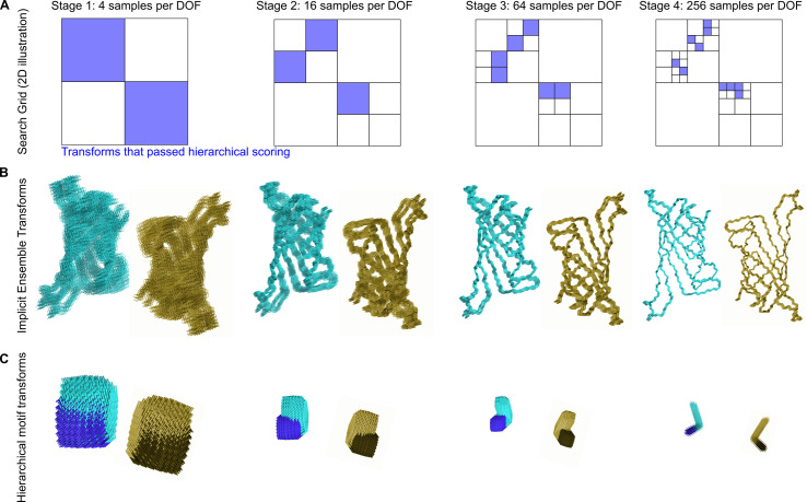
To enable efficient exploration of the search space in such architectures, we implemented an iterative hierarchical search that prunes away areas of the search space unlikely to contain good solutions [41,42]. In this sampling and evaluation scheme, the search begins at low resolution and is repeated with increasing resolution at each iteration. Only the top-scoring regions of the search space are kept for further exploration in the next iteration (Fig 3A and 3B). This reduces the number of samples that must be evaluated at each stage such that the total number of transforms evaluated no longer grows exponentially with dimension. For the simple 2D illustration in Fig 3A, the total number of samples is reduced from 256 to 24. Efficiency gains are roughly exponential with dimensionality and are thus much higher for less constrained docking problems. At each resolution, configurations are scored as an implicit ensemble (Fig 3B) through the use of residue-pair motifs (see “Score functions and motifs” section), tuned to provide an approximation of the best possible score within a corresponding ensemble of residue pair positions (Fig 3C). By evaluating the best possible score within an ensemble, as opposed to an average score, an entire region of docking space can be discarded without missing desirable docks.
Fig 3. Schematic representation of hierarchical sampling.
A. Schematic of a search grid for a single DOF keeping only the transforms that passed hierarchical scoring (blue) at each stage of search. This reduces the space searched at later stages where the search grid is subdivided at increasing resolution. B. A schematic depiction of protein backbones sampled with increasing resolution. The backbones shown would correspond to a single blue box at each stage of the search depicted in panel A; a cloud of such backbones would be sampled for each of the distinct docked configurations corresponding to each blue box. C. Residue-pair motifs are also evaluated at increasing resolution during each iteration of the search.
Due to the reduction in search space, the hierarchical search and scoring of a typical system with the default search space parameters takes approximately 30 seconds on a 4-core cpu. Further reductions or expansions in the number of transforms sampled at each stage of the hierarchical search protocol can be implemented using the ‐‐beam_size option, which defines the maximum number of sampled docks taken to the next stage of a hierarchical search protocol (default 100,000). The beam_size excludes the first, most coarse stage, which samples the entire search space at the lowest assigned resolution, as defined by ‐‐ori_resl (default 30°) and ‐‐cart_resl (default 10 Å).
To reduce low-resolution artifacts, we take the upper bound of scores within each grid square in the hierarchical search rather than its potentially low scoring and non-representative low resolution center (average). This effectively gives each grid square the “benefit of the doubt” during iteration, so that poor scoring regions can be discarded with confidence. We found during empirical testing that the hierarchical search approach did not over-prune substantial numbers of “good” candidates (S1 Fig). Specifically, we compared how efficiently the hierarchical sampling method recovered the top docks identified by enumerative grid sampling (~108 total asymmetric docks) (S1A Fig). The hierarchical search method recovered the top dock in this test set by searching less than 1/105 of the total search space and the top 10 docks by searching less than 1/102 (S1B Fig). To find the top 100 and top 1000 docks, the hierarchical method searched nearly the entire space, although it recovered 80% of the top 100 docks within 1/104 of the total search space. As identifying ~10 top docks per input or input pair is reasonable for most docking problems in practice, the reduction in search space and the consequent reduction in time that the hierarchical search method requires to find top-scoring docks is most likely an acceptable tradeoff.
While a major advantage of RPXDock is utilizing the hierarchical search method (‐‐docking_method hier), it is possible to globally search the conformation space (‐‐docking_method grid). This option may be appropriate for one-component dihedral or polyhedral docking problems that have two or fewer DOFs available. As the global search space is sampled at a single resolution, the user should specify different search resolutions in translational and rotational DOFs (‐‐grid_resolution_cart_angstroms and ‐‐grid_resolution_ori_degrees) across multiple independent trajectories. Nevertheless, grid search is implemented mainly for debugging purposes and is not recommended for production runs.
Specifying termini direction and accessibility
For polyhedral group architectures, the orientation and accessibility of the components’ termini can be important for downstream applications such as multivalent antigen display via genetic fusion [1,39,40,43,44]. Two options are implemented for polyhedral group architectures to (1) restrict the sampling space to docks with termini in the desired orientation (‐‐termini_dir[n]) and (2) evaluate the accessibility of the termini residues (‐‐term_access[n]). The ‐‐termini_dir[n] and ‐‐term_access[n] options mirror the syntax of the ‐‐inputs[n] options, where ‐‐termini_dir1 and ‐‐term_access1 refer to the termini direction and accessibility for ‐‐inputs1, ‐‐termini_dir2 and ‐‐term_access2 for ‐‐inputs2, etc. Both options operate by aligning an ideal 21-residue alpha helix to 7 residues at the user-specified termini.
The ‐‐termini_dir[n] option evaluates the helix orientation by calculating the Z direction of the vector defined by the center of mass of the first and last three residues of the aligned ideal helix (e.g. residues 1–3 to 19–21 for N termini, and inversely for C termini). The option then picks the desired orientation from the required ‐‐flip_components option and disables sampling of the other. The aligned ideal helix is removed before sampling docked transforms.
The ‐‐term_access[n] option evaluates the accessibility of user-defined termini during sampling by adding the aligned ideal helix to the Body class for the BVH to use for clash checking at each step of the search. The aligned helix is omitted in the Score and Result classes for RPX scoring and output. The option syntax is as follows:
‐‐termini_dir1 [‐‐termini_dir2,‐‐termini_dir3]: Accepts a desired orientation as “in”, “out”, or “None”, for the amino terminus followed by the carboxyl terminus (space-delimited) for each corresponding input (‐‐inputs1 for ‐‐termini_dir1,‐‐inputs2 for ‐‐termini_dir2, etc.). “In” restricts sampling to configurations in which the specified terminus points towards the architecture’s center of mass, while “out” restricts sampling to the opposite. This option alternatively accepts a space-delimited pair of booleans where “in” is True, and “out” is False. The option(s) default to “None”. The ‐‐flip_components option must be set to True.
‐‐term_access1 [‐‐term_access2,‐‐term_access3]: Accepts a space-delimited pair of boolean values to enable evaluation of terminus accessibility at the amino and carboxyl termini of each component, respectively. (e.g. ‐‐term_access1 0 1 evaluates accessibility of the carboxyl termini for input .pdb files passed through ‐‐inputs1)
Evaluating docked configurations
Residue-Pair Transform (RPX) Scoring
We employ a 6D implicit side-chain methodology when evaluating residue-pair interactions in a sequence-independent manner. The interaction between two residues is represented by the full 6D rigid-body transformation between their respective backbone N, Cα, and C atoms [17]. Transforms are binned into six dimensional body-centered cubic lattices, with three dimensions each for translation and rotation. The curved space of rotations is divided into 24 relatively flat cells, with one lattice in each cell. A pre-compiled residue-pair transform, or hscore, database of all residue-pair interactions for each amino acid found in structures from the Protein Data Bank (PDB) was binned based on this method and scored using the Rosetta full-atom energy function [45]. During docking, pairs of residues across a docked interface are assigned an RPX score, which is the lowest pre-calculated Rosetta full-atom energy found in the relevant spatial transformation bin of the database. The top-scoring residue pair scores across the interface are evaluated based on a user-defined RPXDock score function (see Ranking dock quality (score functions)). This score was previously found to be more predictive of the interface energy from full-atom sequence-design calculation than the Rosetta centroid energy function or other “coarse-grained” scoring models [17].
Motif-enriched docking
A user may want to diversify or restrict the motifs and secondary structure elements used to score RPXDocked configurations. This can be done using the ‐‐hscore_files and ‐‐hscore_data_dir options. The path suffix in ‐‐hscore_files will be appended to ‐‐hscore_data_dir, which is the default path to search for hscore files. These hscore files can be read in as a tarball zipped .txz format that is slow to load but Python version-agnostic, or in a .pickle format that is fast but Python version-dependent. The ‐‐generate_hscore_pickle_files option can be passed to generate .pickle versions from the .txz file, which can then simply be moved to the corresponding hscore folder before use. Each category of hscore files contains scores for a subset of the full residue-pair motif database, restricted to certain amino acid identities and secondary structure elements. By restricting the database, transforms with no motifs found among the chosen subset result in a score of zero, and are thrown out when proceeding to the next phase of the search, thus biasing against the unselected amino acids and secondary structures (H α-helices, E β-sheets, and L loops). Note that RPXDock is sequence-agnostic, meaning the residue identities of the input .pdb are ignored when placing motif pairs. The default motif set only includes pairs involving isoleucine, leucine, and valine; and only in α-helices. The following hscore files are pre-compiled and provided in the Institute for Protein Design public repository at https://files.ipd.uw.edu/pub/rpxdock/hscores.zip:
ILV_H (default; isoleucine, leucine, valine; helices only)
AILV_H (alanine, isoleucine, leucine, valine; helices only)
AFILMV_EHL (all hydrophobic amino acids; all secondary structures: sheets, helices, and loops)
Restricting regions for scoring
Score only SS. Scoring can be restricted to only certain secondary structure elements using the ‐‐score_only_ss option (any non-delimited combination of ‘EHL’ for sheets, helices, and loops). When active, only residue pairs where at least one of the two motif pairs reside on the desired secondary structure types will be scored. To additionally restrict such that both motif pairs must reside on the designated secondary structure types, the ‐‐score_only_sspair option can be used. Conceptually this results in a similar effect as providing hscore files for only the desired secondary structure types and will enrich for these motifs. Users should note that these restrictions do not explicitly remove or penalize contacts, which contribute to the docking score independently of motifs, at positions on non-desired secondary structure elements.
Masking (Allowed residues). To bias the search towards generating interfaces focused on a specific region of the input structure(s), a list of residue positions can be provided using the ‐‐allowed_residues[n] option. Specifying positions in this way does not prevent other regions of the protein from forming contacts, nor does it affect clash checking. Instead, regions of the protein structure not included as allowed residues simply do not contribute to the score of the docked configuration, thus biasing the search. The ‐‐allowed_residues[n] option mirrors the syntax of the ‐‐inputs[n] options, where ‐‐allowed_residues1 refers to the list of allowed residues for ‐‐inputs1,‐‐allowed_residues2 for ‐‐inputs2, and so forth. The ‐‐allowed_residues[n] options can either be left blank (default), take a single file which applies to all corresponding component inputs, or take a list of files which must have the same length as the list of inputs. The files themselves must contain a whitespace- and/or newline-delimited list of either numbers and/or ranges using Python syntax. For example, a three-lined file:
1 2 3 4 5
7:12
80:-1
will result in specifying residues 1 2 3 4 5 (first line), 7 8 9 10 11 12 (second line), and 80 through the last residue (third line) as “allowed” for all of the corresponding list of inputs. Residue numbering starts from one and numeric gaps in the input .pdb files are ignored and renumbered sequentially. Multi-chain inputs will be concatenated into a single chain by default. It is recommended that users sanitize input .pdb files to these standards prior to using RPXDock to prevent unexpected results.
Ranking dock quality (score functions)
RPXDock evaluates dock quality with a score function that summarizes the number of contacting residue-pairs at an interface (“contacts”) and the RPX score, derived from motif pairs as described above. The RPX score is evaluated for each pair of residues in the docked interface within a maximum distance of each other, as defined by the ‐‐max_pair_dist option (default 8.0 Å at the highest search resolution), which scales with the resolution during the hierarchical search. Afterwards, all the relevant RPX scores are combined according to the score function definition, controlled by the ‐‐function option. The default score function (stnd) is defined as:
where a and b are coefficients set by ‐‐weight_rpx and ‐‐weight_ncontact (default 1 and 0.001, respectively), RPX is the sum of the maximum RPX scores for each pair of contacting residues (i) in the interface (), and ncontact is the number of pairwise contacts in the interface. In this standard score function, RPX is highly covariate with ncontact, and thus it is also highly correlated with the total score. As a result, because RPXDock seeks to maximize the score, the standard algorithm will tend to find the largest possible interfaces.
SASA weighted (sasa_priority) score function. It is likely that there is an optimum interface size for each docking architecture and the subtypes within them, due to the apparent relationship between interface size and interface strength of symmetrical assemblies, the latter of which can be a critical determinant of the fidelity of the assembly process [46,47]. Therefore, the user may wish to bias docked conformations toward a particular interface size. This can be achieved by taking advantage of the correlation between ncontact and interface size, as measured by buried solvent accessible surface area (SASA) [48] (S2 Fig). The sasa_priority score function seeks to find the best docked configuration for a target interface size as measured by the average motif quality XRPX across all residue-pair combinations. For each residue pair, the maximum motif score is considered in this average. Thus, the sasa_priority score function is defined as:
where a is set by ‐‐weight_rpx (default 1) and b by ‐‐weight_ncontact. Note that while the default value of ‐‐weight_ncontact in the standard score function is 0.001, a value of 5 is recommended for the sasa_priority score function. N is the number of contacting residues in the interface, scored based on a log normal distribution with a mean, μ, set by ‐‐weight_sasa (default 1152 Å2) and a tolerance level, σ, set by ‐‐weight_error (default 4). The resultant top-scoring configurations are biased towards the mean (Fig 4A), such that the buried SASA of the top docks at or very close to the weight_sasa, should such docks exist (Fig 4B). An artifact of this score function is that at higher target interface sizes, a set of high-scoring docks with small SASA estimate values emerge as a result of very small interfaces with high average RPX score; these outliers can be removed by the filter_sasa (see Additional Optional Filters below).
Fig 4. Interface size bias by the sasa_priority score function.
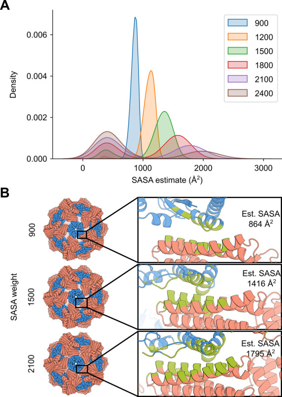
A. 572 pairs of inputs were docked in a two-component icosahedral architecture at a–-weight_sasa value of 900, 1200, 1500, 1800, 2100, and 2400, with total area under each curve normalized to 1. B. The interface of the top-scoring docked configuration for ‐‐weight_sasa value of 900, 1500, and 2100 is highlighted (green). Estimated buried SASA calculated using Rosetta for these docks are 864, 1416, and 1795 Å2, respectively.
The ‐‐weight_sasa parameter may need to be modified depending on the docking problem. For example, cyclic docking might require a different ‐‐weight_sasa parameter than one- or two-component polyhedral group docking. The optimal ‐‐weight_sasa may be determined empirically for each architecture or docking problem by comparing independent docking trajectories and visually inspecting the results. Note that if the value is set to improbably high values (e.g., 99999), the search will fail rather than finding the largest interface, as docks near that SASA value do not exist. Note that this score function was fit using two-component polyhedral group architectures, so other architectures may need additional optimization of the variables. The development and optimization of this score function is described further in the S1 Text.
Other score functions. Additional variants of the standard score function are available, by replacing the sum of the maximum RPX scores at each residue pair considered in the stnd score function with the mean or median (Table 2). These two score functions partially remove the correlation between ncontact and total RPX score. Finally, two more functions were used in development of the sasa_priority score function that empirically estimated the relationship between RPX and ncontact with either a linear or exponential fit.
Table 2. List of additional score functions.
| ‐‐function | Description |
|---|---|
| stnd | score = a * RPX + b * ncontact, where RPX is the sum of the max(motif score) across all residue pairs in a docked interface |
| sasa_priority | Function developed to bias interfaces to a certain size given user requirements. The ‐‐weight_sasa (default = 1152), ‐‐weight_ncontact (default = 0.01 but a value of 5 provides optimum scaling for this score function), and ‐‐weight_error (default = 4) flags must also be specified. |
| mean | Takes the mean of max(motif score), instead of sum() in the standard score function |
| median | Takes the median of max(motif score), instead of sum() in the standard score function |
| exp | scores = RPX‐4.6679 * ncontact0.588 |
| lin | scores = RPX‐0.7514 * ncontact |
Filtering docks
Clustering
After docked configurations are scored, the results are clustered through redundancy filters. Redundancy checking is performed by the filter_redundancy() function, which performs a distance check on the transformed bodies (approximating an unaligned RMSD calculation) and discards similar transforms with distances below a user-defined cutoff set by the ‐‐max_bb_redundancy option (default, 3 Å). Cluster size can be controlled by the ‐‐max_cluster option (default, no limit), which specifies the maximum number of clusters the docked transforms can be sorted into. As docks are sorted by score, only the highest-scoring dock from each cluster is kept. The redundancy filter returns an array of indices corresponding to the docked configurations that pass this filter.
Additional optional filters
We have developed a set of modular filters that can be applied post-docking to remove docks that do not meet certain requirements or to provide more information about the results. Currently available filters are:
filter_sscount: Removes docks below a specified number of secondary structure (SS) elements in the docked interface
filter_sasa: Removes docks outside a specified interface SASA using a similar method to the sasa_priority score function
New filters can be added without having to modify code in the search or scoring modules. At the time of publication, filtering is possible for architectures of the cyclic, dihedral, stacking, wallpaper, and polyhedral groups.
Filter behavior is controlled by a .yaml configuration file passed through the ‐‐filter_config option. This allows facile stacking of an arbitrary number of filters, including multiple instances of the same filter configured in different ways. Filters are defined with a key, or filter label, that can be any arbitrary string without whitespace. All filters have standard and filter-specific parameters. The standard parameters are a “type” parameter and a “confidence” parameter. The “type” parameter must exactly match the name of the filter in the RPXDock main code. The “confidence” parameter is a boolean that controls whether or not the filter will remove docks from the result. If confidence is False, the result will report values for all docks, including those that would have been removed had the confidence been set to True. Note that if confidence is True, a filter can potentially remove all of the results if none of them meet the thresholds, resulting in an empty result object. S2 and S3 Tables provides a list of all available filter-specific parameters.
Result
After RPXDock has been executed, the result class outputs a zipped tarball .txz file and a .pickle file that stores i) the initial body object along with ii) the transforms and iii) associated score and filter values of each docked configuration in a concatenated xarray format. While the .pickle file is faster to access, it is Python version-dependent, so the .txz format is also returned as a version-agnostic output. Each of these output formats can be turned on or off using their respective options: ‐‐save_results_as_tarball and ‐‐save_results_as_pickle, which both default to True. With the the dump_pdb() function, the result object can output the resulting dock in the form of a .pdb file for any given model number, corresponding to the rank of the desired docked configuration by score. We have included an example Python script in the GitHub repository under tools/dump_pdb_from_output.py that demonstrates how to access score and filter information and regenerate docked configurations as .pdb files for any desired dock configuration from either file format. The ‐‐overwrite_existing_results option, which defaults to False, can be passed to overwrite existing outputs for file management purposes.
The top-scoring transforms can be directly output in .pdb format using the ‐‐dump_pdbs option. When used in combination with ‐‐nout_top N, which defaults to 10, the top-scoring N transforms can be output in .pdb format from the RPXDock result object. The user may be interested in saving disk space or for other reasons only saving the asymmetric unit (asu) of the resulting dock; this behavior can be set with the option ‐‐output_asym_only. The ‐‐output_closest_subunits option can be used in combination, which outputs a .pdb file containing the asu chains in positions that exhibit the highest motif contact count from the symmetric result (eg. the asu chains that are closest to each other in space) instead of the default asu chains in positions defined by each symmetry. This can be useful for visualization and for generating inputs for downstream steps in design pipelines.
Results
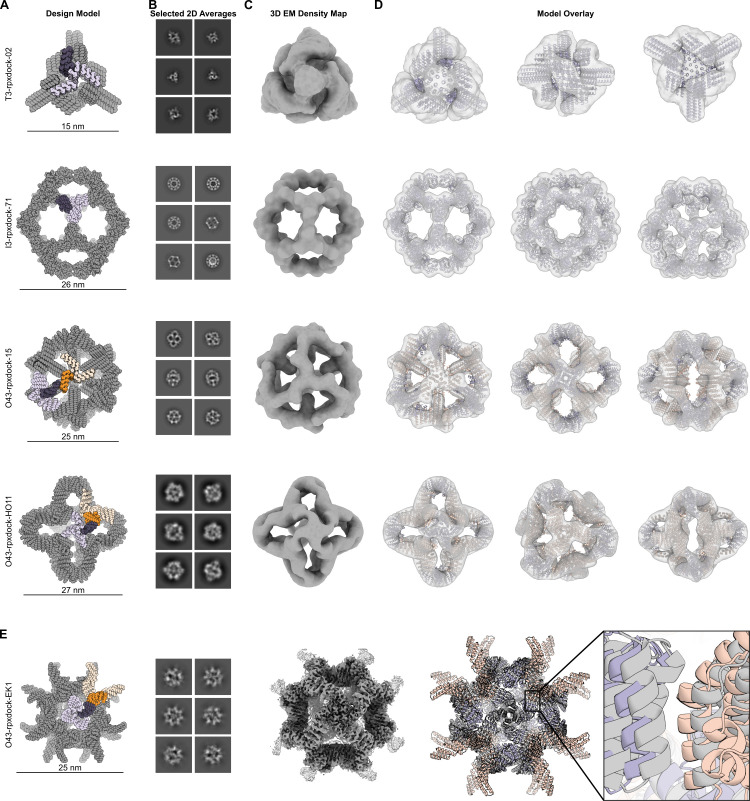
We set out to experimentally evaluate symmetric one- and two-component structures with polyhedral group symmetry generated using RPXDock. Given a set of prevalidated homomeric scaffolds with cyclic symmetry, we generated docks using RPXDock, and the resulting interfaces were sequence-optimized via Rosetta sequence design [49]. Two one-component designs (T3-rpxdock-02, I3-rpxdock-71) and two two-component designs (O43-rpxdock-15, O43-rpxdock-HO11) with tetrahedral, octahedral, or icosahedral symmetry were examined by negative-stain electron microscopy and found to adopt the intended architecture (Figs 5A–5D and S5A). I3-rpxdock-71, while completely independently sampled and designed, resembles a dock previously sampled by RPXDock’s predecessor, tcdock, indicating that the similar top results are identified by the new search algorithm [3]. We obtained a 3.7 Å resolution single-particle reconstruction of the two-component octahedral assembly O43-rpxdock-EK1 (PDB: 8FWD, EMD-29502) using cryogenic electron microscopy and found that it assembles to the intended structure with high accuracy (4 Å Cα root mean square deviation between all 48 chains of the original dock and cryoEM structure, Figs 5E and S5B-S5F and S5 Table). Together, these data confirm that docks generated using RPXDock can be designed to assemble in the intended configurations without disrupting the integrity of the starting scaffolds. Input .pdb files, docking and design scripts, and design models are provided in the tools/ directory available on the RPXDock GitHub page at https://github.com/willsheffler/rpxdock.
Fig 5. Docking and characterization of one- and two-component polyhedral assemblies using RPXDock.
A. Models of one- and two-component docked polyhedral assemblies with the oligomeric building blocks in purple and orange. The asymmetric unit of each assembly, comprising one subunit of each building block, is colored dark purple and dark orange. B. Reference-free 2D class averages from negative stain electron microscopy. Each assembly is viewed along several axes of symmetry. C. 3D density maps reconstructed from selected 2D class averages. D. Overlays of each design model fit into its 3D density map, confirming that each design assembles to the architecture identified by RPXDock. E. Characterization of the two-component octahedral assembly O43-rpxdock-EK1 by cryogenic electron microscopy. The design model is colored as in A). To the right are representative 2D class averages showing different axes of symmetry and a reconstructed 3D map at 3.7 Å resolution. The overlay of the original dock (orange and purple) with the model built from the 3D reconstruction (gray) shows 4 Å Cα root mean square deviation between the original dock and cryoEM structure over 48 chains.
Availability and Future Directions
Setup and installation
At the time of publication, RPXDock has been verified to compile and function correctly on Linux-based operating systems. To set it up, a user must first clone the public repository of the full source code, which can be found at https://github.com/willsheffler/rpxdock, and set up a proper conda environment using the environment.yml file. Note that a user must obtain a pyrosetta license (free for non-profit users) and update the username and password fields for their pyrosetta license in the environment.yml file before creating the environment. Users may need to also install additional packages in their conda environment such as pyyaml to properly build the application. To build and compile the codebase with the newly created conda environment, a user may simply run the pytest command using a gcc>9-compatible compiler.
To verify that the code compiled properly, execute rpxdock/app/dock.py ‐‐help in the new conda environment. The output should provide a list of options that are relevant for docking (S1 Table). Note that several options are still experimental in nature and therefore are not described fully in this publication. For a template of how to set up a simple RPXDock run, please refer to the available example provided in tools/dock.sh in the GitHub repository.
Discussion
RPXdock provides a powerful and general route to modeling, sampling, and scoring symmetric protein complexes across multiple symmetric architectures. Docking monomeric and oligomeric building blocks into higher-order symmetric complexes followed by protein-protein interface design is an established and successful paradigm for accurately creating novel self-assembling protein nanomaterials [2,4,17,19–22]. While deep learning-based generative models have recently proven successful in designing de novo oligomers [50] and small nanocages [51], the ability of RPXdock to use experimentally verified or previously designed scaffolds in a stepwise manner enables the use of specific building blocks that have optimal features for specific applications [1,39,40,43,44]. The RPXdock code can accommodate specific user requirements for complex docking problems, and the efficiency at which high-quality docks are found has been greatly improved compared to its predecessors (tcdock; sicdock; sicaxel [17,19,39,40]) due to the hierarchical search and scoring algorithms. While new capabilities are continuously under development, the core software structure is complete and robust, and has already been successfully applied to a number of symmetric docking and design problems in addition to the structures presented here ([29–31]. Any future modifications and new modules added to the RPXDock application will be updated via the GitHub repository: https://github.com/willsheffler/rpxdock.
Supporting information
A. A 2-dimensional illustration of a hierarchical search grid with samples searched at the highest resolution in blue vs. a complete search grid at the same resolution. In this test dataset, ~108 total docks were sampled. B. A cumulative distribution of the fraction of the total search space that needs to be sampled in order to recover the top 1, 10, 100, and 1000 docks from this dataset.
(TIF)
As such, we parameterized an ncontact score term with respect to computationally measured interface size, SASA.
(TIF)
(TIF)
A. Score as a function of ncontact across various ncontact weights. B. Mean RPX as a function of ncontact. C. Mean RPX as a function of ncontact weighting plotted for interface sizes from Number of unique contacts = 5–55. D. Total number of passing designs out of 960 docks for each weighting and fraction. E-F. Computational design metrics as a function of ncontact weight for top-, middle-, and bottom-ranked designs for E. ddG, and F. SASA. G. The top dock with I32 icosahedral symmetry for, left to right, ncontact weight 1, 3, 5, 7, 9.
(TIF)
A. Representative raw nsEM micrographs of one- and two-component polyhedral self-assembling proteins from RPXDock. Scale bar = 100 nm B. Representative raw CryoEM micrograph showing good particle distribution and contrast of (Scale Bar = 100 nm). C. CryoEM local resolution map of O43-rpxdock-EK1, with the sharpened map at two different contour levels, using a tight mask, and calculated using an FSC value of 0.143. D. Local resolution estimates of the unsharpened map, also at two different contour levels (FSC = 0.143). The protruding arms of the designed cage only start to become visible at very low contour levels. Local resolution estimates range from ~3.2 Å at the core to >4.0 Å along the periphery of the extended arms due to a high degree of flexibility within this region. E. Global resolution estimation plot. F. Orientational distribution plot demonstrating near-complete angular sampling.
(TIF)
(XLSX)
(XLSX)
(XLSX)
(XLSX)
(XLSX)
(DOCX)
(DOCX)
Acknowledgments
We thank Lance Stewart, Christian Richardson, Derrick Hicks, Stacey R. Gerben, Ryan Kibler, George Ueda, and Jorge Fallas for helpful discussions, Shingo Honda for providing feedback on the manuscript, and Luki Goldschmidt and Patrick Vecchiato for computational resource management.
Data Availability
All data are available in the main text or as supplementary materials. Scripts, computational methods, and design models are available on GitHub at https://github.com/willsheffler/rpxdock. For O43-rpxdock-EK1, coordinates are deposited in the Protein Data Bank with the accession code 8FWD; the cryo-EM density map is deposited in the Electron Microscopy Data Bank (EMDB) with the accession code EMD-29502.
Funding Statement
Funding for this work was provided by the Audacious Project at the Institute for Protein Design (N.P.K. and D.B.), The Open Philanthropy Project for Improving Protein Design Fund (D.B.), NSF DGE-1762114 (E.C.Y.), the Bill & Melinda Gates Foundation grant #INV-010680 (N.P.K. and D.B.), a Rosetta Commons Post-Baccalaureate Fellowship (J.S.), National Science Foundation grant CHE-1629214 (N.P.K. and D.B.), grant DE-SC0018940 MOD03 from the U.S. Department of Energy Office of Science (A.J.B., D.B.), the National Institutes of Health grants 1P01AI167966 (N.P.K.) and P50AI150464 (N.P.K.), and the Howard Hughes Medical Institute (D.B.). The funders had no role in study design, data collection and analysis, decision to publish, or preparation of the manuscript.
References
- 1.Ueda G, Antanasijevic A, Fallas JA, Sheffler W, Copps J, Ellis D, et al. Tailored design of protein nanoparticle scaffolds for multivalent presentation of viral glycoprotein antigens. Elife. 2020;9. doi: 10.7554/eLife.57659 [DOI] [PMC free article] [PubMed] [Google Scholar]
- 2.King NP, Sheffler W, Sawaya MR, Vollmar BS, Sumida JP, André I, et al. Computational design of self-assembling protein nanomaterials with atomic level accuracy. Science. 2012;336: 1171–1174. doi: 10.1126/science.1219364 [DOI] [PMC free article] [PubMed] [Google Scholar]
- 3.Hsia Y, Bale JB, Gonen S, Shi D, Sheffler W, Fong KK, et al. Design of a hyperstable 60-subunit protein icosahedron. Nature. 2016. doi: 10.1038/nature18010 [DOI] [PubMed] [Google Scholar]
- 4.Bale JB, Gonen S, Liu Y, Sheffler W, Ellis D, Thomas C, et al. Accurate design of megadalton-scale two-component icosahedral protein complexes. Science. 2016;353: 389–394. doi: 10.1126/science.aaf8818 [DOI] [PMC free article] [PubMed] [Google Scholar]
- 5.Woolfson DN. The design of coiled-coil structures and assemblies. Adv Protein Chem. 2005;70: 79–112. doi: 10.1016/S0065-3233(05)70004-8 [DOI] [PubMed] [Google Scholar]
- 6.Laniado J, Meador K, Yeates TO. A fragment-based protein interface design algorithm for symmetric assemblies. Protein Eng Des Sel. 2021;34: 1–34. doi: 10.1093/protein/gzab008 [DOI] [PMC free article] [PubMed] [Google Scholar]
- 7.Lai Y-T, King NP, Yeates TO. Principles for designing ordered protein assemblies. Trends Cell Biol. 2012;22: 653–661. doi: 10.1016/j.tcb.2012.08.004 [DOI] [PubMed] [Google Scholar]
- 8.Padilla JE, Colovos C, Yeates TO. Nanohedra: using symmetry to design self assembling protein cages, layers, crystals, and filaments. Proc Natl Acad Sci U S A. 2001;98: 2217–2221. doi: 10.1073/pnas.041614998 [DOI] [PMC free article] [PubMed] [Google Scholar]
- 9.Golub E, Subramanian RH, Esselborn J, Alberstein RG, Bailey JB, Chiong JA, et al. Constructing protein polyhedra via orthogonal chemical interactions. Nature. 2020;578: 172–176. doi: 10.1038/s41586-019-1928-2 [DOI] [PMC free article] [PubMed] [Google Scholar]
- 10.Kakkis A, Gagnon D, Esselborn J, Britt RD, Tezcan FA. Metal-Templated Design of Chemically Switchable Protein Assemblies with High-Affinity Coordination Sites. Angew Chem Int Ed Engl. 2020;59: 21940–21944. doi: 10.1002/anie.202009226 [DOI] [PMC free article] [PubMed] [Google Scholar]
- 11.Lin Y-R, Koga N, Vorobiev SM, Baker D. Cyclic oligomer design with de novo αβ-proteins. Protein Sci. 2017;26: 2187–2194. [DOI] [PMC free article] [PubMed] [Google Scholar]
- 12.Grigoryan G, Degrado WF. Probing designability via a generalized model of helical bundle geometry. J Mol Biol. 2011;405: 1079–1100. doi: 10.1016/j.jmb.2010.08.058 [DOI] [PMC free article] [PubMed] [Google Scholar]
- 13.Rhys GG, Wood CW, Beesley JL, Zaccai NR, Burton AJ, Brady RL, et al. Navigating the Structural Landscape of De Novo α-Helical Bundles. J Am Chem Soc. 2019;141: 8787–8797. [DOI] [PubMed] [Google Scholar]
- 14.Huang P-S, Oberdorfer G, Xu C, Pei XY, Nannenga BL, Rogers JM, et al. High thermodynamic stability of parametrically designed helical bundles. Science. 2014;346: 481–485. doi: 10.1126/science.1257481 [DOI] [PMC free article] [PubMed] [Google Scholar]
- 15.Hsia Y, Mout R, Sheffler W, Edman NI, Vulovic I, Park Y-J, et al. Design of multi-scale protein complexes by hierarchical building block fusion. Nat Commun. 2021;12: 2294. doi: 10.1038/s41467-021-22276-z [DOI] [PMC free article] [PubMed] [Google Scholar]
- 16.Divine R, Dang HV, Ueda G, Fallas JA, Vulovic I, Sheffler W, et al. Designed proteins assemble antibodies into modular nanocages. Science. 2021;372. doi: 10.1126/science.abd9994 [DOI] [PMC free article] [PubMed] [Google Scholar]
- 17.Fallas JA, Ueda G, Sheffler W, Nguyen V, McNamara DE, Sankaran B, et al. Computational design of self-assembling cyclic protein homo-oligomers. Nat Chem. 2017;9: 353–360. doi: 10.1038/nchem.2673 [DOI] [PMC free article] [PubMed] [Google Scholar]
- 18.Sahasrabuddhe A, Hsia Y, Busch F, Sheffler W, King NP, Baker D, et al. Confirmation of intersubunit connectivity and topology of designed protein complexes by native MS. Proc Natl Acad Sci U S A. 2018;115: 1268–1273. doi: 10.1073/pnas.1713646115 [DOI] [PMC free article] [PubMed] [Google Scholar]
- 19.King NP, Bale JB, Sheffler W, McNamara DE, Gonen S, Gonen T, et al. Accurate design of co-assembling multi-component protein nanomaterials. Nature. 2014;510: 103–108. doi: 10.1038/nature13404 [DOI] [PMC free article] [PubMed] [Google Scholar]
- 20.Shen H, Fallas JA, Lynch E, Sheffler W, Parry B, Jannetty N, et al. De novo design of self-assembling helical protein filaments. Science. 2018;362: 705–709. doi: 10.1126/science.aau3775 [DOI] [PMC free article] [PubMed] [Google Scholar]
- 21.Gonen S, DiMaio F, Gonen T, Baker D. Design of ordered two-dimensional arrays mediated by noncovalent protein-protein interfaces. Science. 2015;348: 1365–1368. doi: 10.1126/science.aaa9897 [DOI] [PubMed] [Google Scholar]
- 22.Ben-Sasson AJ, Watson JL, Sheffler W, Johnson MC, Bittleston A, Somasundaram L, et al. Design of biologically active binary protein 2D materials. Nature. 2021;589: 468–473. doi: 10.1038/s41586-020-03120-8 [DOI] [PMC free article] [PubMed] [Google Scholar]
- 23.Yan Y, Tao H, Huang S-Y. HSYMDOCK: a docking web server for predicting the structure of protein homo-oligomers with Cn or Dn symmetry. Nucleic Acids Res. 2018;46: W423–W431. doi: 10.1093/nar/gky398 [DOI] [PMC free article] [PubMed] [Google Scholar]
- 24.Schneidman-Duhovny D, Inbar Y, Nussinov R, Wolfson HJ. PatchDock and SymmDock: servers for rigid and symmetric docking. Nucleic Acids Res. 2005;33: W363–7. doi: 10.1093/nar/gki481 [DOI] [PMC free article] [PubMed] [Google Scholar]
- 25.Park T, Baek M, Lee H, Seok C. GalaxyTongDock: Symmetric and asymmetric ab initio protein-protein docking web server with improved energy parameters. J Comput Chem. 2019;40: 2413–2417. doi: 10.1002/jcc.25874 [DOI] [PubMed] [Google Scholar]
- 26.Lyskov S, Gray JJ. The RosettaDock server for local protein-protein docking. Nucleic Acids Res. 2008;36: W233–8. doi: 10.1093/nar/gkn216 [DOI] [PMC free article] [PubMed] [Google Scholar]
- 27.Chauhan VM, Pantazes RJ. MutDock: A computational docking approach for fixed-backbone protein scaffold design. Front Mol Biosci. 2022;9: 933400. doi: 10.3389/fmolb.2022.933400 [DOI] [PMC free article] [PubMed] [Google Scholar]
- 28.Padhorny D, Kazennov A, Zerbe BS, Porter KA, Xia B, Mottarella SE, et al. Protein-protein docking by fast generalized Fourier transforms on 5D rotational manifolds. Proc Natl Acad Sci U S A. 2016;113: E4286–93. doi: 10.1073/pnas.1603929113 [DOI] [PMC free article] [PubMed] [Google Scholar]
- 29.Li Z, Wang S, Nattermann U, Bera AK, Borst AJ, Bick MJ, et al. Accurate Computational Design of 3D Protein Crystals. bioRxiv. 2022. p. 2022.11.18.517014. doi: 10.1101/2022.11.18.517014 [DOI] [Google Scholar]
- 30.Gerben SR, Borst AJ, Hicks DR, Moczygemba I, Feldman D, Coventry B, et al. Design of Diverse Asymmetric Pockets in De Novo Homo-oligomeric Proteins. Biochemistry. 2023. doi: 10.1021/acs.biochem.2c00497 [DOI] [PMC free article] [PubMed] [Google Scholar]
- 31.Wang JY (john), Khmelinskaia A, Sheffler W, Miranda MC, Antanasijevic A, Borst AJ, et al. Improving the secretion of designed protein assemblies through negative design of cryptic transmembrane domains. bioRxiv. 2022. p. 2022.08.04.502842. doi: 10.1101/2022.08.04.502842 [DOI] [PMC free article] [PubMed] [Google Scholar]
- 32.Leaver-Fay A, Tyka M, Lewis SM, Lange OF, Thompson J, Jacak R, et al. ROSETTA3: an object-oriented software suite for the simulation and design of macromolecules. Methods Enzymol. 2011;487: 545–574. doi: 10.1016/B978-0-12-381270-4.00019-6 [DOI] [PMC free article] [PubMed] [Google Scholar]
- 33.Dauparas J, Anishchenko I, Bennett N, Bai H, Ragotte RJ, Milles LF, et al. Robust deep learning-based protein sequence design using ProteinMPNN. Science. 2022; eadd2187. doi: 10.1126/science.add2187 [DOI] [PMC free article] [PubMed] [Google Scholar]
- 34.Chaudhury S, Lyskov S, Gray JJ. PyRosetta: a script-based interface for implementing molecular modeling algorithms using Rosetta. Bioinformatics. 2010;26: 689–691. doi: 10.1093/bioinformatics/btq007 [DOI] [PMC free article] [PubMed] [Google Scholar]
- 35.Larsson T, Akenine-Möller T. A dynamic bounding volume hierarchy for generalized collision detection. Comput Graph. 2006;30: 450–459. [Google Scholar]
- 36.Touw WG, Baakman C, Black J, te Beek TAH, Krieger E, Joosten RP, et al. A series of PDB-related databanks for everyday needs. Nucleic Acids Research. 2015. pp. D364–D368. doi: 10.1093/nar/gku1028 [DOI] [PMC free article] [PubMed] [Google Scholar]
- 37.Kabsch W, Sander C. Dictionary of protein secondary structure: pattern recognition of hydrogen-bonded and geometrical features. Biopolymers. 1983;22: 2577–2637. doi: 10.1002/bip.360221211 [DOI] [PubMed] [Google Scholar]
- 38.Gu Y, He Y, Fatahalian K, Blelloch G. Efficient BVH construction via approximate agglomerative clustering. Proceedings of the 5th High-Performance Graphics Conference. New York, NY, USA: Association for Computing Machinery; 2013. pp. 81–88. [Google Scholar]
- 39.Brouwer PJM, Antanasijevic A, Berndsen Z, Yasmeen A, Fiala B, Bijl TPL, et al. Enhancing and shaping the immunogenicity of native-like HIV-1 envelope trimers with a two-component protein nanoparticle. Nat Commun. 2019;10. doi: 10.1038/s41467-019-12080-1 [DOI] [PMC free article] [PubMed] [Google Scholar]
- 40.Marcandalli J, Fiala B, Ols S, Perotti M, de van der Schueren W, Snijder J, et al. Induction of Potent Neutralizing Antibody Responses by a Designed Protein Nanoparticle Vaccine for Respiratory Syncytial Virus. Cell. 2019;176: 1420–1431.e17. doi: 10.1016/j.cell.2019.01.046 [DOI] [PMC free article] [PubMed] [Google Scholar]
- 41.Morrison DR, Jacobson SH, Sauppe JJ, Sewell EC. Branch-and-bound algorithms: A survey of recent advances in searching, branching, and pruning. Discrete Optim. 2016;19: 79–102. [Google Scholar]
- 42.Ibaraki T. Theoretical comparisons of search strategies in branch-and-bound algorithms. International Journal of Computer & Information Sciences. 1976;5: 315–344. [Google Scholar]
- 43.Boyoglu-Barnum S, Ellis D, Gillespie RA, Hutchinson GB, Park Y-J, Moin SM, et al. Quadrivalent influenza nanoparticle vaccines induce broad protection. Nature. 2021;592: 623–628. doi: 10.1038/s41586-021-03365-x [DOI] [PMC free article] [PubMed] [Google Scholar]
- 44.Walls AC, Fiala B, Schäfer A, Wrenn S, Pham MN, Murphy M, et al. Elicitation of Potent Neutralizing Antibody Responses by Designed Protein Nanoparticle Vaccines for SARS-CoV-2. Cell. 2020;183: 1367–1382.e17. [DOI] [PMC free article] [PubMed] [Google Scholar]
- 45.Alford RF, Leaver-Fay A, Jeliazkov JR, O’Meara MJ, DiMaio FP, Park H, et al. The Rosetta All-Atom Energy Function for Macromolecular Modeling and Design. J Chem Theory Comput. 2017;13: 3031–3048. doi: 10.1021/acs.jctc.7b00125 [DOI] [PMC free article] [PubMed] [Google Scholar]
- 46.Wargacki AJ, Wörner TP, van de Waterbeemd M, Ellis D, Heck AJR, King NP. Complete and cooperative in vitro assembly of computationally designed self-assembling protein nanomaterials. Nat Commun. 2021;12: 883. doi: 10.1038/s41467-021-21251-y [DOI] [PMC free article] [PubMed] [Google Scholar]
- 47.Asor R, Selzer L, Schlicksup CJ, Zhao Z, Zlotnick A, Raviv U. Assembly Reactions of Hepatitis B Capsid Protein into Capsid Nanoparticles Follow a Narrow Path through a Complex Reaction Landscape. ACS Nano. 2019;13: 7610–7626. doi: 10.1021/acsnano.9b00648 [DOI] [PMC free article] [PubMed] [Google Scholar]
- 48.Durham E, Dorr B, Woetzel N, Staritzbichler R, Meiler J. Solvent accessible surface area approximations for rapid and accurate protein structure prediction. J Mol Model. 2009;15: 1093–1108. doi: 10.1007/s00894-009-0454-9 [DOI] [PMC free article] [PubMed] [Google Scholar]
- 49.Leman JK, Weitzner BD, Lewis SM, Adolf-Bryfogle J, Alam N, Alford RF, et al. Macromolecular modeling and design in Rosetta: recent methods and frameworks. Nat Methods. 2020;17: 665–680. doi: 10.1038/s41592-020-0848-2 [DOI] [PMC free article] [PubMed] [Google Scholar]
- 50.Wicky BIM, Milles LF, Courbet A, Ragotte RJ, Dauparas J, Kinfu E, et al. Hallucinating symmetric protein assemblies. Science. 2022; eadd1964. doi: 10.1126/science.add1964 [DOI] [PMC free article] [PubMed] [Google Scholar]
- 51.Lutz ID, Wang S, Norn C, Borst AJ, Zhao YT, Dosey A, et al. Top-down design of protein nanomaterials with reinforcement learning. bioRxiv. 2022. p. 2022.09.25.509419. doi: 10.1101/2022.09.25.509419 [DOI] [Google Scholar]


