Abstract
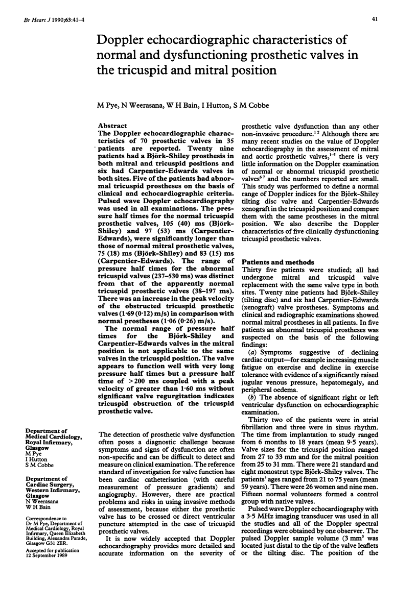
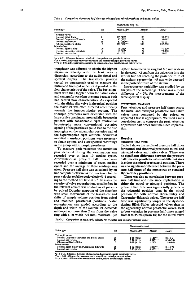
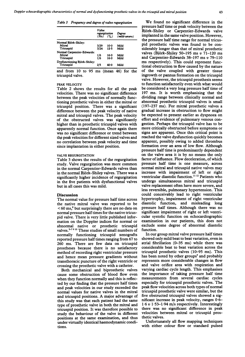
The Doppler echocardiographic characteristics of 70 prosthetic valves in 35 patients are reported. Twenty nine patients had a Björk-Shiley prosthesis in both mitral and tricuspid positions and six had Carpentier-Edwards valves in both sites. Five of the patients had abnormal tricuspid prostheses on the basis of clinical and echocardiographic criteria. Pulsed wave Doppler echocardiography was used in all examinations. The pressure half times for the normal tricuspid prosthetic valves, 105 (40) ms (Björk-Shiley) and 97 (53) ms (Carpentier-Edwards), were significantly longer than those of normal mitral prosthetic valves, 75 (18) ms (Björk-Shiley) and 83 (15) ms (Carpentier-Edwards). The range of pressure half times for the abnormal tricuspid valves (237-530 ms) was distinct from that of the apparently normal tricuspid prosthetic valves (38-197 ms). There was an increase in the peak velocity of the obstructed tricuspid prosthetic valves (1.69 (0.12) m/s) in comparison with normal prostheses (1.06 (0.26) m/s). The normal range of pressure half times for the Björk-Shiley and Carpentier-Edwards valves in the mitral position is not applicable to the same valves in the tricuspid position. The valve appears to function well with very long pressure half times but a pressure half time of greater than 200 ms coupled with a peak velocity of greater than 1.60 ms without significant valve regurgitation indicates tricuspid obstruction of the tricuspid prosthetic valve.
Full text
PDF


Selected References
These references are in PubMed. This may not be the complete list of references from this article.
- Alam M., Rosman H. S., Lakier J. B., Kemp S., Khaja F., Hautamaki K., Magilligan D. J., Jr, Stein P. D. Doppler and echocardiographic features of normal and dysfunctioning bioprosthetic valves. J Am Coll Cardiol. 1987 Oct;10(4):851–858. doi: 10.1016/s0735-1097(87)80280-2. [DOI] [PubMed] [Google Scholar]
- Baseline rest electrocardiographic abnormalities, antihypertensive treatment, and mortality in the Multiple Risk Factor Intervention Trial. Multiple Risk Factor Intervention Trial Research Group. Am J Cardiol. 1985 Jan 1;55(1):1–15. doi: 10.1016/0002-9149(85)90290-5. [DOI] [PubMed] [Google Scholar]
- Dennig K., Kraus F., Rudolph W. Doppler-echokardiographische Bestimmung des Schweregrades der Trikuspidalstenose. Herz. 1986 Dec;11(6):332–336. [PubMed] [Google Scholar]
- Gibbs J. L., Wharton G. A., Williams G. J. Doppler echocardiographic characteristics of the Carpentier-Edwards xenograft. Eur Heart J. 1986 Apr;7(4):353–356. doi: 10.1093/oxfordjournals.eurheartj.a062073. [DOI] [PubMed] [Google Scholar]
- Hatle L., Angelsen B., Tromsdal A. Noninvasive assessment of atrioventricular pressure half-time by Doppler ultrasound. Circulation. 1979 Nov;60(5):1096–1104. doi: 10.1161/01.cir.60.5.1096. [DOI] [PubMed] [Google Scholar]
- Isobe M., Yazaki Y., Takaku F., Hara K., Kashida M., Yamaguchi T., Machii K. Right ventricular filling detected by pulsed Doppler echocardiography during the convalescent stage of inferior wall acute myocardial infarction. Am J Cardiol. 1987 Jun 1;59(15):1245–1250. doi: 10.1016/0002-9149(87)90898-8. [DOI] [PubMed] [Google Scholar]
- Panidis I. P., Ross J., Mintz G. S. Normal and abnormal prosthetic valve function as assessed by Doppler echocardiography. J Am Coll Cardiol. 1986 Aug;8(2):317–326. doi: 10.1016/s0735-1097(86)80046-8. [DOI] [PubMed] [Google Scholar]
- Spirito P., Maron B. J. Doppler echocardiography for assessing left ventricular diastolic function. Ann Intern Med. 1988 Jul 15;109(2):122–126. doi: 10.7326/0003-4819-109-2-122. [DOI] [PubMed] [Google Scholar]
- Wilkins G. T., Gillam L. D., Kritzer G. L., Levine R. A., Palacios I. F., Weyman A. E. Validation of continuous-wave Doppler echocardiographic measurements of mitral and tricuspid prosthetic valve gradients: a simultaneous Doppler-catheter study. Circulation. 1986 Oct;74(4):786–795. doi: 10.1161/01.cir.74.4.786. [DOI] [PubMed] [Google Scholar]
- Williams G. A., Labovitz A. J. Doppler hemodynamic evaluation of prosthetic (Starr-Edwards and Björk-Shiley) and bioprosthetic (Hancock and Carpentier-Edwards) cardiac valves. Am J Cardiol. 1985 Aug 1;56(4):325–332. doi: 10.1016/0002-9149(85)90858-6. [DOI] [PubMed] [Google Scholar]
- Wilson N., Goldberg S. J., Dickinson D. F., Scott O. Normal intracardiac and great artery blood velocity measurements by pulsed Doppler echocardiography. Br Heart J. 1985 Apr;53(4):451–458. doi: 10.1136/hrt.53.4.451. [DOI] [PMC free article] [PubMed] [Google Scholar]


