Abstract
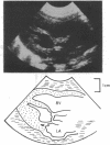
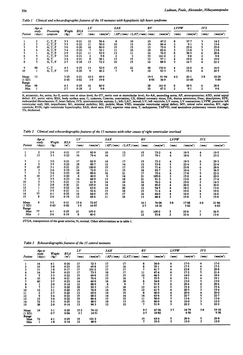
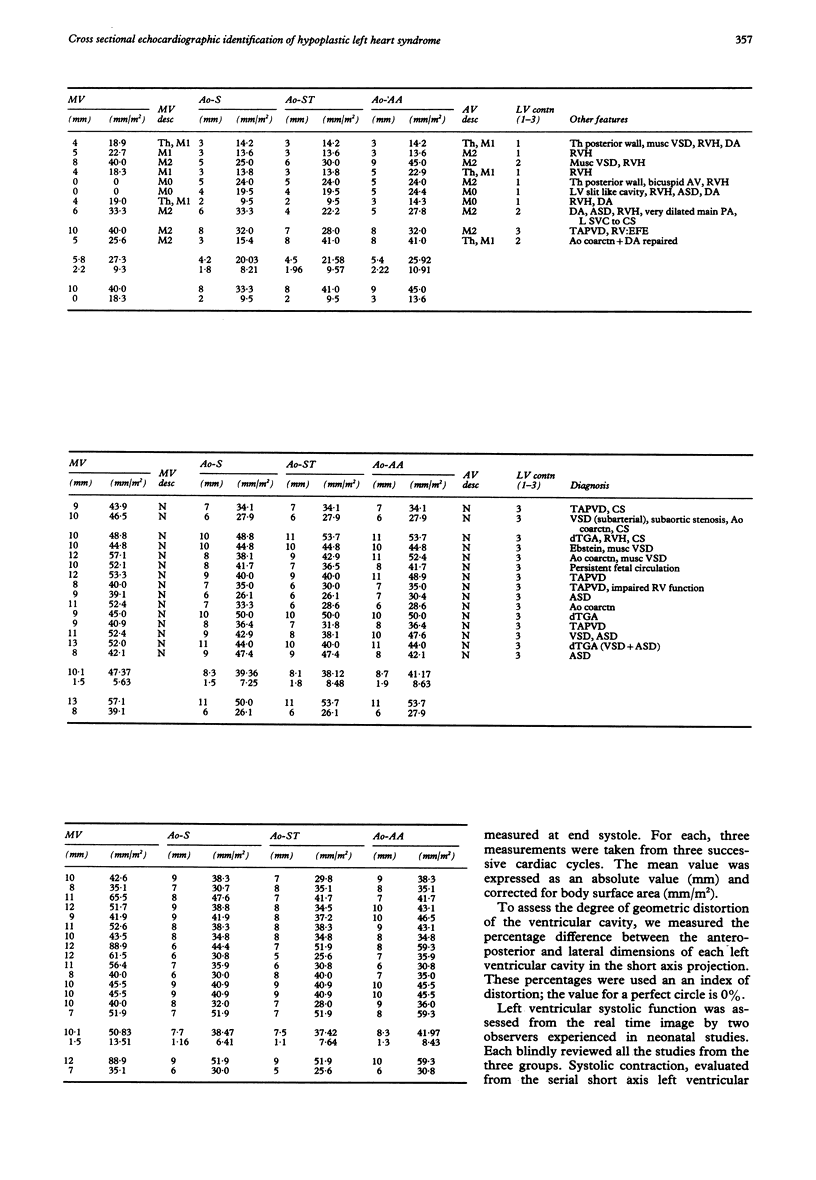
To identify the echocardiographic features that can be used to distinguish between hypoplastic left heart syndrome and other causes of right ventricular overload in the sick neonate cross sectional echocardiographic studies of 10 neonates with hypoplastic left heart syndrome were analysed and compared with those in 15 neonates with other causes of right ventricular overload and 15 normal controls. Left ventricular and right ventricular cavity dimensions and the shape and size of the mitral valve annulus and aortic root were recorded and presented both as absolute values (mm) and corrected for body surface area (mm/m2). Logistic regression was used to produce a classification rule to estimate the probability of a neonate having hypoplastic left heart syndrome. The diameter of the mitral valve annulus was the single most discriminative variable. There was, however, considerable overlap of all the calculated features between neonates with hypoplastic left heart syndrome and those with other causes of right ventricular overload. The diagnosis of hypoplastic left heart syndrome should not be based on any one single echocardiographic feature but the general appearance of abnormal left heart valves, small cavity dimensions, and the size of the mitral valve annulus.
Full text
PDF
Images in this article
Selected References
These references are in PubMed. This may not be the complete list of references from this article.
- Bass J. L., Ben-Shachar G., Edwards J. E. Comparison of M mode echocardiography and pathologic findings in the hypoplastic left heart syndrome. Am J Cardiol. 1980 Jan;45(1):79–86. doi: 10.1016/0002-9149(80)90223-4. [DOI] [PubMed] [Google Scholar]
- Cabrera A., Irurita M., Maíz E., Aguirre J., Pastor E. Hipoplasia del ventrículo izquierdo. Estudio ecocardiográfico y anatómico de 13 casos. Rev Esp Cardiol. 1983;36(2):153–158. [PubMed] [Google Scholar]
- Chesler E., Joffe H. S., Vecht R., Beck W., Schrire V. Ultrasound cardiography in single ventricle and hypoplastic left and right heart syndromes. Circulation. 1970 Jul;42(1):123–129. doi: 10.1161/01.cir.42.1.123. [DOI] [PubMed] [Google Scholar]
- Covitz W., Rao P. S., Strong W. R., Reyes L. Echocardiographic assessment of the aortic root in syndromes with left ventricular hypoplasia. Pediatr Cardiol. 1982;2(1):19–23. doi: 10.1007/BF02265612. [DOI] [PubMed] [Google Scholar]
- Farooki Z. Q., Henry J. G., Green E. W. Echocardiographic sepctrum of the hypoplastic left heart syndrome: a clinicopathologic correlation in 19 newborns. Am J Cardiol. 1976 Sep;38(3):337–343. doi: 10.1016/0002-9149(76)90176-4. [DOI] [PubMed] [Google Scholar]
- Foale R., Nihoyannopoulos P., McKenna W., Kleinebenne A., Nadazdin A., Rowland E., Smith G., Klienebenne A. Echocardiographic measurement of the normal adult right ventricle. Br Heart J. 1986 Jul;56(1):33–44. doi: 10.1136/hrt.56.1.33. [DOI] [PMC free article] [PubMed] [Google Scholar]
- Lundström N. R. Ultrasoundcardiographic studies of the mitral valve region in young infants with mitral atresia, mitral stenosis, hypoplasia of the left ventricle, and cor triatriatum. Circulation. 1972 Feb;45(2):324–334. doi: 10.1161/01.cir.45.2.324. [DOI] [PubMed] [Google Scholar]
- Meyer R. A., Kaplan S. Echocardiography in the diagnosis of hypoplasia of the left or right ventricles in the neonate. Circulation. 1972 Jul;46(1):55–64. doi: 10.1161/01.cir.46.1.55. [DOI] [PubMed] [Google Scholar]
- Mickell J. J., Mathews R. A., Anderson R. H., Zuberbuhler J. R., Lenox C. C., Neches W. H., Park S. C., Fricker F. J. The anatomical heterogeneity of hearts lacking a patent communication between the left atrium and the ventricular mass ('mitral atresia') in presence of a patent aortic valve. Eur Heart J. 1983 Jul;4(7):477–486. doi: 10.1093/oxfordjournals.eurheartj.a061505. [DOI] [PubMed] [Google Scholar]
- Mortera C., Leon G. Detection of persistent ductus in hypoplastic left heart syndrome by contrast echocardiography. Br Heart J. 1980 Nov;44(5):596–598. doi: 10.1136/hrt.44.5.596. [DOI] [PMC free article] [PubMed] [Google Scholar]
- Nihoyannopoulos P., Karas S., Sapsford R. N., Hallidie-Smith K., Foale R. Accuracy of two-dimensional echocardiography in the diagnosis of aortic arch obstruction. J Am Coll Cardiol. 1987 Nov;10(5):1072–1077. doi: 10.1016/s0735-1097(87)80348-0. [DOI] [PubMed] [Google Scholar]
- Plomteux G. Multivariate analysis of an enzymic profile for the differential diagnosis of viral hepatitis. Clin Chem. 1980 Dec;26(13):1897–1899. [PubMed] [Google Scholar]
- Tajik A. J., Seward J. B., Hagler D. J., Mair D. D., Lie J. T. Two-dimensional real-time ultrasonic imaging of the heart and great vessels. Technique, image orientation, structure identification, and validation. Mayo Clin Proc. 1978 May;53(5):271–303. [PubMed] [Google Scholar]


