Abstract
Malignant tumors are complex structures composed of cancer cells and tumor microenvironmental cells. In this complex structure, cells cross-talk and interact, thus jointly promoting cancer development and metastasis. Recently, immunoregulatory molecule-based cancer immunotherapy has greatly improved treatment efficacy for solid cancers, thus enabling some patients to achieve persistent responses or cure. However, owing to the development of drug-resistance and the low response rate, immunotherapy against the available targets PD-1/PD-L1 or CTLA-4 has limited benefits. Although combination therapies have been proposed to enhance the response rate, severe adverse effects are observed. Thus, alternative immune checkpoints must be identified. The SIGLECs are a family of immunoregulatory receptors (known as glyco-immune checkpoints) discovered in recent years. This review systematically describes the molecular characteristics of the SIGLECs, and discusses recent progress in areas including synthetic ligands, monoclonal antibody inhibitors, and Chimeric antigen receptor T (CAR-T) cells, with a focus on available strategies for blocking the sialylated glycan-SIGLEC axis. Targeting glyco-immune checkpoints can expand the scope of immune checkpoints and provide multiple options for new drug development.
Keywords: SIGLEC, sialylated glycan, glyco-immune checkpoint, high affinity SIGLEC-ligands, anti-SIGLEC antibodies
Introduction
Malignant tumors are complex structures composed of cancer cells and various microenvironmental cells1,2, more than 50% of which are tumor-associated macrophages3. Cross-talk between cancer cells and microenvironmental cells facilitates cancer development and metastasis. Therefore, to conquer cancer, the biological behavior of cancer cells and the components of the tumor microenvironment (TME) cells, which greatly enhance treatment efficacy, must be considered.
In recent years, oncologists have recognized the biological importance of the TME in the progression of malignancies, particularly immune cells, and have attempted to ameliorate the immunosuppressive microenvironment of cancers caused by immune checkpoints4,5. Several monoclonal antibodies have been developed to block the PD-1/PD-L1 and CTLA-4 immune checkpoints. According to clinical treatment reports, use of an immunotherapeutic paradigm instead of traditional cytotoxic drugs can effectively reactivate immune cells. Thus, immune checkpoint inhibitors not only protect healthy cells against non-specific killing, but also enable durable response or even cure in patients6,7. Anti-cancer immunotherapies are a promising approach that has brought hope to patients. However, only limited patients show positive responses to PD-1/PD-L1 blockade therapy, owing to the variable expression of PD-1/PD-L1 among human populations and the development of drug-resistance after treatment. To date, the mechanism of primary or secondary resistance is not well understood8,9. Additional immunoregulatory pathways, such as T cell immune checkpoints, are likely to exist10. Consequently, combination strategies have been developed to target multiple immune checkpoints to enhance treatment efficacy11. Among them, sialic acid (Sia)-binding immunoglobulin-like lectins (SIGLECs) have attracted substantial attention as a potential alternative12. Here, we summarize recent progress in targeting the sialylated glycan-SIGLEC axis for cancer immunotherapy.
SIGLEC classification and molecular characteristics
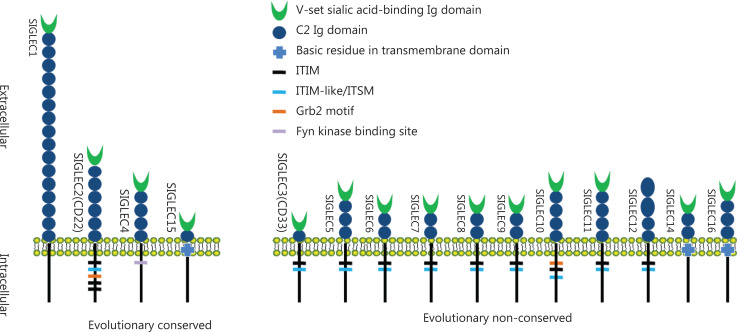
SIGLECs belong to the immunoglobulin superfamily, and are expressed on most immune cells. To date, 15 members of SIGLECs have been identified in humans. According to sequence similarity and evolutionary conservation, SIGLECs are classified into 2 categories. The first category is highly conserved among multiple vertebrate lineages and has low sequence similarity, and comprises SIGLEC1 (CD169, sialoadhesin), SIGLEC2 (CD22), SIGLEC4 (myelin associated glycoprotein, MAG), and SIGLEC15 (CD33L3). The second category lacks evolutionary conservation (i.e., has been identified in humans but not mice) and comprises the SIGLEC3 (CD33) related SIGLECs (CD33rSIGLECs), comprising SIGLEC3, SIGLEC5 (CD170), SIGLEC6 (CD327), SIGLEC7 (CD328), SIGLEC8, SIGLEC9 (CD329), SIGLEC10, SIGLEC11, SIGLEC12, SIGLEC14, and SIGLEC1613–15. The extracellular structure of SIGLECs consists of 1–16 Ig constant-2 set (C2) domains with an additional Ig variable set (V-set) domain at the N terminus, which is responsible for binding sialylated glycan (sialoside) ligands (Figure 1). In the cytoplasmic domain, most CD33rSIGLECs contain either an immunoreceptor tyrosine-based inhibitory motif (ITIM) or immunoreceptor tyrosine-based switch motif (ITSM). After binding sialoside ligands, the ITIM or ITSM recruits SRC homology region 2 domain-containing tyrosine phosphatase-1 and -2 (SHP-1 and SHP-2), and inhibits the activation of tyrosine kinase, thereby participating in immunosuppressive regulation. Several SIGLECs, such as SIGLECs 14, 15, and 16, have positively charged amino acid residues in their transmembrane domains, which interact with DAP12 (also known as transmembrane immune signaling adaptor TYROBP) on immune cells. The intracellular domain of DAP12 contains an immunoreceptor tyrosine-based activation motif (ITAM), which activates spleen tyrosine kinase (SYK) and further catalyzes a downstream immune cascade. Thus, DAP12-paired SIGLECs may participate in the activation of immune cells16.
Figure 1.
The 15 SIGLECs identified in humans. SIGLEC1, SIGLEC2, SIGLEC4, and SIGLEC15 are evolutionarily conserved, and the others are evolutionary non-conserved. SIGLEC1 is the longest SIGLEC without intracellular signaling motif, and human SIGLEC12 has lost the ability to bind Sias.
SIGLECs are expressed on both innate and adaptive immune cells, such as monocytes, neutrophils, natural killer (NK) cells, and B cells. A recent article has indicated that adaptive immune cells such as T lymphocytes also express SIGLECs. Vuchkovska et al.17 have reported that SIGLEC5 is expressed on most activated T cells after antigen receptor stimulation, whereas SIGLEC5 overexpression abrogates the activation of NFAT and AP-1 induced by antigen receptor. The SIGLECs on human or murine leucocytes have diverse functions. Cells expressing SIGLECs are listed in Table 1.
Table 1.
Expression spectrum of SIGLECs on human or murine cells
| SIGLECs | Other names | Expressing cells | Refs |
|---|---|---|---|
| SIGLEC1 | CD169 | Macrophage, Dendritic cell | 14,18,19 |
| SIGLEC2 | CD22 | B cell, cDC*, Mast cell | 14,18 |
| SIGLEC3 | CD33 | Diverse myeloid-derived cells, NK cell, T cell | 14,18,20 |
| SIGLEC4 | MAG | Oligodendrocyte, Schwann cell | 14,18 |
| SIGLEC5 | CD170 | Diverse myeloid-derived cells, T cell, B cell | 14,17,18,21,22 |
| SIGLEC6 | CD327 | Trophoblast, Mast cell, Basophil, B cell, Myeloid leukemia | 14,17,18,23 |
| SIGLEC7 | CD328 | Diverse myeloid-derived cells, NK cell, T cell | 14,18,24,25 |
| SIGLEC8 | – | Eosinophil, Basophil, Mast cell | 14,18 |
| SIGLEC9 | CD329 | Diverse myeloid-derived cells, T cell, NK cell | 14,18,26 |
| SIGLEC10 | – | Macrophage, NK cell, Eosinophil, B cell, T cell | 14,18,27 |
| SIGLEC11 | – | Microglia, Macrophage, Ovarian stromal cell | 14,18,28 |
| SIGLEC12 | Pseudogene | Macrophage, Unknown | 14,18,29 |
| SIGLEC14 | – | Diverse myeloid-derived cells | 14,18,30 |
| SIGLEC15 | CD33L3 | Macrophage, Osteoclast | 14,18,31 |
| SIGLEC16 | – | Macrophage, Microglia | 14,18,21 |
| mSiglec1# | mCD169 | Macrophage, Dendritic cell | 14,18,19,32 |
| mSiglec2 | mCD22 | B cell, cDC*, Mast cell | 14,18,33 |
| mSiglec4 | mMAG | Oligodendrocyte, Schwann cell | 14,18,34 |
| mSiglec15 | mCD33L3 | Macrophage, Osteoclast | 14,18,31 |
| mSiglec3 | mCD33 | Neutrophil, Macrophage, Microglia | 35,36 |
| mSiglecE | Homolog of SIGLEC9 | Diverse myeloid-derived cells, NK cell, Dendritic cell | 37–42 |
| mSiglecF | Homolog of SIGLEC8 | Immature cells of myeloid lineage, Eosinophil, Neutrophil | 35,43–49 |
| mSiglecG | Homolog of SIGLEC10 | Eosinophil | 34,43,44 |
| mSiglecH | Possible human homolog of SIGLEC14 and SIGLEC16? | Plasmacytoid dendritic cell (pDC), Macrophage | 43,50–52 |
#The “m” prefix indicates murine origin. *myeloid-derived DC.
Natural ligands of SIGLECs
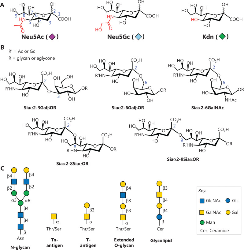
Sias are enriched on the surfaces of mammalian cells, bacteria and viruses, as well as on mucin proteins produced by cancer cells53,54. Sias are a family of sugar derivatives comprising a nine-carbon backbone with a carboxyl group at the C-1 position. The most common Sias in the mammalian glycome are N-acetylneuraminic acid (Neu5Ac), N-glycolylneuraminic acid (Neu5Gc), and the deaminated neuraminic acid 2-keto-3-deoxy-D-glycero-D-galacto-nononic acid (Kdn) (Figure 2A)55. Sias are frequently attached to the penultimate galactose (Gal) or N-acetyl galactosamine (GalNAc) residue through either an α2,3- or an α2,6-linkage. Sias can conjugate to the C-8 or C-9 positions, thus forming α2,8- or α2,9-linked sialosides (Figure 2B). Sialylation, an important glycosylation reaction, is accomplished by the transfer of Sias to the underlying glycan chain by a combination of cytidine monophosphate-Sia synthetases (CMP-Sia synthetases, CSSs) and sialyltransferases (STs). The linkage types are cell- and tissue-specific, and are dynamically regulated by the expression patterns of STs. Sialylated glycans are frequently attached to proteins (N-/O-linked glycoproteins) and lipids (glycolipids) involved in various biological processes, such as pathogen recognition, inflammation, immune responses, and cancer development (Figure 2C).
Figure 2.
Diversity of sialoside structures. (A) Chemical structures of Neu5Ac, Neu5Gc, and Kdn. (B) Common linkage types of sialosides. (C) Underlying glycan backbones for sialylation, including glycoproteins (N-/O-glycan, Tn-, and T-antigen), as well as glycolipids. N-glycan is covalently attached to the amide side chain of the asparagine (Asn) residue, whereas O-glycan is attached to the hydroxyl groups of threonine/serine (Thr/Ser). Glycolipid is linked to the C-1 hydroxyl group of the ceramide. Structures are presented with SNFG symbol nomenclature (https://www.ncbi.nlm.nih.gov/glycans/snfg.html).
Owing to the attachment to the non-reducing end of glycan chains, Sias serve as ligands for certain cell membrane receptors, including SIGLECs56. However, SIGLECs have distinct binding specificity depending on the linkage type of Sias and the underlying sugar. A conserved arginine residue in the V-set domain is believed to ligate the carboxylate group of Sias via a salt bridge57. When essential arginine is mutated, Sia recognition ability is lost58. Further contacts have been observed between SIGLECs and the 4-OH, 5-NAc, and glycerol side chain of Sias. A variable C-C′ loop in the binding site is responsible for recognizing the underlying glycan59. Through interaction with the sialoside ligand, SIGLECs can distinguish “self” and “non-self” molecules, thus preventing unwanted inflammatory responses under homeostatic conditions.
The natural ligands of SIGLECs are sialylated glycans. However, recent studies have shown that lipophilic molecules and proteins mediate binding to SIGLEC receptors in a Sia-independent manner. Suematsu et al.60 have reported that fungal alkanes and triacylglycerols extracted from Trichophyton show ligand activity for SIGLEC5 and SIGLEC14. The lipophilic ligands suppress interleukin-8 (IL-8) production in SIGLEC5-expressing human monocytic cells, whereas the endogenous lipids induce IL-8 production in SIGLEC14-expressing human monocytic cells. These findings suggest that lipophilic ligands modulate innate immune responses, thus expanding understanding of the biological functions and importance of SIGLECs in innate immunity. In addition, Fong et al.61 have found that secreted heat-shock protein 70 (HSP70) acts as a ligand for SIGLEC5 and SIGLEC14, thus inducing either anti-inflammatory signal or pro-inflammatory signals, respectively. Moreover, Nizet and co-workers62 have demonstrated that human neonatal pathogen group B streptococcus engages SIGLEC5 and SIGLEC763 via β protein, thus impairing human leukocytes, increasing bacterial resistance to neutrophil phagocytosis, and suppressing the pyroptosis activity of NK cells. A recent study has suggested that SIGLEC10 interacts with both amino acids and sialic acids of CD24, a protein overexpressed on tumor cells, thus inducing tumor immune escape64.
Association of SIGLECs with cancers
Cancer development is regulated by the crosstalk between cancer cells and other components in the TME, such as cancer-associated fibroblasts, blood vessels, and immune cells. Although numerous immune cells are recruited to the local TME for targeting cancer cells, these abilities are inhibited by cancer-derived suppressive signals. Under suppressive conditions, immune effector cells, such as macrophages, dendritic cells, and T cells, do not have anti-cancer activity but instead facilitate cancer development.
The abnormal expression of some STs in cancer cells significantly affects Sia content and type. For example, a change in ST6GALNAC4 expression has been found to increase the content of disialyl-T antigen [Neu5Acα2,3Galβ1,3(Neu5Acα2,6)GalNAcα–]65. Moreover, hypoxia up-regulates the expression of both STs and the transporter SLC17A5, which transports external Sias into cells66,67. Thus, the cancer cell surface is covered by a dense layer of sialylated glycans, such as polysialic acid, sialylated Lewis antigens, and sialylated Tn/T antigens. Aberrant sialylation is associated with cancer progression and metastasis, and is a hallmark of several cancers including those of the lung, breast, pancreatic, and prostate68. These tumor-associated sialosides have been identified as biomarkers for certain cancers, and used for cancer diagnosis and monitoring. Among them, CA19-9 (also called carbohydrate antigen 19-9 or sialylated Lewis A antigen) is the most commonly used serum marker for pancreatic cancer diagnosis69.
Overexpressed sialosides on cancer cells interact with SIGLECs on immune cells providing an immunosuppressive TME just like the PD-1 does. Therefore, in recent years, SIGLECs have become a new target of anti-cancer immunity70,71. Stanczak et al.12 have reported the upregulation of SIGLECs including SIGLEC9 on tumor-infiltrating T cells from non-small cell lung cancer, colorectal cancer, and ovarian cancer. SIGLEC9-expressing T cells in patients with non-small cell lung cancer correlate with diminished survival, whereas SIGLEC9 polymorphisms are associated with the risk of developing lung and colorectal cancer. Targeting the sialoside-SIGLEC pathway increases anticancer immunity in vitro and in vivo. Moreover, Zhang et al.72 have reported that gastric cancer-specific exitrons significantly increase the expression of PD-1, SIGLEC1, SIGLEC2, SIGLEC3, and SIGLEC7 with high neoantigen load. The exitrons are clinically relevant to sex, age, Lauren classification, tumor stage, and prognosis. Wang et al.73 have constructed a comprehensive immune scoring system including 6 immunosuppressive genes (NECTIN2, CEACAM1, HMGB1, SIGLEC6, CD44, and CD155) to improve prognosis after adjuvant chemotherapy in gastric cancer by supplementing TNM staging. In addition, an interaction of SIGLEC7 and SIGLEC9 from myeloid cells with the elevated Sia in cancer cells has been found in a pancreatic cancer study70.
In our recent studies, we have analyzed the pangenomic characteristics of gastric cancer and identified a set of genes (GSTM1, ACOT1, SIGLEC14, and UGT2B17) with high-frequency absence variation at the whole genome level74–76. Through comparison with whole genome sequencing data for multiracial populations in public databases, we determined that the frequency of absence of the above 4 genes (41%–71%) in the gastric cancer population was much higher than that in European and American healthy populations (4.6%–46%). The absence of SIGLEC14 was first proposed in gastric cancer76. Because SIGLEC14 is an innate immune cell activation receptor, the integrity of the SIGLEC14 gene provides a molecular basis for ensuring the M1 polarization of macrophages or tumor-arresting polarization of neutrophils. Deletion of this gene in cancer is expected to worsen the tumor immunosuppressive microenvironment. A bioinformatic analysis of lung adenocarcinoma has indicated that the expression levels of SIGLEC3, SIGLEC5, SIGLEC7, SIGLEC9, SIGLEC11, and SIGLEC14 correlate with macrophage, neutrophil, and dendritic cell infiltration77.
Strategies to block the sialoside-SIGLEC axis
The above studies have indicated that SIGLECs are involved in the immune evasion of cancers and are potential targets to alleviate the immunosuppressive TME in cancer immunotherapy. In the SIGLEC family, 8 members, SIGLEC3, SIGLEC5, SIGLEC6, SIGLEC7, SIGLEC8, SIGLEC9, SIGLEC10, and SIGLEC11, contain immunosuppressive functional domains in their intracellular domains, which are similar to PD-110,17. Sequence alignment studies have demonstrated that PD-1 shares conserved amino acids in the ITIM and ITSM domains with SIGLEC5, SIGLEC7, and SIGLEC9. The interaction of SIGLECs with sialoside ligands results in inhibitory signaling as does the interaction between PD-1 and PD-L110. Similarly to PD-1 based immunotherapies, blockade of the sialoside-SIGLEC axis provides benefits in cancer treatment.
Multivalent presentation of natural ligands for targeting SIGLECs
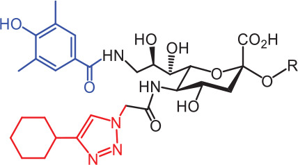
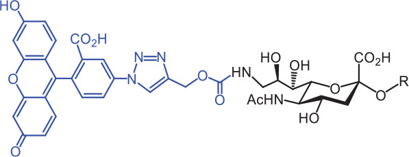
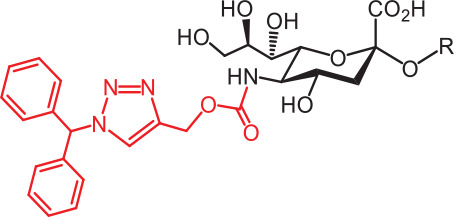
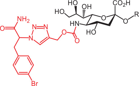
Given that SIGLECs are glyco-immune checkpoints, the ligands or monoclonal antibodies targeting SIGLECs have therapeutic potential. Generally, natural sialosides on glycoproteins and glycolipids exhibit weak monovalent binding affinity toward SIGLECs (Kd = 0.1–3 mM), and this affinity can be increased by presentation of multiple copies to cluster of the SIGLECs78. To mimic the multivalent presentation of sialosides on the cell surface, researchers have prepared libraries of sialosides immobilized on glass slides (sialoside microarrays). Through high-throughput screening with the sialoside microarrays, natural ligands for SIGLECs have been identified (Table 2)56. As the sialylated glycans on traditional biochips cannot fully recapitulate their conformations on the cell surface, and the arrays are expensive, a mammalian living cell screening system has been developed79.
Table 2.
Developed synthetic ligands for corresponding SIGLECs
| SIGLECs | Natural ligands | High affinity synthetic ligands | Refs |
|---|---|---|---|
| SIGLEC1 | Neu5Acα2,3LacNAc Modest |
 TCCNeu5Ac (1) (R = α2,3-LacNAc, IC50 = 0.38 μM) |
87 |
| hCD22* | Neu5Acα2,6LacNAc Strong |
 BPCNeu5Ac (2) (R = α2,6-LacNAc, IC50 = 0.20 μM) |
99 |
 MPBNeu5AcF (3) (R = α2,6-Lac, IC50 = 0.20 μM) |
100 | ||
| mCD22** | Neu5Gcα2,6LacNAc Strong |
 BPANeu5Gc (4) (R = α2,6-LacNAc, IC50 = 0.80 μM) |
99 |
| hCD33*** | Neu5Acα2,6LacNAc Weak |
 (5) (R = α2,6-Lac, IC50 = 11.00 μM) |
100 |
| SIGLEC7 | Neu5Acα2,8Neu5Acα2,3LacNAc Strong |
 FTMCNeu5Ac (6) (R = α2,6-Lac, unknown affinity) |
89 |
| SIGLEC9 | Neu5Acα2,3Galβ1,4(Fucα1,3)-(6-O-SO3)GlcNAc Strong [Ref. 101] |
 (7) (R = α2,6-Lac, unknown affinity) |
102 |
| SIGLEC15 | Neu5Acα2,6GalNAcαThr/Ser (To be further evaluated) [Ref. 103] |
 (8) (R = α2,6-LacNAc, unknown affinity) |
90 |
LacNAc, Galβ1,4GlcNAc; Lac, Galβ1,4Glc; IC50, half maximal inhibitory concentration. *hCD22, human SIGLEC2/CD22. **mCD22, mouse SIGLEC2/CD22. ***hCD33, human SIGLEC3/CD33.
Physiologically, SIGLECs are masked by endogenous cis-ligands, thus aiding in maintenance of cell homeostasis; however, malignant cells show elevated interaction with inhibitory SIGLECs through hypersialylation, and dampened immune surveillance80,81. To block the sialoside-SIGLEC axis, natural sialosides have been incorporated into various polymeric scaffolds to mimic the multivalent presentation of sialosides on glycoproteins and glycolipids82–85. Glycopolymers with a high density of Sia moieties can outcompete the natural sialosides in cancer cells for SIGLEC binding. Thus, sialoside glycopolymers can be used as inhibitors to perturb SIGLECs. To validate early models of hypersialylation-mediated immunoevasion, Bertozzi and coworkers82 have incorporated sialoside-functionalized glycopolymers onto cancer cell surfaces. The results suggest that hypersialylation of cancer cells elicits NK inhibition, and SIGLEC7 can tune the cytotoxicity activation of NK cells according to cancer cell sialylation status82. These results indicate that SIGLEC7 may be a potential therapeutic target for cancer therapy.
Because SIGLECs bind natural ligands with overlapping specificity and lower affinity than synthetic ligands, their regulatory mechanisms may be misinterpreted. Therefore, high affinity synthetic ligands with better specificity for SIGLECs must be developed.
Development of synthetic ligands for SIGLECs
In the past 20 years, various strategies have been used to introduce novel substituents to Sias as synthetic ligands, thus increasing binding affinity to SIGLECs in the sub-micromolar range (Table 2)86.
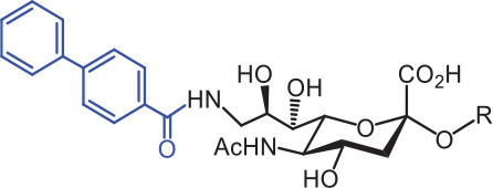
Because of the lack of an intracellular signaling motif, SIGLEC1 (sialoadhesin, Sn) is an ideal receptor for targeted delivery of antigens to macrophages, thereby eliciting a robust humoral response. The crystal structures of murine Sn have been determined, thus providing structural insights into the key features of Sia recognition. A high affinity and specificity ligand TCCNeu5Ac sialoside (1), with sub-micromolar binding affinity (IC50 = 0.38 μM), has been developed87. Through screening with a sialoside analog microarray, several high affinity ligands for SIGLEC2/CD22 have been identified, such as the BPCNeu5Ac (2) and MPBNeu5AcF (3) sialosides for human CD22, and BPANeu5Gc (4) sialoside for murine CD2288. Through the same strategy, FTMCNeu5Ac (6) has been discovered as a high affinity ligand for SIGLEC7, an inhibitory receptor on NK cells89. Moreover, cell-based glycan arrays have been developed to directly probe interactions of glycans with glycan-binding protein on the Chinese hamster ovary cell surface. With this platform, high-affinity glycan ligand 8 was discovered for SIGLEC1590,91. A panel of synthetic ligands has been developed. Examples are listed in Table 2, including SIGLEC1, SIGLEC2, SIGLEC3, SIGLEC7, SIGLEC9, and SIGLEC15.
However, these synthetic ligands alone remain insufficient to unmask the binding sites of endogenous target cell cis-ligands on SIGLECs. Targeting specific SIGLEC on cells requires multivalent presentation of high affinity ligands on various scaffolds, including nanoparticles and polymers92–94. For example, liposomal nanoparticles coated with the high affinity CD22-ligand BPC-Neu5Ac sialoside have been generated to target human malignant B cells92. After binding and endocytosis into acidic endosomes, liposomes are broken, and the encapsulated toxins are released, thus achieving CD22-dependent cytotoxicity in in vitro and in vivo studies. In addition, through metabolic engineering or a chemoenzymatic approach, the high affinity CD22-ligand MPB-Neu5Ac has been incorporated on NK-92 cells and found to enhance anti-tumor activity95,96. Glyco-engineered NK-92 cells exhibit CD22-dependent cytotoxicity to lymphoma cell lines and primary lymphoma cells from human patients. In recent studies, Bertozzi and coworkers97 have incorporated the SIGLEC9 high affinity ligand into a synthetic polypeptide. The artificial glycopeptide serves as a membrane-tethered cis-binding agonist that inhibits macrophage phagocytosis and induces neutrophil apoptosis98.
The above studies have highlighted the potential applications of synthetic SIGLEC ligands as immune modulators with great medicinal value in cancer treatment.
Progress in monoclonal antibodies for SIGLECs
Because cancer cells inhibit immune cell activity and evade immunosurveillance via the sialoside-SIGLEC axis, scientists have developed monoclonal antibodies targeting these inhibitory SIGLECs. By immunizing mice with SIGLEC9-encoding DNA and SIGLEC9 protein, Choi et al.42 have developed the high specificity and functionality monoclonal antibody (8A1E9) against SIGLEC9. The humanized antibody shows anti-tumor immune activity toward ovarian cancer in vitro and in vivo. Similarly, Cyr et al.104 have developed an anti-SIGLEC6 monoclonal antibody achieving highly potent and specific elimination of SIGLEC6 positive leukemic and healthy B cells, thus indicating the potential for cancer immunotherapy.
SIGLEC15 has recently been identified as a critical immune suppressor. Chen and coworkers31 have identified the SIGLEC15 immune suppressor through a genome-scale T-cell activity array. They have found that SIGLEC15 is broadly upregulated on human cancer cells and tumor-infiltrating myeloid cells. Importantly, the expression of SIGLEC15 is mutually exclusive to PD-L1. By binding unknown ligands, SIGLEC15 suppresses antigen-specific T-cell responses in vitro and in vivo. Genetic ablation or antibody (clone m03) blockade of SIGLEC15 amplifies anti-tumor immunity in the TME and inhibits tumor growth in some mouse models31. Xiao et al.105 have reported a monoclonal antibody against SIGLEC15 (S15-4E6A), and evaluated its antitumor effectiveness and modulatory role in macrophages in vitro and in vivo. They have found that S15-4E6A promotes macrophage M1 polarization while inhibiting M2 polarization both in vitro and in vivo, and exerts an efficacious tumor-inhibitory effect on lung adenocarcinoma cells and xenografts. He et al.106 have developed a monoclonal antibody against SIGLEC15 (3D6), which blocks SIGLEC15-mediated suppression of T cell and moderately prevents tumor growth. Wu et al.107 have conducted monoclonal antibody screening on SIGLEC15 and have found that the 3F1 clone antibody has high receptor blocking activity and significantly reverses the inhibitory effect of SIGLEC15 on lymphocyte proliferation. In mouse experiments, the 3F1 monoclonal antibody has shown significant antitumor efficacy when applied alone or in combination with the Erbitux drug. These results have demonstrated that SIGLEC15 is a potential target for normalizing tumor immunity as an alternative to anti-PD-1 therapy.
AMG 330 is a dual specific antibody for CD3 and SIGLEC3/CD33. CD33 is frequently expressed on the surfaces of blasts and leukemic stem cells in acute myelogenous leukemia. AMG 330 binds with low nanomolar affinity to CD33 and CD3ε of both human and cynomolgus monkey origin. In an ex vivo experiment, AMG 330 has been found to mediate autologous depletion of CD33-positive cells from cynomolgus monkey bone marrow aspirates. Thus, AMG 330 is a potential anti-tumor reagent for acute myelogenous leukemia108.
The above studies have indicated that antibodies against SIGLEC checkpoints provide an alternative treatment for patients with cancer refractory to the well-known PD-L1/PD-1-targeting therapies.
Because of the selective expression and endocytosis properties, SIGLECs can be directly targeted to deliver toxic cargo into hematopoietic cancer cells. In 2000, Mylotarg (Gemtuzumab ozogamicin from Pfizer), an anti-CD33 antibody-calicheamicin conjugate, was the first antibody-drug conjugate approved by the U.S. Food and Drug Administration (U.S. FDA). Mylotarg was developed for the treatment of acute myeloid leukemia but was withdrawn because of its high toxicity and low efficacy in 2010. However, with altered dosing, Mylotarg regained approval for treatment of acute myeloid leukemia in 2017109–111. Similarly, in 2017, another antibody-drug conjugate drug, Besponsa (Inotuzumab ozogamicin, Pfizer, NCT01564784), was approved by the U.S. FDA to treat CD22-positive B-cell precursor acute lymphoblastic leukemia112.
Progress in dual functional drugs for desialylation-targeted therapy
During cancer development, tumor cells acquire the ability to evade immunosurveillance; the sialoside-SIGLEC axis between cancer cells and immune cells in the TME plays an important role in this evasion. However, the binding of SIGLECs to Sias is dynamic and reversible. Sialidases (called neuraminidases, NEUs) are enzymes that cleave the terminal Sia resides from glycolipids and glycoproteins, and are involved in several human pathologies such as neurodegenerative disorders, cancers, and infectious and cardiovascular diseases113. The four types of mammalian sialidases, encoded by different genes, are NEU-1, NEU-2, NEU-3, and NEU-4. Mucins (MUCs) are the major substrates of sialidases114. Therefore, sialidase dissociates SIGLECs bound to their ligands. By chemically coupling recombinant sialidases to trastuzumab, human epidermal growth factor receptor 2 (HER2)-specific antibody-sialidase conjugates have been constructed to desialylate tumor cells in a HER2-dependent manner, thus disrupting the sialoside-SIGLEC axis and enhancing antibody-dependent cell-mediated cytotoxicity10,115. Single-cell RNA sequencing has revealed that desialylation repolarizes tumor-associated macrophages and enhances the efficacy of immune checkpoint blockade116. Antibody-sialidase conjugates are thus a promising modality for glyco-immune checkpoint therapy.
Macrophages are important innate immune cells that provide the first line of defense against the invasion of harmful foreign molecules (immune defense) and autologous damaged or dead cells (immune surveillance). Unlike T and B lymphocytes, macrophages can kill foreign microorganisms and tumor cells non-specifically. The polarization status and regulatory mechanisms of macrophages have become major research fields. Macrophages in the TME can polarize in 2 directions depending on external stimuli: M1-type polarization (classical activation of macrophages) and M2-type polarization (alternative activation of macrophages), similarly to Th1 and Th2 activation of T lymphocytes. M1-polarized macrophages are pro-inflammatory cells, which secrete inflammatory factors such as TNF-α and IL-1β, and extend pseudopodia for active phagocytosis. M2-polarized macrophages secrete cytokines such as IL-10 and TGF-β, which induce the production of Treg cells in the TME and promote tumor growth117–119.
Similarly, neutrophils can have either tumor-arresting or tumor-promoting functions40. Recently, new knowledge has been highlighted regarding tumor-infiltrating neutrophils. Xue et al.120 have found that CCL4 and PD-L1 positive tumor-associated neutrophils have a tumor-promoting function in liver cancer. Moreover, the cytokines and chemokines secreted by neutrophils influence innate and adaptive immunity. IL-12, TNF-α, GM-CSF, CXCL10, CCL7, CCL2, and CCL3 are proinflammatory cytokines that serve as T cell and macrophage chemo-attractants. However, CCL17 and CXCL14 are pro-tumor cytokines121. Ligands on pathogens or tumor cells bind SIGLEC9 on neutrophils and limit neutrophil activation122. Thus, CD33rSIGLECs have been recognized as negative regulators of neutrophils. In addition, aberrant sialoglycans on the surfaces of tumor cells can shield potential tumor antigen epitopes and escape recognition, thereby suppressing immunocyte activation. Desialylation on tumor cells can present tumor antigens with Gal/GalNAc residues and thus overcome glyco-immune checkpoints. Huang and colleagues123 have explored whether vaccination with desialylated whole-cell tumor vaccines (ID8 vaccine) might trigger anti-tumor immunity in ovarian cancer. A desialylated tumor cell vaccine has been found to promote anti-tumor immunity and provide a strategy for ovarian cancer immunotherapy in a clinical setting123.
Chimeric antigen receptor T cell (CAR-T) and other approaches
CAR-T approach uses a genetically modified T cell receptor with improved recognition of specific cancer cell antigens and tumor cell killing. CD19 is by far the most targeted biomarker in cancer immunotherapy124. CD19 CAR-T has been used for B-cell acute lymphoblastic leukemia or lymphoma therapy. However, relapse occurs in some cases. Thus, CD22/SIGLEC2 CAR-T and CD33/SIGLEC3 CAR-T were developed for the treatment of refractory leukemia or lymphoma125. A clinical trial has investigated patients with relapsed/refractory large B-cell lymphoma after CD19/22 dual-targeting CAR-T (AUTO3) plus pembrolizumab for relapsed/refractory large B-cell lymphoma (NCT03289455) and observed an overall response rate of 66% (48.9%, CR; 17%, PR)126. Because CD22 is restricted to surfaces of B cells and B lymphoma cells, it is a commonly used target for the treatment of autoimmune diseases and B-cell malignancy. Currently, immunotherapy drugs targeting CD22 include monoclonal antibody drugs, antibody-drug conjugates, and CAR-T therapies. In addition, SIGLEC6 has been reported as a novel target for CAR T-cell therapy in acute myeloid leukemia23.
Given that hypersialylation of cancer cells together with significant upregulation of ST contributes to cancer progression and drug resistance127,128, scientists have designed and constructed long-circulating, self-assembled core-shell nanoparticles carrying a transition state-based ST inhibitor, which inhibits sialoglycans in various cancer cells129. Recently, the Bertozzi group41 has found that the MYC oncogene controls expression of the sialyltransferase ST6GALNAC4 and induces sialosides, which function as a “do not eat me” signal by engaging SIGLEC7 of macrophages, thus hindering cancer cell clearance. Therefore, ST6GALNAC4 is a potential enzyme target for small molecule-mediated immune therapy41. Recently, Wang and coworkers80 have found that classical conventional DCs from cancer patient samples have high expression of several inhibitory SIGLECs including SIGLEC7, SIGLEC9, and SIGLEC10. In subcutaneous murine tumor models, downregulation of the inhibitory mSiglecE receptor on cancer-associated DCs has been found to enhance priming of antigen-specific T cells and induce proliferation. The above studies reveal a potential new target to improve cancer immunotherapy80.
In addition, soluble SIGLECs can function as immunomodulatory molecules, because binding to sialoside ligands interferes with the interaction between membrane SIGLECs and ligands. For example, Tomioka et al.130 have found that transgenic mice expressing the soluble form of mSiglecE show significant suppression of MUC1-expressing tumor proliferation. Related therapeutic interventions might potentially alter the outcomes of certain diseases. Tumor-associated MUC1 binds SIGLEC9, thus mediating tumor cell growth and inducing negative immunomodulation. Ono et al.131 have proposed that soluble SIGLEC9 (sSIGLEC9) competitively inhibits the binding of MUC1 to the receptor SIGLEC9, thus conferring an antitumor benefit against MUC1-expressing tumors. Moreover, soluble SIGLEC14 in the blood has been found to dose-dependently suppress the pro-inflammatory responses of myeloid cells expressing membrane-bound SIGLEC14132.
Conclusions
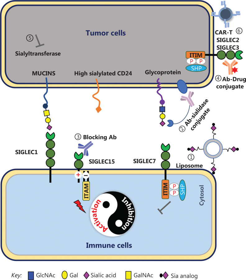
In summary, to overcome the immunosuppressive state of malignancies, scientists have developed various strategies to target the sialylated glycan-SIGLEC axis (Figure 3), although some strategies remain in the conceptual stage or pre-clinical research. As an alternative therapy or combination strategy with immune checkpoint inhibitors, targeting of the sialylated glycan-SIGLEC axis is expected to have a major role in cancer immunotherapy. At present, development of anti-SIGLEC drugs is rapidly progressing, including high affinity ligands, monoclonal antibodies, dual functional reagents of desialylation molecular targeted drugs, and CAR-T cells. The physiological roles of SIGLECs, a new generation of immune checkpoint, continue to expand and are expected to attract greater attention in cancer immunotherapy.
Figure 3.
Strategies for targeting the sialylated glycan-SIGLEC axis. (1) Liposomal nanoparticles coated with high affinity ligands deliver anti-tumor drugs to lymphoma cells, which express SIGLEC2 or SIGLEC3. (2) Antibody-sialidase conjugates destroy the sialic acids on tumor cells and release the SIGLEC receptors. (3) Anti-SIGLEC antibodies block the specific sialoside-SIGLEC axis. (4) Antibody-drug conjugates target and are endocytosed into tumor cells by SIGLECs. (5) Sialyltransferase inhibitors decrease sialyltransferase expression. (6) SIGLEC-specific CAR-T increases the cytotoxicity of immune cells.
Funding Statement
This work was supported by the Shanghai Science and Technology Committee (Grant Nos. 20DZ2201900 to Y.Y. and 23ZR1432500 to W.P.), National Natural Science Foundation of China (Grant Nos. 82072602 to Y.Y.; 91853121, 21977066, and 22177069 to W.P.), Innovation Foundation of Translational Medicine of Shanghai Jiao Tong University School of Medicine (Grant No. TM202001 to Y.Y.), Collaborative Innovation Center for Clinical and Translational Science by Chinese Ministry of Education & Shanghai (Grant No. CCTS-2022202 to Y.Y.), Shanghai Pilot Program for Basic Research-Shanghai Jiao Tong University (Grant No. 21TQ1400210 to W.P.), and Medical-Engineering Interdisciplinary Research Foundation of Shanghai Jiao Tong University (Grant No. YG2022ZD001 to W.P.).
Conflict of interest statement
No potential conflicts of interest are disclosed.
Author contributions
Conceived and designed the analysis: Yingyan Yu, Wenjie Peng.
Collected the data: Yingyan Yu, Wenjie Peng.
Contributed data or analysis tools: Yingyan Yu, Wenjie Peng.
Figure preparation: Yingyan Yu, Wenjie Peng.
Wrote the paper: Yingyan Yu, Wenjie Peng.
References
- 1.Shan Q, Takabatake K, Kawai H, Oo MW, Sukegawa S, Fujii M, et al. Crosstalk between cancer and different cancer stroma subtypes promotes the infiltration of tumor-associated macrophages into the tumor microenvironment of oral squamous cell carcinoma. Int J Oncol. 2022;60:78. doi: 10.3892/ijo.2022.5368. [DOI] [PMC free article] [PubMed] [Google Scholar]
- 2.Xuan W, Khan F, James CD, Heimberger AB, Lesniak MS, Chen P. Circadian regulation of cancer cell and tumor microenvironment crosstalk. Trends Cell Biol. 2021;31:940–50. doi: 10.1016/j.tcb.2021.06.008. [DOI] [PMC free article] [PubMed] [Google Scholar]
- 3.Xu H, Li D, Ma J, Zhao Y, Xu L, Tian R, et al. The IL-33/ST2 axis affects tumor growth by regulating mitophagy in macrophages and reprogramming their polarization. Cancer Biol Med. 2021;18:172–83. doi: 10.20892/j.issn.2095-3941.2020.0211. [DOI] [PMC free article] [PubMed] [Google Scholar]
- 4.Mise Y, Hamanishi J, Daikoku T, Takamatsu S, Miyamoto T, Taki M, et al. Immunosuppressive tumor microenvironment in uterine serous carcinoma via CCL7 signal with myeloid-derived suppressor cells. Carcinogenesis. 2022;43:647–58. doi: 10.1093/carcin/bgac032. [DOI] [PubMed] [Google Scholar]
- 5.Falcomata C, Barthel S, Schneider G, Rad R, Schmidt-Supprian M, Saur D. Context-specific determinants of the immunosuppressive tumor microenvironment in pancreatic cancer. Cancer Discov. 2023;13:278–97. doi: 10.1158/2159-8290.CD-22-0876. [DOI] [PMC free article] [PubMed] [Google Scholar]
- 6.Ariyan CE, Brady MS, Siegelbaum RH, Hu J, Bello DM, Rand J, et al. Robust antitumor responses result from local chemotherapy and CTLA-4 blockade. Cancer Immunol Res. 2018;6:189–200. doi: 10.1158/2326-6066.CIR-17-0356. [DOI] [PMC free article] [PubMed] [Google Scholar]
- 7.Guo Z, Yuan Y, Chen C, Lin J, Ma Q, Liu G, et al. Durable complete response to neoantigen-loaded dendritic-cell vaccine following anti-PD-1 therapy in metastatic gastric cancer. NPJ Precis Oncol. 2022;6:34. doi: 10.1038/s41698-022-00279-3. [DOI] [PMC free article] [PubMed] [Google Scholar]
- 8.Zhao Q, Tong J, Liu X, Li S, Chen D, Miao L. Reversing resistance to immune checkpoint inhibitor by adding recombinant human adenovirus type 5 in a patient with small cell lung cancer with promoted immune infiltration: a case report. J Cancer Res Clin Oncol. 2022;148:1269–73. doi: 10.1007/s00432-022-03931-4. [DOI] [PMC free article] [PubMed] [Google Scholar]
- 9.Dobosz P, Stepien M, Golke A, Dzieciatkowski T. Challenges of the immunotherapy: perspectives and limitations of the immune checkpoint inhibitor treatment. Int J Mol Sci. 2022;23:2847. doi: 10.3390/ijms23052847. [DOI] [PMC free article] [PubMed] [Google Scholar]
- 10.Gray MA, Stanczak MA, Mantuano NR, Xiao H, Pijnenborg JFA, Malaker SA, et al. Targeted glycan degradation potentiates the anticancer immune response in vivo. Nat Chem Biol. 2020;16:1376–84. doi: 10.1038/s41589-020-0622-x. [DOI] [PMC free article] [PubMed] [Google Scholar]
- 11.Yu Y. Multi-target combinatory strategy to overcome tumor immune escape. Front Med. 2022;16:208–15. doi: 10.1007/s11684-022-0922-5. [DOI] [PubMed] [Google Scholar]
- 12.Stanczak MA, Siddiqui SS, Trefny MP, Thommen DS, Boligan KF, von Gunten S, et al. Self-associated molecular patterns mediate cancer immune evasion by engaging Siglecs on T cells. J Clin Invest. 2018;128:4912–23. doi: 10.1172/JCI120612. [DOI] [PMC free article] [PubMed] [Google Scholar]
- 13.Macauley MS, Crocker PR, Paulson JC. Siglec-mediated regulation of immune cell function in disease. Nat Rev Immunol. 2014;14:653–66. doi: 10.1038/nri3737. [DOI] [PMC free article] [PubMed] [Google Scholar]
- 14.Lenza MP, Atxabal U, Oyenarte I, Jimenez-Barbero J, Ereno-Orbea J. Current status on therapeutic molecules targeting Siglec receptors. Cells. 2020;9:2691. doi: 10.3390/cells9122691. [DOI] [PMC free article] [PubMed] [Google Scholar]
- 15.Adams OJ, Stanczak MA, von Gunten S, Laubli H. Targeting sialic acid-Siglec interactions to reverse immune suppression in cancer. Glycobiology. 2018;28:640–7. doi: 10.1093/glycob/cwx108. [DOI] [PubMed] [Google Scholar]
- 16.Lanier LL, Bakker AB. The ITAM-bearing transmembrane adaptor DAP12 in lymphoid and myeloid cell function. Immunol Today. 2000;21:611–4. doi: 10.1016/s0167-5699(00)01745-x. [DOI] [PubMed] [Google Scholar]
- 17.Vuchkovska A, Glanville DG, Scurti GM, Nishimura MI, White P, Ulijasz AT, et al. Siglec-5 is an inhibitory immune checkpoint molecule for human T cells. Immunology. 2022;166:238–48. doi: 10.1111/imm.13470. [DOI] [PMC free article] [PubMed] [Google Scholar]
- 18.Gonzalez-Gil A, Schnaar RL. Siglec ligands. Cells. 2021;10:1260. doi: 10.3390/cells10051260. [DOI] [PMC free article] [PubMed] [Google Scholar]
- 19.Ruffin N, Gea-Mallorqui E, Brouiller F, Jouve M, Silvin A, See P, et al. Constitutive Siglec-1 expression confers susceptibility to HIV-1 infection of human dendritic cell precursors. Proc Natl Acad Sci U S A. 2019;116:21685–93. doi: 10.1073/pnas.1911007116. [DOI] [PMC free article] [PubMed] [Google Scholar]
- 20.Hernandez-Caselles T, Martinez-Esparza M, Perez-Oliva AB, Quintanilla-Cecconi AM, Garcia-Alonso A, Alvarez-Lopez DM, et al. A study of CD33 (Siglec-3) antigen expression and function on activated human T and NK cells: two isoforms of CD33 are generated by alternative splicing. J Leukoc Biol. 2006;79:46–58. doi: 10.1189/jlb.0205096. [DOI] [PubMed] [Google Scholar]
- 21.Schwarz F, Landig CS, Siddiqui S, Secundino I, Olson J, Varki N, et al. Paired Siglec receptors generate opposite inflammatory responses to a human-specific pathogen. EMBO J. 2017;36:751–60. doi: 10.15252/embj.201695581. [DOI] [PMC free article] [PubMed] [Google Scholar]
- 22.Yamanaka M, Kato Y, Angata T, Narimatsu H. Deletion polymorphism of Siglec14 and its functional implications. Glycobiology. 2009;19:841–6. doi: 10.1093/glycob/cwp052. [DOI] [PubMed] [Google Scholar]
- 23.Jetani H, Navarro-Bailon A, Maucher M, Frenz S, Verbruggen C, Yeguas A, et al. Siglec-6 is a novel target for CAR T-cell therapy in acute myeloid leukemia. Blood. 2021;138:1830–42. doi: 10.1182/blood.2020009192. [DOI] [PMC free article] [PubMed] [Google Scholar]
- 24.Yang L, Feng Y, Wang S, Jiang S, Tao L, Li J, et al. Siglec-7 is an indicator of natural killer cell function in acute myeloid leukemia. Int Immunopharmacol. 2021;99:107965. doi: 10.1016/j.intimp.2021.107965. [DOI] [PubMed] [Google Scholar]
- 25.Haas Q, Markov N, Muerner L, Rubino V, Benjak A, Haubitz M, et al. Siglec-7 represents a glyco-immune checkpoint for non-exhausted effector memory CD8+ T cells with high functional and metabolic capacities. Front Immunol. 2022;13:996746. doi: 10.3389/fimmu.2022.996746. [DOI] [PMC free article] [PubMed] [Google Scholar]
- 26.Haas Q, Boligan KF, Jandus C, Schneider C, Simillion C, Stanczak MA, et al. Siglec-9 regulates an effector memory CD8(+) T-cell subset that congregates in the melanoma tumor microenvironment. Cancer Immunol Res. 2019;7:707–18. doi: 10.1158/2326-6066.CIR-18-0505. [DOI] [PubMed] [Google Scholar]
- 27.Zhang C, Zhang J, Liang F, Guo H, Gao S, Yang F, et al. Innate immune checkpoint Siglec10 in cancers: mining of comprehensive omics data and validation in patient samples. Front Med. 2022;16:596–609. doi: 10.1007/s11684-021-0868-z. [DOI] [PubMed] [Google Scholar]
- 28.Wang X, Chow R, Deng L, Anderson D, Weidner N, Godwin AK, et al. Expression of Siglec-11 by human and chimpanzee ovarian stromal cells, with uniquely human ligands: implications for human ovarian physiology and pathology. Glycobiology. 2011;21:1038–48. doi: 10.1093/glycob/cwr039. [DOI] [PMC free article] [PubMed] [Google Scholar]
- 29.Mitra N, Banda K, Altheide TK, Schaffer L, Johnson-Pais TL, Beuten J, et al. Siglec12, a human-specific segregating (pseudo)gene, encodes a signaling molecule expressed in prostate carcinomas. J Biol Chem. 2011;286:23003–11. doi: 10.1074/jbc.M111.244152. [DOI] [PMC free article] [PubMed] [Google Scholar]
- 30.Tsai CM, Riestra AM, Ali SR, Fong JJ, Liu JZ, Hughes G, et al. Siglec-14 enhances NLRP3-inflammasome activation in macrophages. J Innate Immun. 2020;12:333–43. doi: 10.1159/000504323. [DOI] [PMC free article] [PubMed] [Google Scholar]
- 31.Wang J, Sun J, Liu LN, Flies DB, Nie X, Toki M, et al. Siglec-15 as an immune suppressor and potential target for normalization cancer immunotherapy. Nat Med. 2019;25:656–66. doi: 10.1038/s41591-019-0374-x. [DOI] [PMC free article] [PubMed] [Google Scholar]
- 32.Fujiwara Y, Saito Y, Shiota T, Cheng P, Ikeda T, Ohnishi K, et al. Natural compounds that regulate lymph node sinus macrophages: inducing an anti-tumor effect by regulating macrophage activation. J Clin Exp Hematop. 2018;58:17–23. doi: 10.3960/jslrt.17032. [DOI] [PMC free article] [PubMed] [Google Scholar]
- 33.Di Carluccio C, Forgione RE, Montefiori M, Civera M, Sattin S, Smaldone G, et al. Behavior of glycolylated sialoglycans in the binding pockets of murine and human CD22. iScience. 2021;24:101998. doi: 10.1016/j.isci.2020.101998. [DOI] [PMC free article] [PubMed] [Google Scholar]
- 34.Blixt O, Collins BE, van den Nieuwenhof IM, Crocker PR, Paulson JC. Sialoside specificity of the Siglec family assessed using novel multivalent probes: identification of potent inhibitors of myelin-associated glycoprotein. J Biol Chem. 2003;278:31007–19. doi: 10.1074/jbc.M304331200. [DOI] [PubMed] [Google Scholar]
- 35.Brinkman-Van der Linden EC, Angata T, Reynolds SA, Powell LD, Hedrick SM, Varki A. CD33/Siglec-3 binding specificity, expression pattern, and consequences of gene deletion in mice. Mol Cell Biol. 2003;23:4199–206. doi: 10.1128/MCB.23.12.4199-4206.2003. [DOI] [PMC free article] [PubMed] [Google Scholar]
- 36.Bhattacherjee A, Rodrigues E, Jung J, Luzentales-Simpson M, Enterina JR, Galleguillos D, et al. Repression of phagocytosis by human CD33 is not conserved with mouse CD33. Commun Biol. 2019;2:450. doi: 10.1038/s42003-019-0698-6. [DOI] [PMC free article] [PubMed] [Google Scholar]
- 37.McMillan SJ, Sharma RS, Richards HE, Hegde V, Crocker PR. Siglec-E promotes beta2-integrin-dependent NADPH oxidase activation to suppress neutrophil recruitment to the lung. J Biol Chem. 2014;289:20370–6. doi: 10.1074/jbc.M114.574624. [DOI] [PMC free article] [PubMed] [Google Scholar]
- 38.Siddiqui S, Schwarz F, Springer S, Khedri Z, Yu H, Deng L, et al. Studies on the detection, expression, glycosylation, dimerization, and ligand binding properties of mouse Siglec-E. J Biol Chem. 2017;292:1029–37. doi: 10.1074/jbc.M116.738351. [DOI] [PMC free article] [PubMed] [Google Scholar]
- 39.Yu Z, Maoui M, Wu L, Banville D, Shen S. mSiglec-E, a novel mouse CD33-related Siglec (sialic acid-binding immunoglobulin-like lectin) that recruits Src homology 2 (SH2)-domain-containing protein tyrosine phosphatases SHP-1 and SHP-2. Biochem J. 2001;353:483–92. doi: 10.1042/0264-6021:3530483. [DOI] [PMC free article] [PubMed] [Google Scholar]
- 40.Laubli H, Pearce OM, Schwarz F, Siddiqui SS, Deng L, Stanczak MA, et al. Engagement of myelomonocytic Siglecs by tumor-associated ligands modulates the innate immune response to cancer. Proc Natl Acad Sci U S A. 2014;111:14211–6. doi: 10.1073/pnas.1409580111. [DOI] [PMC free article] [PubMed] [Google Scholar]
- 41.Smith BAH, Deutzmann A, Correa KM, Delaveris CS, Dhanasekaran R, Dove CG, et al. MYC-driven synthesis of Siglec ligands is a glycoimmune checkpoint. Proc Natl Acad Sci U S A. 2023;120:e2215376120. doi: 10.1073/pnas.2215376120. [DOI] [PMC free article] [PubMed] [Google Scholar]
- 42.Choi H, Ho M, Adeniji OS, Giron L, Bordoloi D, Kulkarni AJ, et al. Development of Siglec-9 blocking antibody to enhance anti-tumor immunity. Front Oncol. 2021;11:778989. doi: 10.3389/fonc.2021.778989. [DOI] [PMC free article] [PubMed] [Google Scholar]
- 43.Angata T, Hingorani R, Varki NM, Varki A. Cloning and characterization of a novel mouse Siglec, mSiglec-F: differential evolution of the mouse and human (CD33) Siglec-3-related gene clusters. J Biol Chem. 2001;276:45128–36. doi: 10.1074/jbc.M108573200. [DOI] [PubMed] [Google Scholar]
- 44.Aizawa H, Zimmermann N, Carrigan PE, Lee JJ, Rothenberg ME, Bochner BS. Molecular analysis of human Siglec-8 orthologs relevant to mouse eosinophils: identification of mouse orthologs of Siglec-5 (mSiglec-F) and Siglec-10 (mSiglec-G) Genomics. 2003;82:521–30. doi: 10.1016/s0888-7543(03)00171-x. [DOI] [PubMed] [Google Scholar]
- 45.Mao H, Kano G, Hudson SA, Brummet M, Zimmermann N, Zhu Z, et al. Mechanisms of Siglec-F-induced eosinophil apoptosis: a role for caspases but not for SHP-1, SRc kinases, NADPH oxidase or reactive oxygen. PLoS One. 2013;8:e68143. doi: 10.1371/journal.pone.0068143. [DOI] [PMC free article] [PubMed] [Google Scholar]
- 46.Bochner BS. Siglec-8 on human eosinophils and mast cells, and Siglec-F on murine eosinophils, are functionally related inhibitory receptors. Clin Exp Allergy. 2009;39:317–24. doi: 10.1111/j.1365-2222.2008.03173.x. [DOI] [PMC free article] [PubMed] [Google Scholar]
- 47.Zhang JQ, Biedermann B, Nitschke L, Crocker PR. The murine inhibitory receptor mSiglec-E is expressed broadly on cells of the innate immune system whereas mSiglec-F is restricted to eosinophils. Eur J Immunol. 2004;34:1175–84. doi: 10.1002/eji.200324723. [DOI] [PubMed] [Google Scholar]
- 48.Matsui M, Nagakubo D, Satooka H, Hirata T. A novel Siglec-F (+) neutrophil subset in the mouse nasal mucosa exhibits an activated phenotype and is increased in an allergic rhinitis model. Biochem Biophys Res Commun. 2020;526:599–606. doi: 10.1016/j.bbrc.2020.03.122. [DOI] [PubMed] [Google Scholar]
- 49.Nycholat CM, Duan S, Knuplez E, Worth C, Elich M, Yao A, et al. A sulfonamide sialoside analogue for targeting Siglec-8 and -F on immune cells. J Am Chem Soc. 2019;141:14032–7. doi: 10.1021/jacs.9b05769. [DOI] [PMC free article] [PubMed] [Google Scholar]
- 50.Zhang J, Raper A, Sugita N, Hingorani R, Salio M, Palmowski MJ, et al. Characterization of Siglec-H as a novel endocytic receptor expressed on murine plasmacytoid dendritic cell precursors. Blood. 2006;107:3600–8. doi: 10.1182/blood-2005-09-3842. [DOI] [PubMed] [Google Scholar]
- 51.Blasius AL, Colonna M. Sampling and signaling in plasmacytoid dendritic cells: the potential roles of Siglec-H. Trends Immunol. 2006;27:255–60. doi: 10.1016/j.it.2006.04.005. [DOI] [PubMed] [Google Scholar]
- 52.Cao H, Lakner U, de Bono B, Traherne JA, Trowsdale J, Barrow AD. Siglec16 encodes a DAP12-associated receptor expressed in macrophages that evolved from its inhibitory counterpart Siglec11 and has functional and non-functional alleles in humans. Eur J Immunol. 2008;38:2303–15. doi: 10.1002/eji.200738078. [DOI] [PubMed] [Google Scholar]
- 53.Soares CO, Grosso AS, Ereno-Orbea J, Coelho H, Marcelo F. Molecular recognition insights of sialic acid glycans by distinct receptors unveiled by NMR and molecular modeling. Front Mol Biosci. 2021;8:727847. doi: 10.3389/fmolb.2021.727847. [DOI] [PMC free article] [PubMed] [Google Scholar]
- 54.Tamada Y, Nomura H, Aoki D, Irimura T. A possible inhibitory role of sialic acid on MUC1 in peritoneal dissemination of clear cell-type ovarian cancer cells. Molecules. 2021;26:5962. doi: 10.3390/molecules26195962. [DOI] [PMC free article] [PubMed] [Google Scholar]
- 55.Varki A, Cummings RD, Esko JD, Stanley P, Hart GW, Aebi M, et al. Essentials of glycobiology. Cold Spring Harbor, NY: Cold Spring Harbor Laboratory Press; 2022. [PubMed] [Google Scholar]
- 56.Gonzalez-Gil A, Li TA, Kim J, Schnaar RL. Human sialoglycan ligands for immune inhibitory Siglecs. Mol Aspects Med. 2022;90:101110. doi: 10.1016/j.mam.2022.101110. [DOI] [PubMed] [Google Scholar]
- 57.Crocker PR, Paulson JC, Varki A. Siglecs and their roles in the immune system. Nat Rev Immunol. 2007;7:255–66. doi: 10.1038/nri2056. [DOI] [PubMed] [Google Scholar]
- 58.Rodrigues E, Jung J, Park H, Loo C, Soukhtehzari S, Kitova EN, et al. A versatile soluble siglec scaffold for sensitive and quantitative detection of glycan ligands. Nat Commun. 2020;11:5091. doi: 10.1038/s41467-020-18907-6. [DOI] [PMC free article] [PubMed] [Google Scholar]
- 59.Movsisyan LD, Macauley MS. Structural advances of Siglecs: insight into synthetic glycan ligands for immunomodulation. Org Biomol Chem. 2020;18:5784–97. doi: 10.1039/d0ob01116a. [DOI] [PubMed] [Google Scholar]
- 60.Suematsu R, Miyamoto T, Saijo S, Yamasaki S, Tada Y, Yoshida H, et al. Identification of lipophilic ligands of Siglec5 and -14 that modulate innate immune responses. J Biol Chem. 2019;294:16776–88. doi: 10.1074/jbc.RA119.009835. [DOI] [PMC free article] [PubMed] [Google Scholar]
- 61.Fong JJ, Sreedhara K, Deng L, Varki NM, Angata T, Liu Q, et al. Immunomodulatory activity of extracellular Hsp70 mediated via paired receptors Siglec-5 and Siglec-14. EMBO J. 2015;34:2775–88. doi: 10.15252/embj.201591407. [DOI] [PMC free article] [PubMed] [Google Scholar]
- 62.Carlin AF, Chang YC, Areschoug T, Lindahl G, Hurtado-Ziola N, King CC, et al. Group B streptococcus suppression of phagocyte functions by protein-mediated engagement of human Siglec-5. J Exp Med. 2009;206:1691–9. doi: 10.1084/jem.20090691. [DOI] [PMC free article] [PubMed] [Google Scholar]
- 63.Fong JJ, Tsai CM, Saha S, Nizet V, Varki A, Bui JD. Siglec-7 engagement by GBS beta-protein suppresses pyroptotic cell death of natural killer cells. Proc Natl Acad Sci U S A. 2018;115:10410–5. doi: 10.1073/pnas.1804108115. [DOI] [PMC free article] [PubMed] [Google Scholar]
- 64.Barkal AA, Brewer RE, Markovic M, Kowarsky M, Barkal SA, Zaro BW, et al. CD24 signalling through macrophage Siglec-10 is a target for cancer immunotherapy. Nature. 2019;572:392–6. doi: 10.1038/s41586-019-1456-0. [DOI] [PMC free article] [PubMed] [Google Scholar]
- 65.Chang LY, Liang SY, Lu SC, Tseng HC, Tsai HY, Tang CJ, et al. Molecular basis and role of Siglec-7 ligand expression on chronic lymphocytic leukemia B cells. Front Immunol. 2022;13:840388. doi: 10.3389/fimmu.2022.840388. [DOI] [PMC free article] [PubMed] [Google Scholar]
- 66.van Houtum EJH, Bull C, Cornelissen LAM, Adema GJ. Siglec signaling in the tumor microenvironment. Front Immunol. 2021;12:790317. doi: 10.3389/fimmu.2021.790317. [DOI] [PMC free article] [PubMed] [Google Scholar]
- 67.Magesh S, Ando H, Tsubata T, Ishida H, Kiso M. High-affinity ligands of Siglec receptors and their therapeutic potentials. Curr Med Chem. 2011;18:3537–50. doi: 10.2174/092986711796642580. [DOI] [PubMed] [Google Scholar]
- 68.Dobie C, Skropeta D. Insights into the role of sialylation in cancer progression and metastasis. Br J Cancer. 2021;124:76–90. doi: 10.1038/s41416-020-01126-7. [DOI] [PMC free article] [PubMed] [Google Scholar]
- 69.Scara S, Bottoni P, Scatena R. CA 19-9: biochemical and clinical aspects. Adv Exp Med Biol. 2015;867:247–60. doi: 10.1007/978-94-017-7215-0_15. [DOI] [PubMed] [Google Scholar]
- 70.Rodriguez E, Boelaars K, Brown K, Eveline Li RJ, Kruijssen L, Bruijns SCM, et al. Sialic acids in pancreatic cancer cells drive tumour-associated macrophage differentiation via the Siglec receptors Siglec-7 and Siglec-9. Nat Commun. 2021;12:1270. doi: 10.1038/s41467-021-21550-4. [DOI] [PMC free article] [PubMed] [Google Scholar]
- 71.Li B, Zhang B, Wang X, Zeng Z, Huang Z, Zhang L, et al. Expression signature, prognosis value, and immune characteristics of Siglec-15 identified by pan-cancer analysis. Oncoimmunology. 2020;9:1807291. doi: 10.1080/2162402X.2020.1807291. [DOI] [PMC free article] [PubMed] [Google Scholar]
- 72.Zhang Y, Ye G, Yang Q, Zheng B, Zhang G, Hu Y, et al. Landscape of exitrons in gastric cancer. EBioMedicine. 2022;84:104272. doi: 10.1016/j.ebiom.2022.104272. [DOI] [PMC free article] [PubMed] [Google Scholar]
- 73.Wang JB, Li P, Liu XL, Zheng QL, Ma YB, Zhao YJ, et al. An immune checkpoint score system for prognostic evaluation and adjuvant chemotherapy selection in gastric cancer. Nat Commun. 2020;11:6352. doi: 10.1038/s41467-020-20260-7. [DOI] [PMC free article] [PubMed] [Google Scholar]
- 74.Yu Y, Wei C. A powerful HUPAN on a pan-genome study: significance and perspectives. Cancer Biol Med. 2020;17:1–5. doi: 10.20892/j.issn.2095-3941.2019.0317. [DOI] [PMC free article] [PubMed] [Google Scholar]
- 75.Duan Z, Qiao Y, Lu J, Lu H, Zhang W, Yan F, et al. HUPAN: a pan-genome analysis pipeline for human genomes. Genome Biol. 2019;20:149. doi: 10.1186/s13059-019-1751-y. [DOI] [PMC free article] [PubMed] [Google Scholar]
- 76.Yu Y, Zhang Z, Dong X, Yang R, Duan Z, Xiang Z, et al. Pangenomic analysis of Chinese gastric cancer. Nat Commun. 2022;13:5412. doi: 10.1038/s41467-022-33073-7. [DOI] [PMC free article] [PubMed] [Google Scholar]
- 77.Zhang H, Xie Y, Hu Z, Yu H, Xie X, Ye Y, et al. Integrative analysis of the expression of SIGLEC family members in lung adenocarcinoma via data mining. Front Oncol. 2021;11:608113. doi: 10.3389/fonc.2021.608113. [DOI] [PMC free article] [PubMed] [Google Scholar]
- 78.Bull C, Heise T, Adema GJ, Boltje TJ. Sialic acid mimetics to target the Sialic acid-Siglec axis. Trends Biochem Sci. 2016;41:519–31. doi: 10.1016/j.tibs.2016.03.007. [DOI] [PubMed] [Google Scholar]
- 79.Bull C, Nason R, Sun L, Van Coillie J, Madriz Sorensen D, Moons SJ, et al. Probing the binding specificities of human Siglecs by cell-based glycan arrays. Proc Natl Acad Sci U S A. 2021;118:e2026102118. doi: 10.1073/pnas.2026102118. [DOI] [PMC free article] [PubMed] [Google Scholar]
- 80.Wang J, Manni M, Barenwaldt A, Wieboldt R, Kirchhammer N, Ivanek R, et al. Siglec receptors modulate dendritic cell activation and antigen presentation to T cells in cancer. Front Cell Dev Biol. 2022;10:828916. doi: 10.3389/fcell.2022.828916. [DOI] [PMC free article] [PubMed] [Google Scholar]
- 81.Duan S, Paulson JC. Siglecs as immune cell checkpoints in disease. Annu Rev Immunol. 2020;38:365–95. doi: 10.1146/annurev-immunol-102419-035900. [DOI] [PubMed] [Google Scholar]
- 82.Hudak JE, Canham SM, Bertozzi CR. Glycocalyx engineering reveals a Siglec-based mechanism for NK cell immunoevasion. Nat Chem Biol. 2014;10:69–75. doi: 10.1038/nchembio.1388. [DOI] [PMC free article] [PubMed] [Google Scholar]
- 83.Ohira S, Yasuda Y, Tomita I, Kitajima K, Takahashi T, Sato C, et al. Synthesis of end-functionalized glycopolymers containing alpha(2,8) disialic acids via pi-allyl nickel catalyzed coordinating polymerization and their interaction with Siglec-7. Chem Commun (Camb) 2017;53:553–6. doi: 10.1039/c6cc07115e. [DOI] [PubMed] [Google Scholar]
- 84.Yamaguchi S, Yoshimura A, Yasuda Y, Mori A, Tanaka H, Takahashi T, et al. Chemical synthesis and evaluation of a disialic acid-containing dextran polymer as an inhibitor for the interaction between Siglec 7 and its ligand. Chembiochem. 2017;18:1194–203. doi: 10.1002/cbic.201600694. [DOI] [PubMed] [Google Scholar]
- 85.Ohta M, Ishida A, Toda M, Akita K, Inoue M, Yamashita K, et al. Immunomodulation of monocyte-derived dendritic cells through ligation of tumor-produced mucins to Siglec-9. Biochem Biophys Res Commun. 2010;402:663–9. doi: 10.1016/j.bbrc.2010.10.079. [DOI] [PubMed] [Google Scholar]
- 86.Laubli H, Kawanishi K, George Vazhappilly C, Matar R, Merheb M, Sarwar Siddiqui S. Tools to study and target the Siglec-sialic acid axis in cancer. FEBS J. 2021;288:6206–25. doi: 10.1111/febs.15647. [DOI] [PubMed] [Google Scholar]
- 87.Nycholat CM, Rademacher C, Kawasaki N, Paulson JC. In silico-aided design of a glycan ligand of sialoadhesin for in vivo targeting of macrophages. J Am Chem Soc. 2012;134:15696–9. doi: 10.1021/ja307501e. [DOI] [PMC free article] [PubMed] [Google Scholar]
- 88.Blixt O, Han S, Liao L, Zeng Y, Hoffmann J, Futakawa S, et al. Sialoside analogue arrays for rapid identification of high affinity siglec ligands. J Am Chem Soc. 2008;130:6680–1. doi: 10.1021/ja801052g. [DOI] [PMC free article] [PubMed] [Google Scholar]
- 89.Rillahan CD, Schwartz E, Rademacher C, McBride R, Rangarajan J, Fokin VV, et al. On-chip synthesis and screening of a sialoside library yields a high affinity ligand for Siglec-7. ACS Chem Biol. 2013;8:1417–22. doi: 10.1021/cb400125w. [DOI] [PMC free article] [PubMed] [Google Scholar]
- 90.Briard JG, Jiang H, Moremen KW, Macauley MS, Wu P. Cell-based glycan arrays for probing glycan-glycan binding protein interactions. Nat Commun. 2018;9:880. doi: 10.1038/s41467-018-03245-5. [DOI] [PMC free article] [PubMed] [Google Scholar]
- 91.Bull C, Heise T, van Hilten N, Pijnenborg JF, Bloemendal VR, Gerrits L, et al. Steering Siglec-sialic acid interactions on living cells using bioorthogonal chemistry. Angew Chem Int Ed Engl. 2017;56:3309–13. doi: 10.1002/anie.201612193. [DOI] [PubMed] [Google Scholar]
- 92.Chen WC, Completo GC, Sigal DS, Crocker PR, Saven A, Paulson JC. In vivo targeting of B-cell lymphoma with glycan ligands of CD22. Blood. 2010;115:4778–86. doi: 10.1182/blood-2009-12-257386. [DOI] [PMC free article] [PubMed] [Google Scholar]
- 93.Macauley MS, Pfrengle F, Rademacher C, Nycholat CM, Gale AJ, von Drygalski A, et al. Antigenic liposomes displaying CD22 ligands induce antigen-specific B cell apoptosis. J Clin Invest. 2013;123:3074–83. doi: 10.1172/JCI69187. [DOI] [PMC free article] [PubMed] [Google Scholar]
- 94.Duan S, Koziol-White CJ, Jester WF, Jr, Smith SA, Nycholat CM, Macauley MS, et al. CD33 recruitment inhibits IgE-mediated anaphylaxis and desensitizes mast cells to allergen. J Clin Invest. 2019;129:1387–401. doi: 10.1172/JCI125456. [DOI] [PMC free article] [PubMed] [Google Scholar]
- 95.Wang XW, Lang SY, Tian YP, Zhang JH, Yan X, Fang ZH, et al. Glycoengineering of natural killer cells with CD22 ligands for enhanced anticancer immunotherapy. ACS Central Sci. 2020;6:382–9. doi: 10.1021/acscentsci.9b00956. [DOI] [PMC free article] [PubMed] [Google Scholar]
- 96.Hong S, Yu C, Wang P, Shi Y, Cao W, Cheng B, et al. Glycoengineering of NK cells with glycan ligands of CD22 and selectins for B-cell lymphoma therapy. Angew Chem Int Ed Engl. 2021;60:3603–10. doi: 10.1002/anie.202005934. [DOI] [PMC free article] [PubMed] [Google Scholar]
- 97.Delaveris CS, Chiu SH, Riley NM, Bertozzi CR. Modulation of immune cell reactivity with cis-binding Siglec agonists. P Natl Acad Sci USA. 2021;118:e2012408118. doi: 10.1073/pnas.2012408118. [DOI] [PMC free article] [PubMed] [Google Scholar]
- 98.Delaveris CS, Wilk AJ, Riley NM, Stark JC, Yang SS, Rogers AJ, et al. Synthetic Siglec-9 agonists inhibit neutrophil activation associated with COVID-19. ACS Cent Sci. 2021;7:650–7. doi: 10.1021/acscentsci.0c01669. [DOI] [PMC free article] [PubMed] [Google Scholar]
- 99.Kelm S, Gerlach J, Brossmer R, Danzer CP, Nitschke L. The ligand-binding domain of CD22 is needed for inhibition of the B cell receptor signal, as demonstrated by a novel human CD22-specific inhibitor compound. J Exp Med. 2002;195:1207–13. doi: 10.1084/jem.20011783. [DOI] [PMC free article] [PubMed] [Google Scholar]
- 100.Rillahan CD, Macauley MS, Schwartz E, He Y, McBride R, Arlian BM, et al. Disubstituted sialic acid ligands targeting Siglecs CD33 and CD22 associated with myeloid leukaemias and B cell lymphomas. Chem Sci. 2014;5:2398–406. doi: 10.1039/C4SC00451E. [DOI] [PMC free article] [PubMed] [Google Scholar]
- 101.Yu H, Gonzalez-Gil A, Wei Y, Fernandes SM, Porell RN, Vajn K, et al. Siglec-8 and Siglec-9 binding specificities and endogenous airway ligand distributions and properties. Glycobiology. 2017;27:657–68. doi: 10.1093/glycob/cwx026. [DOI] [PMC free article] [PubMed] [Google Scholar]
- 102.Rillahan CD, Schwartz E, McBride R, Fokin VV, Paulson JC. Click and pick: identification of sialoside analogues for siglec-based cell targeting. Angew Chem Int Ed Engl. 2012;51:11014–8. doi: 10.1002/anie.201205831. [DOI] [PMC free article] [PubMed] [Google Scholar]
- 103.Murugesan G, Correia VG, Palma AS, Chai W, Li C, Feizi T, et al. Siglec-15 recognition of sialoglycans on tumor cell lines can occur independently of sialyl Tn antigen expression. Glycobiology. 2021;31:44–54. doi: 10.1093/glycob/cwaa048. [DOI] [PMC free article] [PubMed] [Google Scholar]
- 104.Cyr MG, Mhibik M, Qi J, Peng H, Chang J, Gaglione EM, et al. Patient-derived Siglec-6-targeting antibodies engineered for T-cell recruitment have potential therapeutic utility in chronic lymphocytic leukemia. J Immunother Cancer. 2022;10:e004850. doi: 10.1136/jitc-2022-004850. [DOI] [PMC free article] [PubMed] [Google Scholar]
- 105.Xiao X, Peng Y, Wang Z, Zhang L, Yang T, Sun Y, et al. A novel immune checkpoint Siglec-15 antibody inhibits LUAD by modulating mϕ polarization in TME. Pharmacol Res. 2022;181:106269. doi: 10.1016/j.phrs.2022.106269. [DOI] [PubMed] [Google Scholar]
- 106.He F, Wang N, Li J, He L, Yang Z, Lu J, et al. High affinity monoclonal antibody targeting Siglec-15 for cancer immunotherapy. J Clin Transl Res. 2021;7:739–49. [PMC free article] [PubMed] [Google Scholar]
- 107.Wu J, Peng J, Zhou Y, Zhang R, Wang Z, Hu N, et al. Screening and identification of a novel anti-siglec-15 human antibody 3F1 and relevant antitumor activity. Mol Pharmacol. 2022;102:161–71. doi: 10.1124/molpharm.121.000470. [DOI] [PubMed] [Google Scholar]
- 108.Friedrich M, Henn A, Raum T, Bajtus M, Matthes K, Hendrich L, et al. Preclinical characterization of AMG 330, a CD3/CD33-bispecific T-cell-engaging antibody with potential for treatment of acute myelogenous leukemia. Mol Cancer Ther. 2014;13:1549–57. doi: 10.1158/1535-7163.MCT-13-0956. [DOI] [PubMed] [Google Scholar]
- 109.Baron J, Wang ES. Gemtuzumab ozogamicin for the treatment of acute myeloid leukemia. Expert Rev Clin Pharmacol. 2018;11:549–59. doi: 10.1080/17512433.2018.1478725. [DOI] [PMC free article] [PubMed] [Google Scholar]
- 110.Lamba JK, Chauhan L, Shin M, Loken MR, Pollard JA, Wang YC, et al. CD33 splicing polymorphism determines gemtuzumab ozogamicin response in de novo acute myeloid leukemia: report from randomized phase III children’s oncology group trial AAML0531. J Clin Oncol. 2017;35:2674–82. doi: 10.1200/JCO.2016.71.2513. [DOI] [PMC free article] [PubMed] [Google Scholar]
- 111.Jaramillo S, Krisam J, Le Cornet L, Kratzmann M, Baumann L, Sauer T, et al. Rationale and design of the 2 by 2 factorial design GnG-trial: a randomized phase-III study to compare two schedules of gemtuzumab ozogamicin as adjunct to intensive induction therapy and to compare double-blinded intensive postremission therapy with or without glasdegib in older patients with newly diagnosed AML. Trials. 2021;22:765. doi: 10.1186/s13063-021-05703-w. [DOI] [PMC free article] [PubMed] [Google Scholar]
- 112.Kantarjian HM, DeAngelo DJ, Stelljes M, Liedtke M, Stock W, Gokbuget N, et al. Inotuzumab ozogamicin versus standard of care in relapsed or refractory acute lymphoblastic leukemia: final report and long-term survival follow-up from the randomized, phase 3 INO-VATE study. Cancer. 2019;125:2474–87. doi: 10.1002/cncr.32116. [DOI] [PMC free article] [PubMed] [Google Scholar]
- 113.Toussaint K, Appert-Collin A, Morjani H, Albrecht C, Sartelet H, Romier-Crouzet B, et al. Neuraminidase-1: a sialidase involved in the development of cancers and metabolic diseases. Cancers (Basel) 2022;14:4868. doi: 10.3390/cancers14194868. [DOI] [PMC free article] [PubMed] [Google Scholar]
- 114.Lillehoj EP, Luzina IG, Atamas SP. Mammalian neuraminidases in immune-mediated diseases: mucins and beyond. Front Immunol. 2022;13:883079. doi: 10.3389/fimmu.2022.883079. [DOI] [PMC free article] [PubMed] [Google Scholar]
- 115.Xiao H, Woods EC, Vukojicic P, Bertozzi CR. Precision glycocalyx editing as a strategy for cancer immunotherapy. Proc Natl Acad Sci U S A. 2016;113:10304–9. doi: 10.1073/pnas.1608069113. [DOI] [PMC free article] [PubMed] [Google Scholar]
- 116.Stanczak MA, Rodrigues Mantuano N, Kirchhammer N, Sanin DE, Jacob F, Coelho R, et al. Targeting cancer glycosylation repolarizes tumor-associated macrophages allowing effective immune checkpoint blockade. Sci Transl Med. 2022;14:eabj1270. doi: 10.1126/scitranslmed.abj1270. [DOI] [PMC free article] [PubMed] [Google Scholar]
- 117.Wu Y, Zhang T, Zhang X, Gao Q. Decoding the complexity of metastasis. Cancer Biol Med. 2022;19:284–8. doi: 10.20892/j.issn.2095-3941.2022.0031. [DOI] [PMC free article] [PubMed] [Google Scholar]
- 118.Wissfeld J, Werner A, Yan X, Ten Bosch N, Cui G. Metabolic regulation of immune responses to cancer. Cancer Biol Med. 2022;19:1528–42. doi: 10.20892/j.issn.2095-3941.2022.0381. [DOI] [PMC free article] [PubMed] [Google Scholar]
- 119.Mantovani A, Allavena P, Marchesi F, Garlanda C. Macrophages as tools and targets in cancer therapy. Nat Rev Drug Discov. 2022;21:799–820. doi: 10.1038/s41573-022-00520-5. [DOI] [PMC free article] [PubMed] [Google Scholar]
- 120.Xue R, Zhang Q, Cao Q, Kong R, Xiang X, Liu H, et al. Liver tumour immune microenvironment subtypes and neutrophil heterogeneity. Nature. 2022;612:141–7. doi: 10.1038/s41586-022-05400-x. [DOI] [PubMed] [Google Scholar]
- 121.Raftopoulou S, Valadez-Cosmes P, Mihalic ZN, Schicho R, Kargl J. Tumor-mediated neutrophil polarization and therapeutic implications. Int J Mol Sci. 2022;23:3218. doi: 10.3390/ijms23063218. [DOI] [PMC free article] [PubMed] [Google Scholar]
- 122.Secundino I, Lizcano A, Roupe KM, Wang X, Cole JN, Olson J, et al. Host and pathogen hyaluronan signal through human Siglec-9 to suppress neutrophil activation. J Mol Med (Berl) 2016;94:219–33. doi: 10.1007/s00109-015-1341-8. [DOI] [PMC free article] [PubMed] [Google Scholar]
- 123.Huang J, Li M, Mei B, Li J, Zhu Y, Guo Q, et al. Whole-cell tumor vaccines desialylated to uncover tumor antigenic Gal/GalNAc epitopes elicit anti-tumor immunity. J Transl Med. 2022;20:496. doi: 10.1186/s12967-022-03714-y. [DOI] [PMC free article] [PubMed] [Google Scholar]
- 124.Liu B, Song Y, Liu D. Clinical trials of CAR-T cells in China. J Hematol Oncol. 2017;10:166. doi: 10.1186/s13045-017-0535-7. [DOI] [PMC free article] [PubMed] [Google Scholar]
- 125.Liu D. Cancer biomarkers for targeted therapy. Biomark Res. 2019;7:25. doi: 10.1186/s40364-019-0178-7. [DOI] [PMC free article] [PubMed] [Google Scholar]
- 126.Roddie C, Lekakis LJ, Marzolini MAV, Ramakrishnan A, Zhang Y, Hu Y, et al. Dual targeting of CD19 and CD22 with bicistronic CAR-T cells in patients with relapsed/refractory large B cell lymphoma. Blood. 2023 doi: 10.1182/blood.2022018598. Online ahead of print. [DOI] [PubMed] [Google Scholar]
- 127.Dorsett KA, Marciel MP, Hwang J, Ankenbauer KE, Bhalerao N, Bellis SL. Regulation of ST6GAL1 sialyltransferase expression in cancer cells. Glycobiology. 2021;31:530–9. doi: 10.1093/glycob/cwaa110. [DOI] [PMC free article] [PubMed] [Google Scholar]
- 128.Smithson M, Irwin R, Williams G, Alexander KL, Smythies LE, Nearing M, et al. Sialyltransferase ST6GAL-1 mediates resistance to chemoradiation in rectal cancer. J Biol Chem. 2022;298:101594. doi: 10.1016/j.jbc.2022.101594. [DOI] [PMC free article] [PubMed] [Google Scholar]
- 129.Zhang X, Xu CH, Mo J, Zheng XJ, Chen YF, Yang AQ, et al. Self-assembled core-shell nanoscale coordination polymer nanoparticles carrying a sialyltransferase inhibitor for cancer metastasis inhibition. ACS Appl Mater Interfaces. 2023;15:7713–24. doi: 10.1021/acsami.2c18601. [DOI] [PubMed] [Google Scholar]
- 130.Tomioka Y, Morimatsu M, Nishijima K, Usui T, Yamamoto S, Suyama H, et al. A soluble form of Siglec-9 provides an antitumor benefit against mammary tumor cells expressing MUC1 in transgenic mice. Biochem Biophys Res Commun. 2014;450:532–7. doi: 10.1016/j.bbrc.2014.06.009. [DOI] [PubMed] [Google Scholar]
- 131.Ono E, Uede T. Implication of soluble forms of cell adhesion molecules in infectious disease and tumor: insights from transgenic animal models. Int J Mol Sci. 2018;19:239. doi: 10.3390/ijms19010239. [DOI] [PMC free article] [PubMed] [Google Scholar]
- 132.Huang PJ, Low PY, Wang I, Hsu SD, Angata T. Soluble Siglec-14 glycan-recognition protein is generated by alternative splicing and suppresses myeloid inflammatory responses. J Biol Chem. 2018;293:19645–58. doi: 10.1074/jbc.RA118.005676. [DOI] [PMC free article] [PubMed] [Google Scholar]


