Abstract
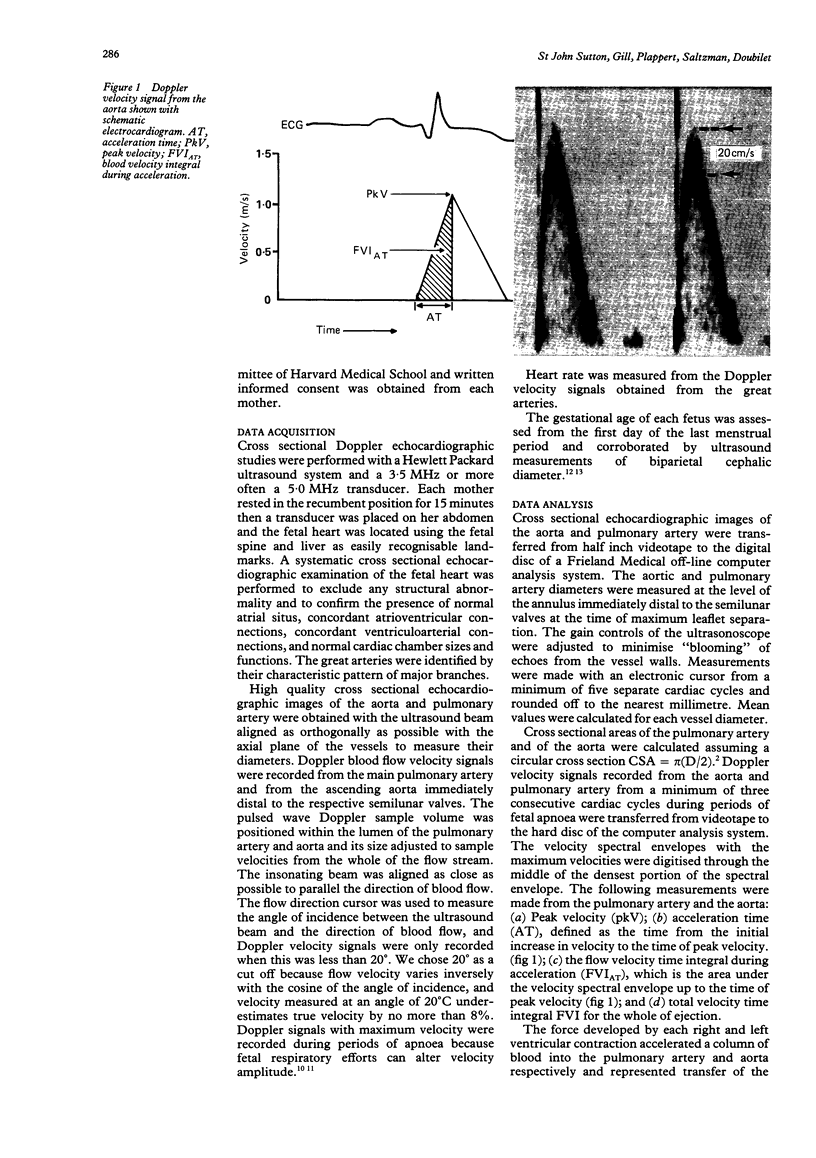
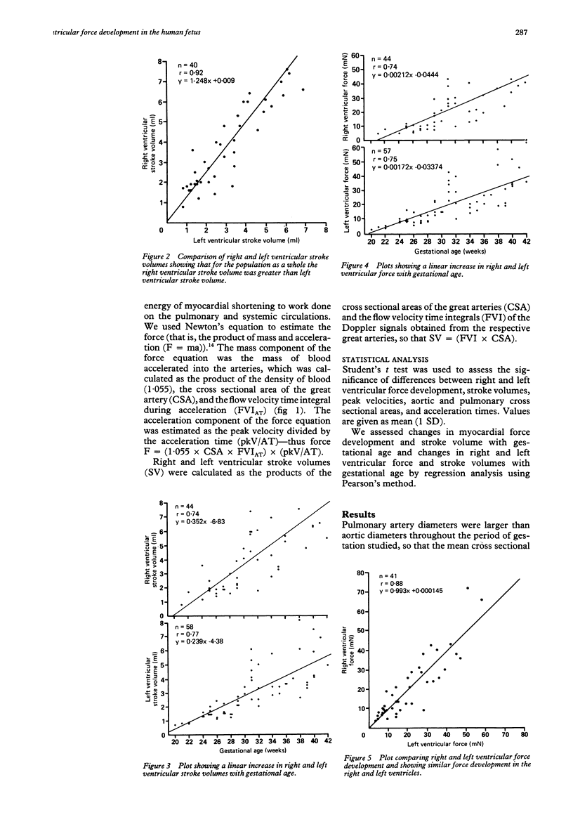
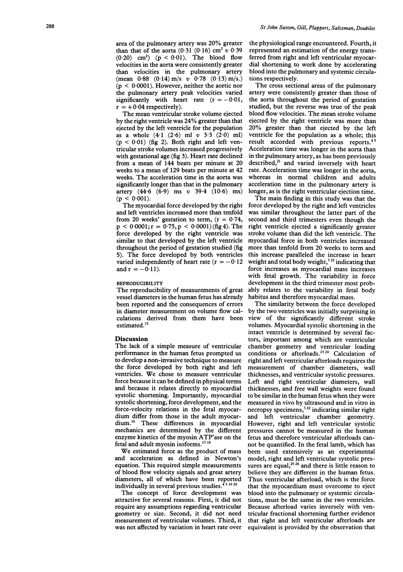
OBJECTIVE--To assess right and left ventricular systolic function in normal human fetuses by cross sectional Doppler echocardiography to calculate the force developed by myocardial shortening. DESIGN--Cross sectional echocardiographic images of the aorta and pulmonary arteries were obtained prospectively in order to measure great vessel diameters and calculate their cross sectional areas. Doppler velocity signals were recorded from the proximal aorta and the proximal pulmonary artery and digitised to obtain peak velocity, acceleration time, flow velocity time integral during acceleration, and the flow velocity time integral for the whole of ejection. Right and left ventricular force development was estimated by Newton's equation in which force is defined as the product of mass and acceleration. PATIENTS--58 normal human fetuses at a gestational age of from 20 to 42 weeks. RESULTS--The cross sectional area of the pulmonary artery was 20% greater than that of the aorta. Aortic acceleration time was longer than that in the pulmonary artery, and peak blood flow velocity in the aorta was consistently greater than that in the pulmonary artery. Right ventricular stroke volume was significantly greater than left ventricular stroke volume. The force developed by the right and left ventricles was, however, similar throughout the gestational period studied, increasing tenfold from 20 weeks' gestation to term (r = 0.74, p less than 0.0001; r = 0.75, p less than 0.0001) respectively. CONCLUSION--The development of right and left ventricular force in the human fetus is similar in spite of the greater volume handled by the right ventricle. This index of ventricular performance does not require calculation of ventricular volume and because it varies independently of ventricular geometry and heart rate it should prove useful in assessing cardiac function in the normal human fetus and in fetuses with ventricular dysfunction.
Full text
PDF
Images in this article
Selected References
These references are in PubMed. This may not be the complete list of references from this article.
- Allan L. D., Joseph M. C., Boyd E. G., Campbell S., Tynan M. M-mode echocardiography in the developing human fetus. Br Heart J. 1982 Jun;47(6):573–583. doi: 10.1136/hrt.47.6.573. [DOI] [PMC free article] [PubMed] [Google Scholar]
- Anderson P. A., Glick K. L., Killam A. P., Mainwaring R. D. The effect of heart rate on in utero left ventricular output in the fetal sheep. J Physiol. 1986 Mar;372:557–573. doi: 10.1113/jphysiol.1986.sp016025. [DOI] [PMC free article] [PubMed] [Google Scholar]
- Angelini A., Allan L. D., Anderson R. H., Crawford D. C., Chita S. K., Ho S. Y. Measurements of the dimensions of the aortic and pulmonary pathways in the human fetus: a correlative echocardiographic and morphometric study. Br Heart J. 1988 Sep;60(3):221–226. doi: 10.1136/hrt.60.3.221. [DOI] [PMC free article] [PubMed] [Google Scholar]
- Borow K. M., Green L. H., Grossman W., Braunwald E. Left ventricular end-systolic stress-shortening and stress-length relations in human. Normal values and sensitivity to inotropic state. Am J Cardiol. 1982 Dec;50(6):1301–1308. doi: 10.1016/0002-9149(82)90467-2. [DOI] [PubMed] [Google Scholar]
- Campbell S., Newman G. B. Growth of the fetal biparietal diameter during normal pregnancy. J Obstet Gynaecol Br Commonw. 1971 Jun;78(6):513–519. doi: 10.1111/j.1471-0528.1971.tb00309.x. [DOI] [PubMed] [Google Scholar]
- Carey R. A., Bove A. A., Coulson R. L., Spann J. F. Correlation between cardiac muscle myosin ATPase activity and velocity of muscle shortening. Biochem Med. 1979 Jun;21(3):235–245. doi: 10.1016/0006-2944(79)90078-4. [DOI] [PubMed] [Google Scholar]
- Cartier M. S., Davidoff A., Warneke L. A., Hirsh M. P., Bannon S., Sutton M. S., Doubilet P. M. The normal diameter of the fetal aorta and pulmonary artery: echocardiographic evaluation in utero. AJR Am J Roentgenol. 1987 Nov;149(5):1003–1007. doi: 10.2214/ajr.149.5.1003. [DOI] [PubMed] [Google Scholar]
- Friedman W. F. The intrinsic physiologic properties of the developing heart. Prog Cardiovasc Dis. 1972 Jul-Aug;15(1):87–111. doi: 10.1016/0033-0620(72)90006-0. [DOI] [PubMed] [Google Scholar]
- Gill R. W. Measurement of blood flow by ultrasound: accuracy and sources of error. Ultrasound Med Biol. 1985 Jul-Aug;11(4):625–641. doi: 10.1016/0301-5629(85)90035-3. [DOI] [PubMed] [Google Scholar]
- Griffin D., Cohen-Overbeek T., Campbell S. Fetal and utero-placental blood flow. Clin Obstet Gynaecol. 1983 Dec;10(3):565–602. [PubMed] [Google Scholar]
- Isaaz K., Ethevenot G., Admant P., Brembilla B., Pernot C. A new Doppler method of assessing left ventricular ejection force in chronic congestive heart failure. Am J Cardiol. 1989 Jul 1;64(1):81–87. doi: 10.1016/0002-9149(89)90657-7. [DOI] [PubMed] [Google Scholar]
- Katz V. L., Seeds J. W. Fetal and neonatal cardiovascular complications from beta-sympathomimetic therapy for tocolysis. Am J Obstet Gynecol. 1989 Jul;161(1):1–4. doi: 10.1016/0002-9378(89)90219-6. [DOI] [PubMed] [Google Scholar]
- Kenny J. F., Plappert T., Doubilet P., Saltzman D. H., Cartier M., Zollars L., Leatherman G. F., St John Sutton M. G. Changes in intracardiac blood flow velocities and right and left ventricular stroke volumes with gestational age in the normal human fetus: a prospective Doppler echocardiographic study. Circulation. 1986 Dec;74(6):1208–1216. doi: 10.1161/01.cir.74.6.1208. [DOI] [PubMed] [Google Scholar]
- Kenny J., Plappert T., Doubilet P., Salzman D., Sutton M. G. Effects of heart rate on ventricular size, stroke volume, and output in the normal human fetus: a prospective Doppler echocardiographic study. Circulation. 1987 Jul;76(1):52–58. doi: 10.1161/01.cir.76.1.52. [DOI] [PubMed] [Google Scholar]
- Kleinman C. S., Donnerstein R. L., DeVore G. R., Jaffe C. C., Lynch D. C., Berkowitz R. L., Talner N. S., Hobbins J. C. Fetal echocardiography for evaluation of in utero congestive heart failure. N Engl J Med. 1982 Mar 11;306(10):568–575. doi: 10.1056/NEJM198203113061003. [DOI] [PubMed] [Google Scholar]
- Kurtz A. B., Wapner R. J., Kurtz R. J., Dershaw D. D., Rubin C. S., Cole-Beuglet C., Goldberg B. B. Analysis of biparietal diameter as an accurate indicator of gestational age. J Clin Ultrasound. 1980 Aug;8(4):319–326. doi: 10.1002/jcu.1870080406. [DOI] [PubMed] [Google Scholar]
- Machado M. V., Chita S. C., Allan L. D. Acceleration time in the aorta and pulmonary artery measured by Doppler echocardiography in the midtrimester normal human fetus. Br Heart J. 1987 Jul;58(1):15–18. doi: 10.1136/hrt.58.1.15. [DOI] [PMC free article] [PubMed] [Google Scholar]
- Sahn D. J., Lange L. W., Allen H. D., Goldberg S. J., Anderson C., Giles H., Haber K. Quantitative real-time cross-sectional echocardiography in the developing normal humam fetus and newborn. Circulation. 1980 Sep;62(3):588–597. doi: 10.1161/01.cir.62.3.588. [DOI] [PubMed] [Google Scholar]
- St John Sutton M. G., Gewitz M. H., Shah B., Cohen A., Reichek N., Gabbe S., Huff D. S. Quantitative assessment of growth and function of the cardiac chambers in the normal human fetus: a prospective longitudinal echocardiographic study. Circulation. 1984 Apr;69(4):645–654. doi: 10.1161/01.cir.69.4.645. [DOI] [PubMed] [Google Scholar]
- St John Sutton M. G., Raichlen J. S., Reichek N., Huff D. S. Quantitative assessment of right and left ventricular growth in the human fetal heart: a pathoanatomic study. Circulation. 1984 Dec;70(6):935–941. doi: 10.1161/01.cir.70.6.935. [DOI] [PubMed] [Google Scholar]
- Sutton M. S., Theard M. A., Bhatia S. J., Plappert T., Saltzman D. H., Doubilet P. Changes in placental blood flow in the normal human fetus with gestational age. Pediatr Res. 1990 Oct;28(4):383–387. doi: 10.1203/00006450-199010000-00016. [DOI] [PubMed] [Google Scholar]
- Weber K. T., Janicki J. S. The dynamics of ventricular contraction: force, length, and shortening. Fed Proc. 1980 Feb;39(2):188–195. [PubMed] [Google Scholar]


