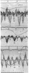
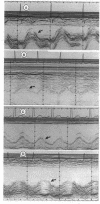
Abstract
OBJECTIVE--To study the time course and underlying mechanisms of right heart filling after cardiac surgery. DESIGN--A prospective observational study of adult patients undergoing cardiac surgery. SETTING--Echocardiography laboratory of the Stanford University Medical Center. PATIENTS--Twenty six patients (mean age 54.9) undergoing cardiac surgery were studied before and two days, one week, six weeks, and six months after cardiac surgery. MAIN OUTCOME MEASURES--Flow in the hepatic veins and superior vena cava, tricuspid and mitral annulus motion, signs of tricuspid regurgitation, and right ventricular size were assessed by echocardiography. RESULTS--Right heart filling, expressed as the ratio of systolic to diastolic forward flow Doppler velocity integrals in the superior vena cava and by tricuspid annulus motion, decreased in parallel from before surgery baseline values of 3.5 (SD 3.1) and 21.9 (3.4) mm, respectively to 0.2 (0.1) and 8.1 (2.3) mm two days after operation. A gradual increase towards baseline values was noted after six months, to 1.4 (1.3) and 15.1 (2.3) mm respectively; however, these values were still significantly less than those before operation. Similar changes were seen in the hepatic venous flow pattern. The decrease in total tricuspid annulus motion was most pronounced in its lateral segment and the atrial component of the tricuspid annulus motion showed similar changes. CONCLUSIONS--The pronounced decrease in tricuspid annulus motion during the early postoperative period suggests right atrial and right ventricular dysfunction as mechanisms responsible for the early changes seen. The progressive return to a normal venous filling pattern and the partial recovery of annular motion six months after operation further support the influence of the above mechanisms, as well as their resolution with time. The persistent flow abnormalities and compromised motion of the free aspects of the tricuspid annulus, however, suggest long term tethering of the right heart wall.
Full text
PDF
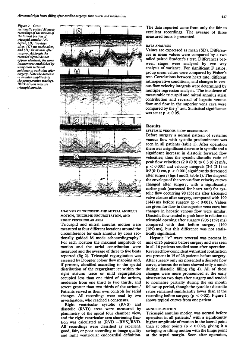
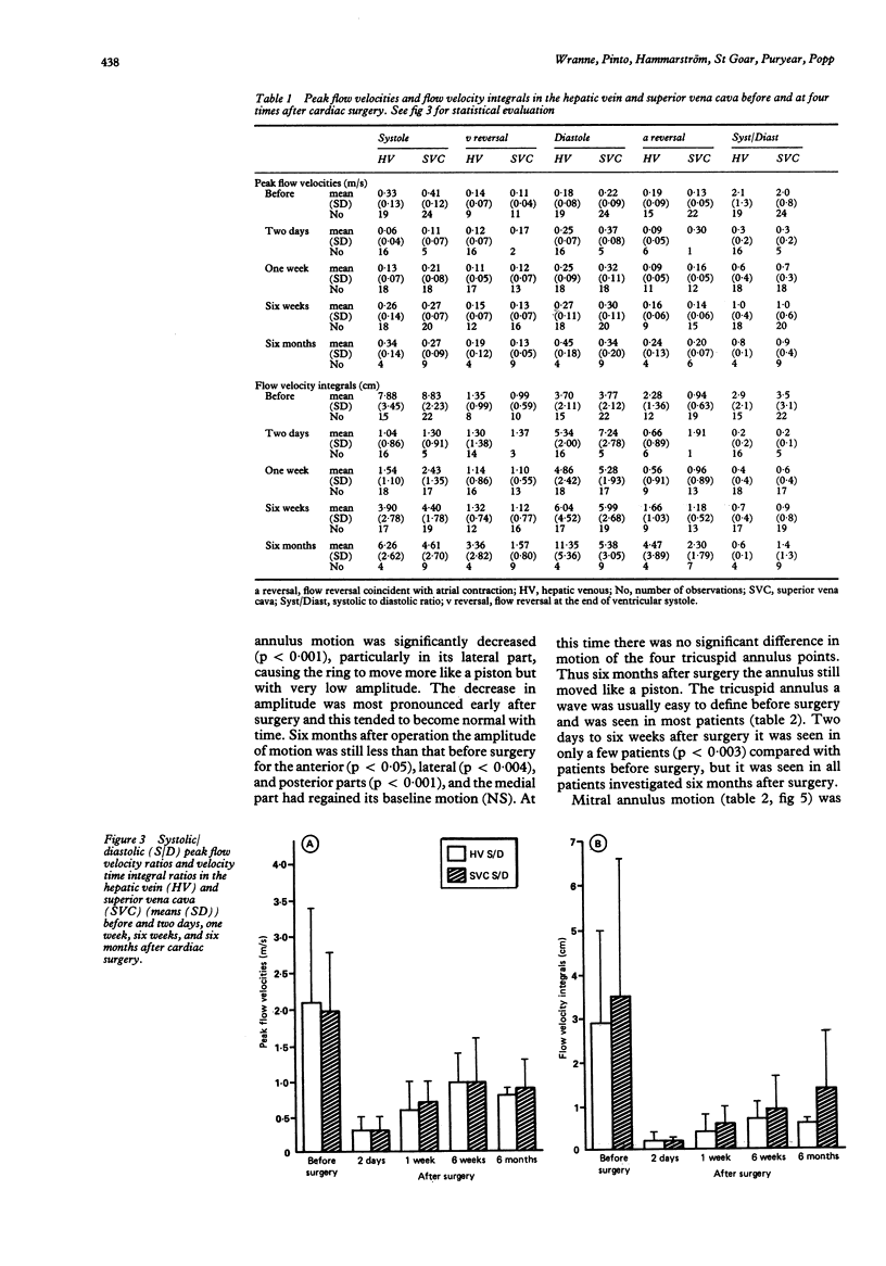
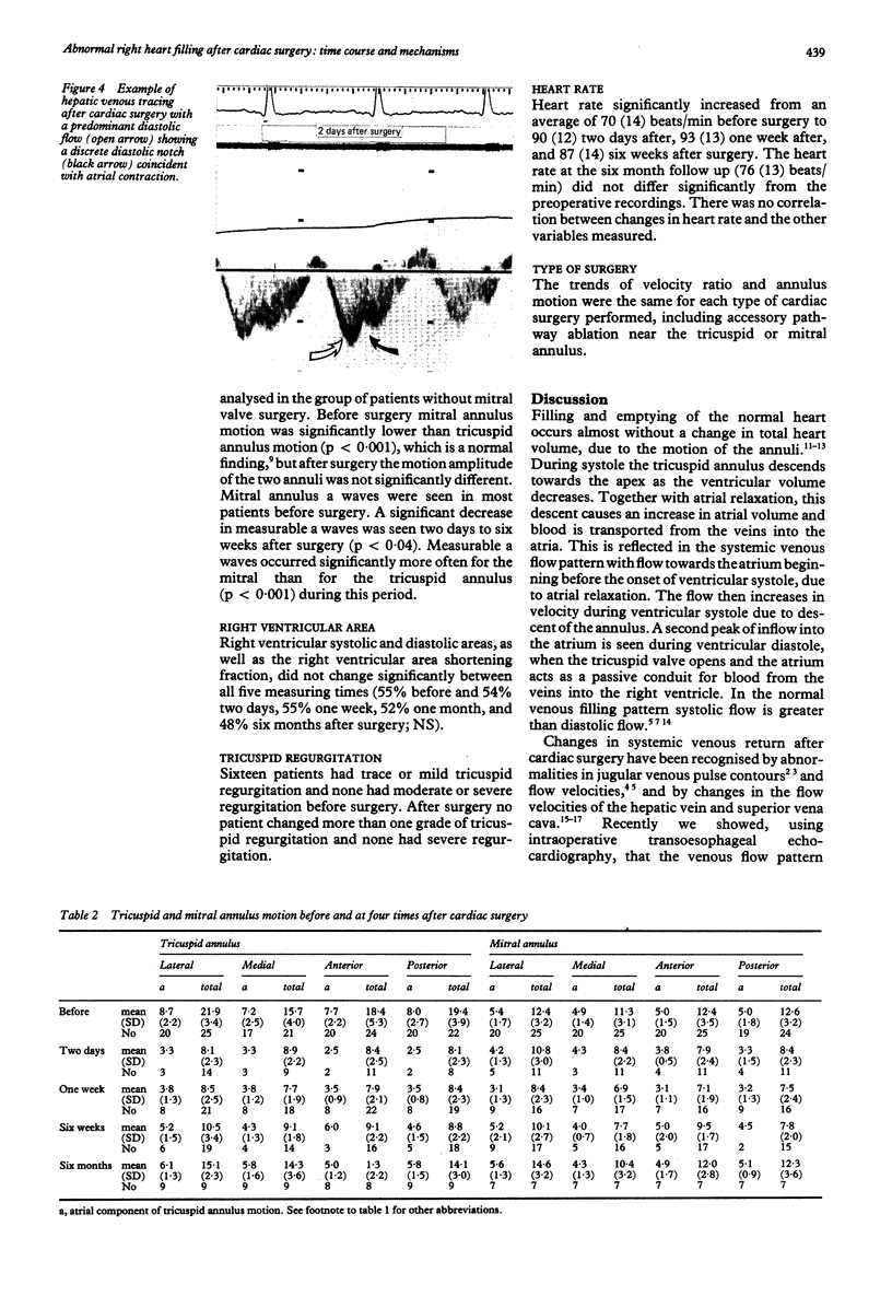
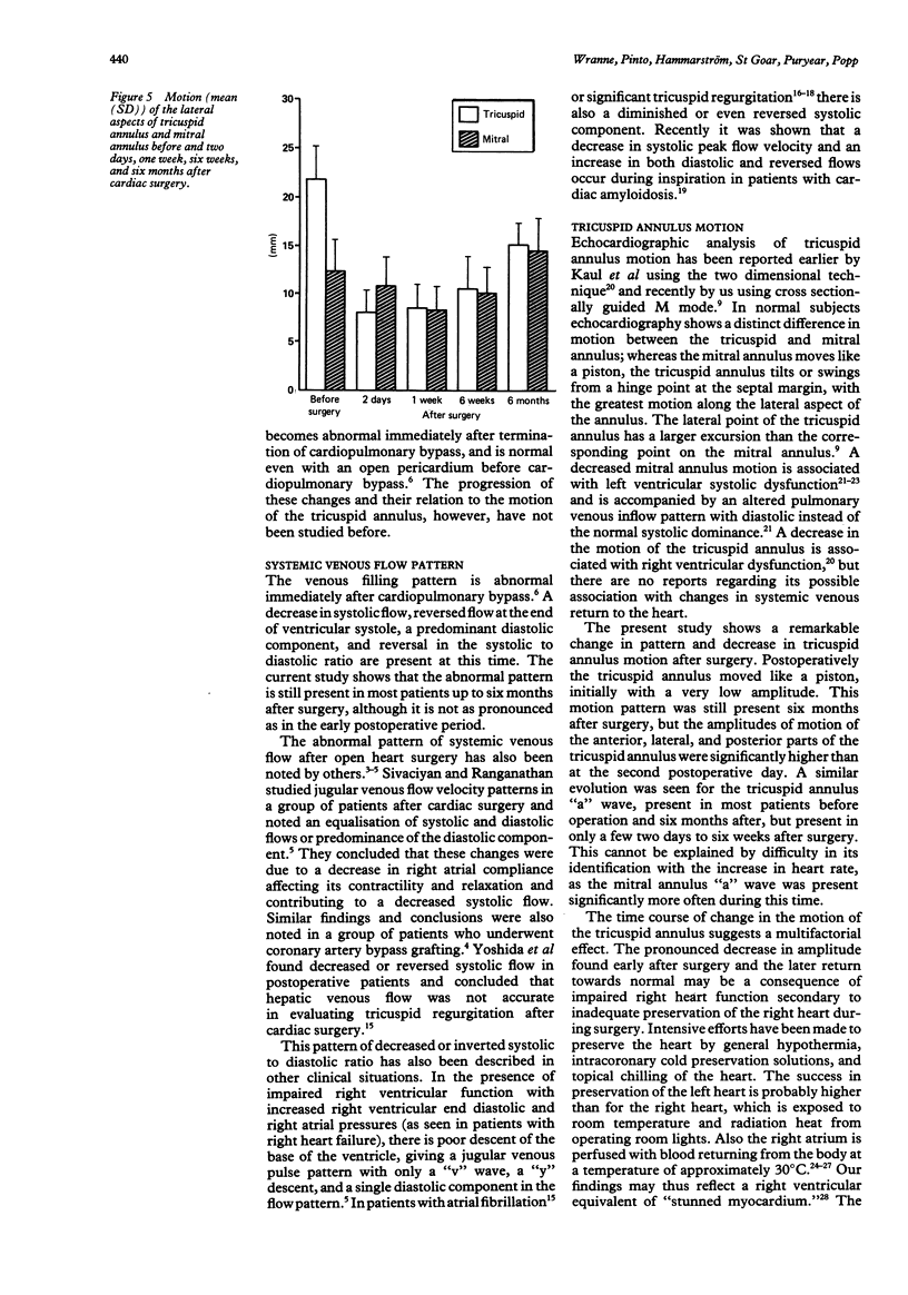
Images in this article
Selected References
These references are in PubMed. This may not be the complete list of references from this article.
- Appleton C. P., Hatle L. K., Popp R. L. Superior vena cava and hepatic vein Doppler echocardiography in healthy adults. J Am Coll Cardiol. 1987 Nov;10(5):1032–1039. doi: 10.1016/s0735-1097(87)80343-1. [DOI] [PubMed] [Google Scholar]
- Bolli R. Mechanism of myocardial "stunning". Circulation. 1990 Sep;82(3):723–738. doi: 10.1161/01.cir.82.3.723. [DOI] [PubMed] [Google Scholar]
- Choong C. Y., Abascal V. M., Thomas J. D., Guerrero J. L., McGlew S., Weyman A. E. Combined influence of ventricular loading and relaxation on the transmitral flow velocity profile in dogs measured by Doppler echocardiography. Circulation. 1988 Sep;78(3):672–683. doi: 10.1161/01.cir.78.3.672. [DOI] [PubMed] [Google Scholar]
- Christakis G. T., Fremes S. E., Weisel R. D., Ivanov J., Madonik M. M., Seawright S. J., McLaughlin P. R. Right ventricular dysfunction following cold potassium cardioplegia. J Thorac Cardiovasc Surg. 1985 Aug;90(2):243–250. [PubMed] [Google Scholar]
- Cohen M. L., Cohen B. S., Kronzon I., Lighty G. W., Winer H. E. Superior vena caval blood flow velocities in adults: a Doppler echocardiographic study. J Appl Physiol (1985) 1986 Jul;61(1):215–219. doi: 10.1152/jappl.1986.61.1.215. [DOI] [PubMed] [Google Scholar]
- Czer L. S., Maurer G., Bolger A., DeRobertis M., Kleinman J., Gray R. J., Chaux A., Matloff J. M. Tricuspid valve repair. Operative and follow-up evaluation by Doppler color flow mapping. J Thorac Cardiovasc Surg. 1989 Jul;98(1):101–111. [PubMed] [Google Scholar]
- Feinstein A. R. T. Duckett Jones Memorial Lecture. The Jones criteria and the challenges of clinimetrics. Circulation. 1982 Jul;66(1):1–5. doi: 10.1161/01.cir.66.1.1. [DOI] [PubMed] [Google Scholar]
- Fisk R. L., Ghaswalla D., Guilbeau E. J. Asymmetrical myocardial hypothermia during hypothermic cardioplegia. Ann Thorac Surg. 1982 Sep;34(3):318–323. doi: 10.1016/s0003-4975(10)62503-9. [DOI] [PubMed] [Google Scholar]
- Gonzalez A. C., Brandon T. A., Fortune R. L., Casano S. F., Martin M., Benneson D. L., Guilbeau E. J., Fisk R. L. Acute right ventricular failure is caused by inadequate right ventricular hypothermia. J Thorac Cardiovasc Surg. 1985 Mar;89(3):386–399. [PubMed] [Google Scholar]
- HARTMAN H. The jugular venous tracing. Am Heart J. 1960 May;59:698–717. doi: 10.1016/0002-8703(60)90511-1. [DOI] [PubMed] [Google Scholar]
- Hammarström E., Wranne B., Pinto F. J., Puryear J., Popp R. L. Tricuspid annular motion. J Am Soc Echocardiogr. 1991 Mar-Apr;4(2):131–139. doi: 10.1016/s0894-7317(14)80524-5. [DOI] [PubMed] [Google Scholar]
- Hoffman E. A., Ritman E. L. Heart-lung interaction: effect on regional lung air content and total heart volume. Ann Biomed Eng. 1987;15(3-4):241–257. doi: 10.1007/BF02584282. [DOI] [PubMed] [Google Scholar]
- Hoffman E. A., Ritman E. L. Invariant total heart volume in the intact thorax. Am J Physiol. 1985 Oct;249(4 Pt 2):H883–H890. doi: 10.1152/ajpheart.1985.249.4.H883. [DOI] [PubMed] [Google Scholar]
- Höglund C., Alam M., Thorstrand C. Effects of acute myocardial infarction on the displacement of the atrioventricular plane: an echocardiographic study. J Intern Med. 1989 Oct;226(4):251–256. doi: 10.1111/j.1365-2796.1989.tb01389.x. [DOI] [PubMed] [Google Scholar]
- Ishida Y., Meisner J. S., Tsujioka K., Gallo J. I., Yoran C., Frater R. W., Yellin E. L. Left ventricular filling dynamics: influence of left ventricular relaxation and left atrial pressure. Circulation. 1986 Jul;74(1):187–196. doi: 10.1161/01.cir.74.1.187. [DOI] [PubMed] [Google Scholar]
- Kaul S., Tei C., Hopkins J. M., Shah P. M. Assessment of right ventricular function using two-dimensional echocardiography. Am Heart J. 1984 Mar;107(3):526–531. doi: 10.1016/0002-8703(84)90095-4. [DOI] [PubMed] [Google Scholar]
- Keren G., Sonnenblick E. H., LeJemtel T. H. Mitral anulus motion. Relation to pulmonary venous and transmitral flows in normal subjects and in patients with dilated cardiomyopathy. Circulation. 1988 Sep;78(3):621–629. doi: 10.1161/01.cir.78.3.621. [DOI] [PubMed] [Google Scholar]
- Klein A. L., Hatle L. K., Burstow D. J., Taliercio C. P., Seward J. B., Kyle R. A., Bailey K. R., Gertz M. A., Tajik A. J. Comprehensive Doppler assessment of right ventricular diastolic function in cardiac amyloidosis. J Am Coll Cardiol. 1990 Jan;15(1):99–108. doi: 10.1016/0735-1097(90)90183-p. [DOI] [PubMed] [Google Scholar]
- Nishimura R. A., Abel M. D., Hatle L. K., Tajik A. J. Relation of pulmonary vein to mitral flow velocities by transesophageal Doppler echocardiography. Effect of different loading conditions. Circulation. 1990 May;81(5):1488–1497. doi: 10.1161/01.cir.81.5.1488. [DOI] [PubMed] [Google Scholar]
- Pennestrí F., Loperfido F., Salvatori M. P., Mongiardo R., Ferrazza A., Guccione P., Manzoli U. Assessment of tricuspid regurgitation by pulsed Doppler ultrasonography of the hepatic veins. Am J Cardiol. 1984 Aug 1;54(3):363–368. doi: 10.1016/0002-9149(84)90198-x. [DOI] [PubMed] [Google Scholar]
- RUSHMER R. F., CRYSTAL D. K. Changes in configuration of the ventricular chambers during the cardiac cycle. Circulation. 1951 Aug;4(2):211–218. doi: 10.1161/01.cir.4.2.211. [DOI] [PubMed] [Google Scholar]
- Ranganathan N., Sivaciyan V., Pryszlak M., Freeman M. R. Changes in jugular venous flow velocity after coronary artery bypass grafting. Am J Cardiol. 1989 Mar 15;63(11):725–729. doi: 10.1016/0002-9149(89)90259-2. [DOI] [PubMed] [Google Scholar]
- Sakai K., Nakamura K., Satomi G., Kondo M., Hirosawa K. Evaluation of tricuspid regurgitation by blood flow pattern in the hepatic vein using pulsed Doppler technique. Am Heart J. 1984 Sep;108(3 Pt 1):516–523. doi: 10.1016/0002-8703(84)90417-4. [DOI] [PubMed] [Google Scholar]
- Sakai K., Nakamura K., Satomi G., Kondo M., Hirosawa K. [Hepatic vein blood flow pattern measured by Doppler echocardiography as an evaluation of tricuspid valve insufficiency]. J Cardiogr. 1983 Mar;13(1):33–43. [PubMed] [Google Scholar]
- Simonson J. S., Schiller N. B. Descent of the base of the left ventricle: an echocardiographic index of left ventricular function. J Am Soc Echocardiogr. 1989 Jan-Feb;2(1):25–35. doi: 10.1016/s0894-7317(89)80026-4. [DOI] [PubMed] [Google Scholar]
- Sivaciyan V., Ranganathan N. Transcutaneous doppler jugular venous flow velocity recording. Circulation. 1978 May;57(5):930–939. doi: 10.1161/01.cir.57.5.930. [DOI] [PubMed] [Google Scholar]
- Velardi A. R., Widmer S. J., Cilley J. H., Jr, Spence R. K., Witkowski T. A., DelRossi A. J. Right ventricular myocardial protection through intracavitary cooling in cardiac operations. J Thorac Cardiovasc Surg. 1989 Dec;98(6):1077–1082. [PubMed] [Google Scholar]
- Wann L. S., Morris S. N., Tavel M. E. Effects of cardiopulmonary bypass on the jugular venous pulse. Am Heart J. 1977 Aug;94(2):262–264. doi: 10.1016/s0002-8703(77)80290-1. [DOI] [PubMed] [Google Scholar]
- Yoshida K., Yoshikawa J., Kato H., Yanagihara K., Okumachi F., Koizumi K., Shiratori K., Asaka T., Suzuki K., Inanami H. [Tricuspid regurgitation evaluated by Doppler hepatic vein flow patterns]. J Cardiogr. 1985 Dec;15(4):1157–1169. [PubMed] [Google Scholar]


