

Abstract
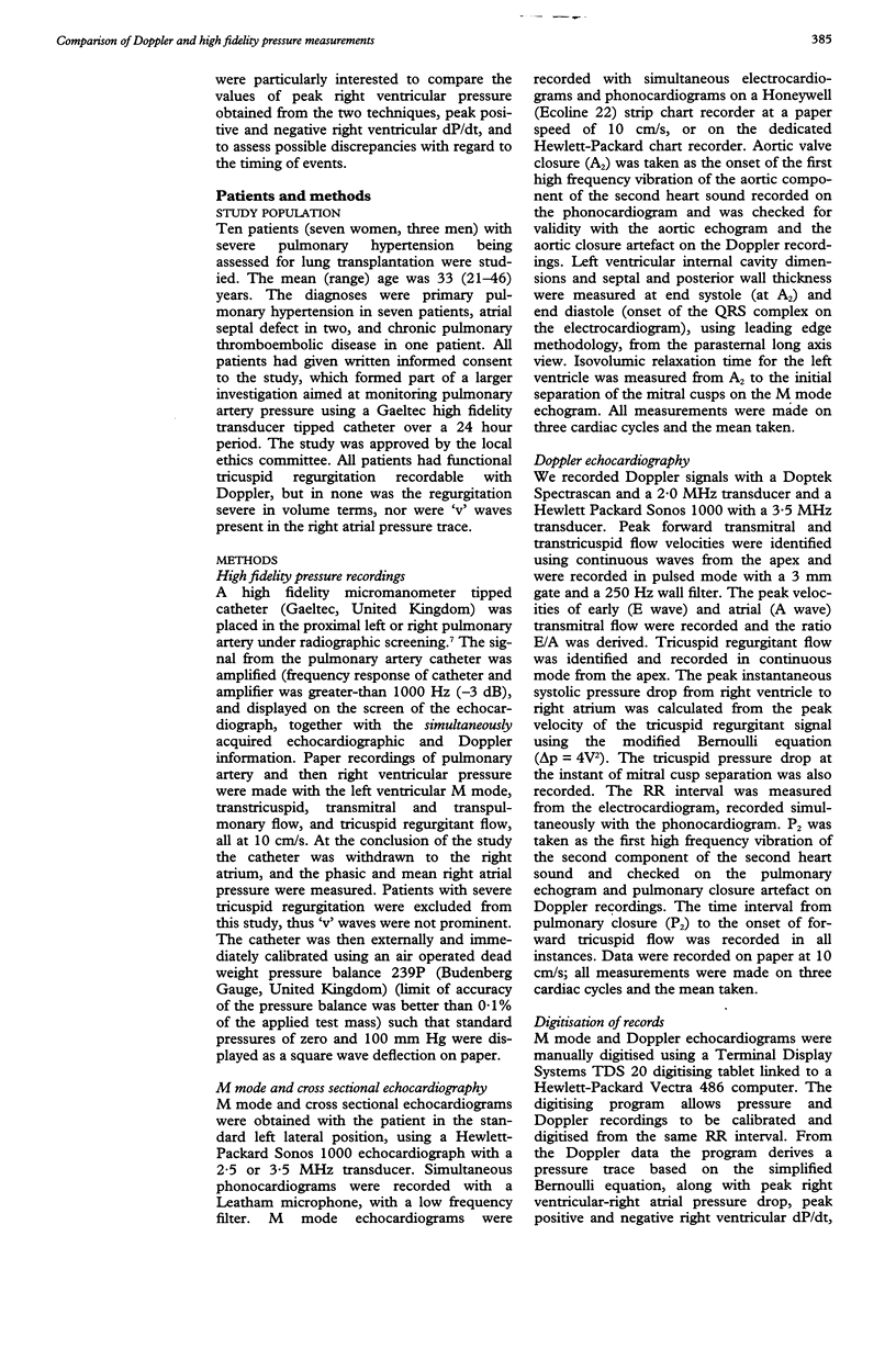
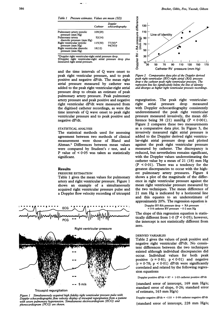
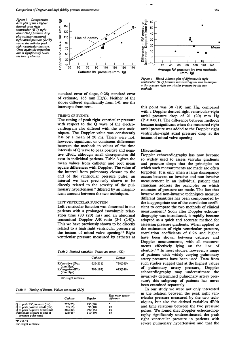
OBJECTIVE--To assess relations between right ventricular pressure measured with a high fidelity transducer tipped catheter and the characteristics of tricuspid regurgitation recorded with Doppler echocardiography. DESIGN--A prospective non-randomised study of patients with severe pulmonary hypertension referred for consideration of lung transplantation. SETTING--A tertiary referral centre for cardiac and pulmonary disease, with facilities for invasive and non-invasive investigation, and assessment for heart and heart-lung transplantation. PATIENTS--10 patients with severe pulmonary hypertension being considered for lung transplantation. ENDPOINTS--Peak right ventricular, pulmonary artery, and right atrial pressures; peak positive and negative right ventricular dP/dt; peak Doppler right ventricular-right atrial pressure drop; Doppler derived peak positive and negative right ventricular dP/dt; and time intervals of Q to peak right ventricular pressure and to peak positive and negative right ventricular dP/dt. RESULTS--The mean (SD) pulmonary artery systolic pressure was 109 (29) mm Hg. The peak Doppler right ventricular-right atrial pressure drop underestimated peak right ventricular pressure by 38 (21) mm Hg, and by 21 (18) mm Hg when the Doppler value was added to the measured right atrial pressure (P values < 0.05). This discrepancy was greater for higher pulmonary artery pressures. The timing of peak right ventricular pressure differed, with the Doppler value consistently shorter (mean difference 16 ms, P < 0.05). Values of peak positive and negative right ventricular dP/dt and the time intervals Q-peak positive right ventricular dP/dt and pulmonary closure to the end of the pressure pulse differed between the two techniques in individual patients, but not in a consistent or predictable way. CONCLUSIONS--Doppler echocardiography significantly underestimates the peak right ventricular pressure and the time interval to peak right ventricular pressure in pulmonary hypertension, particularly when severe. These differences may be related to orifice geometry. Digitisation of Doppler records of tricuspid regurgitation provides useful semiquantitative estimates of absolute values and timing of peak positive and negative right ventricular dP/dt. Clinically significant differences may exist, however, and must be considered in individual patients.
Full text
PDF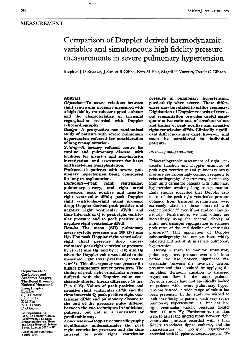
Selected References
These references are in PubMed. This may not be the complete list of references from this article.
- Baumgartner H., Khan S., DeRobertis M., Czer L., Maurer G. Discrepancies between Doppler and catheter gradients in aortic prosthetic valves in vitro. A manifestation of localized gradients and pressure recovery. Circulation. 1990 Oct;82(4):1467–1475. doi: 10.1161/01.cir.82.4.1467. [DOI] [PubMed] [Google Scholar]
- Bland J. M., Altman D. G. Statistical methods for assessing agreement between two methods of clinical measurement. Lancet. 1986 Feb 8;1(8476):307–310. [PubMed] [Google Scholar]
- Brecker S. J., Xiao H. B., Stojnic B. B., Mbaissouroum M., Gibson D. G. Assessment of the peak tricuspid regurgitant velocity from the dynamics of retrograde flow. Int J Cardiol. 1992 Mar;34(3):267–271. doi: 10.1016/0167-5273(92)90023-v. [DOI] [PubMed] [Google Scholar]
- Chen C., Rodriguez L., Levine R. A., Weyman A. E., Thomas J. D. Noninvasive measurement of the time constant of left ventricular relaxation using the continuous-wave Doppler velocity profile of mitral regurgitation. Circulation. 1992 Jul;86(1):272–278. doi: 10.1161/01.cir.86.1.272. [DOI] [PubMed] [Google Scholar]
- Currie P. J., Seward J. B., Chan K. L., Fyfe D. A., Hagler D. J., Mair D. D., Reeder G. S., Nishimura R. A., Tajik A. J. Continuous wave Doppler determination of right ventricular pressure: a simultaneous Doppler-catheterization study in 127 patients. J Am Coll Cardiol. 1985 Oct;6(4):750–756. doi: 10.1016/s0735-1097(85)80477-0. [DOI] [PubMed] [Google Scholar]
- Gibbs J. S., Cunningham D., Sparrow J., Poole-Wilson P. A., Fox K. M. Unpredictable zero drift in intravascular micromanometer tipped catheters during long term pulmonary artery pressure recording: implications for catheter design. Cardiovasc Res. 1989 Feb;23(2):152–158. doi: 10.1093/cvr/23.2.152. [DOI] [PubMed] [Google Scholar]
- Himelman R. B., Stulbarg M., Kircher B., Lee E., Kee L., Dean N. C., Golden J., Wolfe C. L., Schiller N. B. Noninvasive evaluation of pulmonary artery pressure during exercise by saline-enhanced Doppler echocardiography in chronic pulmonary disease. Circulation. 1989 Apr;79(4):863–871. doi: 10.1161/01.cir.79.4.863. [DOI] [PubMed] [Google Scholar]
- Jessup M., Sutton M. S., Weber K. T., Janicki J. S. The effect of chronic pulmonary hypertension on left ventricular size, function, and interventricular septal motion. Am Heart J. 1987 May;113(5):1114–1122. doi: 10.1016/0002-8703(87)90921-5. [DOI] [PubMed] [Google Scholar]
- Louie E. K., Rich S., Brundage B. H. Doppler echocardiographic assessment of impaired left ventricular filling in patients with right ventricular pressure overload due to primary pulmonary hypertension. J Am Coll Cardiol. 1986 Dec;8(6):1298–1306. doi: 10.1016/s0735-1097(86)80300-x. [DOI] [PubMed] [Google Scholar]
- Segal J., Lerner D. J., Miller D. C., Mitchell R. S., Alderman E. A., Popp R. L. When should Doppler-determined valve area be better than the Gorlin formula?: Variation in hydraulic constants in low flow states. J Am Coll Cardiol. 1987 Jun;9(6):1294–1305. doi: 10.1016/s0735-1097(87)80469-2. [DOI] [PubMed] [Google Scholar]
- Skjaerpe T., Hatle L. Noninvasive estimation of systolic pressure in the right ventricle in patients with tricuspid regurgitation. Eur Heart J. 1986 Aug;7(8):704–710. doi: 10.1093/oxfordjournals.eurheartj.a062126. [DOI] [PubMed] [Google Scholar]
- Stewart S. F., Nast E. P., Arabia F. A., Talbot T. L., Proschan M., Clark R. E. Errors in pressure gradient measurement by continuous wave Doppler ultrasound: type, size and age effects in bioprosthetic aortic valves. J Am Coll Cardiol. 1991 Sep;18(3):769–779. doi: 10.1016/0735-1097(91)90801-f. [DOI] [PubMed] [Google Scholar]
- Stojnic B. B., Brecker S. J., Xiao H. B., Helmy S. M., Mbaissouroum M., Gibson D. G. Left ventricular filling characteristics in pulmonary hypertension: a new mode of ventricular interaction. Br Heart J. 1992 Jul;68(1):16–20. doi: 10.1136/hrt.68.7.16. [DOI] [PMC free article] [PubMed] [Google Scholar]
- Xiao H. B., Brecker S. J., Gibson D. G. Effects of abnormal activation on the time course of the left ventricular pressure pulse in dilated cardiomyopathy. Br Heart J. 1992 Oct;68(4):403–407. doi: 10.1136/hrt.68.10.403. [DOI] [PMC free article] [PubMed] [Google Scholar]
- Yock P. G., Popp R. L. Noninvasive estimation of right ventricular systolic pressure by Doppler ultrasound in patients with tricuspid regurgitation. Circulation. 1984 Oct;70(4):657–662. doi: 10.1161/01.cir.70.4.657. [DOI] [PubMed] [Google Scholar]
- Yoganathan A. P., Cape E. G., Sung H. W., Williams F. P., Jimoh A. Review of hydrodynamic principles for the cardiologist: applications to the study of blood flow and jets by imaging techniques. J Am Coll Cardiol. 1988 Nov;12(5):1344–1353. doi: 10.1016/0735-1097(88)92620-4. [DOI] [PubMed] [Google Scholar]