Abstract
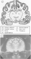
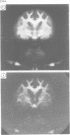
Triethyl tin(TET)-induced cerebral oedema has been studied in cats by magnetic resonance imaging (MRI), and the findings correlated with the histology and fine structure of the cerebrum following perfusion-fixation. MRI is a sensitive technique for detecting cerebral oedema, and the distribution and severity of the changes correlate closely with the morphological abnormalities. The relaxation times, T1 and T2 increase progressively as the oedema develops, and the proportional increase in T2 is approximately twice that in T1. Analysis of the magnetisation decay curves reveals slowly-relaxing and rapidly-relaxing components which probably correspond to oedema fluid and intracellular water respectively. The image appearances taken in conjunction with relaxation data provide a basis for determining the nature of the oedema in vivo.
Full text
PDF
Images in this article
Selected References
These references are in PubMed. This may not be the complete list of references from this article.
- Aleu F. P., Katzman R., Terry R. D. Fine structure and electrolyte analyses of cerebral edema induced by alkyl tin intoxication. J Neuropathol Exp Neurol. 1963 Jul;22(3):403–413. doi: 10.1097/00005072-196307000-00003. [DOI] [PubMed] [Google Scholar]
- Bakay L., Kurland R. J., Parrish R. G., Lee J. C., Peng R. J., Bartkowski H. M. Nuclear magnetic resonance studies in normal and edematous brain tissue. Exp Brain Res. 1975 Sep 29;23(3):241–248. doi: 10.1007/BF00239737. [DOI] [PubMed] [Google Scholar]
- Bakay L. Morphological and chemical studies in cerebral edema: triethyl tin-induced edema. J Neurol Sci. 1965 Jan-Feb;2(1):52–67. doi: 10.1016/0022-510x(65)90062-6. [DOI] [PubMed] [Google Scholar]
- Foster K. R., Resing H. A., Garroway A. N. Bounds on "bound water": transverse nuclear magnetic resonance relaxation in barnacle muscle. Science. 1976 Oct 15;194(4262):324–326. doi: 10.1126/science.968484. [DOI] [PubMed] [Google Scholar]
- Gwan K., Edzes H. T. Water in brain edema. Observations by the pulsed nuclear magnetic resonance technique. Arch Neurol. 1975 Jul;32(7):462–465. doi: 10.1001/archneur.1975.00490490066006. [DOI] [PubMed] [Google Scholar]
- Hirano A., Zimmerman H. M., Levine S. Intramyelinic and extracellular spaces in triethyltin intoxication. J Neuropathol Exp Neurol. 1968 Oct;27(4):571–580. [PubMed] [Google Scholar]
- Mathur-De Vré R. Biomedical implications of the relaxation behaviour of water related to NMR imaging. Br J Radiol. 1984 Nov;57(683):955–976. doi: 10.1259/0007-1285-57-683-955. [DOI] [PubMed] [Google Scholar]
- Partin J. C., Partin J. S., Schubert W. K., McLaurin R. L. Brain ultrastructure in Reye's syndrome. J Neuropathol Exp Neurol. 1975 Sep;34(5):425–444. doi: 10.1097/00005072-197509000-00005. [DOI] [PubMed] [Google Scholar]
- Rees S., Cragg B. G., Everitt A. V. Comparison of extracellular space in the mature and aging rat brain using a new technique. J Neurol Sci. 1982 Feb;53(2):347–357. doi: 10.1016/0022-510x(82)90018-1. [DOI] [PubMed] [Google Scholar]
- Young I. R., Bailes D. R., Burl M., Collins A. G., Smith D. T., McDonnell M. J., Orr J. S., Banks L. M., Bydder G. M., Greenspan R. H. Initial clinical evaluation of a whole body nuclear magnetic resonance (NMR) tomograph. J Comput Assist Tomogr. 1982 Feb;6(1):1–18. doi: 10.1097/00004728-198202000-00001. [DOI] [PubMed] [Google Scholar]


