Abstract
RNA editing is a post-transcriptional modification with a cell-specific manner and important biological implications. Although single-cell RNA-seq (scRNA-seq) is an effective method for studying cellular heterogeneity, it is difficult to detect and study RNA editing events from scRNA-seq data because of the low sequencing coverage. To overcome this, we develop a computational method to systematically identify RNA editing sites of cell types from scRNA-seq data. To demonstrate its effectiveness, we apply it to scRNA-seq data of human hematopoietic stem/progenitor cells (HSPCs) with an annotated lineage differentiation relationship according to previous research and study the impacts of RNA editing on hematopoiesis. The dynamic editing patterns reveal the relevance of RNA editing on different HSPCs. For example, four microRNA (miRNA) target sites on 3ʹ UTR of EIF2AK2 are edited across all HSPC populations, which may abolish the miRNA-mediated inhibition of EIF2AK2. Elevated EIF2AK2 may thus activate the integrated stress response (ISR) pathway to initiate global translational attenuation as a protective mechanism to maintain cellular homeostasis during HSPCs’ differentiation. Besides, our findings also indicate that RNA editing plays an essential role in the coordination of lineage commitment and self-renewal of hematopoietic stem cells (HSCs). Taken together, we demonstrate the capacity of scRNA-seq data to exploit RNA editing events of cell types, and find that RNA editing may exert multiple modules of regulation in hematopoietic processes.
Subject terms: Cell biology, Computational biology and bioinformatics
Introduction
RNA editing alters the sequence of RNA transcripts dynamically and flexibly during cell development and in a cell type-specific manner1. There are two canonical editing types in mammals, A-to-I and C-to-U2. Among them, A-to-I is the most common form in animal cells3. Most known RNA editing sites located in introns, and 5ʹ/3ʹ untranslated regions (UTRs), especially in the ALU repeat. A small proportion takes place in the coding sequences (CDSs), thus altering the sequence and function of their encoded proteins4–6. RNA editing increases the diversity of the transcriptome and expands the ways of regulation7. It has implications for various biological processes such as transcriptional stability and localization, interactions with other primary RNA processing steps such as splicing and polyadenylation, and the biogenesis and functions of small RNAs such as miRNAs and long noncoding RNAs8–10. Because RNA editing often occurs in the 3ʹ UTRs, in which miRNA binding sites are enriched, it may change the native binding sites or introduce new binding sites, and then affect miRNA-mediated gene silencing11,12. Meanwhile, as one of the important post-transcriptional modifications, RNA editing alters base composition at the transcriptional level also exhibits cell-specific features13,14.
Hematopoietic stem cells (HSCs) are defined as a group of cells with the ability to self-renew and reconstitute the hematopoietic system15,16. They then differentiate into multipotent (produce most blood cell subsets), oligopotent (lymphoid or myeloid restricted), and unipotent hematopoietic progenitor cells (HPCs)17. Hematopoietic stem/progenitor cells (HSPCs) require the collaboration of complex pathways including regulation of the cell cycle, apoptosis, and transcription to maintain the internal homeostasis of differentiation and self-renewal18–24. More importantly, only HSCs own the ability of both multi-potency and self-renewal. The multi-potency of HSCs is reflected in the differentiation into different hematopoietic progenitor cells and functional blood cells. The self-renewal of HSCs is to generate HSCs themselves rather than through differentiation of other cells. The HSCs differentiate into hematopoietic progenitor cells to maintain the stability of the hematopoietic system by balancing differentiation and self-renewal25. As one of the most widespread post-transcriptional modifications, RNA editing also plays an indispensable role and is an important regulator of hematopoietic stem cell maintenance of differentiation function. For example, highly edited Azin1 in HSPCs led to an amino acid change and then altered Azin1 protein (AZI) translocation, and finally enhanced AZI binding affinity for DEAD box polypeptide 1. The edited Azin1 maintains the normal differentiation process of HSCs, whereas loss of Azin1 RNA editing makes HSCs proliferate14. MiRNAs also play a role in the stemness maintenance of hematopoietic stem cells, lineage commitment of hematopoietic progenitor cells, and function of mature effector cells26–29. For example, ectopic expression of miRNA-181 in B-lymphoid cells of murine bone marrow led to an increase in the proportion of B-lineage cells in vivo and in vitro30,31. MiR-150 also played important roles in hematopoiesis. Some studies have provided evidence that miR-150 blocks the transition from pro-B to pre-B cells during B-cell maturation32,33.
The rapid development of next-generation sequencing technologies has promoted in-depth research on RNA editing, and the current use of RNA-seq data for RNA editing site detection tools include Multi-Sampled Method34, GIREMI35, RDDpred36, RED-ML37, etc. Since the development of scRNA-seq, most studies focused on gene expression changes in different cell types to explore cellular heterogeneity. Differences in RNA editing events between cell types had been identified in many species. For example in mice, neurons were typically edited at higher levels than glial cells38,39. In Drosophila, RNA editing had been studied in multiple neuronal populations, and each neuronal type had unique RNA editing events40. In human, it was also found that there were cell type-specific RNA editing sites and cell type-enriched RNA editing sites in glutamatergic neurons, medial ganglionic eminence-derived GABAergic neurons, and oligodendrocytes41. These results suggested that RNA editing may give rise to diverse molecular identities of different cell types. Therefore, the exploration of scRNA-seq data cannot be restricted to the expression level. Here, we developed a computational method to detect RNA editing events from scRNA-seq data, and investigated their dynamics and functions in 8 HSPC populations. By integrating aligned reads of all cells of the same cell type, we obtained the pseudo-Bulk RNA-seq of each cell type, which significantly increase the sequencing depth. Meanwhile, as scRNA-seq is strand-specific, we split the aligned reads into forward and reverse strands to improve the accuracy. Using this computational pipeline, we found RNA editing events occurred dynamically during human hematopoiesis, and some sites were co-edited in different cell types. Consistent with previous studies, most of the RNA editing events were located in 3ʹ UTRs, which may change the target sites of miRNAs and then suppress miRNA-mediated gene silencing.
Results
A computational pipeline to identify RNA editing sites of cell types from scRNA-seq data
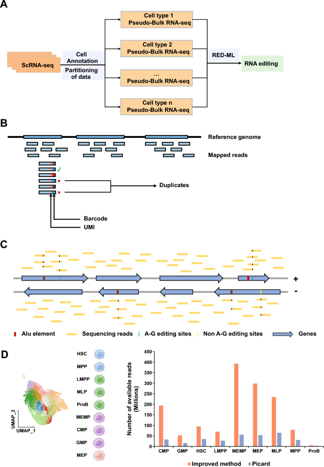
It is a challenge to detect RNA editing events from scRNA-seq data because it has limited sequencing coverages. To overcome this, firstly we integrated the aligned reads of each cell of the same cell type according to the barcode tag in bam files to increase the sequencing coverages, which were merged to a ‘Bulk RNA-seq’ data called pseudo-Bulk RNA-seq data (Fig. 1A) (see details in “Methods” section). Secondly, we improved the method for PCR duplicates removal: only the reads which were aligned to the same genomic coordinate from the same cell with the same unique molecular identifier (UMI) were considered as PCR duplicates (Fig. 1B). Third, as the scRNA-seq data is strand-specific, we split the aligned reads and detect the RNA editing sites of genes transcribed from forward and reverse strands, respectively. In addition, according to the RED-ML method and the features of RNA editing, the sites located in the ALU gene element and the sites mutated as A-G are more likely to be RNA editing sites (Fig. 1C). We applied the improved method to the scRNA-seq data annotated into 8 HSPC populations and found that the improved method could retain more available reads (Fig. 1D).
Figure 1.
A schematic diagram showing the computational method developed in this study. (A) A workflow showing the strategies to identify RNA editing events using scRNA-seq. The cell type annotation information was used to combine the mapped reads of the same cell type in scRNA-seq to obtain pseudo-Bulk RNA-seq for each cell type. (B) The novel threshold used to mark duplicates. Only if the aligned reads with the same alignment position, UMI and barcode are defined as duplicates (see details in “Methods” section). (C) The reads aligned to the reference genome are divided into reads aligned to the forward strand and reverse strand to distinguish RNA editing sites occurring on the forward/reverse strand genes. The edited sites located in ALU element and with the A-G variation are more likely to be identified as RNA editing sites. (D) The scRNA-seq data of hematopoietic stem cells (HSCs), multipotential progenitor cells (MPPs), lymphoid lineage multipotential progenitor cells (LMPPs), multipotential lymphoid lineage progenitor cells (MLPs), megakaryocyte erythroid mast progenitor cells (MEMPs), common myeloid progenitor cells (CMPs), granulocyte monocyte progenitors (GMPs), megakaryocyte erythroid progenitors (MEPs), and B-cell progenitors (ProBs) were used to evaluate improved method and Picard. Bar plot showing the number of available reads using improved method and Picard.
Hematopoietic cell lineage and cell type identification
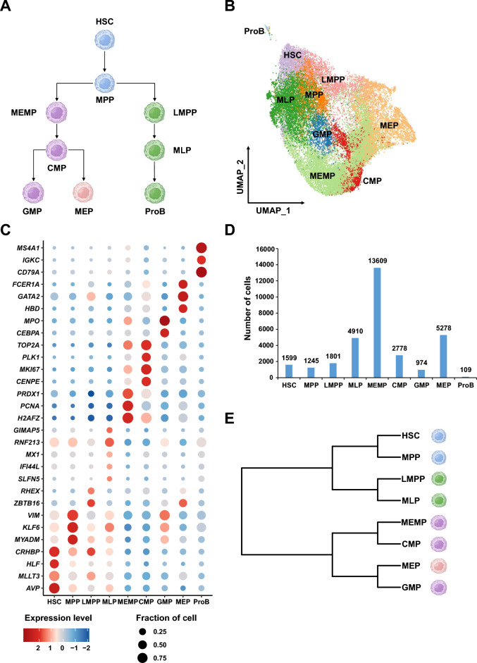
HSPCs is important for maintaining the integrity of the hematopoietic system. To evaluate our method, we used the scRNA-seq data of human HSPCs with defined cell types and lineage relationships according to previous research (Fig. 2A,B). To investigate the conversed RNA editing events of human HSPCs, we collected cord blood (CB) and mobilized peripheral blood (mPB) CD34+ cells and obtained their single-cell transcriptomes. Mobilized peripheral blood (mPB) and umbilical cord blood (CB) are two of the current sources of HSPCs. Researches show that CD34+ cells derived from CB have stronger regeneration capacities than that from mPB. These samples included 6 CB samples without in vitro cultivation, 6 CB samples with 48-h in vitro cultivation, 3 mPB samples without in vitro cultivation and 3 mPB samples with 48-h in vitro cultivation (Supplemental Table 1, Supplemental Fig. S1A). We can find the conversed RNA editing sites in these different samples and cell culture states. We used a previous dataset with accession code CNP000097842 as reference to identify cell types in our data (Fig. 2B). MLP, MEMP, CMP, GMP, MEP and ProB were annotated in the query datasets based on the expression of marker genes reported in other research (Supplemental Fig. S1B)42. Then by integrating the reference and query datasets, we obtained 32,303 cells with specific gene expression profiles (Fig. 2C), including 1599 hematopoietic stem cells (HSCs), 1245 multipotential progenitor cells (MPPs), 1801 lymphoid lineage multipotential progenitor cells (LMPPs), 4910 multipotential lymphoid lineage progenitor cells (MLPs), 13,609 megakaryocyte erythroid mast progenitor cells (MEMPs), 2778 common myeloid progenitor cells (CMPs), 974 granulocyte monocyte progenitors (GMPs), 5278 megakaryocyte erythroid progenitors (MEPs), and 109 B-cell progenitors (ProBs) (Fig. 2D; Supplemental Table 2). As account for only 0.34% (109 of 32,303) of our data, we removed ProBs in further analysis. After generating the pseudo-Bulk RNA-seq data of these cell types, we reconstructed their lineage relationships using unsupervised hierarchical clustering (Fig. 2E), which is consistent with the classical model of lineage determination in human hematopoietic hierarchy (Fig. 2A)42–44. HSC and MPP were found to be clustered together. Myeloid and lymphoid hematopoietic cell populations showed close clustering distinctions, respectively. Taken together, these results demonstrated that our scRNA-seq data exhibited the transcriptome features of human HSPCs.
Figure 2.
9 HSPCs identified using CB/mPB scRNA-seq data and the exhibition of hematopoietic cell lineage. (A) The lineage relationships of HSPCs. According to previous study, HSCs sit at the top of hierarchy. HSCs subsequently differentiate into MPPs. Downstream of MPPs, a strict separation between the myeloid (MEMPs) and lymphoid (LMPPs) branches is the first step in lineage commitment. MEMPs can generate CMPs. CMPs give rise to GMPs and MEPs. LMPPs differentiate into MLPs. MLPs produce ProBs. (B) UMAP embedding projection of 32,303 single-cell transcriptomes (query data and reference data). Cell clusters were colored and annotated based on their transcriptional profile identities (see details in Methods). (C) Dot plot showing the distribution of expression levels of cell-type-specific marker genes across all 9 cell types (see details in “Methods” section). (D) Bar plots showing the total number of detected cells in each cell type. (E) Hierarchical clustering is consistent with the hematopoietic cell lineage in previously studied of 8 cell types by transcriptional profiles.
Dynamic RNA editing events in hematopoietic cell populations
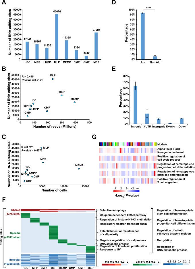
Next we used RED-ML37 to detect RNA editing sites using the pseudo-Bulk RNA-seq data of each cell type. To reduce false positives, we only retained RNA editing sites with sequencing depths greater than 30×. Then a total of 17,841, 15,367, 11,555, 45,626, 19,325, 9,384, 3,742 and 27,094 editing sites in the eight hematopoietic cell populations after filtering (Fig. 3A), and 98,549 unique RNA editing sites among eight hematopoietic cell populations. The investigation of the distance between two adjacent editing sites revealed that global RNA editing density was stable across the eight hematopoietic populations (Supplemental Fig. S2A). There was no significant correlation between the total number of non-redundant reads, the number of cells, and the number of identified RNA editing sites, indicating that the differences in RNA editing events across HSPCs were not due to technical issues (Fig. 3B,C). In addition, consistent with previous reports6, most of the RNA editing sites (94%) were located in the ALU element (Fig. 3D, Supplemental Fig. S2B). To further characterize the RNA editing sites in HSPCs, we investigated the distribution of RNA editing sites in different functional regions of the genome. Most of the RNA editing sites were located in introns (64%) and 3ʹ UTR (17%), and only 1% of the sites were in the coding region (Fig. 3E, Supplemental Fig. S2C), which is in agreement with a previous study14.
Figure 3.
The evaluation of RNA editing sites by distribution in different cell types and genomic elements and their dynamic changes during hematopoiesis. (A) The number of RNA editing sites in each HSPC population. (B) Pearson correlation coefficient between the total reads count and the number of editing sites in each HSPC population. (C) Pearson correlation coefficient between the cell number and the number of editing sites in each HSPC population. The number of RNA editing sites were independent of cell number and sequencing depth. (D) Bar plot showing the proportion of editing sites within and outside Alus in HSPC populations. Most RNA editing sites located in Alu genomics element. Data present mean ± s.e.m. P-value was calculated by T-Test. ****P < 0.0001. (E) Bar plot showing the distribution of editing sites across different genomic elements in HSPC populations. Data present mean ± s.e.m. (F) A heatmap showing confidence scores (P_edit) calculated by RED-ML of all editing sites. The color indicates P_edit (from 0 to 1) for a given site (row) in a population (column). The P_edit equal to 0 means this site is not edited in this population whose coverage is greater than 30×. The right listed the most significant enriched GO pathways in each pattern and the common pathways in all patterns. These GO terms were analysed with genes of these RNA editing sites annotated by ANNOVAR. (G) A heatmap showing the most significant enriched GO pathways in each module related to hematopoiesis. The genes annotated by ANNOVAR were used to do GO analysis.
As RNA editing events are dynamic between cell types (Fig. 3A), to build a comprehensive RNA editing landscape of the eight HSPC populations, we collected RNA editing sites whose read coverage was > 30× in all cell types. A fraction of editing sites (termed shared) was edited in all 8 cell types or specifically edited in lymphoid or myeloid or HSC/MPP hematopoietic cells, while another fraction of editing sites (termed specific) was specifically edited in one cell type (Fig. 3F, Supplemental Tables 3, 4). We then investigated their biological functions and found that RNA editing sites with the above patterns were enriched in different pathways (Fig. 3F). RNA editing may involve in embryonic erythropoiesis45 and adult HSPC differentiation46,47. We found the shared RNA editing sites and specific RNA editing sites both enriched in the pathway relevant with regulation of hematopoietic stem/progenitor cells differentiation and cell cycle (Fig. 3F), which has been proved in several previous studies that cell cycle may have influence on HSPCs differentiation20,48. Those results suggest that RNA editing may regulate the biological processes of HSPCs in multiple ways. In previous studies, it was found that different RNA editing sites may have co-editing modules14. RNA editing sites within the same module are highly correlated. Consistent with these studies, we also identified co-editing modules among different editing sites (Fig. S3A). And different modules may play a role in different cell types (Fig. S3B). The functions of these modules were significantly enriched in the regulation of hematopoietic stem cell differentiation, lineage commitment, stem cell population maintenance, transcription initiation, protein assembly and other HSPC-related biological processes (Fig. 3G, Supplemental Fig. S3C). Therefore, this result suggested that these co-edited modules may have biological functions rather than random events. Together, these observations suggested that RNA editing might play specific roles in different cell types, and might be key to lineage differentiation and self-proliferation during hematopoiesis. Some co-edited modules that occurred in the same biological process may serve as another way for regulating human hematopoiesis.
Possible functions of RNA editing on the maintenance of cell homeostasis during differentiation of HSPCs
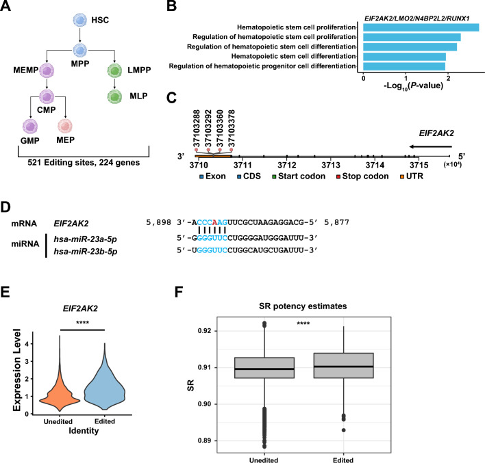
To further explore the global functions of RNA editing on HSPCs, we first analyzed the 521 editing sites shared by all cell types, which were distributed in 224 genes (Fig. 4A, Supplemental Table 5). To explore the role of RNA editing sites in hematopoietic processes, GO enrichment analysis was performed on 224 genes. GO enrichment analysis revealed that EIF2AK2, LMO2, N4BP2L2, and RUNX1 were involved in regulating the differentiation of hematopoietic stem/progenitor cells and the RNA editing sites of those genes located in 3ʹ UTR and exon which may influence the miRNA binding sites and protein translation. (Fig. 4B, Supplemental Table 6). Among them, EIF2AK2 was reported to regulate responses to stress during hematopoiesis49. In our data we identified 37,103,288, 37,103,292, 37,103,360, 37,103,378 four RNA-editing sites in EIF2AK2 3ʹ UTR, which may affect miRNA binding (Fig. 4B,C). Prediction of miRNA targeting sites using TargetScanHuman50 revealed that the binding sites of miR-23a/miR-23b in EIF2AK2 3ʹ UTR were altered due to RNA editing (Fig. 4D, Supplemental Fig. S4A). As miR-23a/miR-23b were expressed in CD34+ cells51, we hypothesized that RNA editing may prevent the binding of miR-23a/miR-23b to EIF2AK2, thereby derepress EIF2AK2. Then we tested this hypothesis and found that as expected, for cells expressing EIF2AK2, the overall expression levels of EIF2AK2 were higher in the cells when EIF2AK2 was edited (log2foldchange: 0.56; P-value < 0.0001) (Fig. 4E). And we found that EIF2AK2 editing occurred only in a small fraction of cells (3.11%) (Supplemental Fig. S4B), which was in agreement with a previous study52.
Figure 4.
RNA editing may have effects on cell homeostasis during hematopoietic cell differentiation in all HSPCs. (A) A summary of the number of editing sites and genes annotated by ANNOVAR shared across 8 HSPC populations. (B) The GO result related with HSPCs proliferation and differentiation of shared genes with RNA editing sites located in 3ʹ UTR or exon coding region. (C) The track of EIF2AK2 shows the loci of RNA editing sites located in the 3ʹ UTR. (D) Predicted site of miR-23a and miR-23b in the EIF2AK2 3ʹ UTR (red: nucleotides edited, blue: binding site). (E) Violin plot showing the expression level of EIF2AK2 in cells with and without EIF2AK2 editing sites. P-value was calculated by Wilcoxon Rank Sum test. ****P < 0.0001 (F) Box plot shows the signaling entropy rate which indicates the differentiation potency of cells in cells without and with EIF2AK2 editing sites. P-value was calculated by T-Test. ****P < 0.0001.
Cell pool integrity is maintained in the blood system by clearing damaged cells. But in order to ensure the longevity of the cells53,54, it is also necessary to ensure that the cells can survive the lower-level stress that often occurs49. Many stressors congregate on the integrated stress response (ISR) pathway, whose function is to balance the pressure signals that activate the cell death pathway in order to protect the cells to restore the cell balance55. PKR encoded by EIF2AK2 is one of the four stress-inducible kinases that maintain the survival-death equilibrium49. The interaction between PKR and the other three kinases (HRI, PERK, GCN2) affects the phosphorylation of eIF2α56, leading to the attenuation of global translation initiation, which saves amino acid synthesis for essential cellular function, reduces the load of chaperones, and lowers the metabolic demand associated with protein synthesis57. Along with the up-regulation of EIF2AK2, We found the expression levels of EIF2AK1, EIF2AK3 and EIF2AK4 (encoding HRI, PERK and GCN2, respectively) were all higher in cells expressing edited EIF2AK2 compared to those expressing unedited EIF2AK2 (Supplemental Fig. S4C), implying that RNA editing of EIF2AK2 may affect the maintenance of cell homeostasis. In addition, we compared the expression levels of EIF2AK2 in eight cell types carrying EIF2AK2 edited sites with non-edited cells. We found that the expression levels of EIF2AK2 in cells with edited sites were higher than those in cells without edited except GMP with no significant difference (Supplemental Fig. S5A). Meanwhile, previous studies found that human HSCs are sensitive to the interference of cell homeostasis. The induction of reactive oxygen species (ROS) and accumulation of DNA damage increase the possibility of apoptosis of HSCs compared to downstream progenitor cells58,59. A study has found high integrated stress response activity in HSC/MPPs compared to progenitors which have lower differentiation potential49. Therefore, we hypothesized that there is a relationship between the activity of ISR pathway and differentiation potency. To test this hypothesis, we used SCENT60 to explore the effect of RNA editing on the differentiation potential of cells. The result also revealed a higher differentiation potential in EIF2AK2 edited cells (Fig. 4F). High differentiation potential was also shown in cells edited with EIF2AK2 among the 8 HSPC populations with significant differences (Supplemental Fig. S5B). Taken together, based on the above analysis, the RNA editing events in EIF2AK2 may play an important role in maintaining cell homeostasis. Besides, we speculated that EIF2AK2 may undergo RNA editing in cells with higher differentiation potential which may more sensitive to interference.
Impact of RNA editing on the lineage commitment of HSPCs and self-renewal of HSCs
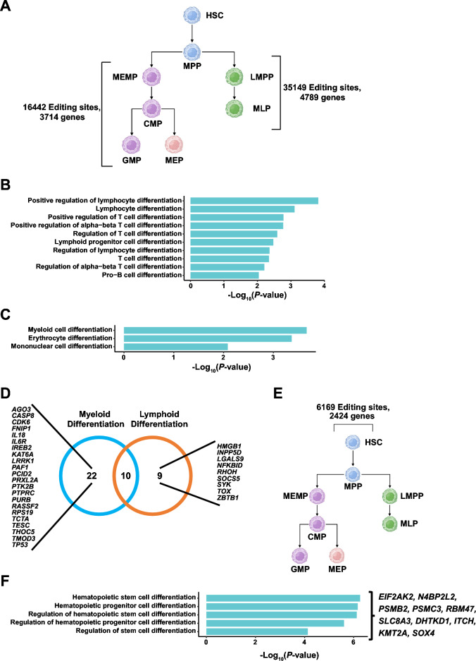
Then we investigate the differences in RNA editing between myeloid and lymphoid HSPCs. We obtained 16,442 lymphoid-specific RNA editing sites distributed in 3714 genes, and 35,149 myeloid-specific RNA editing sites distributed in 4789 genes, respectively (Fig. 5A). GO analysis of genes with lymphoid lineage-specific RNA editing sites revealed a strong enrichment involved in differentiation of lymphocytes, differentiation of T cells, and differentiation of B-cell progenitors (Fig. 5B, Supplemental Table 6). Whereas genes in myeloid lineage-specific editing sites are highly involved in the differentiation of myeloid cells, erythroid cells, and monocytes (Fig. 5C, Supplemental Table 6). All GO terms are provided in supplementary materials. Thus, RNA editing may regulate lineage differentiation of lymphoid and myeloid cells, consistent with a published paper showing that RNA editing may contribute to cell fate decision in the hematopoietic system14.
Figure 5.
RNA editing might influence lineage commitment and HSCs differentiation and self-renewal. (A) A summary of the number of lymphoid-specific and myeloid-specific editing sites and genes annotated by ANNOVAR. (B) The enrichment analysis of genes with lymphoid-specific editing sites involved in the differentiation of lymphoid cells. (C) The enrichment analysis of myeloid-specific edited genes involved in the differentiation of myeloid cells. (D) Venn plot showing the genes edited in lymphoid and myeloid cell lines. The editing sites of those genes are all located in 3ʹ UTR or exon coding region and involved in the differentiation of lymphoid cells, myeloid cells, respectively. (E) A summary of the number of HSC-specific editing sites and genes annotated by ANNOVAR. (F) The enrichment analysis of HSC-specific edited genes involved in hematopoietic stem cell differentiation. And genes involved in those pathways with editing sites are located in 3ʹ UTR or exon coding region.
To further explore the function of genes involved in myeloid and lymphoid HSPCs differentiation pathways, we focused on RNA editing sites located in the 3ʹ UTR and coding regions which may influence the post-translational modification and the function of proteins. We found that 19 and 32 genes were in the pathways regulating lymphoid HSPCs differentiation and myeloid HSPCs differentiation, respectively. And the RNA editing sites of those genes all occurred in the 3ʹ UTR and exon coding regions (Fig. 5D). Many of these genes play important functions in the hematopoietic system. For example, Caspase 8 encoded by CASP8 is a cysteine protease and a key mediator of apoptosis61. Recently, however, many studies have introduced some new aspects to it, citing their significance in cell development and differentiation. The study by Rebe et al. has proved that caspase 8 activity is required for the differentiation of peripheral blood monocytes into macrophages in myeloid cell lines62. Meanwhile, ZBTB1 regulates the development of lymphoid cell lines and myeloid cell lines. Siggs, Owen et al. have shown that a chemically induced mouse with Zbtb1 mutation has a complete and cell-intrinsic T cell deficiency. Besides this, other lymphoid cell lines were also partially impaired. The study shows that ZBTB1 may act as an important transcriptional regulator determining T cell development and lymphopoiesis63.
HSCs are the stem cells that give rise to other hematopoietic cells. We find 6169 sites distributed on 2424 genes specifically edited in HSCs (Fig. 5E). GO Enrichment analysis revealed that genes with RNA editing sites were involved in hematopoietic stem cell differentiation and hematopoietic progenitor cell differentiation, and the RNA editing sites of some genes are located in 3ʹ UTR or exon coding region (Fig. 5F, Supplemental Table 6), such as EIF2AK2, N4BP2L2. According to the GenCards database (https://www.genecards.org/), N4BP2L2 may be involved in the positive regulation of HSC proliferation and negative regulation of HSC differentiation (Table 1).
Table 1.
The function of N4BP2L2 from GeneCards database.
| GO ID | Qualified GO term | Evidence | PubMed IDs |
|---|---|---|---|
| GO:1902035 | Involved in positive regulation of hematopoietic stem cell proliferation | IMP | 19506020 |
| GO:1902037 | Involved in negative regulation of hematopoietic stem cell differentiation | IMP | 19506020 |
Our results showed that RNA editing occurred in genes important for regulating lineage commitment, cell differentiation, and self-renewal during hematopoiesis, suggesting an important role for RNA editing in the hematopoietic process.
Discussion
Differing from previous analysis of scRNA-seq mainly focusing on the gene expression levels, here we provided a novel computational method to study RNA editing using scRNA-seq, and applied it to interpreting hematopoietic differentiation and HSC self-renewal. To perform RNA editing analysis in scRNA-seq data, we made the following three improvements: (i) using cell annotation information to obtain pseudo-Bulk RNA-seq of different cell types; (ii) optimizing the marking duplicates method to increase the sequencing depth; and (iii) using strand information to detect RNA editing sites. Compared with other studies, the distribution of RNA editing sites across different genomic elements, and the proportion of RNA editing sites within ALU elements were consistent with previous research reports14.
We then explore the shared RNA editing sites and specific RNA editing sites among different HSPC populations and found that RNA editing is dynamic between different cell types during HSPCs differentiation and self-renewal, and has specific editing events for different cell types. However, our study did not further resolve the underlying mechanisms conferring to this dynamic change. It seems that RNA editing may serve as a molecular marker for different cell types and have implications for the performance of specific functions of different cell types. Since ADAR enzymes catalyze RNA editing events, there may be some differences in the expression of ADAR enzymes in different cell types, all of which need to be investigated in detail by additional work. Moreover, our study found that EIF2AK2 with the shared RNA editing sites in all HSPC cell populations may have an impact on maintaining self-homeostasis during HSPCs differentiation through ISR pathway. Though a study has found that ISR activation in HSCs/MPPs is more active than in hematopoietic progenitor cells49, our findings may indicate that the ISR pathway not only safeguards HSCs/MPPs but also affects all HSPCs with high differentiation potential. This implies that HSPCs with higher differentiation potential may be more sensitive to external stimulus stress. They perform specific regulation of their own cellular homeostasis through RNA editing to maintain the integrity of the hematopoietic system. Meanwhile, we found lymphoid-specific and myeloid-specific RNA editing sites may involve in lineage commitment and HSPCs differentiation. According to a previous study, RNA editing may influence lineage commitment during hematopoiesis. They found the frequency of RNA editing alters at the branch point of HSPCs differentiation14. Our results indicate that not only changes in editing frequency, but also specific RNA editing sites occur in different lineages during differentiated development. This may suggest that RNA editing sites occur at genes critical to lineage commitment. Besides shared sites, we found specific editing sites in HSCs may have functions on differentiation and self-renew. The capacity to self-renew and to differentiate into other hematopoietic cells are important features of HSCs15. This also suggests that RNA editing may play an important role in maintaining the balance of self-renewal and differentiation of HSCs. However, those results still require more experiments to verify the role of RNA editing for HSPCs.
More broadly, our approach validates the feasibility and usability of RNA editing event studies using high-throughput scRNA-seq. And we showed the overall differences RNA editing of the HSPCs and the possible function. In summary, these efforts confirm the great potential and value of scRNA-seq for the study of biological process mechanisms. Though future studies will be required to confirm and clarify the role of RNA editing.
Methods
Accession numbers
The reference dataset (aligned reads) was downloaded from CNSA (https://db.cngb.org/cnsa/) of CNGBdb with accession code CNP0000978. Both the reference and query dataset were CD34+ cells obtained from human CB and mPB, and the method of cell cultivation, scRNA-seq libraries and pre-processing were as previously described42. The raw sequencing data of query dataset generated in this study was deposited in the CNGB Sequence Archive (CNSA; https://db.cngb.org/cnsa/) of China National GeneBank DataBase (CNGBdb) with accession code CNP0003367.
Ethics statement
This study was performed with the approval of the Institutional Review Board of BGI (BGI-IRB 16089-T3 and BGI-IRB 22090). All methods were performed in accordance with relevant quidelines and regulations.
Enrichment of CD34+ cells from human CB and mPB samples
Human CB and mPB samples were obtained from healthy donors with informed consent. We got mononuclear cells (MNCs) using centrifugation on Lymphoprep medium. CD34 Microbead kits and LS columns using MACS magnet technology (Miltenyi) were used for MNC enrichment for CD34+ cell selection. Downstream experiments were conducted on CD34+ cells after sorting.
Cell culture in vitro and scRNA-seq library
The fresh CD34+ cells were applied to cell culture in vitro or to single-cell RNA-seq (scRNA-seq). CD34+ cells were resuspended in SCGM medium (Cellgenix) using the following recombinant hematopoietic cytokines: recombinant human stem cell factor (rhSCF) 100 ng/ml, recombinant human thrombopoietin (rhTPO) 100 ng/ml, recombinant human fms-related tyrosine kinase-3 ligand (rhFlt3-L) 100 ng/ml and cultured in 24-well tissue culture plates at 37 °C in an atmosphere of 5% CO2 for 48 h (Thermo Fisher). The DNBelab C4 platform was used to perform scRNA-seq. Single-cell suspensions were used for droplet generation, demulsification, microbead collection, reverse transcription, and cDNA amplification to generate barcode libraries. The manufacturer’s protocol was used to construct indexed libraries. QubitTM ssDNA Assay Kit (Thermo Fisher Scientific; #Q10212) was used to quantify the sequencing libraries. DNA nanoballs (DNBs) are loaded into a pattern nanoarray and sequenced at ultra-high throughput with the following read lengths used by the DIPSEQ T1 sequencer: 30 bp for read 1, inclusive of 10 bp cell barcode 1, 10 bp cell barcode 2 and 10 bp unique molecular identifier (UMI), 100 bp of transcript sequence for read 2 and 10 bp for sample index.
Mapping and annotating query datasets
To identify HSPC populations in CD34+ cells, we used “Mapping and annotating query datasets” method of Seurat package (v. 4.1.0) in R (v.4.0.5)64–67. This approach allows the comparison of the similarity of cells in the reference and target datasets, resulting in cell annotation of the target dataset with unknown cell types. The reference data set was obtained from CNGBdb with accession code CNP000097842. Then the query dataset was mapped to the reference data for annotating cell types. First, we imported the final cell-gene expression matrix of the query dataset into the Seurat package to create a Seurat objects. Cells with fewer than 200 detected genes and for which the total mitochondrial gene expression exceeded 5% were removed. Besides, we used the IQR Method of Outlier Detection to remove cells with outlier gene number. Genes expressed in fewer than three cells were also removed.
Downstream analyses were also performed using Seurat package (v. 4.1.0). Normalizing SeuratObject used the NormalizeData function and the ScaleData function. The following functions were run together when mapping the query dataset to the reference dataset: FindIntegrationAnchors, TransferData, IntegrateEmbeddings, ProjectUMAP. The FindAllMarkers function was then used to find the cell type-specific genes. Differential expression analysis was performed based on the Wilcoxon Rank Sum test. Finally, the merge function of Seurat was used to merge the query dataset and reference dataset. All methods use default parameters unless otherwise specified.
The AverageExpression function of Seurat was used to calculate the average expression of 8 HSPC cell types. And then we used hclust function to perform the hierarchical clustering.
Splitting aligned reads based on cell type
To get pseudo-Bulk RNA-seq of 8 cell types, we got the barcode and sample information for each cell of the 8 cell types from SeuratObject. Based on the CB (Cell Barcode) tag in the bam file we can determine the cell from which the reads originated, so we can use the cell barcode of each cell type to obtain aligned reads for each cell barcode. And then, we integrated multiple cells aligned reads of each cell type to obtain a pseudo-Bulk RNA-seq for each cell type. The code of this method is available on Github (https://github.com/Genki-YAN/Cell2Editing).
Marking duplicates and obtaining strand-specific reads
Considering the difference between scRNA-seq data and Bulk RNA-seq data, we designed the procedure of marking duplicates: taking the aligned position, UMI barcode, and cell barcode of reads into account for the definition of duplicates. Only the reads from the same cell with the same unique molecular identifier (UMI) were considered as PCR duplicates. And we retained the read with the highest base quality. The code of this process is available on Github (https://github.com/Genki-YAN/Cell2Editing).
We split strand-specific aligned reads based on flag 16 (read reverse chain) in bam file using Samtools68 with the following commands: samtools view -b -f 16; samtools view -b -F 16.
We compared the number of available reads detected by the improved method with the Picard (“Picard Toolkit.” 2019. Broad Institute, GitHub Repository. https://broadinstitute.github.io/picard/; Broad Institute) method. We counted and compared the non-duplicates reads in the bam files processed by the two methods. The tool locates and marks duplicates in BAM or SAM files, where repeat reads are defined as originating from a single DNA fragment. Duplication may occur during sample preparation, such as library construction using PCR. If the 5’ position, strand and base alignment are the same, Picard compared base alignment quality to mark duplicate reads and available reads.
Detecting RNA editing sites
After the above pre-process of data, we used the RED-ML (https://github.com/BGIHENG/RED-ML)37 with the default parameters to detect the RNA editing sites in the pseudo-Bulk RNA-seq using the GRCh38 genome and GRCh38 SNP database (https://ftp.ncbi.nih.gov/snp/organisms/human_9606_b151_GRCh38p7/VCF/All_20180418.vcf.gz), and removed RNA editing sites with coverage less than 30× to facilitate subsequent analysis. When we investigated cell type-specific RNA editing sites, we used the mpileup function of Samtools for investigating site sequencing coverage, keeping sites with coverage greater than 30× with the following commands: samtools mpileup -B -f -s - output-QNAME -min-MQ 20 -min-BQ 20 -excl-flags DUP.
Annotation of RNA editing sites
RNA editing sites were annotated utilizing ANNOVAR (https://annovar.openbioinformatics.org/en/latest/)69 table_annovar.pl with the reference genome of GRCh38. We used the following commands: -remove -protocol refGene, phastConsElements20way, wgRna, cytoBand -operation g,r,r,r -nastring . -csvout -polish. Based on the strand of the aligned reads, we only retain the RNA editing sites with genes transcribed from the same strand.
Correlation module analysis of RNA editing sites
To identify the RNA editing sites that were co-edited in different cell types, the co-editing modules of filtered RNA editing sites in 8 cell types were analyzed using WGCNA package (v. 1.70.3)70,71. The WGCNAR package was used to estimate the best soft thresholding power for the co-editing module analysis. The minimum power 14, which reached the R2 cut-off of 0.8 for topology model fit, was determined to be the optimal value. The adjacency with the optimal soft-thresholding power estimated above was calculated, and the adjacency was transformed into a topological overlap matrix to calculate the corresponding dissimilarity, and identified 44 co-editing modules with minModuleSize = 30. Then we annotated the RNA editing sites in each module and perform GO enrichment analysis using clusterProfiler R package (v.4.0.5)72,73.
Identification of cells with editing sites
Since RED-ML is a method designed for RNA editing sites by Bulk RNA-seq, and there is no existing method to detect RNA editing sites in individual cells, we used the mpileup function of Samtools to find the aligned reads with RNA editing sites. The cells with at least 1 read supporting RNA editing events were defined as edited cells. Samtools mpileup was performed using the following parameters: samtools mpileup -B -f -s -output-QNAME -min-MQ 20 -min-BQ 20 -excl-flags DUP. To calculate the proportion of cells with edited EIF2AK2, we removed cells that did not express EIF2AK2.
Gene expression and functional analysis of RNA editing sites
The expression of genes with RNA editing sites in cells was quantified using ScaleData function of Seurat. To explore the function of editing sites with different editing patterns, we performed GO enrichment analysis using clusterProfiler package (v.4.0.5)72,73.
Prediction of miRNA target sites
TargetScanHuman (https://www.targetscan.org/vert_80/)50 was used to predict miRNAs that may target and bind to candidate genes. Then we compared the binding region with the RNA editing sites to determine whether RNA editing sites are located in the miRNA binding region. The reference file of UTR sequence was downloaded from TargetScanHuman website. The version of RNA sequence used was GRCh38.
Estimating differentiation potency of single cells
To demonstrate that RNA editing may be correlated with differentiation potency, we first removed cells that do not express genes with the RNA editing, and then we calculated the differentiation potency of cells using SCENT (https://github.com/aet21/SCENT)60 with the default parameters. The P-value was calculated using T-Test.
Supplementary Information
Acknowledgements
This research is supported by grants from National Natural Science Foundation of China (NSFC) (31970816). And this work is supported by China National GeneBank.
Author contributions
H.-X.S., C.L. and S.H. conceived and designed the study. G.D. and W.O. performed the scRNA-seq experiments. Y.W. and X.X. conducted the bioinformatics analysis. H.-X.S., C.L. and S.H. helped with project design and discussions. Y.W. wrote the manuscript. H.-X.S. and C.L. reviewed the manuscript. All authors read and approved the final manuscript.
Data availability
The datasets generated and analysed during the current study are not publicly available due to human genetic resources management and the data in a published article entitled “Stemness-related genes revealed by single-cell profiling of naïve and stimulated human CD34+ cells from CB and mPB” (10.1002/ctm2.1175) being controlled but are available from the corresponding author on reasonable request.
Competing interests
The authors declare no competing interests.
Footnotes
Publisher's note
Springer Nature remains neutral with regard to jurisdictional claims in published maps and institutional affiliations.
Supplementary Information
The online version contains supplementary material available at 10.1038/s41598-023-37325-4.
References
- 1.Rajendren S, et al. Profiling neural editomes reveals a molecular mechanism to regulate RNA editing during development. Genome Res. 2021;31(1):27–39. doi: 10.1101/gr.267575.120. [DOI] [PMC free article] [PubMed] [Google Scholar]
- 2.Chigaev M, et al. Genomic positional dissection of RNA editomes in tumor and normal samples. Front. Genet. 2019;10:211. doi: 10.3389/fgene.2019.00211. [DOI] [PMC free article] [PubMed] [Google Scholar]
- 3.Christofi T, Zaravinos A. RNA editing in the forefront of epitranscriptomics and human health. J. Transl. Med. 2019;17(1):319. doi: 10.1186/s12967-019-2071-4. [DOI] [PMC free article] [PubMed] [Google Scholar]
- 4.Kim DD, et al. Widespread RNA editing of embedded alu elements in the human transcriptome. Genome Res. 2004;14(9):1719–1725. doi: 10.1101/gr.2855504. [DOI] [PMC free article] [PubMed] [Google Scholar]
- 5.Lo Giudice C, et al. Quantifying RNA editing in deep transcriptome datasets. Front. Genet. 2020;11:194. doi: 10.3389/fgene.2020.00194. [DOI] [PMC free article] [PubMed] [Google Scholar]
- 6.Giacopuzzi E, et al. Genome-wide analysis of consistently RNA edited sites in human blood reveals interactions with mRNA processing genes and suggests correlations with cell types and biological variables. BMC Genom. 2018;19(1):963. doi: 10.1186/s12864-018-5364-8. [DOI] [PMC free article] [PubMed] [Google Scholar]
- 7.Bakhtiarizadeh MR, Salehi A, Rivera RM. Genome-wide identification and analysis of A-to-I RNA editing events in bovine by transcriptome sequencing. PLoS ONE. 2018;13(2):e0193316. doi: 10.1371/journal.pone.0193316. [DOI] [PMC free article] [PubMed] [Google Scholar]
- 8.Liu H, et al. Functional Impact of RNA editing and ADARs on regulation of gene expression: Perspectives from deep sequencing studies. Cell Biosci. 2014;4(1):44. doi: 10.1186/2045-3701-4-44. [DOI] [PMC free article] [PubMed] [Google Scholar]
- 9.Hsiao YE, et al. RNA editing in nascent RNA affects pre-mRNA splicing. Genome Res. 2018;28(6):812–823. doi: 10.1101/gr.231209.117. [DOI] [PMC free article] [PubMed] [Google Scholar]
- 10.Tassinari V, et al. The adaptive potential of RNA editing-mediated miRNA-retargeting in cancer. Biochim. Biophys. Acta. 2019;1862(3):291–300. doi: 10.1016/j.bbagrm.2018.12.007. [DOI] [PubMed] [Google Scholar]
- 11.Nishikura K. Functions and regulation of RNA editing by ADAR deaminases. Annu. Rev. Biochem. 2010;79:321–349. doi: 10.1146/annurev-biochem-060208-105251. [DOI] [PMC free article] [PubMed] [Google Scholar]
- 12.Roy-Chaudhuri B, et al. Regulation of microRNA-mediated gene silencing by microRNA precursors. Nat. Struct. Mol. Biol. 2014;21(9):825–832. doi: 10.1038/nsmb.2862. [DOI] [PMC free article] [PubMed] [Google Scholar]
- 13.Wen J, et al. The landscape of coding RNA editing events in pediatric cancer. BMC Cancer. 2021;21(1):1233. doi: 10.1186/s12885-021-08956-5. [DOI] [PMC free article] [PubMed] [Google Scholar]
- 14.Wang F, et al. A comprehensive RNA editome reveals that edited Azin1 partners with DDX1 to enable hematopoietic stem cell differentiation. Blood. 2021;138(20):1939–1952. doi: 10.1182/blood.2021011314. [DOI] [PMC free article] [PubMed] [Google Scholar]
- 15.Hawley RG, Ramezani A, Hawley TS. Hematopoietic stem cells. Methods Enzymol. 2006;419:149–179. doi: 10.1016/S0076-6879(06)19007-2. [DOI] [PMC free article] [PubMed] [Google Scholar]
- 16.Bryder D, et al. Self-renewal of multipotent long-term repopulating hematopoietic stem cells is negatively regulated by Fas and tumor necrosis factor receptor activation. J. Exp. Med. 2001;194(7):941–952. doi: 10.1084/jem.194.7.941. [DOI] [PMC free article] [PubMed] [Google Scholar]
- 17.Granick JL, Simon SI, Borjesson DL. Hematopoietic stem and progenitor cells as effectors in innate immunity. Bone Marrow Res. 2012;2012:165107. doi: 10.1155/2012/165107. [DOI] [PMC free article] [PubMed] [Google Scholar]
- 18.Cheshier SH, et al. In vivo proliferation and cell cycle kinetics of long-term self-renewing hematopoietic stem cells. Proc. Natl. Acad. Sci. 1999;96(6):3120–3125. doi: 10.1073/pnas.96.6.3120. [DOI] [PMC free article] [PubMed] [Google Scholar]
- 19.Zhu J, Emerson SG. Hematopoietic cytokines, transcription factors and lineage commitment. Oncogene. 2002;21(21):3295–3313. doi: 10.1038/sj.onc.1205318. [DOI] [PubMed] [Google Scholar]
- 20.Orford KW, Scadden DT. Deconstructing stem cell self-renewal: Genetic insights into cell-cycle regulation. Nat. Rev. Genet. 2008;9(2):115–128. doi: 10.1038/nrg2269. [DOI] [PubMed] [Google Scholar]
- 21.Afreen S, et al. Concise review: Cheating death for a better transplant. Stem Cells. 2018;36(11):1646–1654. doi: 10.1002/stem.2901. [DOI] [PubMed] [Google Scholar]
- 22.Hsu JI, et al. PPM1D mutations drive clonal hematopoiesis in response to cytotoxic chemotherapy. Cell Stem Cell. 2018;23(5):700–713.e6. doi: 10.1016/j.stem.2018.10.004. [DOI] [PMC free article] [PubMed] [Google Scholar]
- 23.Jeong M, et al. Loss of Dnmt3a immortalizes hematopoietic stem cells in vivo. Cell Rep. 2018;23(1):1–10. doi: 10.1016/j.celrep.2018.03.025. [DOI] [PMC free article] [PubMed] [Google Scholar]
- 24.Li Z, et al. Suppression of m(6)A reader Ythdf2 promotes hematopoietic stem cell expansion. Cell Res. 2018;28(9):904–917. doi: 10.1038/s41422-018-0072-0. [DOI] [PMC free article] [PubMed] [Google Scholar]
- 25.Seita J, Weissman IL. Hematopoietic stem cell: Self-renewal versus differentiation. Wiley Interdiscip. Rev. Syst. Biol. Med. 2010;2(6):640–653. doi: 10.1002/wsbm.86. [DOI] [PMC free article] [PubMed] [Google Scholar]
- 26.Montagner S, Dehó L, Monticelli S. MicroRNAs in hematopoietic development. BMC Immunol. 2014;15:14–14. doi: 10.1186/1471-2172-15-14. [DOI] [PMC free article] [PubMed] [Google Scholar]
- 27.Vasilatou D, et al. The role of microRNAs in normal and malignant hematopoiesis. Eur. J. Haematol. 2010;84(1):1–16. doi: 10.1111/j.1600-0609.2009.01348.x. [DOI] [PubMed] [Google Scholar]
- 28.Havelange V, Garzon R, Croce CM. MicroRNAs: New players in acute myeloid leukaemia. Br. J. Cancer. 2009;101(5):743–748. doi: 10.1038/sj.bjc.6605232. [DOI] [PMC free article] [PubMed] [Google Scholar]
- 29.Kluiver J, et al. The role of microRNAs in normal hematopoiesis and hematopoietic malignancies. Leukemia. 2006;20(11):1931–1936. doi: 10.1038/sj.leu.2404387. [DOI] [PubMed] [Google Scholar]
- 30.Chen CZ, et al. MicroRNAs modulate hematopoietic lineage differentiation. Science. 2004;303(5654):83–86. doi: 10.1126/science.1091903. [DOI] [PubMed] [Google Scholar]
- 31.Fazi F, et al. A minicircuitry comprised of microRNA-223 and transcription factors NFI-A and C/EBPα regulates human granulopoiesis. Cell. 2005;123(5):819–831. doi: 10.1016/j.cell.2005.09.023. [DOI] [PubMed] [Google Scholar]
- 32.Zhou B, et al. miR-150, a microRNA expressed in mature B and T cells, blocks early B cell development when expressed prematurely. Proc. Natl. Acad. Sci. U.S.A. 2007;104(17):7080–7085. doi: 10.1073/pnas.0702409104. [DOI] [PMC free article] [PubMed] [Google Scholar]
- 33.Hu Z, et al. Silencing miR-150 ameliorates experimental autoimmune encephalomyelitis. Front. Neurosci. 2018;12:465–465. doi: 10.3389/fnins.2018.00465. [DOI] [PMC free article] [PubMed] [Google Scholar]
- 34.Ramaswami G, et al. Identifying RNA editing sites using RNA sequencing data alone. Nat. Methods. 2013;10(2):128–132. doi: 10.1038/nmeth.2330. [DOI] [PMC free article] [PubMed] [Google Scholar]
- 35.Zhang Q, Xiao X. Genome sequence-independent identification of RNA editing sites. Nat. Methods. 2015;12(4):347–350. doi: 10.1038/nmeth.3314. [DOI] [PMC free article] [PubMed] [Google Scholar]
- 36.Kim M-S, Hur B, Kim S. RDDpred: A condition-specific RNA-editing prediction model from RNA-seq data. BMC Genom. 2016;17(1):5. doi: 10.1186/s12864-015-2301-y. [DOI] [PMC free article] [PubMed] [Google Scholar]
- 37.Xiong H, et al. RED-ML: A novel, effective RNA editing detection method based on machine learning. Gigascience. 2017;6(5):1–8. doi: 10.1093/gigascience/gix012. [DOI] [PMC free article] [PubMed] [Google Scholar]
- 38.Gal-Mark N, et al. Abnormalities in A-to-I RNA editing patterns in CNS injuries correlate with dynamic changes in cell type composition. Sci. Rep. 2017;7:43421. doi: 10.1038/srep43421. [DOI] [PMC free article] [PubMed] [Google Scholar]
- 39.Lundin E, et al. Spatiotemporal mapping of RNA editing in the developing mouse brain using in situ sequencing reveals regional and cell-type-specific regulation. BMC Biol. 2020;18(1):6. doi: 10.1186/s12915-019-0736-3. [DOI] [PMC free article] [PubMed] [Google Scholar]
- 40.Sapiro AL, et al. Illuminating spatial A-to-I RNA editing signatures within the Drosophila brain. Proc. Natl. Acad. Sci. 2019;116(6):2318–2327. doi: 10.1073/pnas.1811768116. [DOI] [PMC free article] [PubMed] [Google Scholar]
- 41.Cuddleston WH, et al. Cellular and genetic drivers of RNA editing variation in the human brain. Nat. Commun. 2022;13(1):2997. doi: 10.1038/s41467-022-30531-0. [DOI] [PMC free article] [PubMed] [Google Scholar]
- 42.Dong G, et al. Functional stemness-related genes revealed by single-cell profiling of naïve and stimulated human CD34+ cells from CB and mPB. BioRxiv. 2022;2022:481626. doi: 10.1002/ctm2.1175. [DOI] [PMC free article] [PubMed] [Google Scholar]
- 43.Nimmo RA, May GE, Enver T. Primed and ready: Understanding lineage commitment through single cell analysis. Trends Cell Biol. 2015;25(8):459–467. doi: 10.1016/j.tcb.2015.04.004. [DOI] [PubMed] [Google Scholar]
- 44.Aurrand-Lions M, Mancini SJC. Murine bone marrow niches from hematopoietic stem cells to B cells. Int. J. Mol. Sci. 2018;19(8):2353. doi: 10.3390/ijms19082353. [DOI] [PMC free article] [PubMed] [Google Scholar]
- 45.Liddicoat BJ, et al. RNA editing by ADAR1 prevents MDA5 sensing of endogenous dsRNA as nonself. Science. 2015;349(6252):1115–1120. doi: 10.1126/science.aac7049. [DOI] [PMC free article] [PubMed] [Google Scholar]
- 46.Hartner JC, et al. ADAR1 is essential for the maintenance of hematopoiesis and suppression of interferon signaling. Nat. Immunol. 2009;10(1):109–115. doi: 10.1038/ni.1680. [DOI] [PMC free article] [PubMed] [Google Scholar]
- 47.XuFeng R, et al. ADAR1 is required for hematopoietic progenitor cell survival via RNA editing. Proc. Natl. Acad. Sci. U.S.A. 2009;106(42):17763–17768. doi: 10.1073/pnas.0903324106. [DOI] [PMC free article] [PubMed] [Google Scholar]
- 48.Cheshier SH, et al. In vivo proliferation and cell cycle kinetics of long-term self-renewing hematopoietic stem cells. Proc. Natl. Acad. Sci. U.S.A. 1999;96(6):3120–3125. doi: 10.1073/pnas.96.6.3120. [DOI] [PMC free article] [PubMed] [Google Scholar]
- 49.van Galen P, et al. Integrated stress response activity marks stem cells in normal hematopoiesis and leukemia. Cell Rep. 2018;25(5):1109–1117. doi: 10.1016/j.celrep.2018.10.021. [DOI] [PubMed] [Google Scholar]
- 50.McGeary SE, et al. The biochemical basis of microRNA targeting efficacy. Science. 2019;366:6472. doi: 10.1126/science.aav1741. [DOI] [PMC free article] [PubMed] [Google Scholar]
- 51.Georgantas RW, et al. CD34+ hematopoietic stem-progenitor cell microRNA expression and function: A circuit diagram of differentiation control. Proc. Natl. Acad. Sci. U.S.A. 2007;104(8):2750–2755. doi: 10.1073/pnas.0610983104. [DOI] [PMC free article] [PubMed] [Google Scholar]
- 52.Ansell BRE, et al. A survey of RNA editing at single-cell resolution links interneurons to schizophrenia and autism. RNA. 2021;27(12):1482–1496. doi: 10.1261/rna.078804.121. [DOI] [PMC free article] [PubMed] [Google Scholar]
- 53.Ho TT, et al. Autophagy maintains the metabolism and function of young and old stem cells. Nature. 2017;543(7644):205–210. doi: 10.1038/nature21388. [DOI] [PMC free article] [PubMed] [Google Scholar]
- 54.Warr MR, et al. FOXO3A directs a protective autophagy program in haematopoietic stem cells. Nature. 2013;494(7437):323–327. doi: 10.1038/nature11895. [DOI] [PMC free article] [PubMed] [Google Scholar]
- 55.Pakos-Zebrucka K, et al. The integrated stress response. EMBO Rep. 2016;17(10):1374–1395. doi: 10.15252/embr.201642195. [DOI] [PMC free article] [PubMed] [Google Scholar]
- 56.Aitken CE, Lorsch JR. A mechanistic overview of translation initiation in eukaryotes. Nat. Struct. Mol. Biol. 2012;19(6):568–576. doi: 10.1038/nsmb.2303. [DOI] [PubMed] [Google Scholar]
- 57.Wek RC, Jiang HY, Anthony TG. Coping with stress: eIF2 kinases and translational control. Biochem. Soc. Trans. 2006;34(Pt 1):7–11. doi: 10.1042/BST0340007. [DOI] [PubMed] [Google Scholar]
- 58.Milyavsky M, et al. A distinctive DNA damage response in human hematopoietic stem cells reveals an apoptosis-independent role for p53 in self-renewal. Cell Stem Cell. 2010;7(2):186–197. doi: 10.1016/j.stem.2010.05.016. [DOI] [PubMed] [Google Scholar]
- 59.Yahata T, et al. Accumulation of oxidative DNA damage restricts the self-renewal capacity of human hematopoietic stem cells. Blood. 2011;118(11):2941–2950. doi: 10.1182/blood-2011-01-330050. [DOI] [PubMed] [Google Scholar]
- 60.Teschendorff AE, Enver T. Single-cell entropy for accurate estimation of differentiation potency from a cell's transcriptome. Nat. Commun. 2017;8:15599. doi: 10.1038/ncomms15599. [DOI] [PMC free article] [PubMed] [Google Scholar]
- 61.Tummers B, Green DR. Caspase-8: Regulating life and death. Immunol. Rev. 2017;277(1):76–89. doi: 10.1111/imr.12541. [DOI] [PMC free article] [PubMed] [Google Scholar]
- 62.Rébé C, et al. Caspase-8 prevents sustained activation of NF-kappaB in monocytes undergoing macrophagic differentiation. Blood. 2007;109(4):1442–1450. doi: 10.1182/blood-2006-03-011585. [DOI] [PMC free article] [PubMed] [Google Scholar]
- 63.Siggs OM, et al. ZBTB1 is a determinant of lymphoid development. J. Exp. Med. 2011;209(1):19–27. doi: 10.1084/jem.20112084. [DOI] [PMC free article] [PubMed] [Google Scholar]
- 64.Hao Y, et al. Integrated analysis of multimodal single-cell data. Cell. 2021;184(13):3573–3587.e29. doi: 10.1016/j.cell.2021.04.048. [DOI] [PMC free article] [PubMed] [Google Scholar]
- 65.Stuart T, et al. Comprehensive integration of single-cell data. Cell. 2019;177(7):1888–1902. doi: 10.1016/j.cell.2019.05.031. [DOI] [PMC free article] [PubMed] [Google Scholar]
- 66.Butler A, et al. Integrating single-cell transcriptomic data across different conditions, technologies, and species. Nat. Biotechnol. 2018;36(5):411–420. doi: 10.1038/nbt.4096. [DOI] [PMC free article] [PubMed] [Google Scholar]
- 67.Satija R, et al. Spatial reconstruction of single-cell gene expression data. Nat. Biotechnol. 2015;33(5):495–502. doi: 10.1038/nbt.3192. [DOI] [PMC free article] [PubMed] [Google Scholar]
- 68.Danecek P, et al. Twelve years of SAMtools and BCFtools. GigaScience. 2021;10(2):8. doi: 10.1093/gigascience/giab008. [DOI] [PMC free article] [PubMed] [Google Scholar]
- 69.Wang K, Li M, Hakonarson H. ANNOVAR: Functional annotation of genetic variants from high-throughput sequencing data. Nucleic Acids Res. 2010;38(16):e164. doi: 10.1093/nar/gkq603. [DOI] [PMC free article] [PubMed] [Google Scholar]
- 70.Zhang B, Horvath S. A general framework for weighted gene co-expression network analysis. Stat. Appl. Genet. Mol. Biol. 2005;4:17. doi: 10.2202/1544-6115.1128. [DOI] [PubMed] [Google Scholar]
- 71.Langfelder P, Horvath S. WGCNA: An R package for weighted correlation network analysis. BMC Bioinform. 2008;9:559. doi: 10.1186/1471-2105-9-559. [DOI] [PMC free article] [PubMed] [Google Scholar]
- 72.Wu T, et al. clusterProfiler 4.0: A universal enrichment tool for interpreting omics data. Innovation. 2021;2(3):100141. doi: 10.1016/j.xinn.2021.100141. [DOI] [PMC free article] [PubMed] [Google Scholar]
- 73.Yu G, et al. clusterProfiler: An R package for comparing biological themes among gene clusters. OMICS. 2012;16(5):284–287. doi: 10.1089/omi.2011.0118. [DOI] [PMC free article] [PubMed] [Google Scholar]
Associated Data
This section collects any data citations, data availability statements, or supplementary materials included in this article.
Supplementary Materials
Data Availability Statement
The datasets generated and analysed during the current study are not publicly available due to human genetic resources management and the data in a published article entitled “Stemness-related genes revealed by single-cell profiling of naïve and stimulated human CD34+ cells from CB and mPB” (10.1002/ctm2.1175) being controlled but are available from the corresponding author on reasonable request.


