Abstract
Several differentiation protocols enable the emergence of hematopoietic stem and progenitor cells (HSPCs) from human-induced pluripotent stem cells (iPSCs), yet optimized schemes to promote the development of HSPCs with self-renewal, multilineage differentiation, and engraftment potential are lacking. To improve human iPSC differentiation methods, we modulated WNT, Activin/Nodal, and MAPK signaling pathways by stage-specific addition of small-molecule regulators CHIR99021, SB431542, and LY294002, respectively, and measured the impact on hematoendothelial formation in culture. Manipulation of these pathways provided a synergy sufficient to enhance formation of arterial hemogenic endothelium (HE) relative to control culture conditions. Importantly, this approach significantly increased production of human HSPCs with self-renewal and multilineage differentiation properties, as well as phenotypic and molecular evidence of progressive maturation in culture. Together, these findings provide a stepwise improvement in human iPSC differentiation protocols and offer a framework for manipulating intrinsic cellular cues to enable de novo generation of human HSPCs with functionality in vivo.
Keywords: induced pluripotent stem cells, hematopoietic stem/progenitor cells, arterial hemogenic endothelium, Activin/Nodal, WNT and MAPK signaling pathways
Graphical abstract
Graphical Abstract.
Significance Statement.
The ability to produce functional HSPCs by differentiation of human iPSCs ex vivo holds enormous potential for cellular therapy of human blood disorders. However, obstacles still thwart translation of this approach to the clinic. In keeping with the prevailing arterial-specification model, we demonstrate that concurrent modulation of WNT, Activin/Nodal, and MAPK signaling pathways by stage-specific addition of small molecules during human iPSC differentiation provides a synergy sufficient to promote arterialization of HE and production of HSPCs with features of definitive hematopoiesis. This simple differentiation scheme provides a unique tool for disease modeling, in vitro drug screening and eventual cell therapies.
Introduction
Hematopoietic stem and progenitor cell (HSPC) transplantation is the most established cellular replacement therapy for a number of hematological diseases. However, the scarcity of human leukocyte antigen (HLA)-matched donors and insufficient numbers of long-term repopulating hematopoietic stem cells (HSCs) in donor umbilical cord blood (UCB) units often limit the ability to carry-out allogeneic HSPC transplant procedures. In the context of autologous gene therapy applications, sufficient functional HSCs from bone marrow (BM) or mobilized peripheral blood (MPB) cell collections are also commonly unavailable in patients with impaired hematopoiesis, such as BM failure syndromes and chronic inflammatory disorders.1-3 Current protocols to augment cell dose by expansion of human HSCs ex vivo are inefficient.4-6 De novo generation of human HSPCs is a tractable alternative for the development of cell-based therapies when primary HSPCs are limited. One approach relies on reprogramming adult somatic cells to an induced pluripotent stem cell (iPSC) state by enforced expression of a defined set of transcription factors.7-9 For autologous HSPC-based gene therapies, programmable CRISPR/Cas9 nuclease systems may be used for targeted integration of a wild-type therapeutic open reading frame construct within patients’ iPSCs. Although efficiencies of genetic correction are generally low, disease-free iPSC clones can be selected and readily expanded in culture. Induced pluripotent stem cells are then subjected to differentiation into a theoretically infinite supply of HSPCs for transplantation.10,11
Ex vivo iPSC differentiation approaches generally aim at recapitulating the natural hematopoietic developmental process that occurs during ontogeny. Induction of the mesoderm primordial germ layer marks the onset of hematopoiesis in the embryo. Mesodermal progenitors then specify to hematoendothelial fates and establish a series of temporally and spatially distinct blood systems or “waves” to support the growing embryo. The first wave of hematopoietic potential, designated “primitive,” is highly restricted and primarily results in the emergence of erythroid, macrophage and megakaryocyte progenitors. A second wave of hematopoiesis, dubbed “definitive,” supplies lymphoid and erythro-myeloid progenitor cells. The third wave of blood formation is also viewed as “definitive” but is uniquely marked by the appearance of multilineage engrafting HSCs primarily within the ventral wall of the dorsal aorta in the aorto-gonad-mesonephros (AGM) region of the embryo proper.12-15 Within the AGM, HSCs arise from hemogenic endothelium (HE), a unique subset of unipotent vascular endothelium fated to a blood lineage identity.16-18 The mechanism by which HE cells gain hematopoietic potential and morphology at the expense of their endothelial identity is known as endothelial-to-hematopoietic transition (EHT).19-22 During development, definitive HSCs were shown to arise from HE lining arteries, but not veins, evoking the possibility that arterial specification of HE is necessary to initiate the definitive hematopoietic program within the embryo.23-27 Following their emergence in the AGM, nascent HSCs undergo maturation through complex interactions with niche constituents of various hematopoietic sites during development.28 Hence, successful iPSC differentiation protocols are contingent on faithfully reproducing the early stages of mesodermal induction and specification, and the definitive third wave of hematopoiesis during which HE cells emerge, undergo arterial fate specification and progress through an EHT phase for the generation of mature HSCs with functionality in vivo.
Seminal studies have uniformly shown that stepwise addition of cytokines and morphogens alone during differentiation induces pluripotent stem cells (PSCs) to develop a yolk sac-like primitive hematopoietic program, with limited arterial HE activation and inefficient production of HSCs with long-term engraftment capability.29-37 Early mesodermal stage activation of WNT cellular signaling and repression of the Activin/Nodal pathway by one-time addition of CHIR99021 (CHIR) and SB431542 (SB) small molecules, respectively, are now customarily used to block primitive hematopoiesis and enhance the definitive hematopoietic program in human PSC differentiation protocols.38,39 Moreover, formation of HE expressing arterial markers (eg, DLL4 or CXCR4) can be augmented by supplementing culture medium at the mesodermal phase of human PSC differentiation with LY294002 (LY), a small-molecule inhibitor of PI3K kinase, for indirect activation of MAPK cellular signaling.23 The impact of MAPK signaling activation on arterial HE specification is primarily mediated through stimulation of the NOTCH pathway, a critical determinant of arterial specification in embryonic vasculature.40-42 Only arterialized HE populations produce a definitive-type hematopoiesis in culture, but this approach alone is insufficient to promote formation of engraftable HSCs in vitro.
In this study, we assessed whether a combined approach to modulate WNT, Activin/Nodal, and MAPK cellular signaling might provide a synergy sufficient to further promote definitive hematopoiesis and the generation of engrafting HSCs. We found that manipulation of these pathways by addition of CHIR, SB, and LY during human iPSC hematopoietic differentiation significantly enhanced formation of arterial HE and derivation of human HSPCs with self-renewal properties, multilineage differentiation capacity, and evidence of progressive maturation in culture.
Materials and Methods
Generation and Culture of Human iPSCs
Human peripheral blood CD34+ cells were G-CSF mobilized and apheresed from three independent healthy donors after informed consent, in accordance with the Declaration of Helsinki, under an Institutional Review Board-approved clinical protocol (NCT00027274). Human CD34+ cells from each donor were sorted to enrich for the most primitive HSC population (CD34+CD38− cells) using BD FACSAria II or BD FACSAria Fusion instruments with a 100 μm nozzle. Sorted CD34+CD38− cells were reprogrammed into iPSCs (MCND-TEN-S2, HT914, and HT915 iPSC lines) using the integration-free CytoTune 2.0 Sendai virus reprogramming kit (A16517, Thermo Fisher Scientific) as previously reported.43-45 Data reported in this study were obtained with MCND-TEN-S2 (registered at https://hpscreg.eu/cell-line/RTIBDi001-A) and reproducibility of our results was confirmed with HT914 and HT915 where indicated. Pluripotency was confirmed by teratoma formation assay and flow cytometry for TRA-1-60 and NANOG markers as previously described.46,47 Chromosomal integrity was verified by karyotyping using GTW-banded chromosome method at the WiCell Research Institute (Madison, WI, USA). Twenty metaphase cells were analyzed for each iPSC clone. iPSCs were maintained in tissue culture plates coated with Matrigel (#354230, Corning) in Essential 8 (E8) Medium (#A1517001, Thermo Fisher). Culture medium was changed daily and iPSCs were split every 3 to 4 days with 0.5 mM EDTA in phosphate-buffered saline (PBS) at various ratios.
Hematopoietic Differentiation of Human iPSCs
Human iPSCs were differentiated for 12 days using the STEMdiff Hematopoietic Kit (#05310, STEMCELL Technologies, Inc.).43 Briefly, 1 day before differentiation, iPSCs were split into small clusters using PBS/EDTA, and cluster concentrations were calculated. A total of 20-35 clusters were transferred into each well of a Matrigel-coated 12-well plate and cultured overnight in E8 medium with 1.25 μM ROCK inhibitor (#Y0503, Sigma). On day 0 of differentiation, medium A (containing basic fibroblast growth factor [bFGF], bone morphogenetic protein 4 [BMP4], and vascular endothelial growth factor A [VEGFA]) was added to promote mesodermal differentiation and a half medium A change was done on day 2. On day 3 of differentiation, supernatant was removed and hematopoietic differentiation medium B (containing bFGF, BMP4, VEGFA, stem cell factor [SCF], FMS-line tyrosine kinase 3 ligand [Flt3L], and thrombopoietin [TPO]) was added, followed by half-medium changes on days 5, 7, 9, and 11. In select experiments, 3 μM CHIR99021 (#SML1046, Sigma) and 6 μM SB431542 (#S4317, Sigma) were added on day 2 for 24-–36 h, and 3 μM LY294002 (#70920, Cayman Chemicals) was added from days 3 to 6 of differentiation unless otherwise indicated.
Collection of Adherent and Non-adherent Cells Differentiated From iPSCs
On days when flow cytometry analysis was performed, both CD43+CD45+/− hematopoietic (suspension) and CD43−CD45− non-hematopoietic (adherent monolayer) fractions were harvested and combined for analysis. Hematopoietic cells were collected first by vigorous pipetting. The remaining non-hematopoietic population was washed once with PBS, incubated with Accutase (#07920, STEMCELL Technologies, Inc.) for 10 minutes at 37 °C, and vigorously pipetted up and down to ensure complete recovery with PBS/10% fetal bovine serum (FBS). Cells were then combined and filtered through a 40-μm cell strainer, counted, and prepared for analysis.
Flow Cytometry and Fluorescence-Activated Cell Sorting (FACS)
Cells were stained with antibodies (Supplementary Table S1) following manufacturer’s instructions, and analyzed on an LSRII Fortessa analyzer (Becton Dickinson, BD). All flow data were analyzed using FlowJo 10.7 Software. For gene expression and colony forming unit (CFU) assays, cell populations were sorted on BD FACSAria II or BD FACSAria Fusion instrument with a 100 μm nozzle.
CFU Assay
Day 12 suspension hematopoietic cells were collected, sorted, and resuspended in Iscove’s Modified Dulbecco’s Medium (IMDM, Sigma-Aldrich) supplemented with 2% FBS. Human CFU assays were performed as per manufacturer’s instructions (#04445, STEMCELL Technologies, Inc.). Briefly, 9000 CD43+CD45+/−CD34+ cells (NT and LY groups) or 4500 cells (CHIR/SB and CHIR/SB/LY groups) were suspended in 300 μL IMDM/2% FBS, which was then added to 3 mL methylcellulose and vortexed. A volume of 1.1 mL was plated onto 35 mm tissue-culture dishes (#353001, Corning) for a total of 1500 to 3000 cells per plate. Colonies were scored manually 12-14 days following plating. In select experiments, colony-forming activity of iPSC-derived HSPCs was assessed after a 10-day culture period (37 °C, 21% O2, 5% CO2) at a cell concentration of 5 × 10⁵/mL in StemSpan Serum-Free Expansion Medium II (STEMCELL Technologies) supplemented with 100 ng/mL of human recombinant SCF, Flt3L, and TPO (PeproTech).
CFU Replating Assay
For colony replating experiments, 2 weeks after the primary plating, colonies from 2 plates were pooled, washed with PBS, and resuspended in 300 μL IMDM/2% FBS. Cells were then plated in new methylcellulose medium at a concentration of 25 000 cells per mL onto 35 mm tissue-culture dishes. Colonies were scored manually 14 days following secondary plating.
Gene Expression by Real-time qPCR
Total RNA was extracted from sorted cells using RNeasy Micro Kit (#74004, Qiagen). Complementary DNA was synthesized with PrimeScript RT Reagent Kit (#RR037A, Takara) at 37 °C for 15 minutes and de-activated at 85 °C for 5 sec. PCR with reverse transcription was performed using PowerUp SYBR Green Master Mix (#A25777, Thermo Fisher Scientific) on a Bio-Rad C1000 touch system. The fold difference in mRNA expression between treatment groups was determined using the ΔΔCt method. All samples were multiplexed to include an internal GAPDH control. Primers and probes used for real-time qPCR are listed in Supplementary Table S2.
Mouse Transplantation
Six- to 12-week-old NOD.Cg-KitW-41J Tyr+ Prkdcscid Il2rgtm1Wjl/ThomJ mice (NBSGW, stock #026622) were purchased from Jackson Laboratory. Animals were housed and handled in accordance with the guidelines set by the Committee on Care and Use of Laboratory Animals of the Institute of Laboratory Animal Resources, National Research Council (DHHS publication No. NIH 85-23), and the protocol was approved by the Animal Care and Use Committee of the NHLBI. Day 12 CD34+ hematopoietic cells were purified via magnetic-activated cell sorting (MACS) using human CD34 microbeads (#130-046-702, Miltenyi Biotec). As previously reported,48 48 hours prior to the infusion of human cells, mice received 10 mg/kg Busulfex (PDL BioPharma) for conditioning by intraperitoneal injection. For homing studies, 2 × 10⁶ sorted CD34+ hematopoietic cells were resuspended in PBS and injected by intravenous injection into the tail vein of NBSGW mice. Homing of infused cells within the bone marrow and spleen was evaluated 17-24 hours after injection. For long-term transplantation studies, sorted CD34+ hematopoietic cells were resuspended at 2 × 10⁶ cells per 25 μL medium and transplanted by intra-femoral injection. Before transplantation, mice were temporarily anesthetized with isoflurane inhalation. A 26G needle was used to create a tunnel into the bone marrow cavity of the femur. Cells were transplanted into the tunnel using a 28.5G insulin needle. Up to 100 μL peripheral blood was collected every 4 weeks through 16 weeks. Bone marrow was collected 16 weeks post-transplantation and stained with human CD45-PE antibodies (Supplementary Table S1) to quantify human cell engraftment. Investigators were blinded for the analysis of mice.
Statistical Analysis
Results were analyzed with GraphPad Prism Software (version 9.0.2), using 2-tailed unpaired t-test or one-way ordinary ANOVA test with Dunnett correction. Results are displayed as mean ± standard error of the mean (SEM). ns, not significant, *P < .05, ** P < .01, ***P < 0.001, and ****P < 0.0001.
Results
Modulation of WNT, Activin/Nodal, and MAPK Signaling Pathways Enhances Formation of Arterial HE During Human iPSC Differentiation
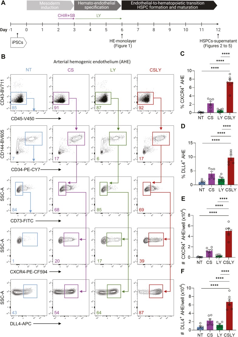
To determine the impact of modulating WNT, Activin/Nodal, and MAPK intracellular signaling on hematoendothelial formation in culture, human iPSCs reprogrammed from CD34+CD38− cells of healthy volunteers were subjected to hematopoietic differentiation for 12 days using our previously reported monolayer culture system43 with or without stage-specific addition of small-molecule regulators of these pathways. In this platform, a non-hematopoietic CD43−CD45− adherent monolayer rapidly forms under conditions that support mesodermal induction (days 0 to 3). With the subsequent addition of hematopoietic cytokines that promote mesodermal specification to hematoendothelial fates and HSPC formation/maturation (days 3 to 12), HE progenitors develop (peak HE production at day 6 of culture) and CD43+CD45+/− hematopoietic cells arise from the monolayer before their eventual release within the supernatant fraction (peak HSPC production at day 12 of culture)43 (Fig. 1A).
Figure 1.
Modulation of WNT, Activin/Nodal, and MAPK signaling pathways during human iPSC differentiation enhances formation of arterial hemogenic endothelium. (A) Schematic of the experimental design for hematopoietic differentiation of human iPSCs, including mesodermal induction (days 0 to 3), hemato-endothelial specification (days 3 to 6), and hematopoietic differentiation (days 6 to 21). Maximal production of hemogenic endothelium (HE) within the CD43−CD45− adherent non-hematopoietic monolayer was observed at day 6 of culture, and peak formation of hematopoietic stem and progenitor cells (HSPCs) was detected within the CD43+CD45+/− supernatant hematopoietic fraction at day 12 of differentiation. Unless otherwise indicated, WNT agonist CHIR99021 (CHIR) and Activin/Nodal antagonist SB431542 (SB) were supplemented on day 2 of differentiation for 24 hours, and MAPK agonist LY294002 (LY) was added from days 3 to 6 of culture. All analyses were performed with cells collected at day 6 of differentiation in the absence of small-molecule adjunct treatment (non-treated, NT) or in the presence of CHIR/SB (CS), LY, or CHIR/SB/LY (CSLY). (B) Representative flow cytometry plots of arterial HE (AHE)-defining markers (CD43−CD45−CD144+CD34+CD73−CXCR4+ or DLL4+). (C) Percentages of CXCR4+ AHE cells. (D) Percentages of DLL4+ AHE cells. (E) Absolute numbers of CXCR4+ AHE cells per culture well. (F) Absolute numbers of DLL4+ AHE cells per culture well. Data shown in this figure were obtained with human iPSC line MCND-TEN-S2. Reproducibility of data was confirmed with human iPSC lines HT914 (Supplementary Fig. S1). In panels C-F, data are displayed as mean ± standard error of the mean (SEM) of 6 independent experiments. One-way ordinary ANOVA test with Dunnett correction was used. ****P ≤ 0.0001. Associated with Supplementary Figs. S1 and S2.
As initially demonstrated in other PSC culture protocols,38,39 we previously confirmed that supplementing CHIR (WNT agonist) and SB (Activin/Nodal repressor) at day 2 of iPSC differentiation for a period of 24 hours suppresses primitive hematopoiesis and promotes definitive HSPC formation in our culture system.43 To assess whether LY-mediated activation of MAPK signaling at the mesodermal stage of development also enhances HE arterial specification in our differentiation approach, we supplemented LY from day 3 through the end of mesodermal induction/specification (day 6). Production of arterial HE was measured by interrogating the CD43−CD45− non-hematopoietic cell fraction for HE-defining markers, including CD144+ (VE-cadherin+), CD34+ and lack of CD73 expression (CD73−), as well as markers previously associated with arterialization of HE (CXCR4+ and DLL4+).23,25,49 Compared to control cultures containing no CHIR/SB/LY (non-treated, NT) or supplemented with CHIR/SB or LY only, combined addition of CHIR, SB, and LY led to a marked increase in percentages (Fig. 1B-D and Supplementary Fig. S1) and numbers (Fig. 1E, F and Supplementary Fig. S1) of CXCR4+ or DLL4+ arterial HE (CD43-CD45−CD144+CD34+CD73−CXCR4+/DLL4+). The peak effect was observed on the sixth day of differentiation and when LY was supplemented from day 3 through day 6 of culture (Supplementary Fig. S2).
Modulation of WNT, Activin/Nodal, and MAPK Signaling Pathways Promotes Definitive HSPC Formation During Human iPSC Differentiation
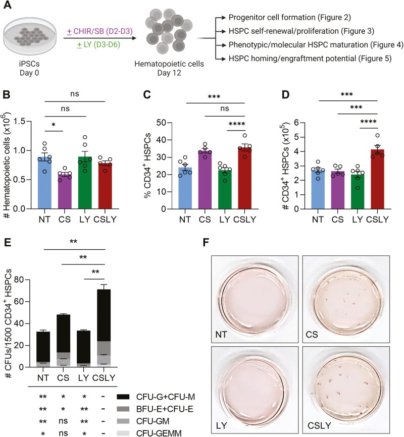
We next investigated whether the early increase in arterial HE formation observed in the presence of CHIR/SB/LY influenced hematopoietic development (Fig. 2A). Addition of CHIR/SB during differentiation decreased overall CD43+CD45+/− hematopoietic cell numbers compared to untreated groups, but supplementation of culture medium with LY in combination with CHIR/SB offset this effect and LY alone had no impact on total cell numbers in culture (Fig. 2B and Supplementary Fig. S3A). Moreover, a rise in percentages (Fig. 2C and Supplementary Fig. S3B) and numbers (Fig. 2D and Supplementary Fig. S3C) of CD34+ progenitors was observed within the hematopoietic population at day 12 of differentiation with CHIR/SB/LY compared to controls. In clonogenic progenitor assays, the total numbers of colony forming units and the frequency of CFUs with unilineage (CFU-G, CFU-M, CFU-E, and BFU-E) and multilineage (CFU-GM and CFU-GEMM) differentiation capacity were similar between control groups but significantly increased in the presence of CHIR/SB/LY (Fig. 2E, F and Supplementary Fig. S3D, E).
Figure 2.
Modulation of WNT, Activin/Nodal, and MAPK signaling pathways during human iPSC differentiation enhances formation of hematopoietic progenitors. (A) Experimental scheme. Human iPSCs were differentiated as illustrated in Fig. 1A in the absence of small-molecule adjunct treatment (non-treated, NT) or in the presence of CHIR/SB (CS), LY or CHIR/SB/LY (CSLY). Hematopoietic progenitor activity of cells released within the culture supernatant at day 12 of differentiation with indicated treatments was evaluated by flow cytometry and colony forming unit (CFU) assay. (B) Absolute numbers of CD43+CD45+/− hematopoietic cells per culture well. (C) Percentages of CD34+ HSPCs. (D) Absolute numbers of CD34+ HSPCs per culture well. (E) Numbers of myeloid (CFU-G, CFU-M, CFU-GM and CFU-GEMM) and erythroid (CFU-E, BFU-E) colonies per 1500 CD34+ cells purified by FACS. The p values shown above the bar graph indicate statistical significance for the total numbers of CFUs in the CSLY group compared to controls. The p values shown below the bar graph indicate statistical significance for the numbers of each colony type in the CSLY group compared to controls. (F) Representative images of CFU plates for data presented in panel E demonstrating a significant increase in colony counts by addition of CSLY during iPSC differentiation. Data shown in this figure were obtained with human iPSC line MCND-TEN-S2. Reproducibility of data was confirmed with human iPSC lines HT914 and HT915 (Supplementary Fig. S3). In panels B-E, data are displayed as mean ± SEM of 3 to 6 independent experiments. One-way ordinary ANOVA test with Dunnett correction was used. ns, not significant, *P ≤ 0.05, **P ≤ 0.01, ***P ≤ 0.001, ****P ≤ 0.0001. Associated with Supplementary Fig. S3.
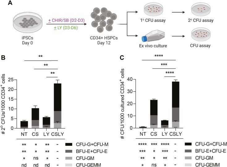
Next, we assessed whether the CHIR/SB/LY combination enabled the emergence of third wave definitive HSPCs with self-renewal (Fig. 3 and Supplementary Fig. S4), progressive maturation (Fig. 4 and Supplementary Fig. S5) and homing/engraftment potential during iPSC differentiation (Fig. 5 and Supplementary Fig. S6). To gain insights into the self-renewal and proliferative capacities of iPSC-derived HSPCs, hematopoietic colonies that formed in primary CFU cultures were pooled and replated in secondary clonogenic assays (Fig. 3A, top). Notably, we observed a significant (2.2- to 6.2-fold) increase in replating efficiency from CD34+ HSPCs arising from CHIR/SB/LY-supplemented cultures compared to control groups (Fig. 3B and Supplementary Fig. S4A, B). To further corroborate these findings, day 12 CD34+ cells derived from each iPSC differentiation condition were cultured for 10 days in cytokine-supplemented medium before quantification of CFU activity in clonogenic progenitor assays (Fig. 3A, bottom). As previously shown, iPSC-differentiated HSPCs that lack self-renewal and proliferative abilities differentiate and lose their colony-forming potential in extended culture.50 Using this experimental scheme, only rare colonies were detected from untreated or LY-supplemented iPSC differentiation conditions. HSPCs derived from CHIR/SB-containing iPSC cultures and differentiated for 10 days in vitro produced increased CFU numbers relative to untreated and LY groups, but the most substantial rise in colony-forming activity after a 10-day culture period was unequivocally observed with CD34+ HSPCs differentiated from iPSCs in the presence of CHIR/SB/LY (Fig. 3C).
Figure 3.
Modulation of WNT, Activin/Nodal, and MAPK signaling pathways during human iPSC differentiation enhances formation of HSPCs with self-renewal capacity. (A) Experimental scheme. Human iPSCs were differentiated as illustrated in Fig. 1A in the absence of small-molecule adjunct treatment (non-treated, NT) or in the presence of CHIR/SB (CS), LY or CHIR/SB/LY (CSLY). Self-renewal capacity of HSPCs released within the culture supernatant at day 12 of differentiation with indicated treatments was evaluated by secondary clonogenic assay or CFU formation following extended (10-day) culture. (B) Secondary clonogenic assay. A total of 1000 CD34+ HSPCs purified by FACS were plated in primary CFU assays. After 12-14 days, colonies were scored, pooled, and equal numbers of cells were replated for each condition. Secondary CFU plates were scored at days 12-14 for myeloid (CFU-G, CFU-M, CFU-GM, and CFU-GEMM) and erythroid (CFU-E, BFU-E) colonies, and counts were normalized to the total number of cells from primary CFU plates. nd, not detected. (C) CFU assay after extended culture. A total of 1000 CD34+ HSPCs purified by FACS were cultured for 10 days and subsequently plated in primary CFU assays. CFU plates were scored at days 12-14 for myeloid and erythroid colonies. Data shown in this figure were obtained with human iPSC line MCND-TEN-S2. Reproducibility of data was confirmed with human iPSC lines HT914 and HT915 (Fig. S4). In panels B and C, data are displayed as mean ± SEM of 3 independent experiments. The P values shown above the bar graphs indicate statistical significance for the total numbers of CFUs in the CSLY group compared to controls. The P values shown below the bar graphs indicate statistical significance for the numbers of each colony type in the CSLY group compared to controls. One-way ordinary ANOVA test with Dunnett correction was used. ns, not significant, *P ≤ 0.05, **P ≤ 0.01, ***P ≤ 0.001, ****P ≤ 0.0001. Associated with Fig. S4.
Figure 4.
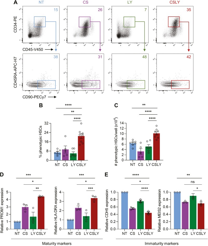
Modulation of WNT, Activin/Nodal, and MAPK signaling pathways during human iPSC differentiation enhances formation of HSPCs with phenotypic and molecular attributes of maturation. Human iPSCs were differentiated as illustrated in Fig. 1A in the absence of small-molecule adjunct treatment (non-treated, NT) or in the presence of CHIR/SB (CS), LY or CHIR/SB/LY (CSLY). All analyses were performed with HSPCs collected at day 12 of differentiation with indicated treatments. (A) Representative flow cytometry plots of phenotypically defined HSCs (CD45+CD34+CD45RA−CD90+). (B) Percentages of phenotypically defined HSCs. (C) Absolute numbers of phenotypically defined HSCs per culture well. (D) Quantitative PCR analysis of PROM1 and HLA-DRA molecular markers associated with hematopoietic maturity in phenotypically defined HSCs. Data were normalized to the NT control group. (E) Quantitative PCR analysis of CDH5 and MEIS2 molecular markers associated with hematopoietic immaturity in phenotypically defined HSCs. Data were normalized to the NT control group. Data shown in this figure were obtained with human iPSC line MCND-TEN-S2. Reproducibility of data was confirmed with human iPSC line HT915 (Supplementary Fig. S5). In panels B-E, data are displayed as mean ± SEM of 3 to 8 independent experiments. One-way ordinary ANOVA test with Dunnett correction was used. ns, not significant, *P ≤ 0.05, **P ≤ 0.01, ***P ≤ 0.001, ****P ≤ 0.0001. Associated with Supplementary Fig. S5.
Figure 5.
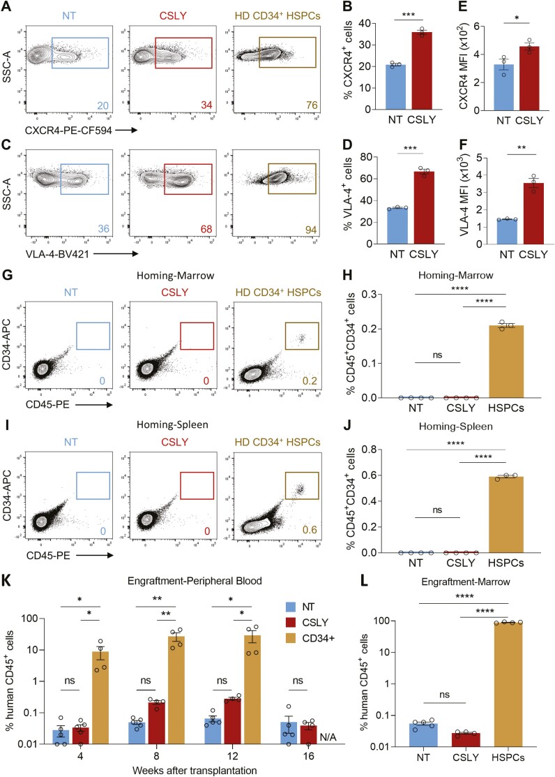
Modulation of WNT, Activin/Nodal, and MAPK signaling pathways during human iPSC differentiation is insufficient to enhance formation of HSPCs with functional properties in vivo. Human iPSCs were differentiated as illustrated in Fig. 1A in the absence of small-molecule adjunct treatment (non-treated, NT) or in the presence of CHIR/SB/LY (CSLY). All analyses were performed using CD34+ HSPCs purified by FACS from the culture supernatant at day 12 of differentiation with indicated treatments or collected by G-CSF mobilization and apheresis of a healthy donor (HD). (A-F) Expression of CXCR4 and VLA-4 homing markers. Panels display representative flow cytometry plots of CXCR4 (A) or VLA-4 (C) expression, percentages of cells expressing CXCR4 (B) or VLA-4 (D), and mean fluorescence intensity (MFI) of CXCR4 (E) or VLA-4 (F) homing markers. (G-J) Early in vivo homing potential. Panels display representative flow cytometry plots and percentages of human CD45+CD34+ cells detected within the marrow (G, H) or spleen (I, J) of NBSGW mice at 24 hours post-injection. (K-L) Long-term in vivo engraftment potential. Panels display percentages of human CD45+ cells detected within the peripheral blood of NBSGW mice at the indicated times post-transplantation (K) or within the murine bone marrow at 4 months post-transplantation (L). Data shown in this figure were obtained with human iPSC line MCND-TEN-S2. In panels B, D, E, and F, data are displayed as mean ± SEM of 3 independent experiments. In panels H, J, K, and L, data are displayed as mean ± SEM of 3 to 5 mice for each group. Two-tailed unpaired t-test (panels B, D, E, and F) and one-way ordinary ANOVA test with Dunnett correction (panels H, J, K and L) were used. ns, not significant, *P ≤ 0.05, **P ≤ 0.01, ***P ≤ 0.001, ****P ≤ 0.0001. Associated with Supplementary Fig. S6.
To determine the impact of CHIR/SB/LY on the extent of HSPC maturation at day 12 of differentiation, we first tracked the CD34+CD38-CD45RA−CD90+ cell surface phenotype customarily accepted to define HSPC populations highly enriched in HSCs with engraftment potential.51 In iPSC cultures treated with CHIR/SB/LY, a notable rise in percentages (Fig. 4A, B and Supplementary Fig. S5A, B) and numbers (Fig. 4C and Supplementary Fig. S5C) of phenotypically defined HSCs was observed within the hematopoietic population at day 12 of differentiation compared to controls. To provide corroborative evidence of the positive effect of CHIR/SB/LY on HSPC maturation during iPSC differentiation, we also compared expression of markers recently pinpointed as landmarks of HSC maturity (eg, PROM1 and HLA-DRA) or immaturity (eg, CDH5 and MEIS2) in a single-cell transcriptomic analysis of human HSPCs at distinct stages of maturation during ontogeny.28 Addition of CHIR/SB/LY induced robust increased expression of the maturation markers PROM1 and HLA-DRA (Fig. 4D), and concurrent downregulation of the immaturity genes CDH5 and MEIS2 (Fig. 4E) compared with control groups. However, this manipulation alone was insufficient to confer a maturation profile comparable to adult HSPCs (Supplementary Fig. S5D, E).
To address whether addition of CHIR/SB/LY during iPSC differentiation conferred enhanced functional properties to HSPCs in vivo, we quantified early homing (24 hours) and long-term engraftment (4 months) of human cells within hematopoietic tissues of NOD.Cg-KitW-41J Tyr+ Prkdcscid Il2rgtm1Wjl/ThomJ (NBSGW) mice transplanted with CD34+ HSPCs differentiated from human iPSCS with or without CHIR/SB/LY small molecules. We first measured expression of the chemokine receptor CXCR4 and the β1-integrin VLA-4, 2 foremost cell surface markers implicated in homing and retention of HSPCs within sites of hematopoiesis after transplantation.52,53 The proportion of human CD34+ HSPCs expressing CXCR4 (Fig. 5A, B) or VLA-4 surface proteins (Fig. 5C, D) significantly increased with addition of CHIR/SB/LY during iPSC differentiation but remained lower than frequencies detected in cultured CD34+ HSPCs collected by G-CSF mobilization and apheresis of a healthy subject (Fig. 5A). With CHIR/SB/LY supplementation, expression of both markers was also upregulated, as measured by mean fluorescence intensity on flow cytometry plots (Fig. 5E, F). However, whereas bona fide human CD34+ cells were readily detectable within marrow and spleen tissues 1 day following tail vein injection of NBSGW mice, human cell chimerism was not measurable in mice transplanted with similar numbers of CD34+ HSPCs derived from iPSC differentiation cultures (Fig. 5G-J). Consistent with the early homing and retention failure in vivo, HSPCs derived from iPSC differentiation cultures in the presence or absence of CHIR/SB/LY did not sustain levels of long-term hematopoietic engraftment comparable to somatic human CD34+ cells after transplantation into NBSGW mouse recipients (Fig. 5K, L and Supplementary Fig. S6).
Discussion
In this study, we investigate the long-standing need for optimized human iPSC differentiation systems to enable production of clinical-grade HSCs with functional properties in vivo for therapeutic applications. This work stemmed from our previous observation that arterial HE within the non-hematopoietic adherent monolayer that forms during the initial phase of our differentiation platform was scarce and likely insufficient to fully support the generation of bona fide engrafting HSCs in culture.43 Thus, in keeping with the prevailing arterial-specification model,23-27 we aimed to augment production of arterial HE during iPSC differentiation and evaluated the impact on HSPC formation in culture. By stage-specific addition of small molecules to modulate key signaling pathways independently shown to control arterial fate during vascular development, including WNT, Activin/Nodal, and MAPK pathways, we made 2 observations. First, we found that combined addition of CHIR/SB/LY during human iPSC hematopoietic differentiation could synergistically promote arterial specification of HE, peaking at day 6 of culture, compared to control groups. Second, we showed that this early increase in arterial HE production promoted formation of definitive HSPCs with improved multilineage differentiation capacity, self-renewal potential, and maturation state at day 12 of culture. We foresee that adjunct treatment with CHIR/SB/LY during human iPSC differentiation may become routinely used to enhance the arterial HE program and boost definitive multilineage hematopoietic formation in culture.
This work is grounded on the hypothesis that arterial specification is essential for definitive hematopoiesis to arise during ontogeny and in ex vivo culture systems. This premise has gained credence based on the observed origin of HSCs within the aortic endothelium during development,20,22,54 the demonstration of shared signaling pathways between arterial fate determination and HSC formation,40,41,55,56 and the selective inhibition of definitive hematopoiesis within the dorsal aorta in a murine knockout model of the artery-specific Ephrin B2 gene.57 Yet, an arterial-independent model has also been endorsed whereby hematopoietic progenitors expressing endothelial markers, rather than bona fide endothelial cells, undergo HSC specification after transitioning through the permissive AGM arterial environment of the developing conceptus. This hypothesis is primarily substantiated by observations of HSC formation in animal models engineered to disrupt normal arterial specification in vivo. However, a direct measure of HSC long-term engraftment activity was not performed in these studies.41,58-62 In our work, although enhanced arterial HE formation by CHIR/SB/LY supplementation was insufficient to enable the emergence of third-wave engraftable HSCs in human iPSC culture, these results do not exclude arterial specification as a central prerequisite to initiate the HSC program. Instead, activation of an arterial program in combination with additional cues uncoupled from arterial specification during human PSC differentiation are likely needed for efficient de novo generation of human HSCs.
Our study focused on modulating WNT, Nodal/Activin, and MAPK intrinsic cellular signaling for hematopoietic development ex vivo, but further fine-tuning of these and other pathways will likely be required to overcome the engraftment deficit. For instance, although WNT signaling activation plays an essential role in the induction of mesoderm during development, inhibition of this pathway at later stages of differentiation may also be necessary for HSPC development from hemogenic endothelium.63 In addition, the impact of MAPK signaling activation on arterial HE specification is primarily mediated through stimulation of NOTCH,23 a critical signaling pathways that requires tight regulation during and following EHT to generate and maintain functional HSCs.25,64,65 Thus, activation of MAPK signaling by LY in our study, although sufficient to enhance the arterial program in combination with CHIR/SB, may not provide the proper intensity of Notch signal permissive for third-wave HSC specification. Stage-specific modulation of additional intrinsic cellular inputs, such as ectopic expression of single or combinations of transcription factors linked to HSC regulation, and inhibition of key epigenetic regulators known to restrict hematopoiesis during development may also be required to promote full maturation and functionality of HSCs engineered ex vivo.11,66,67 Importantly, because hematopoietic cells emerge and mature within the 3-dimensional niches of specialized anatomical sites during development, integration of extrinsic environmental cues during human PSC differentiation ex vivo, namely cell-cell contact, cell-matrix interactions, diffusible factors, and biomechanical forces, may further enhance the emergence of definitive hematopoiesis from arterialized HE in culture (reviewed in Refs 11,68). This concept was unambiguously demonstrated in PSC differentiation strategies based on teratoma formation whereby primitive hematopoietic cells differentiated within teratomas displayed long-term trilineage reconstituting activity upon transplantation in animal models.69-72
Our study also uncovered an early transplantation failure of iPSC-derived HSPCs in xenograft murine models. Although efficient homing, retention, and survival within specialized niches of recipient hematopoietic tissues after transplantation are indispensable for long-term engraftment of HSCs, the early homing failure alone is unlikely sufficient to explain the engraftment deficit observed at 4 months in our study. In a recent investigation,73 ectopic expression of the homing receptor CXCR4 improved short-term marrow retention but was insufficient to confer sustained engraftment. The iPSC differentiation platform used in our study enabled production of human HSPCs with CXCR4 and VLA-4 activity independent of ectopic expression, and medium supplementation with CHIR/SB/LY further enhanced expression of both markers. Hence, the inability of these cells to home and survive after transplantation cannot be attributed to limited expression of the CXCR4 chemokine receptor or VLA-4 integrin. A detailed analysis of their functional integrity, including ligand binding affinity, receptor conformational changes, signal transduction, internalization, and degradation, is needed to fully understand the mechanism underpinning the inadequate homing capabilities of HSPCs engineered ex vivo.74,75 A comprehensive examination of additional surface molecules implicated in cellular adhesion (eg, cadherins, selectins, and tetraspanins) and their interacting ligands may also impart new leads for the generation of human HSCs with short-term retention capability and long-term engraftment competency in vivo.
Conclusion
Here, we provide evidence that combined addition of CHIR/SB/LY during human iPSC hematopoietic differentiation enhances formation of arterial HE and phenotypically defined definitive HSPCs with self-renewal and multilineage differentiation capacity. Our observation that CHIR/SB/LY treatment alone was insufficient to enable the emergence of third-wave engraftable HSCs from human iPSC culture indicates that further refinement is required. The clinically relevant and readily adaptable human iPSC differentiation platform presented in this study offers a unique framework to further define and dependably recapitulate in vitro the diverse signals driving hematopoiesis in vivo to promote formation of bona fide HSCs in vitro.
Supplementary Material
Acknowledgments
We thank: All members of the Larochelle Laboratory for helpful discussions; Dr. David Stroncek and the NIH Department of Transfusion Medicine and Cell Processing Section staff for providing leukapheresis, isolation, and cryopreservation of CD34+ cells; the outpatient clinic nursing staff for recruiting normal volunteers and providing G-CSF administration teaching to healthy subjects; Dr. Julie Holdridge, Dr. James Hawkins and the mouse core facility staff for excellent animal care; Dr. Pradeep Dagur and the NHLBI flow cytometry core facility staff for technical support with sorting; the NHLBI iPSC core facility staff for assistance with iPSC culture. Graphics in Figs. 1A, 2A ,and 3A were created with BioRender.com.
Contributor Information
Yongqin Li, Cellular and Molecular Therapeutics Branch, National Heart, Lung and Blood Institute (NHLBI), National Institutes of Health (NIH), Bethesda, MD, USA.
Jianyi Ding, Cellular and Molecular Therapeutics Branch, National Heart, Lung and Blood Institute (NHLBI), National Institutes of Health (NIH), Bethesda, MD, USA.
Daisuke Araki, Cellular and Molecular Therapeutics Branch, National Heart, Lung and Blood Institute (NHLBI), National Institutes of Health (NIH), Bethesda, MD, USA.
Jizhong Zou, iPSC Core Facility, NHLBI, NIH, Bethesda, MD, USA.
Andre Larochelle, Cellular and Molecular Therapeutics Branch, National Heart, Lung and Blood Institute (NHLBI), National Institutes of Health (NIH), Bethesda, MD, USA.
Funding
This work was supported by the Intramural Research Program of the National Heart, Lung, and Blood Institute, National Institutes of Health (Z99 HL999999 and ZIA HL006217). The content of this publication does not necessarily reflect the views or policies of the Department of Health and Human Services, nor does mention of trade names, commercial products, or organizations imply endorsement by the US Government.
Conflict of Interest
A patent was granted for the iPSC differentiation method used in this manuscript (publication # WO2015050963 A1). A license was also executed with STEMCELL Technologies and this new methodology is now available commercially to the scientific community as STEMdiffTM Hematopoietic Kit (cat # 05310). A.L. declared royalties from this license. The other authors declared no potential conflicts.
Author Contributions
Y.L.: Conception and design, collection and assembly of data, data analysis and interpretation, manuscript writing, final approval of manuscript. J.D., D.A., J.Z.: Collection and assembly of data, final approval of manuscript. A.L.: Conception and design, data analysis and interpretation, manuscript writing, final approval of manuscript.
Data Availability
All data are incorporated into the article and its Supplementary material.
References
- 1. Leonard A, Bonifacino A, Dominical VM, et al. Bone marrow characterization in sickle cell disease: inflammation and stress erythropoiesis lead to suboptimal CD34 recovery. Br J Haematol. 2019;186(2):286-299. 10.1111/bjh.15902. [DOI] [PMC free article] [PubMed] [Google Scholar]
- 2. Sobrino S, Magnani A, Semeraro M, et al. Severe hematopoietic stem cell inflammation compromises chronic granulomatous disease gene therapy. Cell Rep Med 2023;4(2):100919. [DOI] [PMC free article] [PubMed] [Google Scholar]
- 3. Morgan RA, Gray D, Lomova A, Kohn DB.. Hematopoietic stem cell gene therapy: progress and lessons learned. Cell Stem Cell 2017;21(5):574-590. 10.1016/j.stem.2017.10.010. [DOI] [PMC free article] [PubMed] [Google Scholar]
- 4. Haltalli MLR, Wilkinson AC, Rodriguez-Fraticelli A, Porteus M.. Hematopoietic stem cell gene editing and expansion: State-of-the-art technologies and recent applications. Exp Hematol. 2022;107:9-13. 10.1016/j.exphem.2021.12.399. [DOI] [PubMed] [Google Scholar]
- 5. Li J, Wang X, Ding J, et al. Development and clinical advancement of small molecules for ex vivo expansion of hematopoietic stem cell. Acta Pharm Sin B 2022;12(6):2808-2831. 10.1016/j.apsb.2021.12.006. [DOI] [PMC free article] [PubMed] [Google Scholar]
- 6. Walasek MA, van Os R, de Haan G.. Hematopoietic stem cell expansion: challenges and opportunities. Ann N Y Acad Sci. 2012;1266(1):138-150. 10.1111/j.1749-6632.2012.06549.x. [DOI] [PubMed] [Google Scholar]
- 7. Takahashi K, Yamanaka S.. Induction of pluripotent stem cells from mouse embryonic and adult fibroblast cultures by defined factors. Cell. 2006;126(4):663-676. 10.1016/j.cell.2006.07.024. [DOI] [PubMed] [Google Scholar]
- 8. Takahashi K, Tanabe K, Ohnuki M, et al. Induction of pluripotent stem cells from adult human fibroblasts by defined factors. Cell. 2007;131(5):861-872. 10.1016/j.cell.2007.11.019. [DOI] [PubMed] [Google Scholar]
- 9. Yu J, Vodyanik MA, Smuga-Otto K, et al. Induced pluripotent stem cell lines derived from human somatic cells. Science. 2007;318(5858):1917-1920. 10.1126/science.1151526. [DOI] [PubMed] [Google Scholar]
- 10. Ivanovs A, Rybtsov S, Ng ES, et al. Human haematopoietic stem cell development: from the embryo to the dish. Development. 2017;144(13):2323-2337. 10.1242/dev.134866. [DOI] [PubMed] [Google Scholar]
- 11. Ding J, Li Y, Larochelle A.. De novo generation of human hematopoietic stem cells from pluripotent stem cells for cellular therapy. Cells 2023;12(2):321. 10.3390/cells12020321. [DOI] [PMC free article] [PubMed] [Google Scholar]
- 12. Bertrand JY, Chi NC, Santoso B, et al. Haematopoietic stem cells derive directly from aortic endothelium during development. Nature. 2010;464(7285):108-111. [in Eng]. 10.1038/nature08738. [DOI] [PMC free article] [PubMed] [Google Scholar]
- 13. Medvinsky A, Dzierzak E.. Definitive hematopoiesis is autonomously initiated by the AGM region. Cell. 1996;86(6):897-906. 10.1016/s0092-8674(00)80165-8. [DOI] [PubMed] [Google Scholar]
- 14. Muller AM, Medvinsky A, Strouboulis J, Grosveld F, Dzierzak E.. Development of hematopoietic stem cell activity in the mouse embryo. Immunity. 1994;1(4):291-301. 10.1016/1074-7613(94)90081-7. [DOI] [PubMed] [Google Scholar]
- 15. Orkin SH, Zon LI.. Hematopoiesis: an evolving paradigm for stem cell biology. Cell. 2008;132(4):631-644. 10.1016/j.cell.2008.01.025. [DOI] [PMC free article] [PubMed] [Google Scholar]
- 16. Chen MJ, Yokomizo T, Zeigler BM, Dzierzak E, Speck NA.. Runx1 is required for the endothelial to haematopoietic cell transition but not thereafter. Nature. 2009;457(7231):887-891. 10.1038/nature07619. [DOI] [PMC free article] [PubMed] [Google Scholar]
- 17. Eilken HM, Nishikawa S, Schroeder T.. Continuous single-cell imaging of blood generation from haemogenic endothelium. Nature. 2009;457(7231):896-900. 10.1038/nature07760. [DOI] [PubMed] [Google Scholar]
- 18. Lancrin C, Sroczynska P, Stephenson C, et al. The haemangioblast generates haematopoietic cells through a haemogenic endothelium stage. Nature. 2009;457(7231):892-895. [in Eng]. 10.1038/nature07679. [DOI] [PMC free article] [PubMed] [Google Scholar]
- 19. Kumar A, D’Souza SS, Thakur AS.. Understanding the journey of human hematopoietic stem cell development. Stem Cells Int 2019;2019:2141475. 10.1155/2019/2141475. [DOI] [PMC free article] [PubMed] [Google Scholar]
- 20. Zovein AC, Hofmann JJ, Lynch M, et al. Fate tracing reveals the endothelial origin of hematopoietic stem cells. Cell Stem Cell 2008;3(6):625-636. 10.1016/j.stem.2008.09.018. [DOI] [PMC free article] [PubMed] [Google Scholar]
- 21. Jaffredo T, Gautier R, Eichmann A, Dieterlen-Lièvre F.. Intraaortic hemopoietic cells are derived from endothelial cells during ontogeny. Development. 1998;125(22):4575-4583. 10.1242/dev.125.22.4575. [DOI] [PubMed] [Google Scholar]
- 22. Boisset JC, van Cappellen W, Andrieu-Soler C, et al. In vivo imaging of haematopoietic cells emerging from the mouse aortic endothelium. Nature. 2010;464(7285):116-120. 10.1038/nature08764. [DOI] [PubMed] [Google Scholar]
- 23. Park MA, Kumar A, Jung HS, et al. Activation of the arterial program drives development of definitive hemogenic endothelium with lymphoid potential. Cell Rep 2018;23(8):2467-2481. 10.1016/j.celrep.2018.04.092. [DOI] [PMC free article] [PubMed] [Google Scholar]
- 24. Slukvin II, Uenishi GI.. Arterial identity of hemogenic endothelium: a key to unlock definitive hematopoietic commitment in human pluripotent stem cell cultures. Exp Hematol. 2019;71:3-12. 10.1016/j.exphem.2018.11.007. [DOI] [PMC free article] [PubMed] [Google Scholar]
- 25. Uenishi GI, Jung HS, Kumar A, et al. NOTCH signaling specifies arterial-type definitive hemogenic endothelium from human pluripotent stem cells. Nat Commun. 2018;9(1):1828. 10.1038/s41467-018-04134-7. [DOI] [PMC free article] [PubMed] [Google Scholar]
- 26. Gordon-Keylock S, Sobiesiak M, Rybtsov S, Moore K, Medvinsky A.. Mouse extraembryonic arterial vessels harbor precursors capable of maturing into definitive HSCs. Blood. 2013;122(14):2338-2345. 10.1182/blood-2012-12-470971. [DOI] [PMC free article] [PubMed] [Google Scholar]
- 27. Yzaguirre AD, Speck NA.. Insights into blood cell formation from hemogenic endothelium in lesser-known anatomic sites. Dev Dyn. 2016;245(10):1011-1028. 10.1002/dvdy.24430. [DOI] [PMC free article] [PubMed] [Google Scholar]
- 28. Calvanese V, Capellera-Garcia S, Ma F, et al. Mapping human haematopoietic stem cells from haemogenic endothelium to birth. Nature. 2022;604(7906):534-540. 10.1038/s41586-022-04571-x. [DOI] [PMC free article] [PubMed] [Google Scholar]
- 29. Chadwick K, Wang L, Li L, et al. Cytokines and BMP-4 promote hematopoietic differentiation of human embryonic stem cells. Blood. 2003;102(3):906-915. 10.1182/blood-2003-03-0832. [DOI] [PubMed] [Google Scholar]
- 30. Kennedy M, D’Souza SL, Lynch-Kattman M, Schwantz S, Keller G.. Development of the hemangioblast defines the onset of hematopoiesis in human ES cell differentiation cultures. Blood. 2007;109(7):2679-2687. 10.1182/blood-2006-09-047704. [DOI] [PMC free article] [PubMed] [Google Scholar]
- 31. Pick M, Azzola L, Mossman A, Stanley EG, Elefanty AG.. Differentiation of human embryonic stem cells in serum-free medium reveals distinct roles for bone morphogenetic protein 4, vascular endothelial growth factor, stem cell factor, and fibroblast growth factor 2 in hematopoiesis. Stem Cells. 2007;25(9):2206-2214. 10.1634/stemcells.2006-0713. [DOI] [PubMed] [Google Scholar]
- 32. Keller G, Kennedy M, Papayannopoulou T, Wiles M V.. Hematopoietic commitment during embryonic stem cell differentiation in culture. Mol Cell Biol. 1993;13(1):473-486. 10.1128/mcb.13.1.473-486.1993. [DOI] [PMC free article] [PubMed] [Google Scholar]
- 33. Muller AM, Dzierzak EA.. ES cells have only a limited lymphopoietic potential after adoptive transfer into mouse recipients. Development. 1993;118(4):1343-1351. 10.1242/dev.118.4.1343. [DOI] [PubMed] [Google Scholar]
- 34. Vodyanik MA, Bork JA, Thomson JA, Slukvin Igor I.. Human embryonic stem cell-derived CD34+ cells: efficient production in the coculture with OP9 stromal cells and analysis of lymphohematopoietic potential. Blood. 2005;105(2):617-626. 10.1182/blood-2004-04-1649. [DOI] [PubMed] [Google Scholar]
- 35. Ng ES, Davis RP, Azzola L, Stanley EG, Elefanty AG.. Forced aggregation of defined numbers of human embryonic stem cells into embryoid bodies fosters robust, reproducible hematopoietic differentiation. Blood. 2005;106(5):1601-1603. 10.1182/blood-2005-03-0987. [DOI] [PubMed] [Google Scholar]
- 36. Zambidis ET, Peault B, Park TS, Bunz F, Civin Curt I.. Hematopoietic differentiation of human embryonic stem cells progresses through sequential hematoendothelial, primitive, and definitive stages resembling human yolk sac development. Blood. 2005;106(3):860-870. 10.1182/blood-2004-11-4522. [DOI] [PMC free article] [PubMed] [Google Scholar]
- 37. Davis RP, Ng ES, Costa M, et al. Targeting a GFP reporter gene to the MIXL1 locus of human embryonic stem cells identifies human primitive streak-like cells and enables isolation of primitive hematopoietic precursors. Blood. 2008;111(4):1876-1884. 10.1182/blood-2007-06-093609. [DOI] [PubMed] [Google Scholar]
- 38. Sturgeon CM, Ditadi A, Awong G, Kennedy M, Keller G.. Wnt signaling controls the specification of definitive and primitive hematopoiesis from human pluripotent stem cells. Nat Biotechnol. 2014;32(6):554-561. 10.1038/nbt.2915. [DOI] [PMC free article] [PubMed] [Google Scholar]
- 39. Kennedy M, Awong G, Sturgeon CM, et al. T lymphocyte potential marks the emergence of definitive hematopoietic progenitors in human pluripotent stem cell differentiation cultures. Cell Rep 2012;2(6):1722-1735. 10.1016/j.celrep.2012.11.003. [DOI] [PubMed] [Google Scholar]
- 40. Lawson ND, Scheer N, Pham VN, et al. Notch signaling is required for arterial-venous differentiation during embryonic vascular development. Development. 2001;128(19):3675-3683. 10.1242/dev.128.19.3675. [DOI] [PubMed] [Google Scholar]
- 41. Kumano K, Chiba S, Kunisato A, et al. Notch1 but not Notch2 is essential for generating hematopoietic stem cells from endothelial cells. Immunity. 2003;18(5):699-711. 10.1016/s1074-7613(03)00117-1. [DOI] [PubMed] [Google Scholar]
- 42. Burns CE, Traver D, Mayhall E, Shepard JL, Zon LI.. Hematopoietic stem cell fate is established by the Notch-Runx pathway. Genes Dev. 2005;19(19):2331-2342. 10.1101/gad.1337005. [DOI] [PMC free article] [PubMed] [Google Scholar]
- 43. Ruiz JP, Chen G, Haro Mora JJ, et al. Robust generation of erythroid and multilineage hematopoietic progenitors from human iPSCs using a scalable monolayer culture system. Stem Cell Res. 2019;41:101600. 10.1016/j.scr.2019.101600. [DOI] [PMC free article] [PubMed] [Google Scholar]
- 44. Beers J, Linask KL, Chen JA, et al. A cost-effective and efficient reprogramming platform for large-scale production of integration-free human induced pluripotent stem cells in chemically defined culture. Sci Rep. 2015;5:11319. 10.1038/srep11319. [DOI] [PMC free article] [PubMed] [Google Scholar]
- 45. Chen G, Gulbranson DR, Hou Z, et al. Chemically defined conditions for human iPSC derivation and culture. Nat Methods. 2011;8(5):424-429. 10.1038/nmeth.1593. [DOI] [PMC free article] [PubMed] [Google Scholar]
- 46. Baskfield A, Li R, Beers J, et al. An induced pluripotent stem cell line (TRNDi009-C) from a Niemann-Pick disease type A patient carrying a heterozygous p.L302P (c.905 T>C) mutation in the SMPD1 gene. Stem Cell Res. 2019;38:101461. 10.1016/j.scr.2019.101461. [DOI] [PMC free article] [PubMed] [Google Scholar]
- 47. Qanash H, Li Y, Smith RH, et al. Eltrombopag improves erythroid differentiation in a human induced pluripotent stem cell model of diamond Blackfan Anemia. Cells 2021;10(4):734. 10.3390/cells10040734. [DOI] [PMC free article] [PubMed] [Google Scholar]
- 48. Leonard A, Yapundich M, Nassehi T, et al. Low-dose busulfan reduces human CD34(+) cell doses required for engraftment in c-kit mutant immunodeficient mice. Mol Ther Methods Clin Dev. 2019;15:430-437. 10.1016/j.omtm.2019.10.017. [DOI] [PMC free article] [PubMed] [Google Scholar]
- 49. Ditadi A, Sturgeon CM, Tober J, et al. Human definitive haemogenic endothelium and arterial vascular endothelium represent distinct lineages. Nat Cell Biol. 2015;17(5):580-591. 10.1038/ncb3161. [DOI] [PMC free article] [PubMed] [Google Scholar]
- 50. Doulatov S, Vo LT, Chou SS, et al. Induction of multipotential hematopoietic progenitors from human pluripotent stem cells via respecification of lineage-restricted precursors. Cell Stem Cell 2013;13(4):459-470. 10.1016/j.stem.2013.09.002. [DOI] [PMC free article] [PubMed] [Google Scholar]
- 51. Majeti R, Park CY, Weissman IL.. Identification of a hierarchy of multipotent hematopoietic progenitors in human cord blood. Cell Stem Cell 2007;1(6):635-645. 10.1016/j.stem.2007.10.001. [DOI] [PMC free article] [PubMed] [Google Scholar]
- 52. Lapidot T, Kollet O.. The essential roles of the chemokine SDF-1 and its receptor CXCR4 in human stem cell homing and repopulation of transplanted immune-deficient NOD/SCID and NOD/SCID/B2m(null) mice. Leukemia. 2002;16(10):1992-2003. 10.1038/sj.leu.2402684. [DOI] [PubMed] [Google Scholar]
- 53. Peled A, Kollet O, Ponomaryov T, et al. The chemokine SDF-1 activates the integrins LFA-1, VLA-4, and VLA-5 on immature human CD34(+) cells: role in transendothelial/stromal migration and engraftment of NOD/SCID mice. Blood. 2000;95(11):3289-3296. [PubMed] [Google Scholar]
- 54. Kissa K, Herbomel P.. Blood stem cells emerge from aortic endothelium by a novel type of cell transition [in eng]. Nature. 2010;464(7285):112-115. 10.1038/nature08761. [DOI] [PubMed] [Google Scholar]
- 55. Lawson ND, Vogel AM, Weinstein BM.. sonic hedgehog and vascular endothelial growth factor act upstream of the Notch pathway during arterial endothelial differentiation. Dev Cell. 2002;3(1):127-136. 10.1016/s1534-5807(02)00198-3. [DOI] [PubMed] [Google Scholar]
- 56. Gering M, Patient R.. Hedgehog signaling is required for adult blood stem cell formation in zebrafish embryos. Dev Cell. 2005;8(3):389-400. 10.1016/j.devcel.2005.01.010. [DOI] [PubMed] [Google Scholar]
- 57. Chen II, Caprioli A, Ohnuki H, et al. EphrinB2 regulates the emergence of a hemogenic endothelium from the aorta. Sci Rep. 2016;6:27195. 10.1038/srep27195. [DOI] [PMC free article] [PubMed] [Google Scholar]
- 58. Burns CE, Galloway JL, Smith AC, et al. A genetic screen in zebrafish defines a hierarchical network of pathways required for hematopoietic stem cell emergence. Blood. 2009;113(23):5776-5782. 10.1182/blood-2008-12-193607. [DOI] [PMC free article] [PubMed] [Google Scholar]
- 59. Krebs LT, Xue Y, Norton CR, et al. Notch signaling is essential for vascular morphogenesis in mice. Genes Dev. 2000;14(11):1343-1352. [PMC free article] [PubMed] [Google Scholar]
- 60. Nakagawa M, Ichikawa M, Kumano K, et al. AML1/Runx1 rescues Notch1-null mutation-induced deficiency of para-aortic splanchnopleural hematopoiesis. Blood. 2006;108(10):3329-3334. 10.1182/blood-2006-04-019570. [DOI] [PubMed] [Google Scholar]
- 61. Robert-Moreno A, Guiu J, Ruiz-Herguido C, et al. Impaired embryonic haematopoiesis yet normal arterial development in the absence of the Notch ligand Jagged1. EMBO J. 2008;27(13):1886-1895. 10.1038/emboj.2008.113. [DOI] [PMC free article] [PubMed] [Google Scholar]
- 62. Monteiro R, Pinheiro P, Joseph N, et al. Transforming growth factor beta drives hemogenic endothelium programming and the transition to hematopoietic stem cells. Dev Cell. 2016;38(4):358-370. 10.1016/j.devcel.2016.06.024. [DOI] [PMC free article] [PubMed] [Google Scholar]
- 63. Chanda B, Ditadi A, Iscove NN, Keller G.. Retinoic acid signaling is essential for embryonic hematopoietic stem cell development. Cell. 2013;155(1):215-227. 10.1016/j.cell.2013.08.055. [DOI] [PubMed] [Google Scholar]
- 64. Gama-Norton L, Ferrando E, Ruiz-Herguido C, et al. Notch signal strength controls cell fate in the haemogenic endothelium. Nat Commun. 2015;6:8510. 10.1038/ncomms9510. [DOI] [PMC free article] [PubMed] [Google Scholar]
- 65. Guiu J, Shimizu R, D’Altri T, et al. Hes repressors are essential regulators of hematopoietic stem cell development downstream of Notch signaling. J Exp Med. 2013;210(1):71-84. 10.1084/jem.20120993. [DOI] [PMC free article] [PubMed] [Google Scholar]
- 66. Vo LT, Kinney MA, Liu X, et al. Regulation of embryonic haematopoietic multipotency by EZH1. Nature. 2018;553(7689):506-510. 10.1038/nature25435. [DOI] [PMC free article] [PubMed] [Google Scholar]
- 67. Soto RA, Najia MAT, Hachimi M, et al. Sequential regulation of hemogenic fate and hematopoietic stem and progenitor cell formation from arterial endothelium by Ezh1/2. Stem Cell Rep. 2021;16(7):1718-1734. 10.1016/j.stemcr.2021.05.014. [DOI] [PMC free article] [PubMed] [Google Scholar]
- 68. Sugden WW, North TE.. Making Blood from the Vessel: Extrinsic and Environmental Cues Guiding the Endothelial-to-Hematopoietic Transition. Life (Basel) 2021;11(10). [DOI] [PMC free article] [PubMed] [Google Scholar]
- 69. Amabile G, Welner RS, Nombela-Arrieta C, et al. In vivo generation of transplantable human hematopoietic cells from induced pluripotent stem cells. Blood. 2013;121(8):1255-1264. 10.1182/blood-2012-06-434407. [DOI] [PMC free article] [PubMed] [Google Scholar]
- 70. Suzuki N, Yamazaki S, Yamaguchi T, et al. Generation of engraftable hematopoietic stem cells from induced pluripotent stem cells by way of teratoma formation. Mol Ther. 2013;21(7):1424-1431. 10.1038/mt.2013.71. [DOI] [PMC free article] [PubMed] [Google Scholar]
- 71. Tsukada M, Ota Y, Wilkinson AC, et al. In vivo generation of engraftable murine hematopoietic stem cells by Gfi1b, c-Fos, and Gata2 overexpression within teratoma. Stem Cell Rep. 2017;9(4):1024-1033. 10.1016/j.stemcr.2017.08.010. [DOI] [PMC free article] [PubMed] [Google Scholar]
- 72. Morvan MG, Teque F, Ye L, et al. Genetically edited CD34(+) cells derived from human iPS cells in vivo but not in vitro engraft and differentiate into HIV-resistant cells. Proc Natl Acad Sci USA. 2021;118(20):e2102404118. [DOI] [PMC free article] [PubMed] [Google Scholar]
- 73. Reid JC, Tanasijevic B, Golubeva D, et al. CXCL12/CXCR4 signaling enhances human PSC-derived hematopoietic progenitor function and overcomes early in vivo transplantation failure. Stem Cell Rep. 2018;10(5):1625-1641. 10.1016/j.stemcr.2018.04.003. [DOI] [PMC free article] [PubMed] [Google Scholar]
- 74. Busillo JM, Benovic JL.. Regulation of CXCR4 signaling. Biochim Biophys Acta. 2007;1768(4):952-963. 10.1016/j.bbamem.2006.11.002. [DOI] [PMC free article] [PubMed] [Google Scholar]
- 75. Imai Y, Shimaoka M, Kurokawa M.. Essential roles of VLA-4 in the hematopoietic system. Int J Hematol. 2010;91(4):569-575. 10.1007/s12185-010-0555-3. [DOI] [PubMed] [Google Scholar]
Associated Data
This section collects any data citations, data availability statements, or supplementary materials included in this article.
Supplementary Materials
Data Availability Statement
All data are incorporated into the article and its Supplementary material.


